Article

Antioxidant Properties of a Supercritical Fluid Extract of the Halophyte Mesembryanthemum nodiflorum L. from Sicilian Coasts: Nutraceutical and Cosmeceutical Applications

1
Laboratorio di Biochimica Marina ed Ecotossicologia, Dipartimento di Scienze della terra e del Mare DiSTeM, Università degli Studi di Palermo, Via G. Barlotta 4, 91100 Trapani, Italy
2
School of Biological Sciences, University of Portsmouth (United Kingdom), PO1 2UP Portsmouth, UK
3
Laboratory of Analysis, treatment and valorization of pollutants of the environment and products, Faculty of 11 Pharmacy, University of Monastir, Avicenne St., Monastir 5000, Tunisia
4
Istituto di Biologia Marina, Consorzio Universitario della Provincia di Trapani, Via G. Barlotta 4, 91100 Trapani, Italy
*
Author to whom correspondence should be addressed.
Appl. Sci. 2020, 10(7), 2374; https://doi.org/10.3390/app10072374
Submission received: 21 February 2020 / Revised: 29 March 2020 / Accepted: 30 March 2020 / Published: 31 March 2020
(This article belongs to the Special Issue Bioactive Substances: Properties, Applications and or Toxicities)

Abstract

:
The aim of this paper was to obtain different extracts from the aerial parts of Mesembryanthemum nodiflorum, comparing traditional extraction (using solvents with increasing polarity such as hexane, ethanol 80%, acetone 70% and water) with an eco-friendly technique (supercritical fluid extraction (SFE)); to evaluate which extract showed a higher amount of antioxidants and then evaluate the bioactive properties in vitro, in human fibroblast (HS68). From the six extracts obtained it was observed that the solvent with the highest extraction efficiency was water, but the extracts in ethanol, N-hexane and SFE are those that showed the highest antioxidant activity (polyphenols, 1,1-Diphenyl-2-picrylhydrazyl (DPPH) and reducing power). On the basis of these results, the SFE extract was chosen to evaluate the antioxidant and anti-aging activity in vitro, by assessing cell vitality and molecular markers (MTT and immunoblotting assays). The results showed that the SFE extract exerted antioxidant activity in vitro, protecting cells from mortality induced by oxidative stress; this protection was also confirmed at the molecular level, by the levels of the protein integrin α-1, that is able to prevent the negative effects of a stress situation, such as oxidative stress, that could promote aging and related diseases. This extract, obtained with an eco-friendly technique, given its beneficial properties, could be used for application in nutraceuticals and cosmeceuticals.

Graphical Abstract

1. Introduction

Halophytes, that include more than 2500 species worldwide, are known to tolerate and grow in high salt concentration environments. Species belonging to the genus Mesembryanthemum, which are facultative halophytes, have the ability to grow under saline stress conditions, but also without salt or at least in an environment where the salt concentration in the soil is quite low [1,2].
Under stress conditions, halophytes increase the production of secondary metabolites, such as polyphenolic compounds (antioxidants, anti-inflammatory), to protect cell structures from oxidative effects [3,4,5]. These are produced in plant cells to prevent the accumulation of Reactive Oxygen Species (ROS) synthesized during environmental stress, which would otherwise be detrimental to tissues [5]. It has been shown that the growth of these plants in gradients of salt concentration, for example, plant zonation in salt marshes, yields different types of antioxidants (including total phenols, flavonoids, ascorbate, reduced/oxidized glutathione and reactive oxygen species scavenging enzymes) in various quantities [6,7,8].
Mesembryanthemum species are part of the Aizoaceae family, commonly referred to as ice plants.
The genus Mesembryanthemum comprises about 70 species and is native to the Mediterranean region, the Atlantic Islands, Saudi Arabia, South Africa, South Australia and California [1].
Mesembryanthemus species are known as halophytic models due to the methods of salt tolerance utilized by these plants [9]. The Mesembryanthemum genus has three edible species of interest: M. edule, M. crystallinum and M. nodiflorum [10,11].
Several studies have shown that species belonging to the Mesembrianthemums genus such as M. crystallinum possess bioactive molecules (e.g., antioxidant, antiviral and antimicrobial) [12,13]. M. nodiflorum and M. crystallinum have been used in both food and medical fields. In traditional Tunisian medicine, these species have been used for the treatment of eye infections [13]. The seeds of M. forsskaolii, due to their high protein content, have been used as flours replacing them with wheat [13,14].
M. nodiflorum, in addition to the aforementioned use to combat eye infection [10], leaf extracts from this plant are used for various treatments such as soaps and antiseptic poultices that can be applied to skin to treat burns and sores [12]. It was also observed that of M. nodiflorum extract showed extensive bactericidal activity [15] and anti-tumor properties [16].
Given its multiple beneficial effects, M. nodiflorum is a good candidate for the extraction of bioactive compounds, useful for pharmaceutical or cosmetic applications.
Bioactive compounds can be extracted using traditional or innovative methods. Traditional extraction methods, often, involve the use of toxic solvents and have low selectivity and/or low extraction yields with very long extraction times [17]. Innovative methods, on the contrary, try to overcome these drawbacks. Extraction with supercritical fluids (SFE) is an innovative, clean and environmentally friendly technology, that has been shown to obtain high-quality extracts using CO2 [7,17,18].
Knowing the beneficial effects of this species and the presence of bioactive compounds, it would be important to study the protective and anti-aging effect of this plant by evaluating some related biomarkers. In particular, alpha-1 Integrin and Sirtuin 1 (Sirt 1) have been studied as proteins linked to oxidative stress and aging.
Integrin alpha-1 is a transmembrane glycoprotein, which plays an important role in the adhesion of the cell to the matrix and in the transduction of the signal to the cell, regulating events such as growth, division, cell survival and apoptosis [19].
Sirtuin 1 (Sirt 1) is a nuclear deacetylase, with protective action against various types of stress: it has an anti-inflammatory, anti-carcinogenic action and contributes to the cellular regulation of both growth and metabolism, acting positively on longevity [20].
In consideration of what has been shown in the literature, the aim of this work has been to obtain different extracts M. nodiflorum with different traditional extraction techniques and with ecofriedly techniques, to evaluate which extraction procedure is more effective and more suitable for a possible large-scale use and to evaluate the bioactive properties of M. nodiflorum in vitro, in human fibroblast (HS68), in particular, to evaluate the real protective and anti-aging effects.

2. Materials and Methods

2.1. Collection and Preparation of Plant Material

Samples of aerial parts of M. nodiflorum were collected along the coast North of Trapani in Sicily (Italy), in the pre-flowering period (March 2019). Specimens were kept in a herbarium and taxonomic identification was performed at the University of Palermo (Italy).
After harvesting, the plants were taken to the laboratory where they were processed. The aerial parts were dried for 48 h at 40 °C, and grounded into fine powders [7]. These dried samples were then used to extract antioxidants in specified solvents.

2.1.1. Extraction with Solvents

The extraction yield and antioxidant activity of an extract is influenced by the polarity of the solvent. Due to the different nature of the polyphenols present in the samples, different solvents should be used; in addition, the presence of hydroxyl groups, the length of the hydrocarbons and the molecular size mean that the polyphenols have a different solubility [7,21].
As proposed by Messina et al. [7], several solvents with increasing polarity hexane, ethanol 80%, acetone 70%, (Merck KGaA, Darmstadt, Germany) and water were used for the extraction [7,8,22].
The extraction of the dried aerial parts of M. nodiflorum was performed as follows: 10 mL of solvent (hexane, 80% ethanol, 70% acetone and water) was added to one gram of dry matrix [8]. The sample was homogenized for five minutes at 4 °C, using an Ultra-Turrax (IKA, Werke Staufen, Germany) at 24,000 rpm according to an established protocol [23,24,25].
The yield was calculated as described by Manuguerra et al. [26]. The following Equation (1) was used:
Yield of extract (%) = weight of extract/weight of sample × 100.
The matrices extracted with water were centrifuged at 500 g for 10 min at 4 °C three times. They were then filtered (Whatman® qualitative filter paper, Grade 93–10 µm, Merck KGaA Darmstadt, Germany) and freeze-dried [7,8].

2.1.2. Extraction with Supercritical Fluid Extraction (SFE)

To the dry matrix obtained from M. nodiflorum, supercritical technology was applied using supercritical (SC)-CO2 with a standardized protocol [7,27,28].
A supercritical extraction unit (SFE System model HELIX, Applied Separations Allentown, PA, USA) equipped with a CO2 pump unit and a steel vessel with a volume of 50 mL was used.
The extraction was done as follows: five grams of the dried matrix of M. nodiflorum with a hydroscopic dispersant reagent (Applied Separations, Allentown, PA, USA) (1:2 (w/w)) were in the extraction vessel sandwiched with defatted glass wool forming a fixed bed in the vessel. The unit was pressurized and the sample was kept in contact with SC–CO2 (60 °C) and pressure (150 bar) for 30 min in static mode [29]. Dynamic extraction was carried out with a CO2 flow of 2.5 lpm (liter per minute) for 90 min. The extract was obtained in the sample vial collector and stored at −20 °C.

2.1.3. Total Phenolic Content

Folin-Ciocalteu (Merck KGaA, Darmstadt, Germany) reagent assay [30] was carried out to determine the total phenolic content of samples [31] of M. nodiflorum. Gallic acid (Merck KGaA, Darmstadt, Germany) was used as the standard for calibration (5–500 mg/mL) and results were expressed as mg of gallic acid equivalents (GAE) per g of the M. nodiflorum dw [7,8,26,32]. The absorbance of each dilution was measured at 725 nm, using a microplate reader (Multiscan-Sky Microplate Reader, Thermo-Scientific TM, USA). Each sample was analyzed in triplicate.

2.1.4. DPPH Assay

The 1,1-Diphenyl-2-picrylhydrazyl (DPPH) free radical scavenging assay was used to determine the free radical scavenging activity of the samples of M. nodiflorum. DPPH is a stable free radical with an unpaired valence electron at a nitrogen atom which is reduced by receiving a hydrogen atom [33]. This decolourises the violet DPPH solution when a free radical scavenger (hydrogen donor) is added [31]. In this way the ability of compounds to be free radical scavengers, such as antioxidants, was measured.
The DPPH (1,1-diphenyl-2-picryhydrazyl) radical scavenging activity of different extracts was evaluated as described by Bernatoniene et al. [34] slightly modified. Briefly, 400 µL of various concentrations (1–10 mg/mL) of the extracts and 1.6 mL of 0.1 mM DPPH (Merck KGaA, Darmstadt, Germany) in ethanol [7,34] were incubated for 30 min in the dark.
The absorbance was read against the blank at 517 nm using a microplate reader (Multiscan-Sky Microplate Reader, Thermo-Scientific TM, USA). Gallic acid was used as the reference standard. The following formula (2) was used to calculate the percentage of inhibition (I%) of free radicals DPPH:
I% = 1 − (Asample/Ablank) × 100,
where Ablank is the absorbance of the control reaction, and Asample is the absorbance of the test sample.
The inhibition percentage was calculated for all concentrations (1 to 10 mg/mL) used.
The IC 50%, which is the concentration when 50% of the antioxidant is reduced [7,35], was calculated as described by Yeddes et al. [35].

2.1.5. Iron-Reducing Power

The method of Oyaizu [36] was followed to analyze the reducing power of samples of M. nodiflorum. In this assay, the ability to reduce Fe3+ to Fe2+ was tested to measure the reducing power of the plant extractions [10].
Different concentrations of M nodiflorum extracts (0.1 to 10 mg/mL) (300 µL) were mixed with phosphate buffer (300 µL, 0.2 M, pH 6.6) and potassium ferricyanide (K3Fe(CN)6, Carlo Erba reagents, Milano, Italy) (300 µL, 1%); the mixture was incubated at 50 °C for 20 min.
After incubation 300 µL of trichloroacetic acid (Carlo Erba reagents, Milano, Italy) (10%) was added to the mixture. The samples were centrifuged at 3000 rpm for 10 min at 4 °C.
An aliquot (300 µL) of the supernatant was taken, 300 µL distilled water and 600 µL, FeC13 (0.1%) (Merck KGaA Darmstadt, Germany) were added.
Absorbance was measured at 700 nm using a microplate reader (Multiscan-Sky Microplate Reader, Thermo-Scientific TM, USA). Gallic acid was used as standard.
The increase in absorbance of the reaction mixture indicated an increase in reducing power. EC50 value (mg mL−1), expressed as the effective concentration of the extract at which the absorbance was 0.5, was determined from linear regression analysis [7,12,26].

2.2. Evaluation of Cytotoxicity and Protective Effect in Fibroblast Cell Line HS-68

In order to verify, at the cellular and molecular level, the beneficial effects determined by the SFE extract, three experiments were conducted: the first was aimed to evaluate the possible dose-dependent toxicity of the extract and, on the basis of the response, to identify the ideal concentrations to be used in the second trial. In the second experiment, the cells were treated with doses that, in the first trial, did not cause death by toxicity; in this second trial, cells, pre-treated or not with extract, were subjected to oxidative stress with a standardized protocol that induces cell mortality, to verify whether or not, the extract has a protective effect.
The last step was to reproduce the second experiment in larger volumes, in order to collect cells for analysis of molecular markers by immunoblotting.

2.2.1. Hs68 Cell Culture

Human skin fibroblasts (ECACC n. 89051701, Sigma®, Sigma-Aldrich, Saint Louis, MO, USA) were seeded in 25 cm2 plastic tissue culture flasks (Nunc, Germany) cultured in Dulbecco’s Modified Eagle’s Medium (DMEM) (Merck KGaA, Darmstadt, Germany)supplemented with 10% fetal bovine serum (Merck KGaA, Darmstadt, Germany), 2 mM glutamine (Merck KGaA, Darmstadt, Germany), and 100 µg/mL penicillin–streptomycin (Merck KGaA, Darmstadt, Germany), and incubated in a humidified atmosphere at 5% CO2, 95% air and 37°C. All cell culture methods were performed under sterile conditions using a grade II flow hood.
Exponentially growing cells were detached from culture flasks by brief exposure to 0.05% of trypsin in PBS, pH 7.2–7.4 and pelleted by centrifugation (1000 rpm, 10 min, 25° C).

2.2.2. Cytotoxicity Assay and Protective Effect of M. nodiflorum Extract

For the first trial, cells were seeded in a 96-well plate at a concentration of 7 × 10³ cells/well and incubated for 24 h. After 24 h, the cells were treated with SFE extract, for 24 h and 48 h. The extract was dissolved in ethanol and utilized at a concentration ranging from 5–50 µg/mL in the medium, with a final solvent concentration of 0.1% (v/v).
The exposure of cells to ethanol alone, at the final concentration employed in the test, was previously assessed, as a routine procedure in the lab [7] and it was demonstrated that it did not exert any negative effects on vitality (data not shown).
Cell vitality was determined using the (3-(4,5-dimethylthiazol-2-yl)-2,5- diphenyltetrazolium bromide (MTT, Sigma-Aldrich) assay according to Mosmann [37] as reported in Messina et al. [38]. Results were expressed as percentage of viable cells with respect to the control. Each experiment of vitality was carried out in triplicate.
The next step was to evaluate the antioxidant effect of the extract at the cellular level, both in terms of vitality (trial two) and in terms of molecular markers These last two tests were carried employed two concentrations of the extract that, in the trial one, dose-dependent, did not induce significant cytotoxicity (cell mortality).
For the vitality test cells were detached as described previously, seeded in a 96-well plate and treated with a concentration of 5 and 10 µg/mL of M. nodiflorum SFE extract. After 24 and 48 h, all samples, except the control, were exposed to the chemical promoter of oxidative stress, hydrogen peroxide, H2O2 (HP) (Carlo Erba reagents, Milano, Italy), according to a previous standardized protocol [26,38,39] and left to incubate at 37°C for 2 h. Afterwards, the cell vitality was determined by MTT assay [37,38].

2.2.3. Evaluation of Molecular Markers by Immunoblotting

For the evaluation of molecular markers, cells were seeded in a 6-plate well at a concentration of 2 × 10⁵ cells/well, treated with the SFE extract (10µg/mL) and exposed to hydrogen peroxide as previously described [26,38,39]. After the treatment, the cells were lysed in 500 μL of RIPA buffer, as described by Thaieran et al. [40]. The concentration of total proteins, in the lysate, was determined according to Lowry et al. [41]. Experiments were carried in duplicate.
Equivalent amounts of total proteins (30 μg) were loaded on pre-cast gel (Bio-Rad, Hercules, CA, USA) for SDS-polyacrylamide electrophoresis (SDS-PAGE) and blotted using a Trans Blot Turbo Transfer System (Bio-Rad, Hercules, CA, USA).
The correct amount of loaded proteins was confirmed by Ponceau red staining and by the levels of actin. The primary antibodies (AbI), specifics for Integrin α1 (AB1934- Ab polyclonal from rabbit, Merck KGaA, Darmstadt, Germany) Sirtuin 1 (Sirt1 NBP1-49540- Ab polyclonal from rabbit, Merck KGaA, Darmstadt, Germany) and Actin (A4700- Ab Monoclonal from mouse Merck KGaA, Darmstadt, Germany), were diluted in buffer as suggested by the company for each AbI. In relation to the origin of the AbI, the appropriate secondary antibodies were used (anti-mouse or anti-rabbit secondary AbII) conjugated with horseradish peroxidase (GAM/R-HRP, Bio-Rad Hercules, CA, USA).
The signals originated by immunoreaction were detected using enhanced chemo-luminescent (ECL) reagents (SDS-PAGE, Bio-Rad, Hercules, CA, USA). Images were obtained, photographed and digitalized by Chemi Doc XRS (Bio-Rad, Hercules, CA, USA), and further analyzed with Image Lab software (Bio-Rad, Hercules, CA, USA). The showed results, for each protein, represent the mean value of three separate immunoblotting and were expressed as fold increase of the target protein, in each treatment, vs the control [42].

2.3. Statistical Analysis

Statistical analysis was performed using the computer application SPSS for Windows® (version 20.0, SPSS Inc., Chicago, IL, USA). All the analyses were carried out in triplicate. The results are expressed as Mean ± Standard deviation. The homogeneity of variance was confirmed by the Levene test. Data were subjected to one-way analysis of variance (ANOVA), and Student–Newman–Keuls or Games–Howell post-hoc tests were performed in order to make multiple comparisons between experimental groups. The significance level was 95% in all cases (p < 0.05).

3. Results and Discussions

3.1. Extraction and Evaluation of Antioxidant Power

One of the objectives of this work was to find the most effective solvent and procedure to extract antioxidants from M. nodiflorum species. The tested solvents were N-hexane, 80% (v/v) ethanol 70% (v/v) acetone, water and SFE.
The results of the yield of M. nodiflorum extracts are shown in Table 1. The aqueous extract showed the highest yield (p < 0.05). Significantly lower yields (p < 0.05) were obtained with N-hexane and SFE.
The extraction is fundamental for the recovery of antioxidants and depends on the solvents and on the chemical properties of the samples [43,44]. As it is known from the literature [21], methanol, ethanol and, their mixtures with water, are solvents with a higher extraction capacity, because the quality and quantity of the extracted antioxidant compounds depends on the polarity of the solvent [7,8,21]. In addition to ethanol, water, and other solvents with different polarities such as n-hexane and acetone, we wanted to test the effectiveness of SFE. This technique was chosen because it allows to obtain a non-toxic and solvent-free extract [27,28].

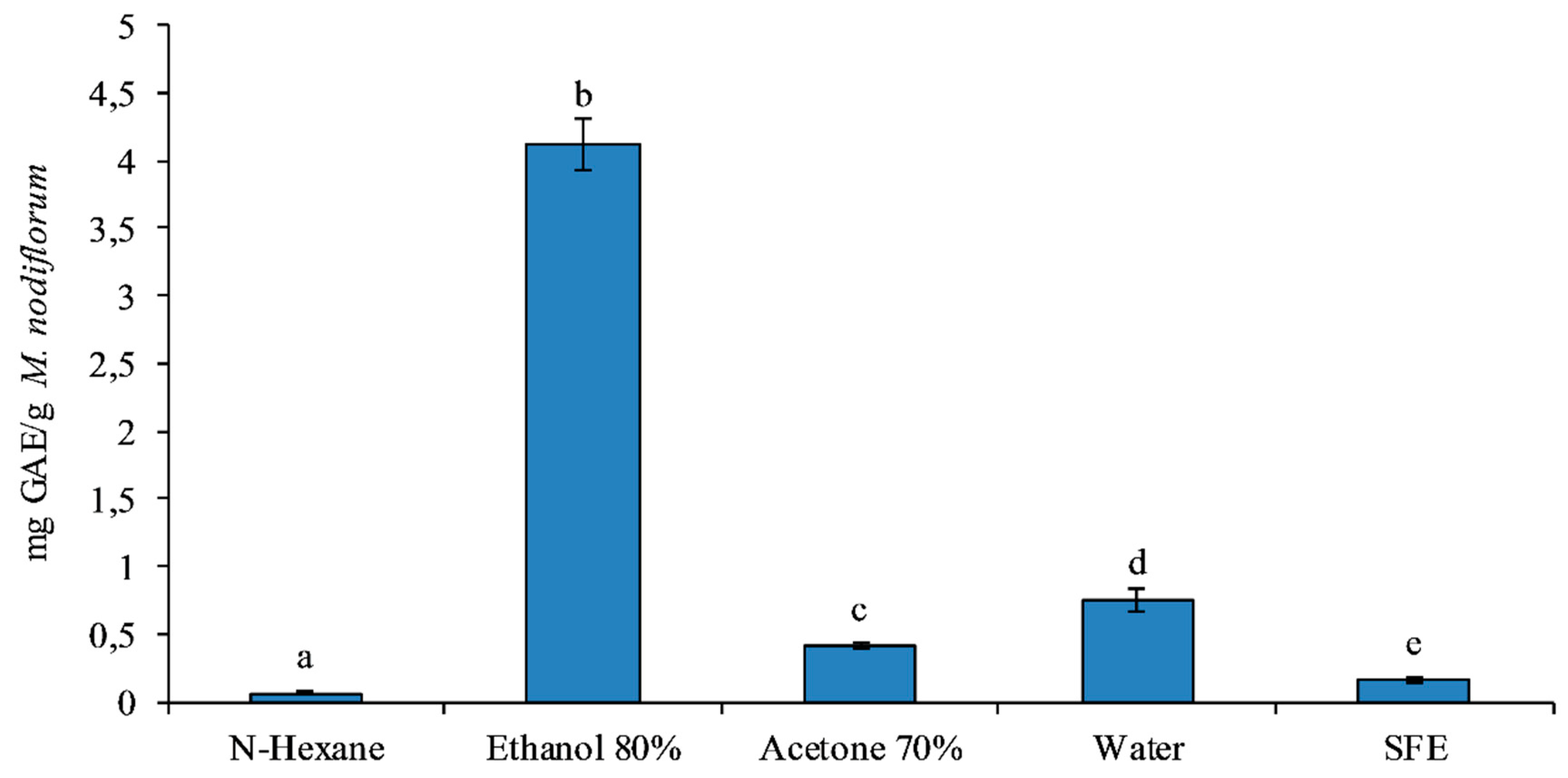
3.2. Total Phenolic Content Analysis

The Folin–Ciocalteu reagent assay is directed to assess the total phenolic content [30], measured as mg GAE equivalents per mg of raw material, extracted by each solvent. Our results showed that the highest yield (p < 0.05) of phenolics, in M. nodiflorum, was obtained by ethanol 80% (v/v), while the n-hexane gave the lowest yield (p < 0.05) (Figure 1), together with SFE, in relation to the yield (Figure 1).
Wang et al. [45] showed 70% (v/v) ethanol extracts to have a higher content of phenolics when compared to 70% (v/v) acetone extracts in wheat bran. This study found ethanol to have lower toxicity, and additionally, extracts could be recovered by reduced pressure distillation [45]. However, it was also shown that when the concentration of ethanol was increased above 80% (v/v), the total phenolic content decreased due to lipid components also being extracted [45].

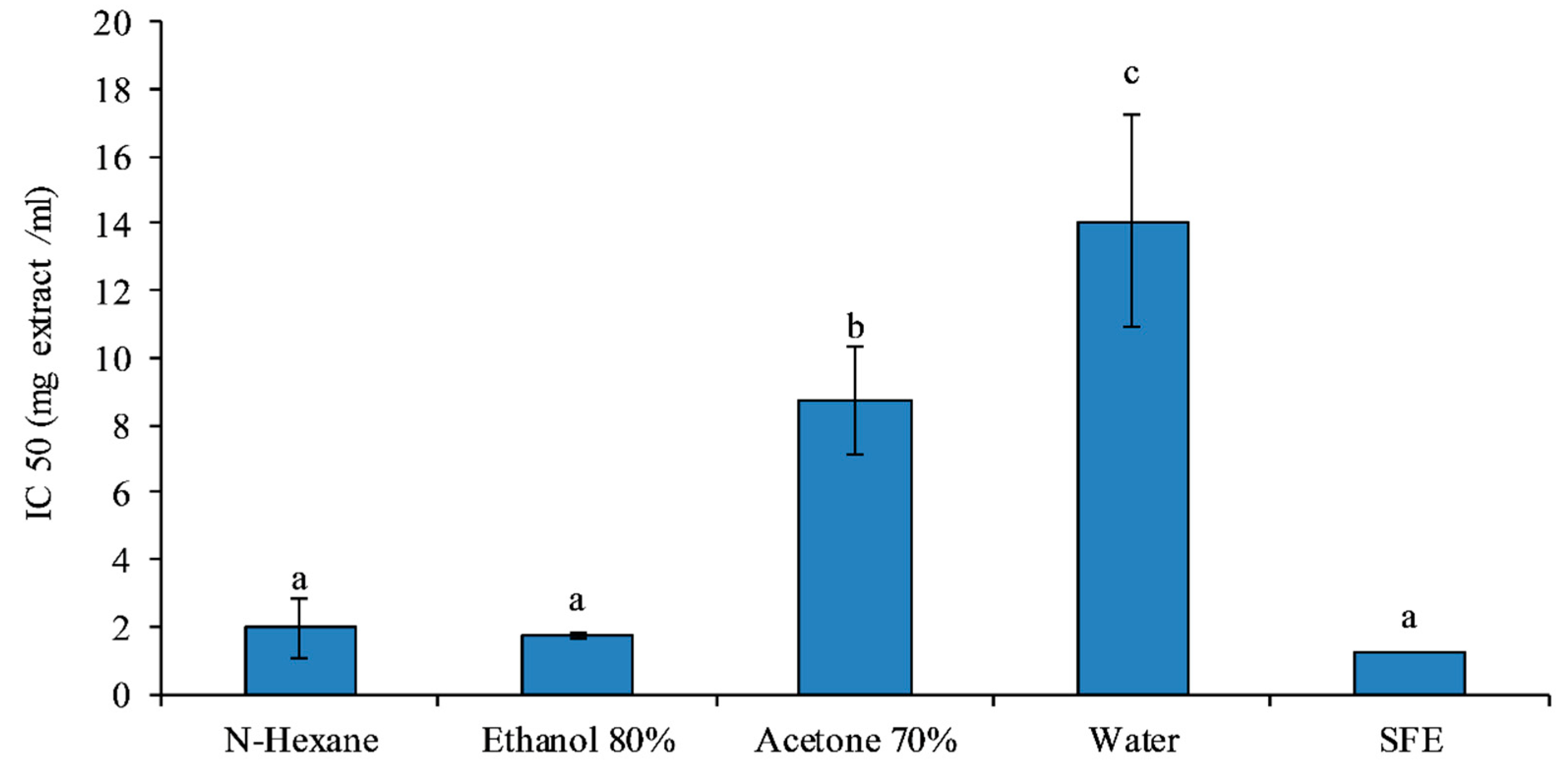
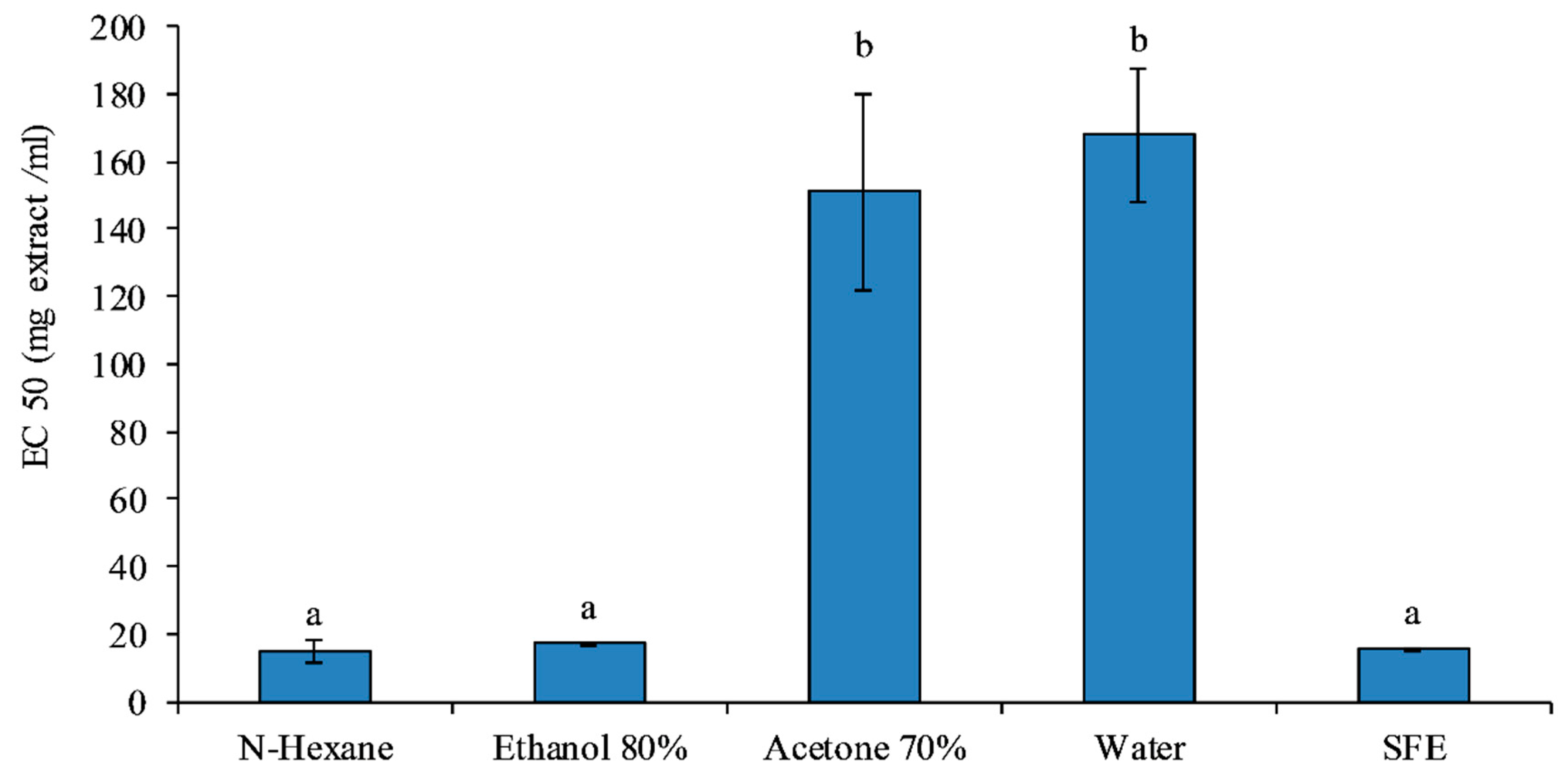
3.3. DPPH Assay Analysis

The DPPH assay measures the ability of an antioxidant to inhibit the free radicals DPPH and could be referred to as mg of raw sample, necessary to inhibit the radical. Our results, showed in Figure 2, are expressed as IC50, indicating which concentration of each extract, it is necessary to inhibit the 50% of the radical) [35]). For this reason, lower is the value, higher is the antioxidant power.
The extracts in Ethanol, N-hexane and SFE showed the highest antioxidant activity (p < 0.05), attested by the lower values of IC50 (1.73 ± 0.11, 1.97 ± 0.90 and 1.22 ± 0.03 mg/mL) (Figure 2). Similar results were obtained from Falleh et al. [10] that, in methanol extracts of M. nodiflorum, found an IC50 equal to 0.11 mg/mL [10].
We experienced a limitation in comparing our data with literature referred to this species, as both solvents and systems of extraction are different, determining variations in yield and quality of the extracts.
In the congener species, M. edule, Falleh et al. [46] showed that the stronger antiradical activity was present in the extract obtained with the most polar solvents, such as 20% and 40% aqueous alcohol. According to several studies, increasing the polarity of the solvent, with the addition of water, the antioxidant activity of the resulting extracts improved [46,47,48].
Our results (Figure 2) showed that SFE extracts have the same antioxidant capacity of ethanolic extracts. The effectiveness of the SFE technique for the extraction of bioactive antioxidant components has been evaluated in other vegetable sources such as the plant marigold [7], grape (Vitis labrusca B.) peel [49], and bee pollen [50], where a high scavenging activity was attested.
Although the SFE extract showed, among the others, some of the lower yield (p < 0.05) (Table 1), its high scavenging activity, combined with the sustainability of the technique, that avoid the use of toxic solvents, suggests the importance of this eco-friendly technique for the sustainable production of M. nodiflorum extracts.

3.4. Iron-Reducing Power Analysis

The iron-reducing power was measured as the EC50. A lower value of EC50 indicates a higher reduction power as it requires less mg of raw material to achieve a value of reducing power equal to 0.5. The ability of M. nodiflorum extracts to chelate ferrous ion was shown in Figure 3.
The showed results are in accord with the previous (Figure 2) and confirm that the extracts of M. nodiflorum, obtained with ethanol (EC50 17.10 ± 0.20), N-hexane (EC50 15.11±3.27) and SFE (EC50 15.44 ± 0.45), are stronger as antioxidants, in respect to the extract in water and acetone (p < 0.05).
It was difficult to compare these data with the literature because, as observed by Falleh et al. [10], the reducing power of Fe3+ extracts differs considerably, depending on the species of Mesembryanthemun genera [10].
Our results showed a comparable reducing activity among the SFE, ethanol and hexane extracts, respect tp water and acetone (p < 0.05) (Figure 3), suggesting to use the SFE technique to obtain ecofriendly M. nodiflorum extracts [7].

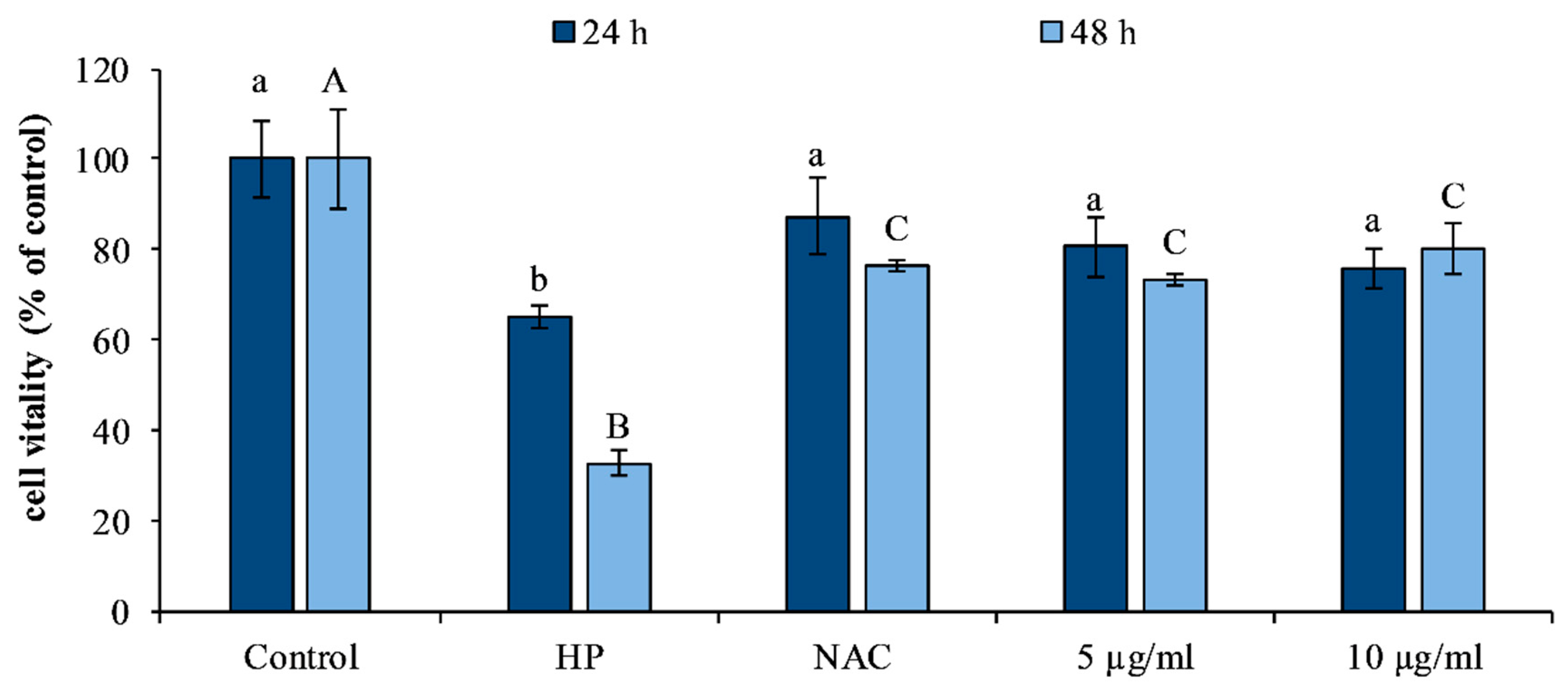
3.5. Evaluation of Cytotoxicity and Protective Effect in Fibroblast Cell Line HS-68

HS68 cell line was used as in vitro model to investigate the cytotoxic and protective effect of M. nodiflorum SFE extract. The effects of the treatments on the cells were preliminarily evaluated on a wide range of concentrations (5–50 µg/mL), through dose-dependent assays, to check the possible toxicity of the compounds. The treatment for 48 h did not induce any mortality for toxicity compared to control (data not shown).
Similar results have been obtained from Deters et al. [51] in a keratinocyte cell line (HaCaTs) treated with different doses of ice plant extract.
The protective effect of our M. nodiflorum SFE extract, against oxidative stress, was tested at two concentrations, 5 and 10 µg/mL. For the induction of oxidative stress, a standardized protocol, that uses 10 µM hydrogen peroxide (HP), to induce toxicity in this cell line [26,38,39], was used. A comparison also with the synthetic antioxidant N-acetyl cysteine (NAC) (Merck KGaA, Darmstadt, Germany) was done.
The cell vitality, determined by the MTT test, showed, as expected, a significant vitality reduction (p < 0.05) in cells exposed to the HP, compared to the control (Figure 4). On the contrary, cells pretreated with the synthetic antioxidant NAC and then exposed to HP, resulted protected against mortality, respect to the treatment with HP alone, reporting only 10% and 25% of the vitality reduction at 24 and 48 h (Figure 4). Similarly, pre-treating cells with M. nodiflorum SFE extract, before the exposure to HP, determined a significant protection against mortality, maintaining the vitality of cells compared to the control, also after exposure to the HP. These results indicate that the natural antioxidant is able to protect cells from oxidative stress, as the synthetic antioxidant.
The increased vitality of cells treated with M. nodiflorum SFE extract and subsequently subjected to stressors, can be related to the antioxidant power of the plant, attested by the DPPH radical scavenging activities [10] and reducing power. Similar results were obtained by Lin et al. [52], who demonstrated that the treatment of human dermal fibroblas, with ice plant callus extract, before UV exposure, positively affected the antioxidant activity.
Our data suggest that M. nodiflorum SFE extract given its significant antioxidant capacities protecting cells against oxidative stress-induced mortality.

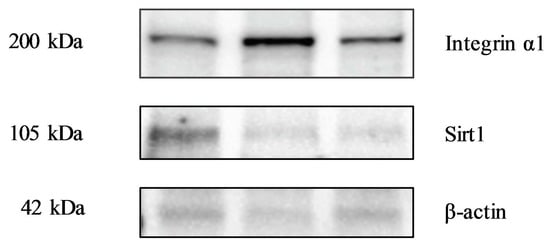
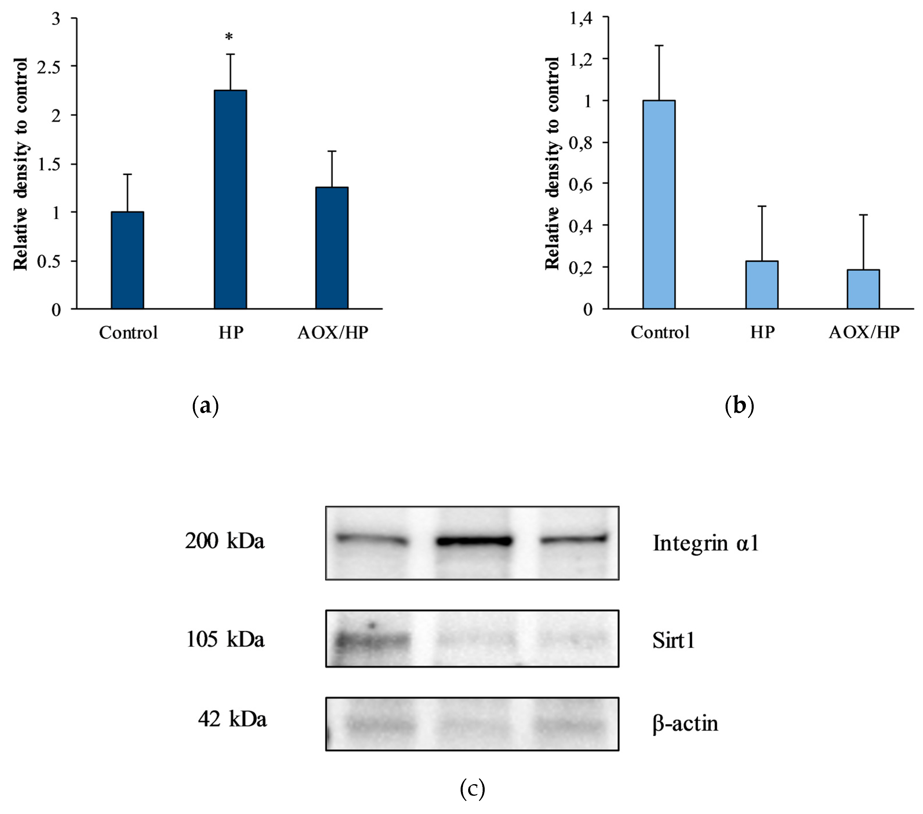
3.6. Evaluation of Biomolecular Markers by Immunoblotting

Immunoblot analysis was carried out on HS68 cells in order to evaluate the variations of two proteins related to oxidative stress, in response to the treatment with M. nodiflorum SFE extract (AOX) and hydrogen peroxide (HP). The results are shown in Figure 5.
Integrins are heterodimeric transmembrane glycoproteins that participate in different cell processes, such as cell differentiation, migration, survival and proliferation [19,53,54]. In fibroblasts, α1 is known to activate the RAS/ERK proliferative pathway and has a pro-invasive function in certain cancers and it is generally considered as a stress marker [19].
In our experiment, the protein integrin α- 1 significantly increased, respect to the control, (p < 0.05), in cells induced to oxidative stress by HP, without antioxidant protection. On the contrary, cells of the treatment AOX/HP, that were pretreated with SFE extract, prior to the induction of oxidative stress by hydrogen peroxide (HP), showed a reduction of its levels, compared to the control, highlighting the protective effect of the extract against oxidative stress.
Integrin α1β1 is involved in the process of remodeling the extracellular matrix and reduces the expression of the type I collagen gene, which is the most abundant component of connective tissue. Matrix remodeling is an important event, characterized by the deposition of new collagen, a process that is activated for normal organogenesis, wound healing, but also during the invasion of metastasis [55]. Cellular stress influences matrix remodeling, deregulating the biological pathways in which integrins are involved [56].
Other studies reported the increased expression of integrins in a stress situation, such as in mouse skin exposed to UV, that showed the expression of integrins α2, α5 and α1 [56], or in human endothelial cells of the umbilical vein, in which the expression of the α 1 subunit resulted induced by factors such as Tumor necrosis factor alpha (TNF-α) and retinoic acid [57].
Sirtuins are a family of nicotine adenine dinucleotide (NAD) dependent protein deacetylases that regulate cellular function through deacetylation of a wide range of target proteins [20] and are also involved in stress resistance, apoptosis, senescence, aging and inflammation. They regulate several physiological and pathological events, including neurodegeneration, age-related disorders, obesity, heart disease, inflammation and cancer [58].
Our results showed that, independently from the treatment with M. nodiflorum SFE extract, the protein Sirt 1 evidenced a significant decrease (p < 0.05) after the exposition to HP, respect to the control, indicating its sensitivity to situation related to oxidative stress.
In particular, sirtuin 1 has anti-inflammatory, anticancerous effects and regulates growth modulation and cellular metabolism function [59]. SIRT1 regulates inflammation mediators, and its activation inhibits the production of factors such as TNF-α, MCP-1 and IL-8. The suppression of pro-inflammatory cytokine production by SIRT1, is related to its negative regulation of NF-kB activity by deacetylation of the RelA/p65 lysine 310 subunit [59].
Based on these properties, SIRT1 could be considered as a potential pharmacological target against major aging diseases [60].
However, the precise mechanisms whereby sirtuins exert their anti-aging effects remain elusive.
Further investigations are needed to understand how sirtuins modulate the acetylation and deacetylation processes of various target proteins associated with oxidative stress.

4. Conclusions

M. nodiflorum, is a plant with a good polyphenolic content and high antioxidant activity, as observed in congeneric species. Our study highlighted the antioxidant power of the SFE extract obtained in an eco-friendly way, without the use of toxic solvents.
The quality of this extract and its beneficial effects have been confirmed at the cellular level, by the protection against cell death induced by oxidative stress and by the modulation of molecular markers, in particular the protein integrin α 1, able to keep cells protected in a situation of oxidative stress. The extract, obtained with an eco-friendly technique, given its beneficial properties, could be used for applications in the pharmaceutical, cosmeceutical and nutraceutical fields.

Author Contributions

Conceptualization, C.M.M. and A.S.; data curation, C.M.M.; formal analysis, R.A., S.M., E.C., A.M. and G.R.; investigation, C.M.M. supervision, C.M.M. and A.S.; writing—original draft, R.A., S.M., E.C.; writing—review and editing, C.M.M. and A.S. All authors have read and agreed to the published version of the manuscript.

Funding

This research received no external funding.

Acknowledgments

Edward Collins, from the School of Biological Sciences of the University of Portsmouth (United Kingdom), was awarded an Erasmus+ grant to complete an internship in the Marine Biochemistry and Ecotoxicology Laboratory of the University of Palermo.

Conflicts of Interest

The authors declare no conflict of interest.

References

  1. Sabovljevic, M.; Sabovljevic, A. Contribution to the coastal bryophytes of the Northern Mediterranean: Are there halophytes among bryophytes? Phytol. Balc. 2007, 13, 131–135. [Google Scholar]
  2. Aslam, R.; Bostan, N.; Nabgha-e-Amen; Maria, M.; Safdar, W. A critical review on halophytes: Salt tolerant plants. J. Med. Plant Res. 2011, 5, 7108–7118. [Google Scholar]
  3. Flowers, T.J.; Colmer, T.D. Salinity tolerance in halophytes. New Phytol. 2008, 179, 945–963. [Google Scholar] [CrossRef] [PubMed]
  4. Lopes, A.; Rodrigues, M.J.; Pereira, C.; Oliveira, M.; Barreira, L.; Varela, J.; Trampetti, F.; Custódio, L. Natural products from extreme marine environments: Searching for potential industrial uses within extremophile plants. Ind. Crops Prod. 2016, 94, 299–307. [Google Scholar] [CrossRef]
  5. Ksouri, R.; Megdiche, W.; Falleh, H.; Trabelsi, N.; Boulaaba, M.; Smaoui, A.; Abdelly, C. Influence of biological, environmental and technical factors on phenolic content and antioxidant activities of Tunisian halophytes. C. R. Biol. 2008, 331, 865–873. [Google Scholar] [CrossRef]
  6. Boestfleisch, C.; Wagenseil, N.B.; Buhmann, A.K.; Seal, C.E.; Wade, E.M.; Muscolo, A.; Papenbrock, J. Manipulating the antioxidant capacity of halophytes to increase their cultural and economic value through saline cultivation. AoB Plants 2014, 6, 1–16. [Google Scholar] [CrossRef]
  7. Messina, C.M.; Troia, A.; Arena, R.; Manuguerra, S.; Ioannou, T.; Curcuraci, E.; Renda, G.; Hellio, C.; Santulli, A. Species-Specific Antioxidant Power and Bioactive Properties of the Extracts Obtained from Wild Mediterranean Calendula spp. (Asteraceae). Appl. Sci. 2019, 9, 4627. [Google Scholar] [CrossRef] [Green Version]
  8. Messina, C.M.; Renda, G.; Laudicella, V.A.; Trepos, R.; Fauchon, M.; Hellio, C.; Santulli, A. From ecology to biotechnology, study of the defense strategies of algae and halophytes (from Trapani Saltworks, NW Sicily) with a focus on antioxidants and antimicrobial properties. Int. J. Mol. Sci. 2019, 20, 881. [Google Scholar] [CrossRef] [Green Version]
  9. Yang, J.; Yen, H.E. Early salt stress effects on the changes in chemical composition in leaves of ice plant and Arabidopsis. A Fourier transform infrared spectroscopy study. Plant Physiol. 2002, 130, 1032–1042. [Google Scholar] [CrossRef] [Green Version]
  10. Falleh, H.; Ksouri, R.; Oueslati, S.; Guyot, S.; Magné, C.; Abdelly, C. Interspecific variability of antioxidant activities and phenolic composition in Mesembryanthemum genus. Food Chem. Toxicol. 2009, 47, 2308–2313. [Google Scholar]
  11. Lee, S.Y.; Choi, H.D.; Yu, S.N.; Kim, S.H.; Park, S.K.; Ahn, S.C. Biological Activities of Mesembryanthemum crystallinum (Ice plant) Extract. J. Life Sci. 2015, 25, 638–645. [Google Scholar] [CrossRef] [Green Version]
  12. Falleh, H.; Ksouri, R.; Medini, F.; Guyot, S.; Abdelly, C.; Magné, C. Antioxidant activity and phenolic composition of the medicinal and edible halophyte Mesembryanthemum edule L. Ind. Crops Prod. 2011, 34, 1066–1071. [Google Scholar] [CrossRef]
  13. Soliman, M.I.; Zaghloul, M.S.; Heikal, Y.M. Genetic variation within and among three Egyptian Mesembryanthemum species using different genetic markers. Egypt. J. Basic Appl. Sci. 2014, 1, 127–135. [Google Scholar] [CrossRef] [Green Version]
  14. Mustafa, A.I.; Al-Jassir, M.S.; Nawawy, M.A.; Ahmed, S.E. Studies on samh seeds (Mesembryanthemum forsskalei Hochst) growing in Saudi Arabia: 3. Utilization of samh seeds in bakery products. Plant Foods Hum. Nutr. 1995, 48, 279–286. [Google Scholar] [CrossRef] [PubMed]
  15. El-Amier, Y.A.; Haroun, S.A.; El-Shehaby, O.A.; Al-Hadithy, O.N. Antioxidant and Antimicrobial Properties of Some Wild Aizoaceae Species Growing in Egyptian Desert. J. Environ. Sci. 2016, 45, 1–10. [Google Scholar]
  16. Doudach, L.; Meddah, B.; Benbacer, L.; Hammani, K.; El Mzibri, M.; Verité, P.; Elomri, A.; Cherrah, Y. Ethnopharmacological studies of Mesembryanthemum nodiflorum. Phytopharmacology 2013, 4, 246–258. [Google Scholar]
  17. Herrero, M.; Cifuentes, A.; Ibañez, E. Sub-and supercritical fluid extraction of functional ingredients from different natural sources: Plants, food-by-products, algae and microalgae: A review. Food Chem. 2006, 98, 136–148. [Google Scholar] [CrossRef] [Green Version]
  18. Fornari, T.; Vicente, G.; Vázquez, E.; García-Risco, M.R.; Reglero, G. Isolation of essential oil from different plants and herbs by supercritical fluid extraction. J. Chromatogr. A 2012, 1250, 34–48. [Google Scholar] [CrossRef] [Green Version]
  19. Boudjadi, S.; Beaulieu, J.F. ITGA1 (integrin, alpha 1). Atlas Genet. Cytogenet. Oncol. Haematol. 2017, 19, 86–89. [Google Scholar] [CrossRef] [Green Version]
  20. Law, I.K.M.; Liu, L.; Xu, A.; Lam, K.S.L.; Vanhoutte, P.M.; Che, C.M.; Leung, P.T.Y.; Wang, Y. Identification and characterization of proteins interacting with SIRT1 and SIRT3: Implications in the antiaging and metabolic effects of sirtuins. Proteomics 2009, 9, 2444–2456. [Google Scholar] [CrossRef]
  21. Franco, D.; Sineiro, J.; Rubilar, M.; Sánchez, M.; Jerez, M.; Pinelo, M.; Costoya, N.; Núñez, M.J. Polyphenols from plant materials: Extraction and antioxidant power. Electron. J. Environ. Agric. Food Chem. 2008, 7, 3210–3216. [Google Scholar]
  22. Trabelsi, N.; Megdiche, W.; Ksouri, R.; Falleh, H.; Oueslati, S.; Soumaya, B.; Hajlaoui, H.; Abdelly, C. Solvent effects on phenolic contents and biological activities of the halophyte Limoniastrum monopetalum leaves. LWT Food Sci. Technol. 2010, 43, 632–639. [Google Scholar] [CrossRef]
  23. Gharbi, S.; Renda, G.; La Barbera, L.; Amri, M.; Messina, C.M.; Santulli, A. Tunisian tomato by-products, as a potential source of natural bioactive compounds. Nat. Prod. Res. 2017, 31, 626–631. [Google Scholar] [CrossRef] [PubMed]
  24. Fish, W.W.; Perkins-Veazie, P.; Collins, J.K. A Quantitative Assay for Lycopene That Utilizes Reduced Volumes of Organic Solvents. J. Food Compos. Anal. 2002, 15, 309–317. [Google Scholar] [CrossRef] [Green Version]
  25. Musa, K.H.; Abdullah, A.; Jusoh, K.; Subramaniam, V. Antioxidant Activity of Pink-Flesh Guava (Psidium guajava L.): Effect of Extraction Techniques and Solvents. Food Anal. Methods 2011, 4, 100–107. [Google Scholar] [CrossRef]
  26. Manuguerra, S.; Caccamo, L.; Mancuso, M.; Arena, R.; Rappazzo, A.C.; Genovese, L.; Santulli, A.; Messina, C.M.; Maricchiolo, G. The antioxidant power of horseradish, Armoracia rusticana, underlies antimicrobial and antiradical effects, exerted In Vitro. Nat. Prod. Res. 2018, 1–4. [Google Scholar] [CrossRef]
  27. Messina, C.M.; Manuguerra, S.; Renda, G.; Santulli, A. Biotechnological Applications for the Sustainable Use of Marine By-products: In Vitro Antioxidant and Pro-apoptotic Effects of Astaxanthin Extracted with Supercritical CO2 from Parapeneus longirostris. Mar. Biotechnol. 2019, 21, 565–576. [Google Scholar] [CrossRef]
  28. Messina, C.; Renda, G.; Randazzo, M.; Laudicella, A.; Gharbi, S.; Pizzo, F.; Morghese, M.; Santulli, A. Extraction of bioactive compounds from shrimp waste. Bull. Inst. Natl. Sci. Technol. Mer Salammbô 2015, 42, 27–29. [Google Scholar]
  29. Anaëlle, T.; Serrano Leon, E.; Laurent, V.; Elena, I.; Mendiola, J.A.; Stéphane, C.; Nelly, K.; Stéphane, L.B.; Luc, M.; Valérie, S.P. Green improved processes to extract bioactive phenolic compounds from brown macroalgae using Sargassum muticum as model. Talanta 2013, 104, 44–52. [Google Scholar] [CrossRef]
  30. Singleton, V.L.; Rossi, J.A. Colorimetry of Total Phenolics with Phosphomolybdic-Phosphotungstic Acid Reagents. Am. J. Enol. Vitic. 1965, 16, 144–158. [Google Scholar]
  31. Krishnaiah, D.; Sarbatly, R.; Nithyanandam, R. A review of the antioxidant potential of medicinal plant species. Food Bioprod. Process. 2011, 89, 217–233. [Google Scholar] [CrossRef]
  32. Oki, T.; Masuda, M.; Furuta, S.; Nishiba, Y.; Terahara, N.; Suda, I. Involvement of anthocyanins and other phenolic compounds in radical-scavenging activity of purple-fleshed sweet potato cultivars. J. Food Sci. 2002, 67, 1752–1756. [Google Scholar] [CrossRef]
  33. Kedare, S.B.; Singh, R.P. Genesis and development of DPPH method of antioxidant assay. J. Food Sci. Technol. 2011, 48, 412–422. [Google Scholar] [CrossRef] [PubMed] [Green Version]
  34. Bernatoniene, J.; Masteikova, R.; Davalgiene, J.; Peciura, R.; Gauryliene, R.; Bernatoniene, R.; Majiene, D.; Lazauskas, R.; Civinskiene, G.; Velziene, S.; et al. Topical application of Calendula officinalis (L.): Formulation and evaluation of hydrophilic cream with antioxidant activity. J. Med. Plants Res. 2011, 5, 868–877. [Google Scholar]
  35. Yeddes, N.; Chérif, J.; Guyot, S.; Sotin, H.; Ayadi, M. Comparative Study of Antioxidant Power, Polyphenols, Flavonoids and Betacyanins of the Peel and Pulp of Three Tunisian Opuntia Forms. Antioxidants 2013, 2, 37–51. [Google Scholar] [CrossRef] [Green Version]
  36. Oyaizu, M. Studies on products of browning reactions: Antioxidative activities of products of browning reaction prepared from Glucosamine. Jpn. J. Nutr. 1986, 44, 307–315. [Google Scholar] [CrossRef] [Green Version]
  37. Mosmann, T. Rapid colorimetric assay for cellular growth and survival: Application to proliferation and cytotoxicity assays. J. Immunol. Methods 1983, 65, 55–63. [Google Scholar] [CrossRef]
  38. Messina, C.M.; Pizzo, F.; Santulli, A.; Bušelić, I.; Boban, M.; Orhanović, S.; Mladineo, I. Anisakis pegreffii (Nematoda: Anisakidae) products modulate oxidative stress and apoptosis-related biomarkers in human cell lines. Parasit. Vectors 2016, 9, 1–10. [Google Scholar] [CrossRef] [Green Version]
  39. Abbes, M.; Baati, H.; Guermazi, S.; Messina, C.; Santulli, A.; Gharsallah, N.; Ammar, E. Biological properties of carotenoids extracted from Halobacterium halobium isolated from a Tunisian solar saltern. BMC Complement. Altern. Med. 2013, 13, 1–8. [Google Scholar] [CrossRef] [Green Version]
  40. Taherian, A.; Li, X.; Liu, Y.; Haas, T.A. Differences in integrin expression and signaling within human breast cancer cells. BMC Cancer 2011, 11, 1–15. [Google Scholar] [CrossRef] [Green Version]
  41. Lowry, O.H.; Rosebrough, N.J.; Farr, A.L.; Randall, R.J. Protein measurement with the Folin phenol reagent. J. Biol. Chem. 1951, 193, 265–275. [Google Scholar] [PubMed]
  42. Manuguerra, S.; Ruiz, C.E.; Santulli, A.; Messina, C.M. Sub-lethal doses of polybrominated diphenyl ethers, In Vitro, promote oxidative stress and modulate molecular markers related to cell cycle, antioxidant balance and cellular energy management. Int. J. Environ. Res. Public Health 2019, 16, 588. [Google Scholar] [CrossRef] [PubMed] [Green Version]
  43. Kalpna, R.; Mital, K.; Sumitra, C. Vegetable and fruit peels as a novel source of antioxidants. J. Med. Plants Res. 2011, 5, 63–71. [Google Scholar]
  44. Shimada, K.; Fujikawa, K.; Yahara, K.; Nakamura, T. Antioxidative Properties of Xanthan on the Autoxidation of Soybean Oil in Cyclodextrin Emulsion. J. Agric. Food Chem. 1992, 40, 945–948. [Google Scholar] [CrossRef]
  45. Wang, J.; Sun, B.; Cao, Y.; Tian, Y.; Li, X. Optimisation of ultrasound-assisted extraction of phenolic compounds from wheat bran. Food Chem. 2008, 106, 804–810. [Google Scholar] [CrossRef]
  46. Falleh, H.; Oueslati, S.; Guyot, S.; Dali, A.B.; Magné, C.; Abdelly, C.; Ksouri, R. LC/ESI-MS/MS characterisation of procyanidins and propelargonidins responsible for the strong antioxidant activity of the edible halophyte Mesembryanthemum edule L. Food Chem. 2011, 127, 1732–1738. [Google Scholar] [CrossRef]
  47. Zhao, G.R.; Xiang, Z.J.; Ye, T.X.; Yuan, Y.J.; Guo, Z.X. Antioxidant activities of Salvia miltiorrhiza and Panax notoginseng. Food Chem. 2006, 99, 767–774. [Google Scholar] [CrossRef]
  48. Zhou, K.; Yu, L. Effects of extraction solvent on wheat bran antioxidant activity estimation. LWT Food Sci. Technol. 2004, 37, 717–721. [Google Scholar] [CrossRef]
  49. Ghafoor, K.; Park, J.; Choi, Y.H. Optimization of supercritical fluid extraction of bioactive compounds from grape (Vitis labrusca B.) peel by using response surface methodology. Innov. Food Sci. Emerg. Technol. 2010, 11, 485–490. [Google Scholar] [CrossRef]
  50. Messina, C.M.; Panettieri, V.; Arena, R.; Renda, G.; Espinosa Ruiz, C.; Morghese, M.; Piccolo, G.; Santulli, A.; Bovera, F. The Inclusion of a Supercritical Fluid Extract, Obtained From Honey Bee Pollen, in the Diet of Gilthead Sea Bream (Sparus aurata), Improves Fish Immune Response by Enhancing Anti-oxidant, and Anti-bacterial Activities. Front. Vet. Sci. 2020, 7, 95. [Google Scholar] [CrossRef] [PubMed] [Green Version]
  51. Deters, A.M.; Meyer, U.; Stintzing, F.C. Time-dependent bioactivity of preparations from cactus pear (Opuntia ficus indica) and ice plant (Mesembryanthemum crystallinum) on human skin fibroblasts and keratinocytes. J. Ethnopharmacol. 2012, 142, 438–444. [Google Scholar] [CrossRef] [PubMed]
  52. Lin, Y.H.; Lin, Y.K.; Lin, Y.H. Photoprotective Effects of Ice Plant (Mesembryanthemum crystallinum) Callus Extract on Gene Expression of Human Dermal Fibroblast Against UV Exposure. J. Biobased Mater. Bioenerg. 2019, 13, 570–575. [Google Scholar] [CrossRef]
  53. Giancotti, F.G. Complexity and specificity of integrin signalling. Nat. Cell Biol. 2000, 2, E13–E14. [Google Scholar] [CrossRef]
  54. Beauséjour, M.; Noël, D.; Thibodeau, S.; Bouchard, V.; Harnois, C.; Beaulieu, J.F.; Demers, M.J.; Vachon, P.H. Integrin/Fak/Src-mediated regulation of cell survival and anoikis in human intestinal epithelial crypt cells: Selective engagement and roles of PI3-K isoform complexes. Apoptosis 2012, 17, 566–578. [Google Scholar] [CrossRef] [PubMed] [Green Version]
  55. Langholz, O.; Röckel, D.; Mauch, C.; Kozlowska, E.; Bank, I.; Krieg, T.; Eckes, B. Collagen and collagenase gene expression in three-dimensional collagen lattices are differentially regulated by α1β1 and α2β1 integrins. J. Cell Biol. 1995, 131, 1903–1915. [Google Scholar] [CrossRef] [PubMed]
  56. Jean, C.; Gravelle, P.; Fournie, J.J.; Laurent, G. Influence of stress on extracellular matrix and integrin biology. Oncogene 2011, 30, 2697–2706. [Google Scholar] [CrossRef] [Green Version]
  57. Defilippi, P.; Van Hinsbergh, V.; Bertolotto, A.; Rossino, P.; Silengo, L.; Tarone, G. Differential distribution and modulation of expression of Alpha1/beta1 integrin on human endothelial cells. J. Cell Biol. 1991, 114, 855–863. [Google Scholar] [CrossRef] [Green Version]
  58. Lin, Z.; Fang, D. The Roles of SIRT1 in Cancer. Genes Cancer 2013, 4, 97–104. [Google Scholar] [CrossRef] [PubMed] [Green Version]
  59. Zhu, X.; Liu, Q.; Wang, M.; Liang, M.; Yang, X.; Xu, X.; Zou, H.; Qiu, J. Activation of Sirt1 by resveratrol inhibits TNF-α induced inflammation in Fibroblasts. PLoS ONE 2011, 6, e27081. [Google Scholar] [CrossRef] [PubMed]
  60. Huang, J.; Gan, Q.; Han, L.; Li, J.; Zhang, H.; Sun, Y.; Zhang, Z.; Tong, T. SIRT1 overexpression antagonizes cellular senescence with activated ERK/S6k1 signaling in human diploid fibroblasts. PLoS ONE 2008, 3, e1710. [Google Scholar] [CrossRef]
Figure 1. Total phenolic content (mg GAE/g) in different extracts of dried leaves of M. nodiflorum. Lowercase letters (a–e) indicate significant differences between different samples (p < 0.05)
Applsci 10 02374 g001
Figure 2. The 1,1-Diphenyl-2-picrylhydrazyl (DPPH) radical scavenging activity (IC 50, mg/mL) in different extracts of dried leaves of M. nodiflorum. Lowercase letters (a–c) indicate significant differences between different solvents (p < 0.05).
Applsci 10 02374 g002
Figure 3. Reducing power (EC50, mg/mL) in different extracts of dried leaves of M. nodiflorum. Lowercase letters (a–b) indicate significant differences between different solvents (p < 0.05).
Applsci 10 02374 g003
Figure 4. Effect of M. nodiflorum supercritical fluid extract (SFE) on the vitality of fibroblast cells exposed to oxidative stress, induced by hydrogen peroxide (HP). Control: cells maintained in standard culture conditions; HP: cells exposed to hydrogen peroxide (10 µM); NAC: cells treated with N-acetyl cystein (5mM) and subsequently exposed to HP (10 µM); 5 and 10 µg/mL: cells treated with 5 and 10 µg/mL of M. nodiflorum SFE and then exposed to HP (10 µM). Lowercase letters (a–b) indicate superscript letters indicate a significant difference compared to the control at 24 h. Capital letters (A–C) indicate significant differences compared to the control at 48 h (p < 0.05).
Applsci 10 02374 g004
Figure 5. Results of the immunoblot of Integrin α1 and Sirt 1 in HS68 cells. Control: cells maintained in standard culture conditions; HP: cells exposed to hydrogen peroxide (10 µM); AOX/HP: cells treated with 10 µg/mL of M. nodiflorum SFE and then exposed to HP (10 µM). β actin was used as internal control. The immunoblot image, in (c), is one representative of the three separate experiments. The related protein quantifications are represented in the graphics (* p < 0.05 vs. control): (a) relative density of Integrin α1 protein; (b) relative density of Sirt 1 protein.
Applsci 10 02374 g005
Table 1. Extract yield (g/100g) obtained, with different solvents, from dried leaves of M. nodiflorum.
Solvent Yield (g/100)
N-Hexane0.43 ± 0.28 a
Ethanol18.39 ± 1.69 b
Acetone12.30 ± 2.45 c
Water33.32 ± 8.37 d
SFE0.79 ± 0.27 a
Lowercase letters (a–d) indicate significant differences between different solvents (p < 0.05).

