Article

Preliminary Study of the Use of Root with Sedline® EEG Monitoring for Assessment of Anesthesia Depth in 6 Horses

1
Department of Large Animals Diseases with Clinic, Faculty of Veterinary Medicine, Warsaw University of Life Sciences, Nowoursynowska 100, 02-797 Warsaw, Poland
2
National Geriatrics, Rheumatology and Rehabilitation Institute, Spartańska 1, 02-637 Warszawa, Poland
3
Emeritus Professor of Anesthesiology and Pain Management, Department of Clinical Sciences, College of Veterinary Medicine, Cornell University, Ithaca, NY 14850, USA
*
Author to whom correspondence should be addressed.
Appl. Sci. 2020, 10(3), 1050; https://doi.org/10.3390/app10031050
Submission received: 2 January 2020 / Revised: 30 January 2020 / Accepted: 3 February 2020 / Published: 5 February 2020

Abstract

:

Featured Application

The use of EEG during equine anesthesia can be a new complementary method for clinical use in order to achieve more precise anesthesia depth assessment due to brain awareness measurement, visible in real time during surgery. Can it be used to assure the desired management of potential pain from surgical stimulation? Purpose of the study: The purpose of this study is to determine whether the Root with Sedline EEG monitor could be adapted to use during equine anesthesia and surgery for commonly diagnosed conditions. This was indicated prior to in-depth studies comparing this technology to other parameters currently used during various anesthetic protocols in both minor- and major-risk surgeries.

Abstract

Management of equine anesthesia monitoring is still a challenge. Careful monitoring to provide guidelines for anesthesia depth assessment currently relies upon eye signs, cardiopulmonary responses, and the level of muscle relaxation. Electroencephalography, as a non-invasive brain activity monitor, may be used to complement the routinely monitored physiologic parameters. Six horses, undergoing various surgical procedures and anesthesia protocols, were monitored with the use of a Root with Sedline EEG monitor and a routine monitor of life parameters. The life parameters were compared to the changes on the EEG density spectral array observed live during anesthesia. During all procedures the level of awareness was monitored using the EEG, with higher frequency and power of waves indicating a higher level of awareness. It was evident from this that there were variations according to the type of procedure and the anesthetic protocol. Cerebral activity was elevated during painful moments of the surgery and recovery, requiring adjustments in anesthetic concentrations. Evaluation of changes in the spectral edge frequency (SEF) could show the periods when the patient is stabilized. EEG monitoring has the potential to be used in clinical anesthesiology of horses. It was shown that this system may be used in horses under general anesthesia but is currently less effective in a standing horse for diagnostic or minor procedures.

1. Introduction

Assessment of adequate depth of anesthesia in equine patients during general anesthesia protocols, especially with the use of balanced anesthesia that combines inhalation and intravenous drugs, is still a challenge. The lack of neurologic-based research causes anesthetists to base their decisions regarding the quality of anesthesia upon hemodynamics, respiratory, and other parameters (e.g., muscle tone, eye reflexes).
Electroencephalography is known as a non-invasive technique of measuring the electrical potential and, therefore, the awareness of the Central Neurological System [1,2]. In horses, it has been used primarily in studies concerning sleep and neurological disorders [3,4,5]. Recently, it has been presumed to be possibly the most objective method for anesthesia depth assessment in medicine [6,7] and in veterinary medicine [8,9,10]. To date, there have been several studies performed to investigate this potential in horses [11,12,13,14,15]. These, however, did not investigate intraoperative analysis of the raw EEG wave. The only algorithms (mathematical analysis of EEG that transformed the raw EEG into a unified number) available, which could work during anesthesia, were adapted from human medicine, and were subsequently confirmed in research as not applicable for horses [16,17,18]. Possibly, the most interesting for the equine practitioner might be the recording of the spectral edge frequency of EEG waves for the left and right hemisphere. Quite promising are also the results of studies recording the DSA (density spectral area), which show the changes in the power of EEG waves in different frequency bands (delta (δ) 0.1-4 Hz, theta (θ) 4-8 Hz, alpha (α) 8-12 Hz, beta (β) 12–25 Hz, gamma (λ) >30 Hz) with colorful diagrams. The presence of waves of a certain frequency reflects the awareness; the main rule describes the lowest awareness with a presence of low-frequency waves [13,19,20].
The aim of this pilot study was to investigate the method of EEG measurements in horses, starting from electrode mounting, checking device data collection in potential clinical conditions (including different positions of a horse during surgery, different anesthesia protocols, and different types of surgeries). Comparisons were also made by using various electrode placement locations and during subsequent anesthesia phases—sedation, maintenance, recovery. This would be used as the basics for further studies including methodical statistical analysis and comparison of these measurements to monitoring techniques commonly used in clinical practice.
The study was approved by the Local Ethical Committee.

2. Materials and Methods

Six horses were used for the study—an 11-month-old Arabian mare (A), a 9-year-old half-blood gelding (B), a 3-year-old half-blood stallion (C), a 3-year-old English blood stallion (D), a 7-year-old half-blood gelding (E), and a 12-year-old half-blood gelding (F). The types of procedures, positions of the horses, and types of anesthesia protocol, including drugs and their relevant doses, are shown in Table 1.
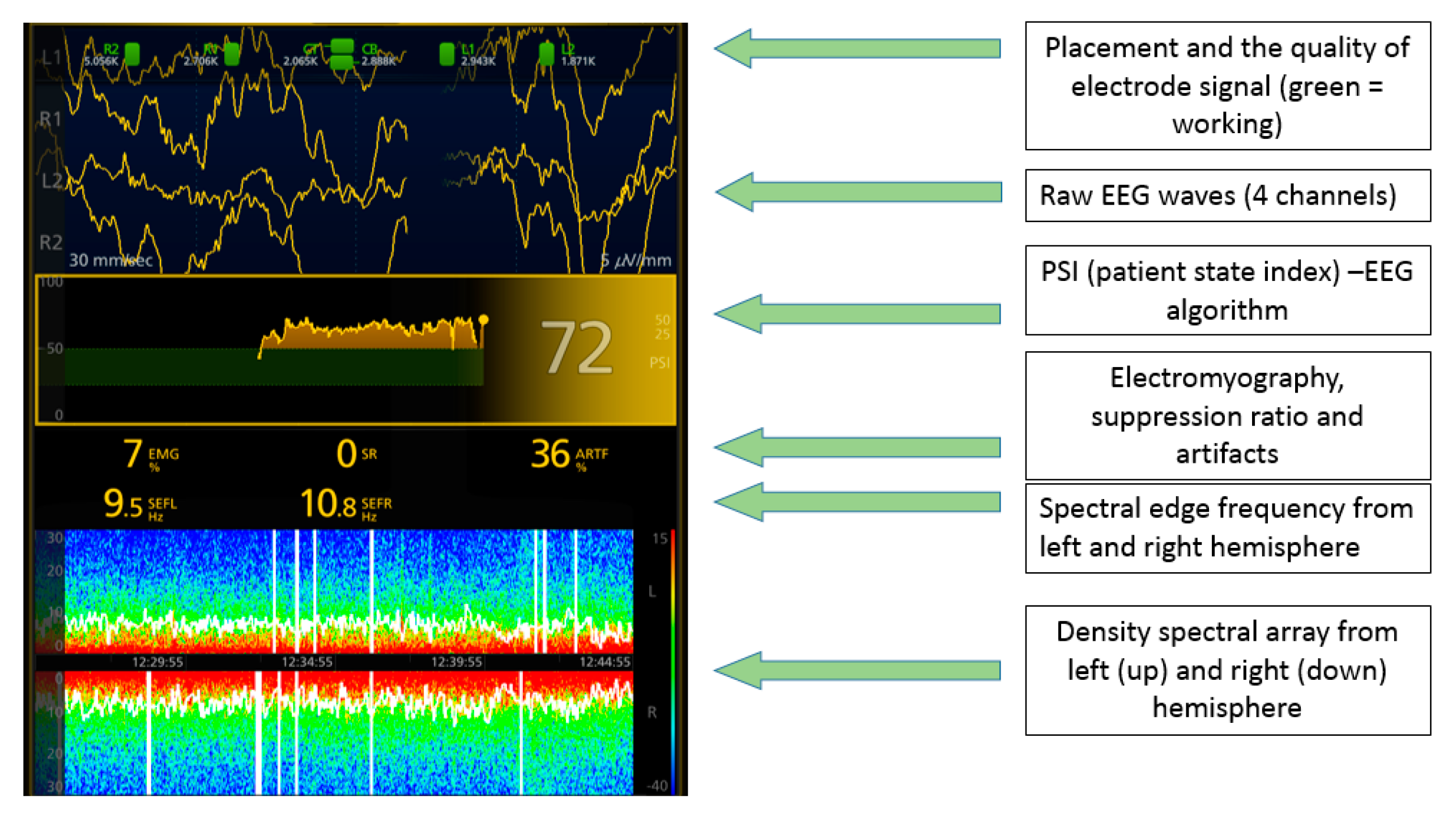
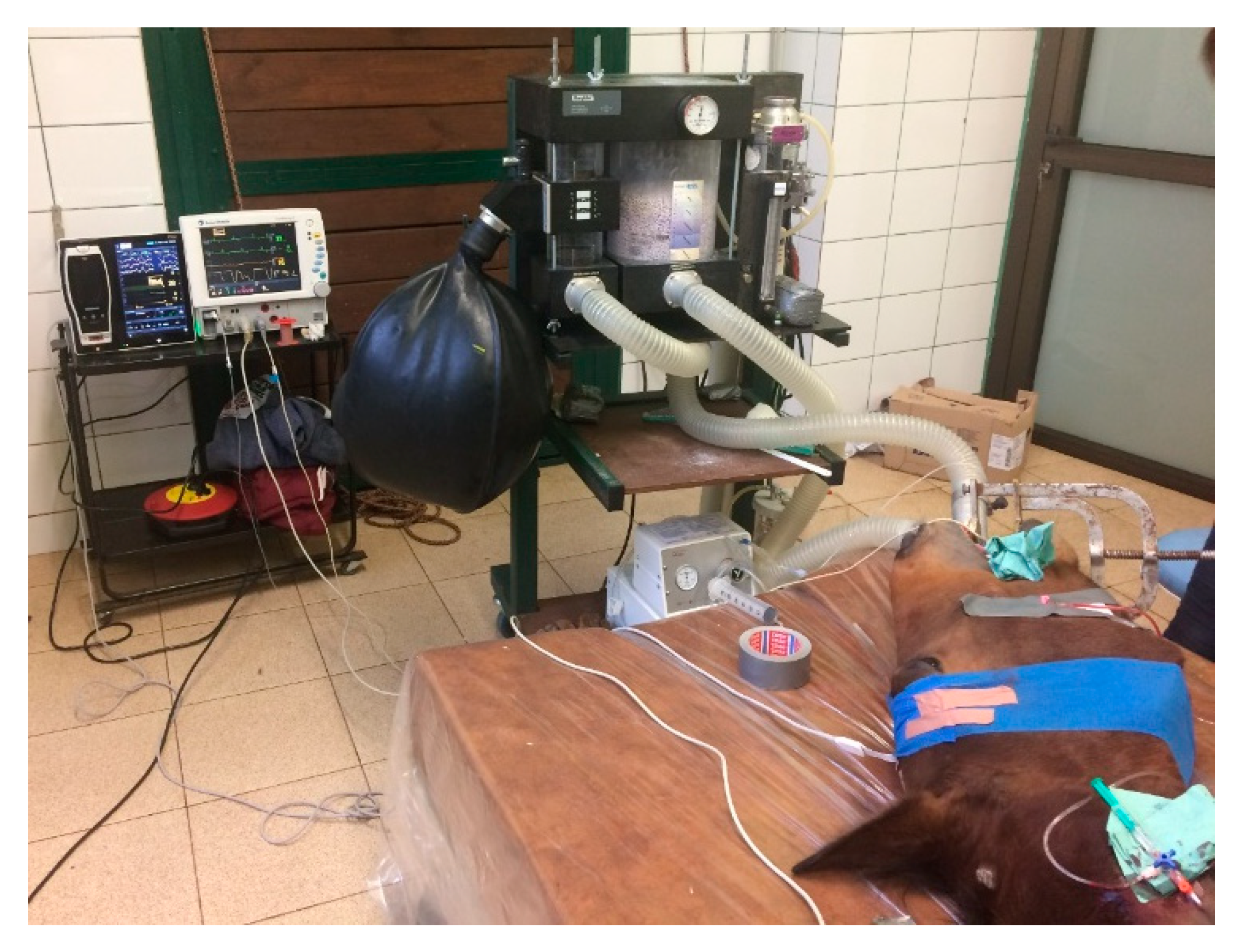
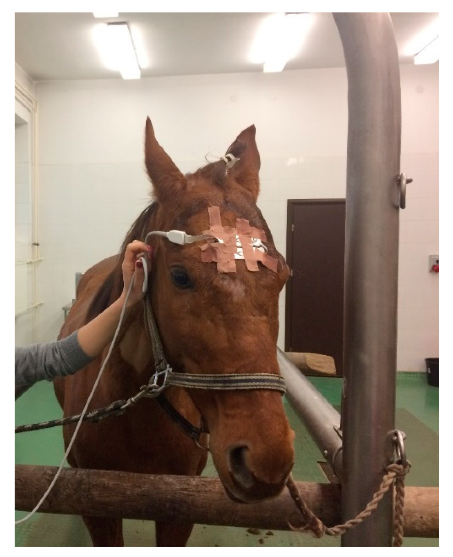
In each case, electrodes were placed on the frontal part of the horse skull (according to Giovagnoli 1996) with the use of gel and tape—two left (L1 and L2), two right (R1, R2), one ground electrode (GB) and one reference electrode (CT) in the center position. The placement area was not shaved. Once the electrodes were in place, the recording device (Root with Sedline®, Masimo, Irvine CA, USA) was attached and the measurements commenced (Figure 1). On the monitor, the raw EEG waves from 4 channels (L1, L2, R1, R2), spectral edge frequency (SEF) of the left and right hemisphere, EMG (electromyography) of the muscles that could influence the measurements, Patient State Index (mathematical algorithm counted from raw EEG), burst suppression ratio (SR), and density spectral area (showing the power of waves in certain frequency bands) could be seen. The impedance was also measured, and when it exceeded the marginal acceptable level and interfered with the EEG measurement (indicated by the electrode color on the screen changing from green to yellow), the electrodes were pressed slightly and additional gel was applied between the electrode and the skin surface. For each procedure, a life parameter monitor (Datex Ohmeda Cardiocap 5 or Veterinarian Patient Monitor Vs2000v) was attached for the measurement of pulse oximetry, ECG, capnography, breaths per minute count, and, in 3 cases, end tidal isoflurane and invasive blood pressure (Figure 2). These measurements were conducted during sedation (Case D), maintenance of anesthesia, and recovery (Cases A, B, C, E, and F). The results were recorded as numbers for hemodynamic parameters on the anesthesia record chart and compared in the time of recording during anesthesia with changes visible on the DSA. This was to mimic the situation when the practitioner would monitor the patient during anesthesia and, at the same time, investigate the possibility of occurrence of any visible changes in response to inadequate anesthetic/analgesic administration (seen as changes in life parameters) during the surgical procedure.
During all procedures, visual readings of the DSA were made, including the hue of the color that indicated the power of the wave (red color—high power, blue color—low power) and the role of the color on the scale—frequency. The scale was always visible on the monitoring screen. The DSA was shown as two horizontal stripes, representing the two hemispheres (Figure 3).

3. Results

During all procedures it was clearly demonstrated that the levels of awareness, as illustrated by the high frequency and power of the waves shown by the EEG, were different according to the anesthetic protocol and the surgical procedure.
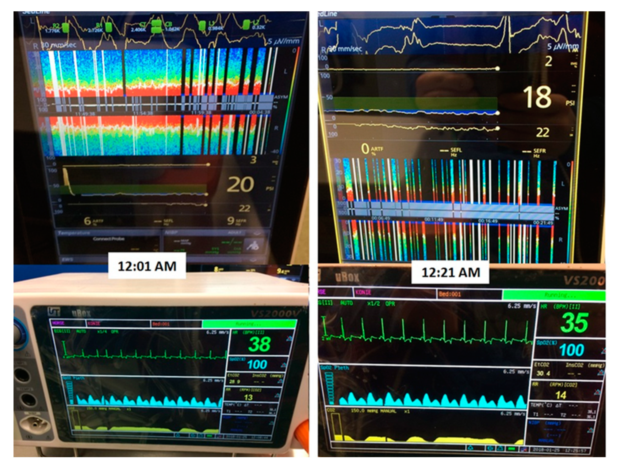
In the young patient brought to the CT (Horse A), the hemodynamic parameters were stable and within the normal range throughout the entire procedure. This underlined the changes visible on the DSA—while the SEF was quite stable, with time, there was a gradual rise in the power of the low-frequency (<3 Hz) waves. Apart from that, low power of higher-frequency waves (5–15 Hz) could be seen, with the power gradually dropping with elevating frequency. This was correlated with the high level of isoflurane set on the vaporizer (3%). Eventually, the strong and frequent burst suppression (flattening of the EEG wave) could be seen as black stripes within the DSA (and on the raw EEG) that correlated with maintaining a high isoflurane level. During this change, of all related parameters, only the heart rate dropped slightly (Figure 4).
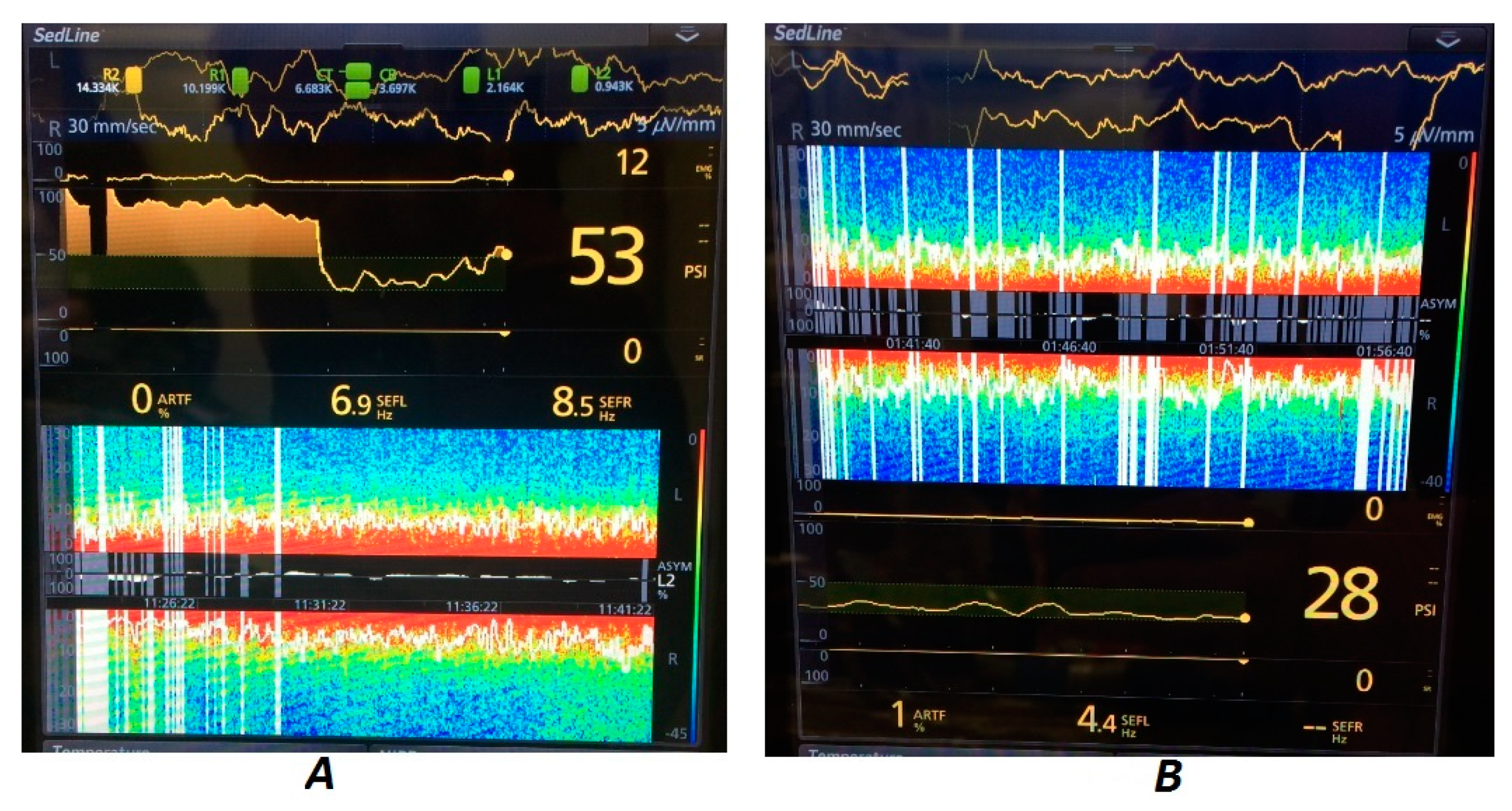
During colic surgery (Horse B), which lasted 3 h, there was extensive variability in the frequency and power of the EEG waves recorded. The horse was in a critical clinical condition before the surgery and its hemodynamic parameters changed significantly during the maintenance of anesthesia. The DSA was recorded without artifacts. In-tidal isoflurane concentrations and supplemental medications were adjusted as needed to maintain cardiopulmonary function and assure desirable pain management. Significant jumps in the SEF and power of higher-frequency waves could be seen during more painful moments—skin cut, enterotomy, and skin suturing at the end. Also, periods of high power of high-frequency waves could be seen (Figure 5).
In the case of castration (Horse C), the SEF was also variable but the waves of significant (for overall arousal) power were present only within short frequencies. There were no periods of higher-frequency waves emerging during the whole procedure. The hemodynamic parameters were stable. The horse was kept in a light plane of anesthesia. There were sporadic artifacts visible. During the whole procedure, the appearing waves were stable in power within their frequency bands (Figure 5).
In standing surgery (Horse D), the DSA was not demonstrated clearly enough to provide adequate interpretation as there was a large impedance between electrodes that caused the occurrence of the artifacts due to movement of the head, which seems to be a significant drawback in EEG measurements. The total power and amplitude of the wave was high, which reflected high awareness (Figure 6).
During anesthesia for arthroscopy (Horse E), the DSA was visible without any artifacts. The SEF was stable and there were minor changes during maintenance. The power of the waves was greatest at low frequencies and was stable during the entire procedure. The hemodynamic parameters were also stable. The recovery showed a gradual return of increased brain activity by elevation of the power of the higher-frequency waves with spectral edge frequency growth in the same time period (Figure 7).
In the case of total intravenous anesthesia (Horse F), the changes in the SEF were the most significant, particularly during the induction and recovery phases. The frequency of the waves was low, and the most commonly observed waves were very low frequency and in the delta frequency band. Of all procedures, in this horse the power in the frequency band was the most stable. There were no problems with recording the density spectral array, nor were there artifacts interfering with the readings. Anesthetic concentrations were adjusted as indicated (Figure 8).
The most important findings of DSA changes during all procedures are shown in Table 2.

4. Discussion

These preliminary studies, conducted on six different patients, showed that EEG monitoring with the use of surface electrodes in the Root with Sedline® system may be used in equine patients. The electrodes can be attached without shaving the skin and the presence of coating do not disturb the signal transmission. The glue on the electrodes might not be enough to maintain good contact, especially since the horse is able to sweat over the whole of the skin surface; thus, additional tape or bandaging is necessary for mounting. However, the measurements on DSA divided into two hemispheres do not provide as much information as desired, since it is difficult to attach the electrodes in perfect symmetrical placement.
DSA measurement might be difficult to obtain without general anesthesia in horses due to skin and muscle movement, which interferes with signal transmission and electrode mounting. The artifacts were disturbing especially during standing surgeries as the head was not still. However, during recoveries it was possible to observe the DSA up to the moment the horse started to stand, which might make this system useful for recovery predictions. This was also shown for TIVA anesthesia in Horse F—a situation where monitoring is normally limited and recovery prediction, especially in the field, is highly desirable for practitioners. The matter of successful recovery is one of the most difficult, particularly after long anesthesia during colics.
Observations of DSA revealed changes connected with procedures, possible pain, and the depth of anesthesia. In Horse A, the occurrence of burst suppression, independent of hemodynamic parameters, revealed too deep a level of anesthesia. It is worth mentioning that in the case of CT the horse did not receive any nociceptive stimuli. The variability of SEF in Horse B is an indicator of unstable anesthesia and possible difficulties that might occur during further anesthesia, which is understandable in a case of colic surgery in a horse in critical clinical condition. Also, the occurrence of higher power in higher-frequency bands can indicate periodical arousal due to surgical stimulation and/or inappropriate depth and analgesia level. There may be major changes in cardiopulmonary parameters occurring during various stages of the colic symptoms and adjustments during surgery. These anticipated levels of visceral pain create major challenges to maintaining the desired level of anesthesia and analgesia. Horses C and E are good examples of routine procedures in horses with good physical condition, and they showed a stable SEF matched with stable hemodynamic parameters. The level of anesthesia can be assessed in such cases by changes in the power of low- and medium-frequency waves. A procedure on a horse in a good clinical condition is described by the occurrence of stable frequency waves, which is the target of anesthesia depth assessment. Case D demonstrated the problem with standing surgery measurements due to head movement, but this problem might be better solved by head immobilization.
During field anesthesia, such as the case of Horse F, DSA might be especially helpful as in such cases there are limited possibilities of anesthesia monitoring compared to hospital conditions. Some modification of current monitors might be helpful. A comparison of Horse F and the other horses anesthetized with inhalant agent shows significant differences in the influence of the drugs on the Central Nervous System. Inhalants revealed the occurrence of various frequency waves, while total intravenous anesthesia seems to stabilize the waves on low and medium frequencies without significant changes. This also shows the complicated route of action of inhalant anesthetics, which has not been described until now.
The vital signs monitored and recorded were not as responsive as the EEG changes during the surgical procedures. Heart rate changes were more likely to respond sooner to potential surgical pain than blood pressure. Neither eye movement/position nor muscle relaxation were indicative of pain during the levels of surgical anesthesia maintained in these horses.
Comparing the changes between the phases of anesthesia can be a problem. The surface electrodes can be mounted before sedation in some horses, although during sedation there is not as much need for EEG monitoring as after induction of anesthesia. However, there is a need among the practitioners who perform standing surgery to be sure about the level of anesthesia in those procedures, so possibly the methodology of measurements in those cases should be improved. These kinds of surgeries are more and more widely used due to lower anesthetic risk.
The recording of EEG during induction is still a challenge as the movement of the horse from the standing to the recumbent position interferes with measurements, which likely results in artifacts. However, from the observations, during inductions, the frequency of the waves drops, which is understandable. The most important readings are recorded during anesthetic maintenance, which can show significant changes in the wave frequencies, thus indicating inappropriate levels of anesthetic during surgery.
There are some drawbacks to the method that was used in this research. There is still a need for better software for the EEG DSA machine, as the DSA can be saved only as a picture screenshot from the device during anesthesia but cannot be transferred to the computer and then mathematically analyzed. That is the reason that the study was based upon observations, rather than statistics. Moreover, making observations of the DSA and understanding the changes requires practice and the establishment of the relation to other parameters.
From a physiological point of view there are different expected responses to medications selected in balanced anesthesia and analgesia. This was demonstrated in this study.

5. Conclusions

EEG DSA measurements during general anesthesia have the potential to help clinicians provide better monitoring and anesthesia depth assessment in horses. Further investigation is needed to systematically record all the observations characteristic of specific events, protocols and surgeries. This preliminary study on different types of anesthesia protocols during multiple surgeries revealed its usefulness in monitoring changes, possibly faster than those shown by hemodynamic parameters. EEG monitoring is not a new concept, but as new monitors are available their recordings should be analyzed together with other parameters due to the complex nervous system responses to anesthetic agents and species differences. The Root with Sedline EEG monitor shows promising results; however, more investigation into the unique issues of equine anesthesia and surgery is necessary.

Animal Consent

The Local Ethical Committee was asked to verify the research and claimed that the research does not need the Official Ethical Committee Consent and application for it.
Author Contributions

O.D. designed the study and led it practically, and wrote the paper in general. C.E.S. helped with study design, advised throughout the process of measurements, and gave final remarks. B.L. reviewed the paper and gave remarks concerning anesthesiology. B.T. reviewed the paper and corrected the manuscript. All authors have read and agreed to the published version of the manuscript.

Funding

The study was funded by the Warsaw University of Life Sciences Internal Grant for Young Scientists No 505-10-023600-P00387-99.

Acknowledgments

The authors would like to thank the Masimo company for providing us with the equipment needed to perform the measurements and Horse Clinic Służewiec for allowing us to conduct part of the research within their facility.

Conflicts of Interest

The authors declare no conflict of interest.

References

  1. Noachtar, S.; Binnie, C.; Ebersole, J.; Mauguiere, F.; Sakamoto, A.; Westmoreland, B. A glossary of terms most commonly used by clinical electroencephalographers and proposal for the report form for the EEG findings. Klin. Neurophysiol. 2004, 35, 5–21. [Google Scholar]
  2. Constant, I.; Sabourdin, N. The EEG signal: A window on the cortical brain activity. Pediatric Anesth. 2012, 22, 539–552. [Google Scholar] [CrossRef] [PubMed]
  3. Klemm, W.R. Electroencephalography in the diagnosis of epilepsy. Probl. Vet. Med. 1989, 1, 535–557. [Google Scholar] [PubMed]
  4. Giovagnoli, G.; de Feo, M.R.; Frascarelli, M. The use of EEG and ECG ambulatory technique in horses: Preliminary observations. Pferdeheilkunde 1996, 12, 446–449. [Google Scholar]
  5. Lacombe, V.A.; Podell, M.; Furr, M. Diagnostic validity of electroencephalography in equine intracranial disorders. J. Vet. Intern. Med. 2001, 15, 385–393. [Google Scholar] [CrossRef] [PubMed]
  6. Purdon, P.L.; Sampson, A.; Pavone, K.J.; Brown, E.N. Clinical electroencephalography for anesthesiologists part I: Background and basic signatures. Anesthesiol. J. Am. Soc. Anesthesiol. 2015, 123, 937–960. [Google Scholar]
  7. Choi, B.M. Characteristics of electroencephalogram signatures in sedated patients induced by various anesthetic agents. J. Dent. Anesth. Pain Med. 2017, 17, 241–251. [Google Scholar] [CrossRef] [PubMed] [Green Version]
  8. Otto, K.A.; Mally, P. Noxious stimulation during orthopaedic surgery results in EEG ‘arousal’ and ‘paradoxical arousal’ reaction in isoflurane-anaesthetised sheep. Res. Vet. Sci. 2003, 75, 103–112. [Google Scholar] [CrossRef]
  9. Haskins, S.C. Monitoring the anesthetized patient. In Lumb and Jones Veterinary Anesthesia, 3rd ed.; Thurmon, J.C., Tranquilli, W.J., Benson, G.J., Eds.; Williams and Wilkins: Baltimore, MD, USA, 1996; pp. 409–424. [Google Scholar]
  10. Grint, N.J.; Johnson, C.B.; Lorena, S.D.S.; Luna, S.; Hussni, C.A.; Whay, H.R.; Murrell, J.C. Electroencephalographic responses to a noxious surgical stimulus in mules, horses, and ponies. J. Equine Vet. Sci. 2014, 34, 955–962. [Google Scholar] [CrossRef]
  11. Auer, J.A.; Amend, J.F.; Garner, H.E. Electroencephalographic responses during volatile anesthesia in domestic ponies: A comparative study of isoflurane, enflurane, methoxyflurane, and halothane. J. Equine Med. Surg. 1979, 3, 130–134. [Google Scholar]
  12. Miller, S.M.; Short, C.E.; Ekstrom, P.M. Quantitative electroencephalographic evaluation to determine the quality of analgesia during anesthesia of horses for arthroscopic surgery. Am. J. Vet. Res. 1995, 56, 374–379. [Google Scholar] [PubMed]
  13. Otto, K.A.; Voigt, S.; Piepenbrock, S.; Deegen, E. Effects of low dose ketamine on haemodynamic and electroencephalographic variables during surgery in isoflurane anaesthetised horses. Vet. Anaesth. Analg. 1998, 25, 8–12. [Google Scholar] [CrossRef]
  14. Haga, H.A.; Dolvik, N.I. Electroencephalographic and cardiovascular variables as nociceptive indicators in isoflurane-anaesthetized horses. Vet. Anaesth. Analg. 2005, 32, 128–135. [Google Scholar] [CrossRef] [PubMed]
  15. Williams, D.C.; Aleman, M.R.; Brosnan, R.J.; Fletcher, D.J.; Holliday, T.A.; Tharp, B.; Kass, P.H.; Steffey, E.P.; Lecouteur, R.A. Electroencephalogram of healthy horses during inhaled anesthesia. J. Vet. Intern. Med. 2016, 30, 304–308. [Google Scholar] [CrossRef] [PubMed] [Green Version]
  16. Johnson, C.B.; Taylor, P.M. Comparison of the effects of halothane, isoflurane and methoxyflurane on the electroencephalogram of the horse. Br. J. Anaesth. 1998, 81, 748–753. [Google Scholar] [CrossRef] [Green Version]
  17. Murrell, J.C.; Johnson, C.B.; White, K.L. Changes in the EEG during castration in horses and ponies anaesthetized with halothane. Vet. Anaesth. Analg. 2003, 30, 138–146. [Google Scholar] [CrossRef]
  18. Tünsmeyer, J.; Hopster, K.; Kästner, S.B. Clinical Use of a Multivariate electroencephalogram (narcotrend) for assessment of anesthetic Depth in horses during isoflurane–Xylazine anesthesia. Front. Vet. Sci. 2016, 3, 25. [Google Scholar] [CrossRef] [PubMed] [Green Version]
  19. Ekstrom, P.M.; Short, C.E.; Geimer, T.R. Electroencephalography of detomidine-ketamine-halothane and detomidine-ketamine-isoflurane anesthetized horses during surgery: A comparison. Vet. Surg. 1993, 22, 414–418. [Google Scholar] [CrossRef] [PubMed]
  20. Akeju, O.; Westover, M.B.; Pavone, K.J.; Sampson, A.L.; Hartnack, K.E.; Brown, E.N.; Purdon, P.L. Effects of sevoflurane and propofol on frontal electroencephalogram power and coherence. Anesthesiology 2014, 121, 990–998. [Google Scholar] [CrossRef] [PubMed] [Green Version]
Figure 1. Electrode placement on the frontal part of the skull on a sedated patient. The area of the strongest EEG reading (and where the brain is closest to the skull surface) can be drawn from the line between eyes and above to the line between the ears and 10 cm below. The side borders can be drawn as the lines parallel to the midline, going through the inner canthus of the eye. It is important, particularly for readings of each hemisphere separately, to place the ground and reference electrodes exactly on the midline.
Applsci 10 01050 g001
Figure 2. Constant monitoring with a vital signs monitor and a Root with Sedline EEG monitor during TIVA anesthesia in Horse F. It is important to follow the observations during anesthesia in parallel on both monitors to note down the changes and refer them to the other parameters. While the raw EEG shown on the monitor is the most current, the density spectral area (DSA) better shows the overall trends of the changes. For life parameters, specifically in field situations as shown, a portable monitor is the most suitable.
Applsci 10 01050 g002
Figure 3. Explanation of reading the results on Root with Sedline® during measurements.
Applsci 10 01050 g003
Figure 4. Measurements during CT of Patient A. Within 20 min only slight changes in heart rate could be observed, while on DSA strong frequent burst suppression can be seen, which indicated significant central nervous system depression. This shows the importance of constant parallel observations on both EEG and life parameter monitors during anesthesia. The EEG results are the fastest and most current; however, the raw EEG line is difficult to instantly interpret, and DSA is more helpful in that case.
Applsci 10 01050 g004
Figure 5. EEG DSA during colic surgery (Horse B) (A) and castration (Horse C) (B). Note the differences between the power levels in various frequency bands during colic surgery, which indicates changing levels of awareness as well as possible pain sensation, while during castration the highest power was focused on low-frequency waves, which indicated a low level of awareness. Even though the SEF was not stable during castration, the waves of higher frequency occurred sporadically and had such a low power that they did not influence the overall anesthetic depth.
Applsci 10 01050 g005
Figure 6. EEG DSA during sedation (Horse D) is difficult to interpret due to artifacts (white vertical lines) caused by the head movement. Although readings during standing sedation would be most helpful, there is still a challenge connected with the artifacts. Currently, the new type of monitoring is believed to eliminate the influence of movement on the readings, but this technology remains to be tested on horses in order to prove its usefulness in that species.
Applsci 10 01050 g006
Figure 7. Parallel monitoring of vital signs and the EEG during arthroscopy on Horse E. The most efficient method of observation is to place both monitors—EEG and life parameters—together in order to perform life observations. It is crucial to have well-trained staff to place all the sensors swiftly after induction and to ensure proper organization of the cables. Because of the possibility of electrode detachment when moving the head or sweating, it is important that one person is responsible only for making sure that all the sensors work and the measurements are read without problems. This excludes the possibility that one person is responsible for the anesthesia and measurements at the same time (as it is done routinely in clinics).
Applsci 10 01050 g007
Figure 8. Full EEG DSA recorded from induction to the moment of the horse standing up during recovery in Horse F, sedated with TIVA. The phases of anesthesia are well visible—the first part with pronounced power of low-frequency waves and then the maintenance, up to the elevation of all frequency waves (and, therefore, SEF) during recovery. Note the low power of low-frequency waves but stable medium power of all the delta frequency band. The significant changes within the recovery phase are promising to predict the quality and time of recovery, particularly important in horses.
Applsci 10 01050 g008
Table 1. Description of patients, procedures, and types of anesthesia. S, sedation; I, induction; M, maintenance; PIVA, partial intravenous anesthesia; TIVA, total intravenous anesthesia; CRI, constant-rate infusion.
PatientProcedureType of AnesthesiaPositionAnesthesia Protocol
Horse AComputed TomographyTotal inhalantLeft lateralS: xylazine 1 mg/kg
I: ketamine 2,2 mg/kg, diazepam 0,1 mg/kg
M: isoflurane
Horse BColic surgeryPIVABackS: xylazine 1 mg/kg
I: ketamine 2,2 mg/kg, diazepam 0,1 mg/kg
M: isoflurane, CRI xylazine 0,5 mg/kg/h + ketamine 1 mg/kg/h
Horse CCastrationPIVABackS: xylazine 1 mg/kg
I: ketamine 2,2 mg/kg, diazepam 0,1 mg/kg
M: isoflurane, CRI xylazine 0,5 mg/kg/h + ketamine 1 mg/kg/h
Horse DSinusoscopyStanding anesthesiaStandingLoading dose: detomidine 0,02 mg/kg + butorphanol 0,01 mg/kg
M: CRI detomidine 0,05 mg/kg
Horse EArthroscopyPIVAbackS: xylazine 1 mg/kg
I: ketamine 2,2 mg/kg, diazepam 0,1 mg/kg
M: isoflurane, CRI xylazine 0,5 mg/kg/h + ketamine 1 mg/kg/h
Horse FRemoval of hoof cancerTIVALeft lateralS: detomidine 0,02 mg/kg + butorphanol 0,01 mg/kg
I: ketamine 2,2 mg/kg, diazepam 0,1 mg/kg
M: Triple drip
Table 2. Electroencephalography findings during anesthesia that were recorded in every patient.
PatientMost Important Findings on Density Spectral Array during Procedures
Horse AStable SEF during inhalant maintenance was observed
Gradual changes of the level of low-frequency waves were observed
Burst suppression as a sign of too-deep anesthesia was observed
Horse BPresence of waves of different frequencies was common
Horse CChanges only within the low-frequency waves
Horse DArtifacts impeded the observations
High and changeable SEF
Waves of different frequencies were observed
Sudden jump of the frequency during recovery
Horse EStable SEF
Low-frequency waves were predominant
Horse FStable frequency band power waves
Very-low-frequency waves present during the procedure