Article

Plasma-Treated Flammulina velutipes-Derived Extract Showed Anticancer Potential in Human Breast Cancer Cells

1
Plasma Bioscience Research Center/Applied Plasma Medicine Center, Department of Electrical and Biological Physics, Kwangwoon University, Seoul 01897, Korea
2
College of Engineering, Department of Biotechnology, University of Suwon, Hwaseong 18323, Korea
3
College of Science, Department of Zoology, King Saud University, Riyadh 11451, Saudi Arabia
4
Centre for Innovation Competence (ZIK) Plasmatis, Leibniz Institute for Plasma Science and Technology (INP Greifswald), 17489 Greifswald, Germany
*
Authors to whom correspondence should be addressed.
These authors equally contributed to this work.
Appl. Sci. 2020, 10(23), 8395; https://doi.org/10.3390/app10238395
Submission received: 9 October 2020 / Revised: 20 November 2020 / Accepted: 22 November 2020 / Published: 25 November 2020
(This article belongs to the Special Issue Plasma Medicine Technologies)

Abstract

:
Natural products with medicinal properties are among alternative therapies of interest due to their high body tolerance. We aimed to determine whether nonthermal gas plasma could enhance the medicinal value of Flammulina velutipes mushrooms. Generated gas plasma was characterized by its emission spectrum in ambient air, pH, temperature, and H2O2 and NOx concentrations after exposure for various periods. Phenolic and flavonoid contents in the extracts were measured using antioxidant assays and Fourier transform infrared and ultraviolet-visible spectroscopy. We analyzed the effects of the plasma-treated mushroom-derived extracts against breast carcinoma using the MCF7 and MDA-MB231 cell lines. The extracts significantly and concentration dependently inhibited the growth of breast cancer cells without inducing toxicity in normal MCF10A cells, and induced apoptosis via oxidative stress, evidenced by DNA damage (γ-H2AX foci formation), and increased the population of MCF7 breast cancer cells arrested in the G2/M phase of the cell cycle. The extracts also induced mitochondrion-mediated apoptosis of MCF7 cells through cytochrome c release and caspase cleavage activity. The plasma improved the biological activity of mushrooms by increasing their phenolic compounds that prevented the growth of breast cancer cells in vitro.

1. Introduction

Breast cancer remains one of the biggest challenges to medical research because it is the most common malignancy and leading cause of death among women. Conventional treatments can cause several side effects and generate resistance toward therapeutics, and therefore alternative approaches to cancer treatment are garnering more attention, and natural products in alternative therapies and cancer management strategies are gaining more popularity [1]. Mushrooms are a readily available, functional food with several health benefits and phytoconstituents. Currently, Flammulina velutipes (F. velutipes) is one of the four most widely cultivated mushrooms worldwide, because of its taste and high nutritional value. Additionally, its consumption can confer a wide variety of health-promoting effects, including immunity enhancement and reduced blood cholesterol levels and pressure, as well as chemopreventative benefits. This is owing to its bioactive constituents [2]. Although extracts of this mushroom are significantly active against different breast cancer cell lines due to its antioxidant properties [3], it has not been comprehensively studied. The nutritional value of mushrooms is highly dependent on the cultivation method. The primary objective behind the cultivation strategy of any organic product is to boost its yield. Currently, an agricultural support system that helps increase production with minimal environmental side effects is required. Plasma has practical applications in the agricultural and food industries, and the contributory role of nonthermal plasma to agriculture has been established [4,5,6]. An increase in mushroom growth induced by nonthermal gas plasma (NTP) might be exploited to further enhance crop yield.
Generally, reactive species and other plasma-induced components contribute to initiating growth mechanism or signaling direct plasma treatment. Plasma discharge can create neutral atoms and molecules, radicals, excited states, ions, and electrons, together with electrical energy [7]. These energetic electrons can generate radicals, charged species, excited states, and ultraviolet photons. With these unique properties, NTP sources can create a chemically productive environment. Without increasing the temperature, plasma generates an exclusive environment in which highly reactive plasma species can be delivered in a nondestructive and beneficial way to biological samples [8,9]. Studies have highlighted the various pharmacological properties of F. velutipes such as antimicrobial [10], anti-inflammatory [11], antioxidant [2], and immunomodulatory [12] and anticancer [2,12] activities. Water-based extracts of F. velutipes can inhibit growth and induce apoptosis in breast cancer cells [13]. Moreover, whole extracts or their purified components, such as polysaccharides and sterols, can affect cancer cell migration, inhibit growth, and exert immunomodulatory activities [14,15,16]. Due to the safety associated with plasma, exposing natural products such as F. velutipes to gas plasma to enhance its phytochemical properties, as well as yield, is rapidly gaining popularity. This study aimed to determine the effectiveness of plasma-treated mushroom-derived extracts on breast cancer cells with safety considerations for their normal counterparts.

2. Materials and Methods

2.1. Characterization of Gas Plasma

Figure 1A shows a schematic of the soft plasma jet employed in this study, which used ambient air as the feeding gas, at an operating voltage of 2 kV. Optical emission spectroscopy (OES) of the soft plasma jet plume was measured using an Ocean Optic HR4000CG-UV-NIR spectrometer (Ocean Insight, Orlando, FL, USA). Figure 1B shows the plasma OES spectrum with strong emission lines of reactive oxygen and nitrogen species (RONS). For instance, the prominent emission of nitrogen second positive system (N2 SPS), nitrogen first negative system (N2 FNS), and NO were observed in the 310–380 nm, 400–450 nm, and 200–300 nm range, respectively. The emission of OH radicals was detected at 306.67 nm. In addition, several prominent atomic emission lines were observed, including Hα (656.13 nm), N (746.68, 821.17, and 867.88 nm), and O (777.08 and 844.08 nm). To standardize plasma discharge conditions, we measured the media pH and temperature, and the results showed no significant increase with prolonging treatment time (Figure 1C,D).

2.2. H2O2 Level, NOx Level, Temperature, and pH Measurement

To investigate the effect of plasma treatment on oxidative and acidic modification, ROS (H2O2 and NOx) concentration and pH of the water were measured at different treatment times. In our experiment, pH was measured using a pH meter. The QuantiChromTM Nitric Oxide Assay Kit (BioAssay Systems, Hayward, CA, USA) and the Amplex™ Red Hydrogen Peroxide/Peroxidase Assay Kit (Molecular Probes, Eugene, OR, USA) were used, respectively, to determine NOx and H2O2 concentrations. All detection steps were performed, according to the manufacturers’ protocols. Temperature and pH in PBS were recorded using an IR digital camera (TI90-Fluke Instruments, Everett, WA, USA) and pH meter (pH spear by Coretech), respectively.

2.3. Mushroom Cultivation and Plasma Application

Mushrooms were cultivated in the laboratory in a temperature-controlled chamber (Sejong Plus, SJP 110308-7, Seoul, Korea) using potato dextrose agar (PDA) as a growth medium. Cultivation was at 25 °C and 50% humidity for the first six weeks, and at 4 °C for the following four weeks. To maintain humidity, water was occasionally sprayed, and water-soaked paper towels were stored inside the chamber. A plasma jet device using air gas (1.5 L min−1) was employed in this study. Three different treatment times (30, 60, and 120 s) were used to treat the culture samples every week, and the distance from the plasma actuator to the sample surface was maintained at 5 cm. The sample was treated once a week for up to 10 weeks.

2.4. Collection of Material

After 10 weeks, the matured fruiting bodies from all culture plates were harvested by slicing the base just above the surface of the medium using a sharp blade.

2.5. Mushroom Extract Preparation

Harvested mushrooms were freeze-dried using liquid nitrogen and ground to a fine powder. Powdered plant material (1000 mg) from each of the two different plasma treatment groups (30 s and 60 s) and from the untreated group was weighed and soaked in 5 mL of 5% ethanol for 48 h. Then, the resulting extracts were filtered using Whatman No. 1 filter paper and dried (Figure 2A). Finally, 50 mg of dried extracts were weighed for each sample and a 25 mg mL−1 stock solution was prepared and stored at 4 °C. This stock solution was further diluted with distilled water to prepare a sub-stock solution of 1 mg mL−1. The mushroom extract stock solution was used at concentrations of 1000, 500, 200, 100, 50, and 10 µg mL−1 to treat cancer and normal cells, and cells were harvested based on the relevant experimental conditions.

2.6. Characterization of Mushroom Extract

2.6.1. FTIR and UV-Vis Analysis

Fourier transform infrared spectroscopy (FTIR) and ultraviolet-visible (UV-Vis) spectroscopy were used to investigate the presence of active constituents in the extract. The mushroom extract was placed in the infrared beam output of the FTIR spectrometer (Jasco FT/IR 4000). Analysis of the obtained oscillatory spectra determined the functional groups with which the radiation interacted. UV-Vis spectrum of the extract was recorded from diluted extract (1 mg mL−1). The mixture was scanned using a circular dichroism spectrophotometer (Jasco J-815, JASCO, Tokyo, Japan).

2.6.2. Phenol Content

The total phenol content of the extract was determined using the Folin–Ciocalteu colorimetric method [17,18]. Gallic acid was used as a standard. Different concentrations of gallic acid were prepared as standard solutions (0–300 g L−1), and the calibration curve was established using gallic acid. The test sample (0.2  mL) was mixed with 0.6  mL of water and 0.2  mL of Folin–Ciocalteu phenol reagent (1:1). After 5 min, 1  mL of saturated sodium carbonate solution (8% w/v in water) was added to the mixture, and the volume brought to 3  mL with distilled water. The reaction was incubated at room temperature for 30 min in the dark, and the absorbance from different samples was measured at 750 nm. Results were expressed as micrograms of gallic acid equivalent (GAE)/50 mg dry weight.

2.6.3. Flavonoid Content

The aluminum chloride colorimetric method was used to determine the total flavonoid content of each sample [19]. Quercetin was used to prepare the standard curve for calibration. A stock solution was prepared by dissolving 5.0 mg of quercetin in 1.0 mL of methanol, and then standard solutions were prepared by serial dilutions using methanol. Crude extract (100 μL) was made up to 1 mL with methanol and mixed with 4 mL of distilled water. Then, 0.3 mL of 5% NaNO2 solution was added and incubated for 5 min. The addition of 0.3 mL of 10% AlCl3 solution followed, and the mixture was allowed to stand for 6 min. Next, 2 mL of 1 mol L−1 NaOH solution was added, and the final volume was brought to 10 mL with double-distilled water. The mixture was allowed to stand for 15 min, and absorbance was measured at 510 nm. The concentration of total flavonoid content in the test samples was calculated from the calibration plot and expressed as μg quercetin equivalent (QE)/mg of dried plant material.

2.7. DPPH Activity Assay

Antioxidant activity of the extract was measured using the scavenging effect of radicals on DPPH (2,2-diphenyl-1-picrylhydrazyl) [20]. To examine the antioxidant enzyme activity of mushroom extracts from the different treatment groups, the extracts were mixed homogenously with the DPPH reagent solution and incubated for 30 min, in the dark, at room temperature. The absorbance spectra of the mixtures were measured using a spectrophotometer (Synergy HT, BioTek Instruments, Winooski, VT, USA) at 517 nm. The following formula was used to calculate radical scavenging activity (% inhibition):
% Inhibition = (ABlank − ASample)/ABlank × 100

2.8. Cell Culture

Breast cancer cell lines MCF7 (ATCC® HTB-22™) and MDA-MB231 (ATCC® HTB-26™) were procured from American Type Culture Collection (Manassas, VA, USA). The normal breast epithelial cell line MCF10A (ATCC® CRL-10317™) was obtained from Seoul National University, Seoul, Korea. MCF7 and MDA-MB231 cells were grown in DMEM medium (Welgene LM001-05, Gyeongsan-si, Korea) supplemented with 10% serum, penicillin, and streptomycin. MCF10A cells were cultured in MEGM medium (Lonza CC-3150). All cell lines were maintained in a humidified incubator with 5% CO2 at 37 °C.

2.9. Cytotoxicity of Extracts

The cytotoxicity of mushroom extracts was determined using Alamar Blue dye (DAL1025, Thermo Fisher Scientific, Waltham, MA, USA). Briefly, 1 × 10⁴ cells per well (100 µL) were seeded in triplicate in 96-well cell culture plates. Each experimental set consisted of the control extract (untreated groups, 0 s) and plasma-treated (30 and 60 s) groups at different concentrations (10, 50, 100, 200, 500, and 1000 µg mL−1). Untreated cells served as a negative control for each group. Alamar Blue dye (10% v/v in media) was added to each well, as described [21]. The fluorescence emission after dye conversion was measured using a 540 nm excitation and 595 nm emission in a BioTek microplate reader.

2.10. Flow Cytometry Analysis

To measure cell death induced by mushroom extract treatment, 2 × 10⁵ cells were seeded in a 35 mm dish. Following 24 h of treatment, cells were washed, harvested, and stained with Annexin V-FITC and propidium iodide using a BD Pharmingen™ FITC Annexin V Apoptosis Detection Kit I (BD 556547; BD Biosciences, San Diego, CA, USA), according to the manufacturer’s protocol. For cell cycle assessment, 2 × 10⁵ cells were seeded in 35 mm dishes for treatment with extracts. The following day, cells were washed with cold 1 × PBS containing 2% serum, fixed with 70% ethanol for 1 h, centrifuged, and resuspended in PI staining solution (20 µg mL−1 PI, 50 µg mL−1 RNase A) for overnight incubation at 4 °C. Cells were washed, centrifuged, and resuspended in 1× PBS before acquisition and analysis using the BD FACSVerse instrument. For ROS measurements, cells were cultured in 60 mm cell culture dishes and treated with mushroom extracts. Cells were harvested after 24 h and incubated with 2′,7’-dichlorodihydroflourescein diacetate (H2DCFDA) (#D-399, Life Technologies, Invitrogen, Burlington, ON, Canada), followed by washing and resuspension with cold PBS, for immediate analysis using the BD FACSVerse instrument. For DNA damage determination, the total γ-H2AX level was measurement using FACS analysis. Briefly, 2 × 10⁵ cells were cultured in 35 mm dishes in triplicates. Following 24 h treatment, cells were harvested and stained with primary H2AX antibody (7631s, Cell Signaling, Danvers, MA, USA) and fluorescent secondary Alexafluor (A11009, Invitrogen, Burlington, ON, Canada) antibody for 45 min, followed by acquisition and analysis with BD FACS verse.

2.11. Primer Design and Quantitative Real-Time PCR

To determine changes in gene expression, total RNA was extracted from untreated cells, as well as from cells treated with plasma-exposed mushroom extract, using Trizol reagents (RNAiso Plus, Takara, Shiga, Japan), according to the manufacturer’s protocol. Template cDNA from 2 µg of RNA was prepared using reverse transcription supermix (MMLV reverse transcriptase, RT buffer (500 mM Tris-HCl pH 8.3, 30 mM MgCl2, 100 mM DTT, and 750 mM KCl), RNase inhibitor, and dNTPs) according to the Enzynomics protocol. PCR reactions were carried out in an iCycler IQ real-time detection system (Bio-Rad laboratories, Hercules, CA, USA) using iQ SYBR Green Supermix (Bio-Rad). The primers used in this study are listed in Table 1. All primers were designed and purchased from DNA Macrogen, Seoul, Korea.

2.12. Confocal Microscopy for DNA Damage Assessment

Microscopic evaluations of DNA damage were carried out using DAPI and γ-H2AX (BD Pharmingen, #560447, San Diego, CA, USA) staining. Briefly, 2 × 10⁵ cells were cultured on coverslips inside 35 mm dishes. After 24 h of treatment, cells were washed with 1X cold PBS, fixed with 4% paraformaldehyde (30 min at room temperature), followed by staining with H2AX antibody, according to the manufacturer’s protocol. Then, the cells were assessed by confocal microscopy for DNA and cellular damage.

2.13. Statistical Analysis

Results are expressed as the means ± SD of triplicates from three independent experiments. Significant differences between groups were analyzed using Student’s t-tests. Multiple group comparisons were made using one-way ANOVA test. Significance was considered to be * p < 0.05, ** p < 0.01, and *** p < 0.001.

3. Results

3.1. Detection of RONS in Medium Exposed to Plasma

Since plasma generates various reactive species in medium, we assayed H2O2 and NOx assays using commercially available kits. Concentrations of H2O2 and NOx time-dependently increased after plasma exposure (Figure 1E,F). An increased concentration of RONS eventually creates the oxidative stress in biological samples.

3.2. Phytochemical and Antioxidant Activities of Extracts Derived from Plasma-Exposed Mushrooms

Total phenolic content and total flavonoid content of the mushroom fruiting body extract were calculated from calibration curves. Phenolic compounds have redox properties that allow them to act as antioxidants [22]. Flavonoids, including flavones, flavonols, and condensed tannins, are plant secondary metabolites, the antioxidant activity of which depends on the presence of free OH groups, especially 3-OH. These flavonoids exhibit antioxidant activity in vitro and act as antioxidants in vivo [23]. To determine whether plasma could affect these biological components, we measured the levels of phenols and flavonoids in untreated/plasma-treated mushroom-derived extracts using Folin–Ciocalteu and aluminum chloride colorimetric methods, respectively. Here, the 30 s plasma-treated mushroom groups produced the highest levels of phenolic and flavonoid compounds as compared with the 60 s and 0 s treatment groups (Figure 2B,C). These findings suggest that plasma exposure for 30 s increases phenol and flavonoid content in the mushroom extracts, which might possess antioxidant properties. Therefore, we assayed DPPH radical scavenging that relies on color changes due to free radicals. Decreasing absorption values are directly proportional to the amount of produced DPPH [24]. The DPPH inhibition activity was notably higher in the samples from the 30 s groups than in those from the 60 s and 0 s groups. These results were consistent with the increased phenol and flavonoid contents under similar exposure conditions (Figure 2D). These results suggest that plasma exposure can improve phytoconstituent levels and antioxidant properties in mushroom extracts.

3.3. Analysis of Active Constituents by FTIR and UV-Vis

For analyzing the types of functional groups present in plant extracts, FTIR spectroscopy is an important characterization that provides the structural information in plants. In this experiment, dried powders of mushroom extracts were used. The 900–4000 cm−1 complete FTIR spectra are shown in Figure 3A, denoting several functional groups that are normally observed in mushrooms, such as broad bands at 3300 cm−1 of -OH, and at 1640 cm−1 of C = C stretching, respectively. The -OH functional group indicates phenol. Figure 3B shows the magnified spectra in the 900–1500 cm−1 range. New functional groups in this range were generated after exposure to plasma. For instance, the band at 1421 cm−1 can be attributed to -OH bending, while the bands at 1090 cm−1 and 1039 cm−1 can be attributed to C-O stretching.
We also analyzed the extracts using UV-Vis spectroscopy. The color of chemicals directly affects absorption in visible and ultraviolet ranges. Figure 3C shows absorption peaks mainly in the region of 230–400 nm in the UV-Vis spectra of all three extracts. This region is closely associated antioxidant components such as phenols, flavonoids, and aromatic compounds [25]. Natural polyphenols and flavonoid components have absorbance maxima in the ranges of 260–350 [25] and 280–350 [26] nm, respectively. Although all samples produced peaks in these regions, absorbance was higher for the 30 and 60 s samples as compared with control samples at the same wavelength. The peak was more obvious for the 30 s than the 60 s group, which was consistent with results obtained from the phytochemical analysis.

3.4. Antiproliferative Activity of Plasma-Treated Mushroom-Derived Extracts in Breast Cancer Cells

We investigated the antiproliferative activity of 1000, 500, 200, 100, 50, and 10 μg mL−1 mushroom extracts against the breast cancer cell lines, MCF7 and MDA-MB231. Various concentrations were tested on the three plasma treatment groups (0 (control), 30, and 60 s) at 24 h post-incubation, using Alamar Blue assays of emitted fluorescence. Extracts derived from the 30 and 60 s groups concentration dependently inhibited the growth of both cells (Figure 4A,B). However, the effect of the 30 s group was more prominent in MCF7 than in MDA-MB231 cells. Growth inhibition of both cell lines was similar in the case of the extracts from the 60 s group. The control extracts had no inhibitory effect on the growth of breast cancer cells, at any of the tested concentrations. We compared toxicity by assessing MCF10A normal mammary epithelial cell viability using the same extracts (i.e., 0, 30, and 60 s) and conditions. The MCF10A cells remained unaffected by all three extracts at all tested concentrations (Figure 4C). These results indicated that extracts derived from plasma-treated mushrooms can inhibit the growth of breast cancer cells in vitro (Supplementary Figure S1).

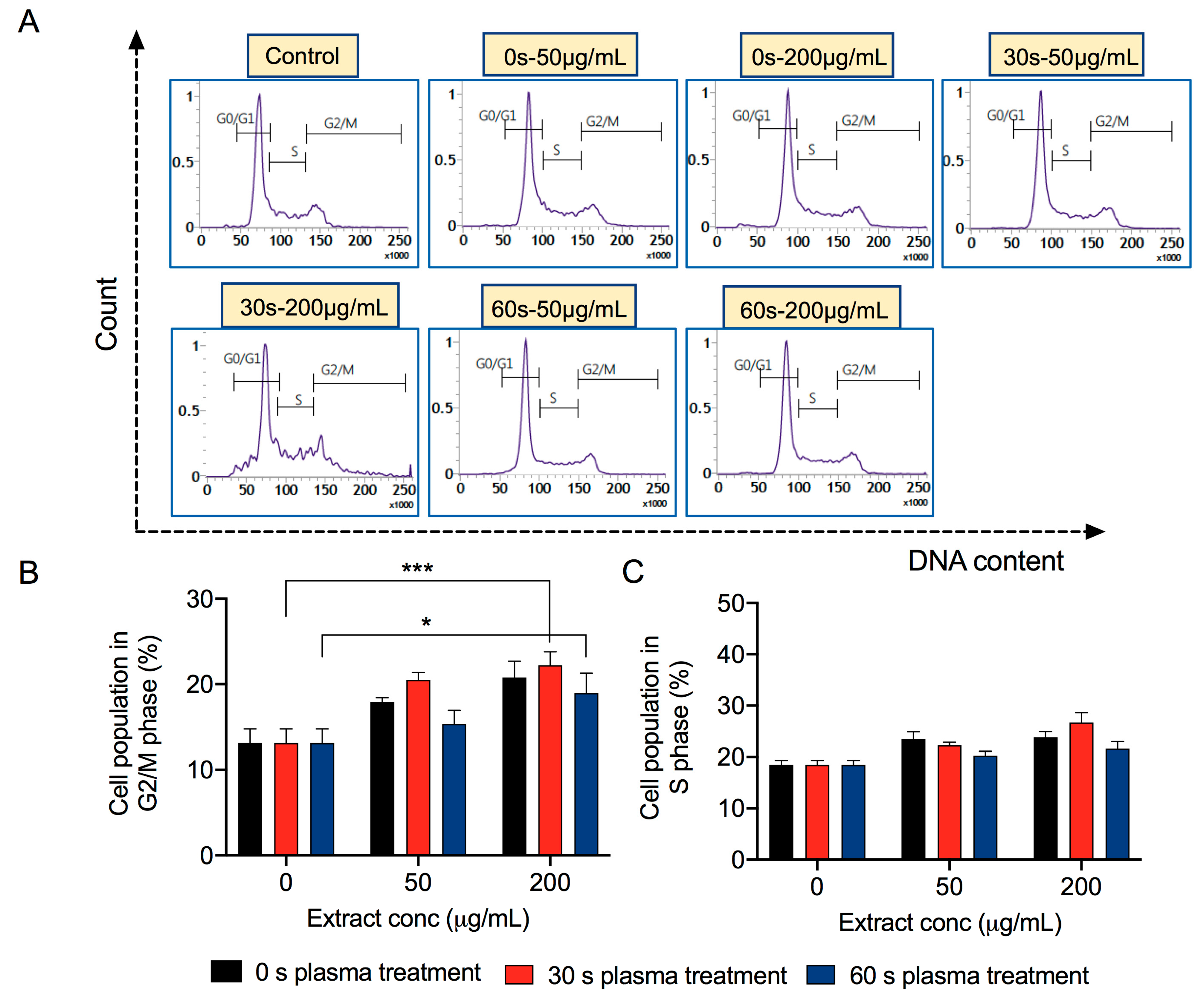
3.5. Extracts Derived from Plasma-Treated Mushrooms Induce Cancer Cell Death due to Cell Cycle Arrest

The results of the Alamar Blue activity assay showed that the viability of cancer cells concentration dependently decreased when incubated with the extracts of plasma-exposed mushrooms. Annexin-V/PI provides a sensitive approach for determining cellular apoptosis, as well as necrotic or late apoptotic cells. Thus, we further assessed cell death in breast cancer cells, at extract concentrations of 50 (low) and 200 (high) μg mL−1, using flow cytometry. The Annexin-V/PI findings revealed that the plasma-treated mushroom-derived extracts for 30 and 60 s caused the maximum death of MCF7 cells as compared with the controls (Figure 5A,B). The extracts exposed to plasma for 30 and 60 s (at a concentration of 200 μg mL−1) resulted in 30.25% (double-positive cells) and 20.65% cell death, respectively. The control extracts were relatively toxic toward MCF7 cancer cells (14.93% at 50 μg mL−1 and 12.13% at 200 μg mL−1), but minimally toxic toward MCF10A cells (5.77% at 50 μg mL−1 and 4.36% at 200 μg mL−1, Figure 5C,D). The findings were similar in MDA-MB231 cancer cells but were more prominent in MCF7 cells (Supplementary Figure S2).
Many natural products inhibit cell proliferation and induce death by inducing arrest at different phases of the cell cycle [27]. Cell cycle arrest (G1 phase) and apoptosis are different means of cellular growth arrest that lead to the failure of a cell to move into the S phase. Therefore, we analyzed MCF7 cancer cell cycles, after incubation with 50 and 200 μg mL−1 of extracts. Flow cytometry results revealed an arrest in the G2/M phase of MCF7 cells by PI/RNase A staining with plasma-treated mushroom-derived extracts, while S-phase was also affected at 30 s plasma-treated mushroom-derived extract at 200 μg mL−1. The effects were similar in MCF7 cells incubated with control extract (Figure 6A–C), indicating that the untreated mushrooms contain several phenolic compounds that affect the cell cycle, rather than promoting proliferation in cancer cells. However, this requires further investigation. The results indicated that the inhibitory effects induced by the extracts on breast cancer cell growth were due to the incidence of cell death associated with cell cycle arrest.

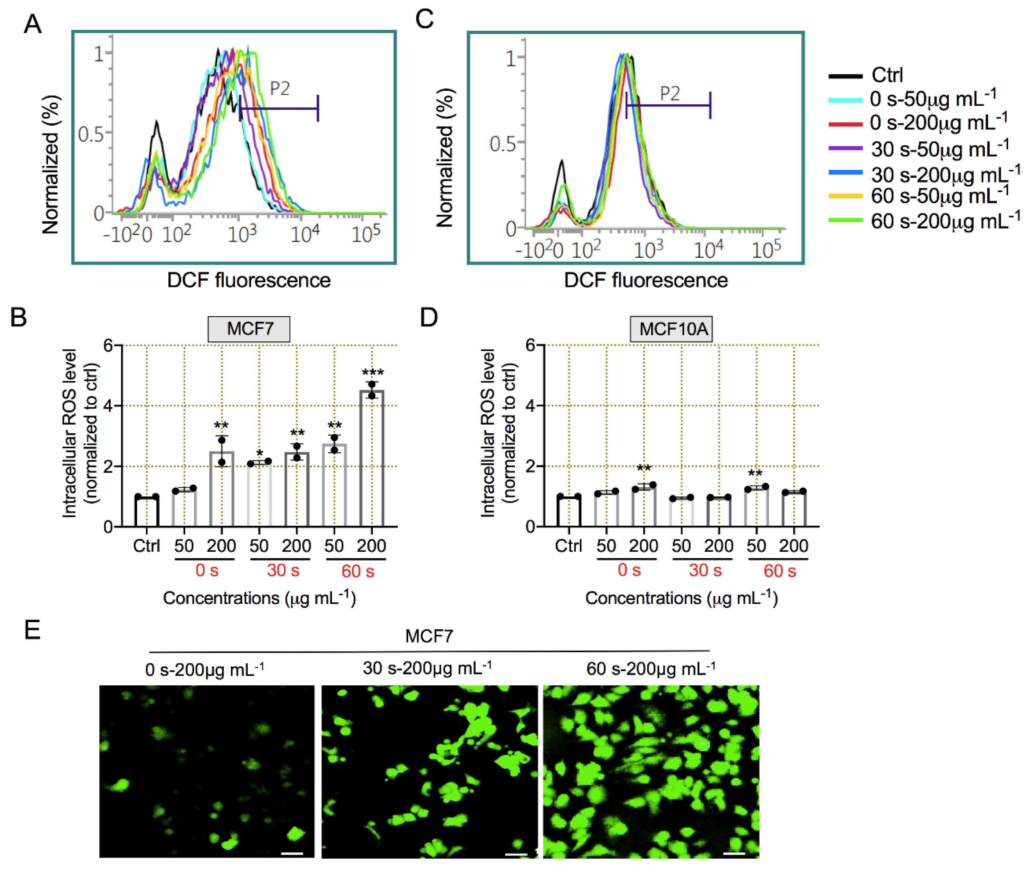
3.6. Plasma-Exposed Mushroom-Derived Extracts Induced ROS Generation for Apoptosis

Several natural compounds from plant extracts can increase intracellular ROS levels as primary mechanisms to stimulate cancer cell death [28,29]. Consequently, phytocomponents released from plasma-treated mushrooms might increase intracellular ROS levels as a precursor to cell death. We measured ROS levels in MCF7 cancer and MCF10A normal cells using H2DCFDA, which emits green fluorescence in the presence of ROS upon oxidation. Figure 7A shows that intracellular ROS levels significantly increased in MCF7 cells incubated with 50 and 200 μg mL−1 extracts derived from plasma-treated mushrooms using flow cytometry. The untreated controls also induced ROS induction at 200 μg mL−1 of extract, which was consistent with the cell cycle data (Figure 7B). Levels of ROS were slightly affected in MCF10A cells subjected to similar treatments (Figure 7C,D). Microscopy analysis further confirmed the induction of oxidative stress in MCF7 cells incubated with extracts visualized as increased green fluorescence (Figure 7E).
These findings show that ROS induction can influence cancer cell apoptosis, which is regulated by several signaling pathways and related molecules. Therefore, to verify the ability of the mushroom extracts to induce breast cancer cell apoptosis, we measured the expression of apoptosis-related gene markers in MCF7 cells under all three treatment groups (0, 30, and 60 s) at a 200 μg mL−1 extract concentration. The expression of proapoptotic BAX, p53, Puma, CytC, Casp3, Casp7, and Casp8 genes were upregulated in the 30 and 60 s groups, whereas the antiapoptotic Bcl-xL gene expression was not significantly altered as compared with the controls. Expression of the Bcl2 gene was increased in the 30 and 60 s groups. This was notable because Bcl2 belongs to the Bcl-2 family of proteins that regulate apoptotic cell death, either by inhibiting (antiapoptotic Bcl-xL, Mcl-1, Bcl-w, and A-1) or inducing (proapoptotic Bax, Bik, Bak, and Bok) apoptosis [30,31]. These effects were more prominent in MCF7 breast cancer cells incubated with 30 s extracts. We also found that H2AX expression increased, indicating double-strand breaks in the DNA of MCF7 cells incubated with 30 and 60 s extracts (Figure 8A–J). These results were further confirmed by immunofluorescence staining, which revealed γH2AX foci formation in MCF7 cells treated with 30 and 60 s extracts (200 μg mL−1) (Figure 9). To further confirm whether this induction of γH2AX was due to generally increased levels of H2AX, we measured total levels of γH2AX. The levels of γH2AX did not considerably change in the cells incubated with 30 and 60 s extracts (200 μg mL−1) as compared with controls (Supplementary Figure S3).

4. Discussion

Natural products, such as functional foods, have gained popularity as they provide alternatives to conventional cancer therapies, which are often accompanied by side effects and drug resistance. Mushrooms are rich in phytometabolic and bioactive constituents, and thus comprise an excellent alternative therapy that has health-promoting and tumor preventive effects [32]. Possible roles of various mushroom species in cancer treatment have been investigated [33,34], while others involving the optimization and improvisation of their constituents have already been directed towards anticancer therapeutics. In this regard, both direct and indirect treatment with NTP increases the yield and quality of plants including mushrooms, and improves other aspects of agriculture [35,36]. Here, we applied an air plasma jet for 30 and 60 s from an optimized device to enhance the yield and properties of F. velutipes, and then assessed characteristics and amounts of metabolic constituents such as phenols and flavonoids determined by FTIR and UV-Vis. Higher peak intensities in the UV-Vis spectra of extracts of plasma-treated mushrooms, indicated higher concentrations of constituents. Higher levels of total phenols, flavonoids, and antioxidant activities indicated that plasma enhanced the properties of mushroom extracts and might further improve their functions or applications such as cancer cell attenuation.
Natural products exhibit anticancer activity primarily via selective cytotoxicity toward cancer cells. The cell viability results indicated that the mushroom extracts could selectively affect the metabolic growth of cancer cells at specific doses, while normal mammary breast cells remained largely unharmed. Furthermore, this effect was significantly higher in MCF-7s than in MDA-MB-231 breast cancer cells. Water extracts of F. velutipes can inhibit both MCF-7 and MDA-MB-231 cells at different IC50 doses [13]. Water extracts of F. velutipes decrease the anticancer activity more prominently in MDA-MB-231 than MCF7 cells. However, we found that ethanol extracts of were highly cytotoxic to MCF-7 cancer cells. This might have been due to extract solvents differently affecting cancer cell subtypes. Differences in intrinsic molecular signaling patterns within subtypes of cancer and susceptibility towards certain bioactive compounds might also be involved [37]. Further investigation should provide more clarity into this aspect. We also found that reduced cell viability was associated with cell death and changes in the cell cycle. Consistent with these findings, more MCF7 than normal cells died, indicating that the mushroom extract effectively induced cell death in breast cancer as compared with normal mammary cells. The Annexin-V/PI analysis revealed that mushroom extracts prepared from plasma-treated mushrooms could induce cell death by apoptosis (Figure 5), due to changes in cell death markers. Furthermore, apoptotic cell death was associated with cell cycle arrest, as MCF7 cells were induced into the G2/M phase, while the S-phase also seemed to be affected by the 30 s extract (200 μg mL−1) in MCF7 cells (Figure 6). At similar concentrations, control (untreated) groups also had the intrinsic ability to alter cell cycle phases.
Several mushrooms contain high amounts of antioxidants such as polyphenol, and vital compounds that exert anticancer effects that might be realized through mechanisms similar to those of some drugs, for instance, Nrf2 inhibitors. Being rich in antioxidants, extracts of mushrooms can prevent tissue damage and apoptosis by scavenging ROS [38]. However, some mushroom extracts can induce ROS and subsequent apoptosis as anticancer effects [39]. We found that ROS levels were dose-dependently enhanced in groups incubated with mushroom extracts. Natural product-derived inhibitors of key pathways can induce ROS activity that can result in cell death [40,41,42]. However, cytotoxicity was maximal in the cells incubated with 30 s extracts, indicating ROS independent mechanisms. Apoptosis can be activated caspase-dependently without increasing ROS levels in cells [43]. Mushrooms are natural product with various metabolic constituents that can act as antioxidants and pro-oxidants depending on cellular milieu with increased ROS levels, which can also be a mechanism underlying cancer cell death [44]. Increased oxidative stress positively correlates with cell death by apoptosis and DNA damage [39]. Moreover, extracts from other mushroom species can alter the expression of apoptotic and DNA repair genes [45]. Consistent with these findings, our extracts of mushrooms exposed to plasma also significantly induced proapoptotic genes without affecting antiapoptotic gene expression. Furthermore, the upregulation of genes associated with DNA damage were upregulated, which might further promote apoptosis in cancer cells. In particular, extracts derived from mushrooms exposed to plasma for 30 s were relatively better at reducing cell viability (Figure 4), apoptosis (Figure 5), and at altering the expression of cell death-related genes. (Figure 8). Conversely, the 60 s plasma-treated mushroom-derived extracts were better at increasing ROS levels (Figure 7) and DNA damage (Figure 9). Therefore, we postulate that exposure to plasma can enhance the active properties and yield of mushroom constituents that could be useful for suppressing cancer cell growth. The mechanism of anticancer activity in extracts of plasma-treated mushrooms is primarily through the increased expression of apoptotic genes and DNA damage that results in the death of cancer cells.

5. Conclusions

In this study, we attempted to determine the anticancer activity of enoki mushrooms grown under plasma treatment against malignant breast cancer cells. To establish the role of plasma treatment on the enhancement of anticancer activity, we produced extracts from both plasma-treated and untreated mushrooms to evaluate their effects on breast cancer cells using various assays. Treating cancer cells with extracts of mushroom could be improved by exposing mushrooms to plasma. The 30 s extracts exerted more prominent effects on cancer cell death and induced apoptotic cell death markers. Further studies of the mechanisms related to this activity are required for validation. The anticancer potential of specific mechanisms of extracts from various mushroom species exposed to plasma should also be evaluated in future studies.

Supplementary Materials

The following are available online at https://www.mdpi.com/2076-3417/10/23/8395/s1, Figure S1: Plasma-treatment stimulates cytotoxicity of mushroom derived extracts against breast cancer cells, Figure S2: Plasma treatment improves effect of mushroom derived extracts on apoptotic cancer cell death, Figure S3: Levels of H2AX total protein does not alter in plasma-treated mushroom derived extracts exposed breast cancer cells.

Author Contributions

Conceptualization, S.M., N.K., and N.K.K.; methodology, S.M., P.B., N.K., R.W., S.B., and L.N.N.; software, N.K., S.B., and R.W.; validation, N.K.K.; formal analysis, S.M., P.B., N.K., and L.N.N.; investigation, S.M., P.B., N.K., and N.K.K.; writing—original draft preparation S.M., P.B., N.K., and N.K.K.; writing—review and editing, N.K., R.W., S.B., N.K.K., and E.H.C.; supervision, N.K.K., and E.H.C. All authors have read and agreed to the published version of the manuscript.

Funding

This study was supported by the National Research Foundation (NRF) of Korea, funded by the Korean government (NRF-2016K1A4A3914113), MSIT (Ministry of Science and ICT), Korea under the ITRC (Information Technology Research Center) support program (IITP-2020-0-01846) supervised by the IITP (Institute of Information & Communications Technology Planning & Evaluation), and by Kwangwoon University and the University of Suwon in 2020.

Conflicts of Interest

The authors declare no conflict of interest.

References

  1. Mitra, S.; Dash, R. Natural Products for the Management and Prevention of Breast Cancer. Evid. Based Complement. Altern. Med. 2018, 2018, 8324696. [Google Scholar] [CrossRef] [PubMed]
  2. Wu, M.; Luo, X.; Xu, X.; Wei, W.; Yu, M.; Jiang, N.; Ye, L.; Yang, Z.; Fei, X. Antioxidant and immunomodulatory activities of a polysaccharide from Flammulina velutipes. J. Tradit. Chin. Med. 2014, 34, 733–740. [Google Scholar] [CrossRef] [Green Version]
  3. Ukaegbu, C.I.; Shah, S.R.; Hazrulrizawati, A.H.; Alara, O.R. Acetone extract of Flammulina velutipes caps: A promising source of antioxidant and anticancer agents. Beni-Suef Univ. J. Basic Appl. Sci. 2018, 7, 675–682. [Google Scholar] [CrossRef]
  4. Scholtz, V.; Šerá, B.; Khun, J.; Šerý, M.; Julák, J. Effects of Nonthermal Plasma on Wheat Grains and Products. J. Food Qual. 2019, 2019, 7917825. [Google Scholar] [CrossRef] [Green Version]
  5. Dobrin, D.; Magureanu, M.; Mandache, N.B.; Ionita, M.-D. The effect of non-thermal plasma treatment on wheat germination and early growth. Innov. Food Sci. Emerg. 2015, 29, 255–260. [Google Scholar] [CrossRef]
  6. Bourke, P.; Ziuzina, D.; Boehm, D.; Cullen, P.J.; Keener, K. The potential of cold plasma for safe and sustainable food production. Trends Biotechnol. 2018, 36, 615–626. [Google Scholar] [CrossRef] [Green Version]
  7. Neyts, E.C. Plasma-Surface Interactions in Plasma Catalysis. Plasma Chem. 2016, 36, 185–212. [Google Scholar] [CrossRef]
  8. Hirst, A.M.; Frame, F.M.; Arya, M.; Maitland, N.J.; O’Connell, D. Low temperature plasmas as emerging cancer therapeutics: The state of play and thoughts for the future. Tumor Biol. 2016, 37, 7021–7031. [Google Scholar] [CrossRef] [Green Version]
  9. Laroussi, M. Plasma Medicine: A Brief Introduction. Plasma 2018, 1, 5. [Google Scholar] [CrossRef] [Green Version]
  10. Dong, Y.R.; Cheng, S.J.; Qi, G.H.; Yang, Z.P.; Yin, S.Y.; Chen, G.T. Antimicrobial and antioxidant activities of Flammulina velutipes polysaccharides and polysaccharide-iron(III) complex [corrected]. Carbohydr. Polym. 2017, 161, 26–32. [Google Scholar] [CrossRef]
  11. Wu, D.-m.; Duan, W.-q.; Liu, Y.; Cen, Y. Anti-inflammatory effect of the polysaccharides of Golden needle mushroom in burned rats. Int. J. Biol. Macromol. 2010, 46, 100–103. [Google Scholar] [CrossRef] [PubMed]
  12. Chang, H.-H.; Hsieh, K.-Y.; Yeh, C.-H.; Tu, Y.-P.; Sheu, F. Oral administration of an Enoki mushroom protein FVE activates innate and adaptive immunity and induces anti-tumor activity against murine hepatocellular carcinoma. Int. Immunopharmacol. 2010, 10, 239–246. [Google Scholar] [CrossRef] [PubMed]
  13. Gu, Y.H.; Leonard, J. In vitro effects on proliferation, apoptosis and colony inhibition in ER-dependent and ER-independent human breast cancer cells by selected mushroom species. Oncol. Rep. 2006, 15, 417–423. [Google Scholar] [CrossRef] [PubMed]
  14. Leung, M.Y.; Fung, K.P.; Choy, Y.M. The isolation and characterization of an immunomodulatory and anti-tumor polysaccharide preparation from Flammulina velutipes. Immunopharmacology 1997, 35, 255–263. [Google Scholar] [CrossRef]
  15. Yi, C.; Zhong, H.; Tong, S.; Cao, X.; Firempong, C.K.; Liu, H.; Fu, M.; Yang, Y.; Feng, Y.; Zhang, H.; et al. Enhanced oral bioavailability of a sterol-loaded microemulsion formulation of Flammulina velutipes, a potential antitumor drug. Int. J. Nanomed. 2012, 7, 5067–5078. [Google Scholar]
  16. Chang, Y.C.; Hsiao, Y.M.; Wu, M.F.; Ou, C.C.; Lin, Y.W.; Lue, K.H.; Ko, J.L. Interruption of lung cancer cell migration and proliferation by fungal immunomodulatory protein FIP-fve from Flammulina velutipes. J. Agric. Food Chem. 2013, 61, 12044–12052. [Google Scholar] [CrossRef]
  17. Singleton, V.L.; Rossi, J.A. Colorimetry of Total Phenolics with Phosphomolybdic-Phosphotungstic Acid Reagents. Am. J. Enol. Vitic. 1965, 16, 144. [Google Scholar]
  18. Chandra, S.; Khan, S.; Avula, B.; Lata, H.; Yang, M.H.; ElSohly, M.A.; Khan, I.A. Assessment of Total Phenolic and Flavonoid Content, Antioxidant Properties, and Yield of Aeroponically and Conventionally Grown Leafy Vegetables and Fruit Crops: A Comparative Study. Evid. Based Complement. Altern. Med. 2014, 2014, 253875. [Google Scholar] [CrossRef]
  19. Chang, C.-C.; Yang, M.-H.; Wen, H.-M.; Chern, J.-C. Estimation of total flavonoid content in propolis by two complementary colorimetric methods. J. Food Drug Anal. 2002, 10, 178–182. [Google Scholar]
  20. Brand-Williams, W.; Cuvelier, M.-E.; Berset, C. Use of a free radical method to evaluate antioxidant activity. LWT Food Sci. Technol. 1995, 28, 25–30. [Google Scholar] [CrossRef]
  21. Adhikari, M.; Adhikari, B.; Kaushik, N.; Lee, S.-J.; Kaushik, N.K.; Choi, E.H. Melanoma Growth Analysis in Blood Serum and Tissue Using Xenograft Model with Response to Cold Atmospheric Plasma Activated Medium. Appl. Sci. 2019, 9, 4227. [Google Scholar] [CrossRef] [Green Version]
  22. Baba, S.A.; Malik, S.A. Determination of total phenolic and flavonoid content, antimicrobial and antioxidant activity of a root extract of Arisaema jacquemontii Blume. J. Taibah Univ. Sci. 2015, 9, 449–454. [Google Scholar] [CrossRef] [Green Version]
  23. Shimoi, K.; Masuda, S.; Shen, B.; Furugori, M.; Kinae, N. Radioprotective effects of antioxidative plant flavonoids in mice. Mutat. Res. Fund. Mol. M 1996, 350, 153–161. [Google Scholar] [CrossRef]
  24. Alam, M.N.; Bristi, N.J.; Rafiquzzaman, M. Review on in vivo and in vitro methods evaluation of antioxidant activity. Saudi Pharm. J. 2013, 21, 143–152. [Google Scholar] [CrossRef] [Green Version]
  25. Engida, A.M.; Faika, S.; Nguyen-Thi, B.T.; Ju, Y.H. Analysis of major antioxidants from extracts of Myrmecodia pendans by UV/visible spectrophotometer, liquid chromatography/tandem mass spectrometry, and high-performance liquid chromatography/UV techniques. J. Food Drug Anal. 2015, 23, 303–309. [Google Scholar] [CrossRef] [Green Version]
  26. Zhao, M.; Yang, B.; Wang, J.; Li, B.; Jiang, Y. Identification of the major flavonoids from pericarp tissues of lychee fruit in relation to their antioxidant activities. Food Chem. 2006, 98, 539–544. [Google Scholar] [CrossRef]
  27. Lin, H.-W.; Hsieh, M.-J.; Yeh, C.-B.; Hsueh, K.-C.; Hsieh, Y.-H.; Yang, S.-F. Coronarin D induces apoptotic cell death through the JNK pathway in human hepatocellular carcinoma. Environ. Toxicol. 2018, 33, 946–954. [Google Scholar] [CrossRef]
  28. Ryu, S.; Lim, W.; Bazer, F.W.; Song, G. Chrysin induces death of prostate cancer cells by inducing ROS and ER stress. J. Cell. Physiol. 2017, 232, 3786–3797. [Google Scholar] [CrossRef]
  29. Zhang, S.; He, Y.; Tong, Q.; Chen, Q.; Wu, X.; Huang, W. Deltonin induces apoptosis in MDA-MB-231 human breast cancer cells via reactive oxygen species-mediated mitochondrial dysfunction and ERK/AKT signaling pathways. Mol. Med. Rep. 2013, 7, 1038–1044. [Google Scholar] [CrossRef] [Green Version]
  30. Leibowitz, B.; Yu, J. Mitochondrial signaling in cell death via the Bcl-2 family. Cancer Biol. Ther. 2010, 9, 417–422. [Google Scholar] [CrossRef] [Green Version]
  31. Youle, R.J.; Strasser, A. The BCL-2 protein family: Opposing activities that mediate cell death. Nat. Rev. Mol. Cell Biol. 2008, 9, 47–59. [Google Scholar] [CrossRef] [PubMed]
  32. Patel, S.; Goyal, A. Recent developments in mushrooms as anti-cancer therapeutics: A review. 3 Biotech 2012, 2, 1–15. [Google Scholar] [CrossRef] [PubMed] [Green Version]
  33. Ayeka, P.A. Potential of Mushroom Compounds as Immunomodulators in Cancer Immunotherapy: A Review. Evid. Based Complement. Altern. Med. 2018, 2018, 7271509. [Google Scholar] [CrossRef] [PubMed]
  34. Blagodatski, A.; Yatsunskaya, M.; Mikhailova, V.; Tiasto, V.; Kagansky, A.; Katanaev, V.L. Medicinal mushrooms as an attractive new source of natural compounds for future cancer therapy. Oncotarget 2018, 9, 29259–29274. [Google Scholar] [CrossRef] [Green Version]
  35. Pérez-Pizá, M.C.; Prevosto, L.; Grijalba, P.E.; Zilli, C.G.; Cejas, E.; Mancinelli, B.; Balestrasse, K.B. Improvement of growth and yield of soybean plants through the application of non-thermal plasmas to seeds with different health status. Heliyon 2019, 5, e01495. [Google Scholar] [CrossRef] [Green Version]
  36. Randeniya, L.K.; de Groot, G.J.J.B. Non-Thermal Plasma Treatment of Agricultural Seeds for Stimulation of Germination, Removal of Surface Contamination and Other Benefits: A Review. Plasma Process. Polym. 2015, 12, 608–623. [Google Scholar] [CrossRef]
  37. Dampier, K.; Hudson, E.A.; Howells, L.M.; Manson, M.M.; Walker, R.A.; Gescher, A. Differences between human breast cell lines in susceptibility towards growth inhibition by genistein. Br. J. Cancer 2001, 85, 618–624. [Google Scholar] [CrossRef]
  38. Hu, H.; Zhang, Z.; Lei, Z.; Yang, Y.; Sugiura, N. Comparative study of antioxidant activity and antiproliferative effect of hot water and ethanol extracts from the mushroom Inonotus obliquus. J. Biosci. Bioeng. 2009, 107, 42–48. [Google Scholar] [CrossRef]
  39. Jeong, J.W.; Jin, C.Y.; Park, C.; Hong, S.H.; Kim, G.Y.; Jeong, Y.K.; Lee, J.D.; Yoo, Y.H.; Choi, Y.H. Induction of apoptosis by cordycepin via reactive oxygen species generation in human leukemia cells. Toxicol. In Vitro 2011, 25, 817–824. [Google Scholar] [CrossRef]
  40. Sznarkowska, A.; Kostecka, A.; Meller, K.; Bielawski, K.P. Inhibition of cancer antioxidant defense by natural compounds. Oncotarget 2017, 8, 15996. [Google Scholar] [CrossRef] [Green Version]
  41. Stepanic, V.; Gasparovic, A.C.; Troselj, K.G.; Amic, D.; Zarkovic, N. Selected attributes of polyphenols in targeting oxidative stress in cancer. Curr. Top. Med. Chem. 2015, 15, 496–509. [Google Scholar] [CrossRef] [PubMed]
  42. Chian, S.; Li, Y.-Y.; Wang, X.-J.; Tang, X.-W. Luteolin sensitizes two oxaliplatin-resistant colorectal cancer cell lines to chemotherapeutic drugs via inhibition of the Nrf2 pathway. Asian Pac. J. Cancer Prev. 2014, 15, 2911–2916. [Google Scholar] [CrossRef] [PubMed] [Green Version]
  43. Chien, S.Y.; Wu, Y.C.; Chung, J.G.; Yang, J.S.; Lu, H.F.; Tsou, M.F.; Wood, W.G.; Kuo, S.J.; Chen, D.R. Quercetin-induced apoptosis acts through mitochondrial- and caspase-3-dependent pathways in human breast cancer MDA-MB-231 cells. Hum. Exp. Toxicol. 2009, 28, 493–503. [Google Scholar] [CrossRef] [PubMed]
  44. Lambert, J.D.; Elias, R.J. The antioxidant and pro-oxidant activities of green tea polyphenols: A role in cancer prevention. Arch. Biochem. Biophys. 2010, 501, 65–72. [Google Scholar] [CrossRef] [Green Version]
  45. Wang, Y.; Xu, G.; Tang, X.; Chen, H. Polysaccharide lentinan extracted from the stipe of Lentinus edodes mushroom exerts anticancer activities through the transcriptional regulation of cell cycle progression and metastatic markers in human colon cancer cells. Faseb. J. 2017, 31, lb391. [Google Scholar]
Figure 1. Characterization and standardization of plasma device. (A) Schematic of air gas jet plasma device; (B) Optical emission spectrum of plasma-generated species in ambient air; (C,D) pH and temperature of media after exposure to plasma for various periods, respectively; (E,F) Determination of H2O2 and NOx concentrations in medium after plasma exposure for indicated periods. ** p < 0.01 and *** p < 0.001.
Applsci 10 08395 g001
Figure 2. Extract preparation from plasma-treated/untreated mushrooms (F. velutipes) and their characterization. (A) Experimental scheme for extract preparation from plasma-exposed and unexposed mushroom; (BD) Measurement of phenolic, flavonoid, and antioxidant properties in extracts prepared from plasma-treated (30 s and 60 s) and untreated (0 s) mushrooms. * p < 0.05 and *** p < 0.001. Untreated sample was used as a negative control for normalization.
Applsci 10 08395 g002
Figure 3. Fourier transform infrared spectra and UV-Vis analysis of extracts of nonthermal gas plasma-treated mushroom-derived extracts. Fourier transform infrared spectra of F. velutipes extract (A) 900–4000 cm−1 and (B) 900–1500 cm−1. Black line, control (0 s not exposed to plasma); red line, exposed to plasma for 30 s; blue line, exposed to plasma for 60 s; (C) UV-Vis absorption spectra of F. velutipes extracts. Black line, control (0 s not exposed to plasma); red line, exposed to plasma for 30 s; and blue line, exposed to plasma for 60 s.
Applsci 10 08395 g003
Figure 4. Extracts derived from plasma-treated mushrooms are cytotoxic to breast cancer cells. (AC) Results of Alamar Blue assays performed using MDA-MB231, MCF7 cancer, and MCF10A normal cells incubated for 24 h with 0, 10, 50, 100, 200, 500, and 1000 μg mL−1 of extracts derived from plasma-treated mushrooms for 30 s, 60 s, or left unexposed (0 s, control). * p < 0.05, ** p < 0.01, and *** p < 0.001. Untreated sample was used as a control in all tested plant extracts groups for normalization.
Applsci 10 08395 g004
Figure 5. Plasma-treated mushroom-derived extracts stimulate apoptotic cell death in breast cancer cells. (AD) Cell death analysis in MCF7 breast cancer and MCF10A normal mammary epithelial cells treated with extracts prepared from 30 and 60 s plasma-treated, and untreated (0 s) mushrooms at 50 and 200 μg mL−1 after 24 h post-incubation using Annexin-V/PI staining.
Applsci 10 08395 g005
Figure 6. Cell cycle arrest is associated with cancer cell death. (AC) Flow cytometry analysis of MCF7 breast cancer cells treated with extracts prepared from 0 s (untreated), 30 and 60 s plasma-treated mushrooms at 50 and 200 μg mL–1 using PI/RNase A, after 24 h post-incubation. Representative graph shows percentage of cell population in G2/M and S phase. * p < 0.05, and *** p < 0.001.
Applsci 10 08395 g006
Figure 7. Plasma-treated mushroom-derived extracts induced intracellular oxidative stress. (AD) Flow cytometric detection of intracellular ROS in MCF7 breast cancer and MCF10A normal cells treated with extracts prepared from 0 s (untreated), 30 and 60 s plasma-treated (0 s) mushrooms, after 24 h post-incubation at 50 and 200 μg mL−1 using fluorescent H2DCFDA dye, respectively; (E) Microscopic images of ROS detection in MCF7 cells treated in similar conditions as above. Untreated sample (no extract) was used as control for normalization. * p < 0.05, ** p < 0.01, and *** p < 0.001.
Applsci 10 08395 g007
Figure 8. Induction of cell death markers by plasma-treated mushroom-derived extracts. (AJ) Quantification of mRNA expression of BAX, Bcl-xL, BCl2, PUMA, Casp3, p53, CytC, H2AX, Casp8, and Casp7 apoptosis-related markers, respectively in MCF7 cells treated at 200 μg mL−1 concentration. * p < 0.05, ** p < 0.01, and *** p < 0.001.
Applsci 10 08395 g008
Figure 9. Plasma-treated mushroom-derived extracts provoke DNA damage in breast cancer cells. Quantification of H2AX foci formation in MCF7 cells by immunofluorescence staining treated as indicated. Scale bar, 20 µm.
Applsci 10 08395 g009
Table 1. List of primer sequences used in this study.
Gene NameSequence (5′-3′)Size (bp)
ACTIN-forwardGGC ATC CTC ACC CTG AAG TA82
ACTIN-reverseAGG TGT GGT GCC AGA TTT TC
BAX-forwardAAG AAG CTG AGC GAG TGT CTC186
BAX-reverseGCT GGC AAA GTA GAA AAG GGC
BCL2-forwardTTC TTT GAG TTC GGT GGG GTC208
BCL2-reverseTCT TCA GAG ACA GCC AGG AGA
BCL-XL-forwardTCA GTG AGT GAG CAG GTG TT170
BCL-XL-reverseGGC CTC AGT CCT GTT CTC TT
TP53-forwardGCC CCT CCT CAG CAT CTT ATC246
TP53-reverseAAA GCT GTT CCG TCC CAG TAG
H2AX-forwardCAA CAA GAA GAC GCG AAT CA223
H2AX-reverseCGG GCC CTC TTA GTA CTC CT
Casp3-forwardATG TCG ATG CAG CAA ACC TC173
Casp3-reverseTCC TTC TTC ACC ATG GCT CA
Casp7-forwardAGA AGC TGT TAC CAC ACC CA185
Casp7-reverseACT CCA TCT CAG TCA GTG GC
Casp8-forwardCCC AAA TCA ACA AGA GCC TGC219
Casp8-reverseTCA GAC AGT ATC CCC GAG GTT
CytC-forwardCAA CAC CTC TCA CAT CTT ACA GA185
CytC-reverseGGC AAA AGC ACC ATT CCC AA
PUMA-forwardCCC ACC ACC ATC TCA GGA AA185
PUMA-reverseGTG GTC ACG TTT GGC TCA TT
Publisher’s Note: MDPI stays neutral with regard to jurisdictional claims in published maps and institutional affiliations.

