Article

Simulation of Chronic Intoxication in Rats Exposed to Cadmium and Mercury

by
Iveta Cimboláková
1,
Tatiana Kimáková
2,
Henrieta Pavolová
3,
Tomáš Bakalár
3,*,
Dušan Kudelas
3 and
Andrea Seňová
3
1
Institute of Physical Education and Sport, Pavol Jozef Šafárik University, Ondavská 21, 040 11 Košice, Slovakia
2
Department of Public Health and Hygiene, Faculty of Medicine, Pavol Jozef Šafárik University, Trieda SNP 1, 040 11 Košice, Slovakia
3
Institute of Earth Resources, Faculty of Mining, Ecology, Process Control and Geotechnologies, Technical University of Košice, Letná 9, 042 00 Košice, Slovakia
*
Author to whom correspondence should be addressed.
Appl. Sci. 2020, 10(22), 8066; https://doi.org/10.3390/app10228066
Submission received: 14 October 2020 / Revised: 5 November 2020 / Accepted: 11 November 2020 / Published: 13 November 2020

Abstract

:
Cadmium and mercury are widespread and non-biodegradable pollutants of great concern to human and animal health. In this study, the influence of exposure to low doses of cadmium and mercury on Wistar rats was investigated. The experiments aimed to identify suitable markers of chronic intoxication with heavy metals in rats. The subjects were 48 naive young rats (24 females and 24 males), four weeks old, grouped randomly into three distinct groups—control group, group exposed to cadmium and group exposed to mercury. The control group received sham treatment—clean untreated water. Cd exposed group received water containing cadmium chloride dihydrate and Hg exposed group received water with mercury dichloride. Both cadmium and mercury were administered to experimental rats in drinking water in concentrations exceeding the maximum acceptable concentration of these metals 500 times, i.e., 0.5 mg Hg and 2.5 mg Cd per liter of water. The results were evaluated quarterly during the experiment (52 weeks). Selected physiological parameters (life span, body weight changes and intake of food and water), reproductive parameters (number of births (litters), number of born pups and number of raised pups) and toxicological parameters (average daily dose, total dose received and the amount of toxic metal received) were studied. The results of the experiments indicate differences between both individual groups and between males and females, which confirmed that these parameters are essential in such experiments of chronic exposure to subtoxic doses of heavy metals.

1. Introduction

Heavy metals naturally occur in the environment, but anthropogenic activities can increase the background levels in the environment, raising scientific and public health concerns for human health. Heavy metals can be associated with an unprecedented expansion of chemicals in industrial and agricultural production that consequently causes an increase in the concentration of toxic substances in the environment [1]. Although modern civilization is bringing an enhancement of the living standards, it is also accompanied by negative phenomena in the form of disturbed ecology, natural and biological conditions of life [2]. German pharmacologists first described the effect of very low doses of toxic substances as a Schulz-Arndt’s Law, and, since then, this phenomenon has become an important part of toxicology. At present, this phenomenon is known as “hormesis” or hormesis effect. Hormesis is a biophysical, dose–response phenomenon characterized by low doses of a substance having a stimulating effect and high doses having an inhibitory effect. Calabrese [3] claimed that hormesis can be encountered in the entire plant and animal kingdoms virtually in all substances if their doses are small enough. This occurrence has been documented across a wide range of biological models, diverse types of exposures and a variety of outcomes [4]. Currently, more attention is paid to the existence of a hormetic effect and the exposure (dose)–effect (response) relationship that is a subject of many experiments as discussed in the article.
The study of the effect of low doses initiated the transformation of the views on heavy metal toxicology and the assessment of their biological response to exposure. The interest is shifting from investigating the mechanism of toxicity towards the monitoring of the adaptation mechanisms. Current theories [4,5,6] reveal biological processes and mechanisms that develop only at low doses that change the view on this matter. Although organisms are daily exposed to chemicals, radiation or infectious agents, most of the exposure has no apparent health consequences to the organisms; however, possible apparent pathogenetic consequences are related to the extent, frequency and mode of contact. These factors have a critical impact on the exposure risk degree. Exposure assessment, dose estimation, optimization for an individual or population, at large, play an important role in exposure toxicology. “Daily Intake of the Substance—Exposure Dose” is the result of a quantitative exposure assessment. Investigation of chronic exposure to very low (subtoxic) doses is currently a focus of numerous studies. In the past, environmental pollution was an acute problem of specific location or site; however, at present, this is a widespread and comprehensive environmental and social issue worldwide, which can pose a serious problem that can affect the health of organisms including humans in large territories in the future. Finally, this not only applies for current but also future generations where the consequences are difficult to predict.
The critical question is the exposure estimation which should include the method of entry, dosing, time course and contact with the toxic material. The way the toxic substance is received (route of exposure) is essential. If it is ingested, it can be bound to food ingredients, while the possibility of such binding is significantly lower by water intake. Furthermore, the difference between single and fractionated doses of contaminant also needs to be considered. In that context, exposure–response is crucial. As reported by Budnik et al. [7], exposure estimates are critical in epidemiological studies. Checkoway et al. [8] pointed out that inadequate estimates can lead to incorrect conclusions. The response from the exposed subjects to toxic substances is another matter. Factors (e.g., intake, absorption, perceptiveness, toxicity and biokinetics) that are influenced by a host of other factors of an exposed subject (e.g., age, gender, weight, nutritional status and genetic predisposition) can be included here.
The aim of the study was to demonstrate the effect of low doses of cadmium and mercury administered to Wistar rats in drinking water at subtoxic concentrations on selected physiological, reproductive and toxicological parameters as possible markers of exposure of chronic intoxication by heavy metals in doses higher than the maximum permissible concentration (MPC).

2. Experimental Section

The study was conducted at Faculty of Medicine of Pavol Jozef Šafárik University in Košice (experimental bio-models laboratory). The research center has the accreditation for the conduction of the breeding of laboratory animals and empirical, evidence-based research on the animals according to the legislative provision. Before the experiment, approval from the corresponding ethical committee of Faculty of Medicine of Pavol Jozef Šafárik University in Košice, ŠVPPSR (No. Ro-2448/09-221) was obtained.

2.1. Exposure Methods

The experiments included a general monitoring of the effect of toxic substances on selected physiological parameters of both male and female Wistar rats during the entire experiment, and a reproduction monitoring of reprotoxicity. For the experiments, two heavy metals, cadmium (Cd) and mercury (Hg), were selected. The experiments included 48 naive young rats (24 females and 24 males), 4 weeks old, of mean body weight 120 ± 19 g. The rats were housed in cages in same sex pairs, with ad libitum access to water and food (standard Larsen diet), at 22 ± 2 °C, 50% humidity with 12 h light and 12 h dark. Heavy metals were administered to Wistar rats in drinking water at concentrations of 0.5 mg·L−1 for Hg and 2.5 mg·L−1 for Cd, which are 500 times higher than MPC in drinking water, 0.001 and 0.005 mg·L−1 for Hg and Cd, respectively, given by the Decree of the Ministry of Health of the Slovak Republic No. 247/2017 Coll. The water used for Hg and Cd salts was tap water and the concentration of Hg and Cd was verified by Atomic adsorption spectroscopy.
The control (K) group—16 rats (8 males and 8 females)—received untreated drinking water, the Cd treated group—16 rats (8 males and 8 females)—received cadmium chloride (CdCl2 2 H2O) at a concentration of 4.88 mg·L−1 (i.e., 2.5 mg Cd·L−1) in water and the Hg treated group—16 rats (8 males and 8 females) received water containing mercuric chloride (HgCl2) at a concentration of 0.68 mg·L−1 (i.e., 0.5 mg Hg·L−1). In all groups, the following parameters were monitored and evaluated:
  • Animal survival
  • Weight changes
  • Food intake
  • Water intake
The reproductive experiment also included 48 animals which were kept in pairs (1 male and 1 female). By random selection, they were divided into the K group and treated groups, Cd and Hg, as described above. The born pups were left with their mothers until weaning and the males were isolated from females with pups for that period. During the experiment, the following reproductive parameters were monitored and evaluated:
  • Number of births (litters), number of born pups, and number of raised pups (live pups assessed on the 21st day after delivery);
  • Number of born pups per litter and number of raised pups per litter
In the toxicological experiment, the following parameters were monitored and evaluated:
  • Average daily dose (ADD);
  • Total dose received in 52 weeks in mg·kg−1 of live weight of a rat;
  • The amount of toxic metal received in relation to the LD50 dose.
ADD and LD50 were counted based on the standards and in accordance with the Methodological instruction of Ministry of the Environment of the Slovak Republic on preparation of risk analysis of polluted areas.

2.2. Statistical Analysis

For statistical data processing, Microsoft Excel and Arcus Bio were used. In accordance with the approval from the corresponding ethical committee (which may limit the number of animals included in the experiment), the Directive 2010/63/EU of the European Parliament and the Council of 22 September 2010 on the protection of animals used for scientific purposes and the Slovak legislation, the total number of animals used for the experiment was 48 + 48, as mentioned above, while the minimum number necessary is 6 (for animals) for statistical significance. In the study, two concentration groups were used as the study was concentrated on chronic exposure to very low (subtoxic) doses not to toxic doses that require three or more concentration groups of animals. The results in figures and tables are average values ± standard deviation (SD). As in the experiments there were measurements in more sets (males and females before the experiment, after each quarter, at the end of the experiment, etc.), the single-factor ANOVA with Tukey–Kramer post-test was used for analysis of variance for repeated measurements and the evaluation of the statistical significance of the obtained values for repeated measurements. The normality of distribution and homogeneity of variance of each sample set was tested in addition to using the ANOVA test. The statistical significance was set at p < 0.05. All data generated and analyzed during the study are included in the study. All animals were weighed at the start of the treatment, once a week for the first 13 weeks and once a month thereafter. Measurement of food and water consumption was made once a week for the first 13 weeks and once a month thereafter. The results are statistically processed and presented as means on the beginning of the experiment and at the end of quarters.

3. Results

The death of the rats was not observed in any of the studied groups during the whole course of the study. The survival was 100%, together with confirmation, that the listed ration indeed belongs to the category of low doses.

3.1. General Experiment

The dynamics of body weight progress was different in the Cd and Hg groups compared to the K group depending on the sex of the animals in stages (quarters). In the Cd group, the weight in individual stages was lower in both the female and male groups (except for the second quarter) than in the K group. In the Hg/male group, the weight was lower than in the K/male group during all stages. However, in Hg/female group, the weight was higher than in the K/female group from the first to the third quarter and lower in the fourth quarter. Comparing the Cd and Hg groups, the weight in the Hg group was higher than in the Cd group in both sexes. Despite these trends in weight development in the comparison of the K group and the Cd and Hg groups in the individual stages of the experiment, no statistical significance was recorded. The average weight in the female and male groups during the experiments are given in Table 1 and Table 2, respectively. At the end of the experiment (after 52 weeks), the weight of the animals in both the Cd and Hg groups was lower compared to the K group, although statistical significance was not observed. When comparing the male and female groups, the body weight was significantly higher in all the male groups than that in female groups (p < 0.001). When comparing the Cd and Hg groups, the wight of the animals was higher in Hg group than in Cd group for both males and females, although there was no statistical significance observed in the exposed and K groups. A comparison of weights is presented in Figure 1.
The water intake had a fluctuating trend in both Cd and Hg groups and K group of the animals of both sexes in the stages (quarters). In Cd/female group, water intake was lower than in K/female group during the first to third quarters, but higher in the fourth quarter. However, in Cd/male group, the water intake was higher than in K/male group in all quarters. In Hg/female group, the water intake was lower than in K/female group in the second and third quarters and higher in the first and fourth quarters. In Hg/male group, the water intake was lower than in K/male group in all quarters except for the first quarter. Comparing the exposed groups, the water intake was higher in the Hg/female group than in the Cd/female group during all stages of the experiment. The water intake in the Cd/male group was higher than in the Hg/male group. However, no statistical significance was noticed.
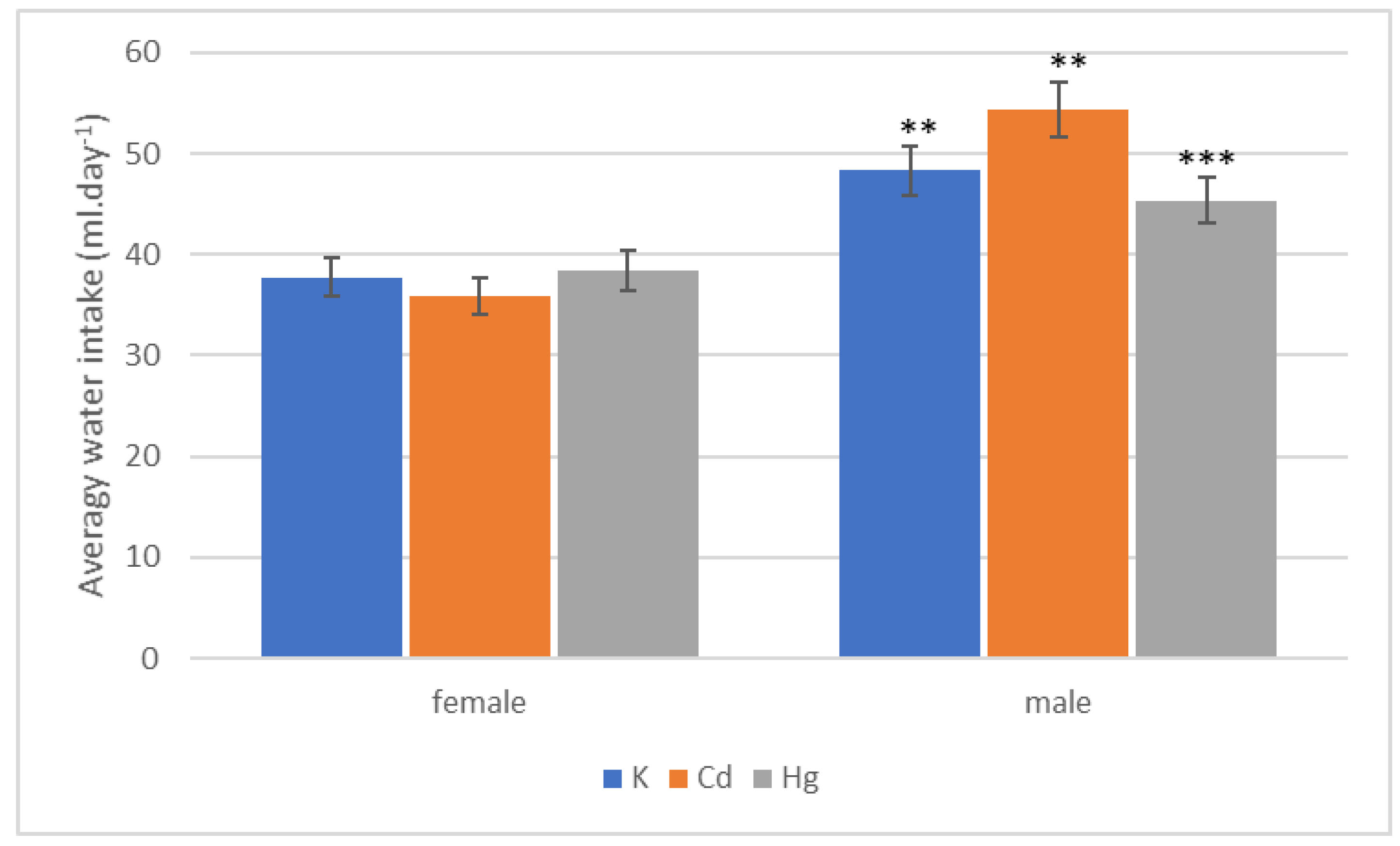
The average water intake in the female and male groups during the quarters and the whole experiment are given in Table 3 and Table 4, respectively. When evaluating the trends during the whole period of 52 weeks, the water intake in Cd/male group was higher than in both K and Hg male groups. On the other hand, water intake in Hg/female group was higher than in K and Cd female groups. A statistically significant difference was recorded between Cd/female and Cd/male groups (p < 0.001) and K/female and K/male groups (p < 0.001). In the male groups, a statistical significance was recorded between the Cd/male and Hg/male group (p = 0.0145). The water intake is presented in Figure 2.
The food intake was also different between the Cd and Hg groups and the K group depending on the sex of the animals in the stages (quarters). In the Cd/male group, the food intake was higher than in the K/male group during all stages. However, in Cd/female group, the food intake was lower than in the K/female group in the first to third quarters and higher in the fourth quarter. A statistically significant difference in food intake was recorded between the Cd and K female groups in the third quarter of the experiment (p = 0.037). In the Hg/male group, the food intake was lower than in the K/male group in all stages except for the first quarter. However, in Hg/female group, the food intake was lower than in the K/female group in the first three quarters and higher in the fourth quarter. Comparing the exposed groups, food intake was higher in the Cd group than in the Hg group in both sexes. When comparing the intake between the groups in the individual stages of the experiment (except for the third quarter in female group), no statistical significance was observed. The average food intake in the female and male groups during the quarters and the whole experiment are given in Table 5 and Table 6, respectively.
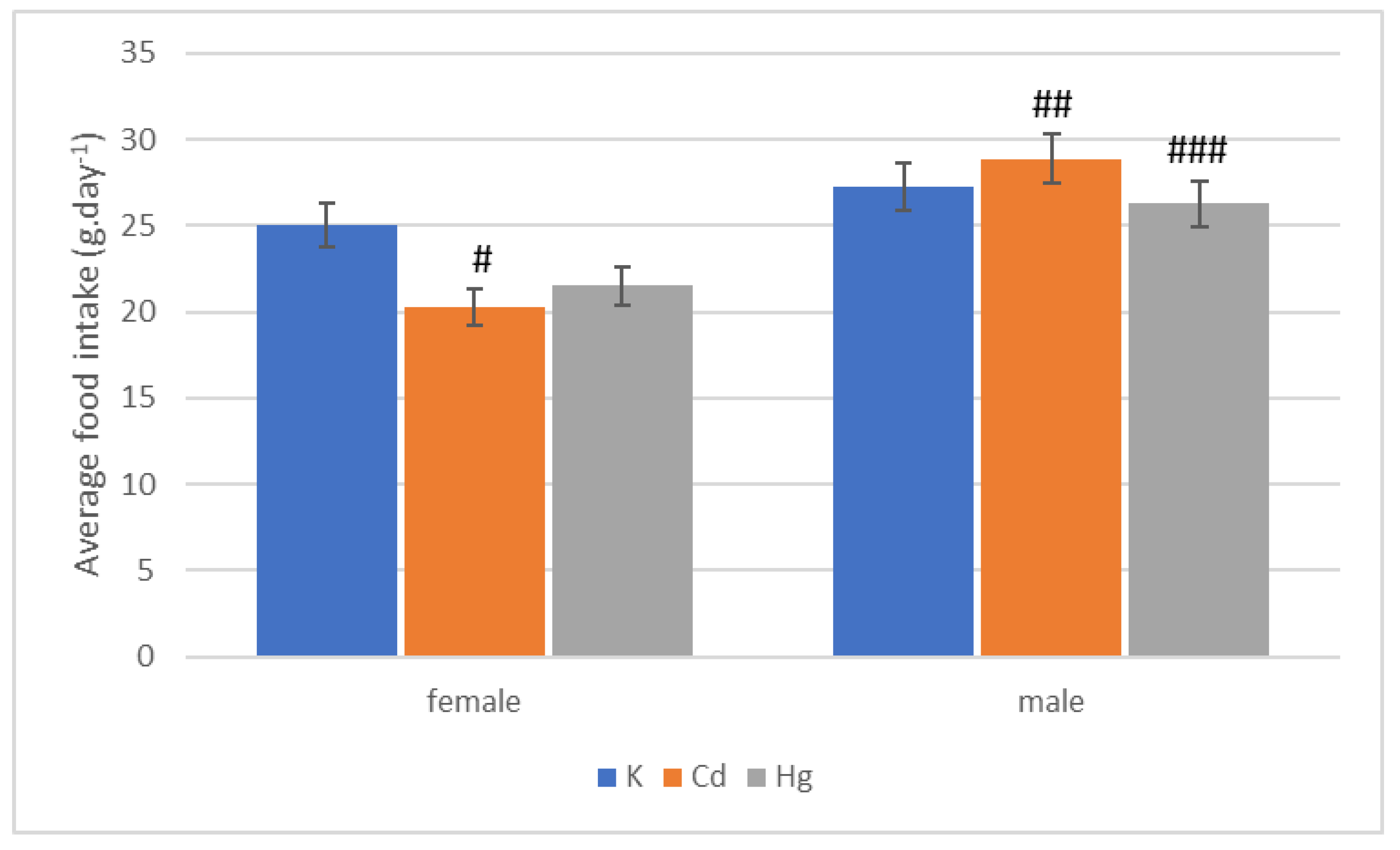
When evaluating the trends during the whole period of 52 weeks, the food intake in Cd and Hg female groups was lower than in the K/female group. In Cd/male group, the food intake was higher than in K/male group. In the Hg/male group, the intake was lower than in K/male group. In Cd and Hg female groups, a higher trend of food intake was recorded in the Hg group than in the Cd group, while, in male groups, a higher food intake was found in the Cd group than in the Hg group. A statistically significant difference was recorded between the Cd/female and Cd/male groups (p < 0.001), Hg/female and Hg/male groups (p = 0.023), and the Cd/female and K/female groups (p < 0.021). The food intake is presented in Figure 3.

3.2. Reproductive Experiment

The number of litters during the 52 weeks of the experiment was lower in the Cd and Hg groups than the K group. Among the exposed groups, a higher number of litters was recorded in the Cd group than in the Hg group. However, no statistically significant difference was noticed between the groups. The average number of litters is shown in Figure 4.
The number of born and raised pups during the whole period was lower in the Cd and Hg groups than in the K group. Among the exposed groups, a lower number of born and raised pups was recorded in the Cd group than in the Hg group, but no statistical significance was noted between the groups. The average number of pups born and raised during the experiment and the percentage of raised pups out of the total number of pups are presented in Figure 5.
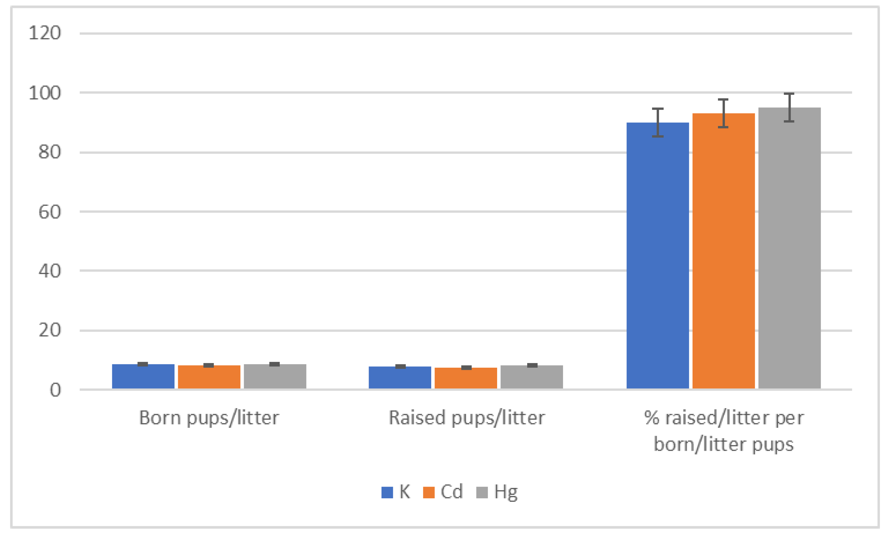
The number of born pups per litter and raised pups per litter was higher but insignificantly in the Hg group than in the K and Cd groups. The average number of born pups per litter and raised pups per litter and the percentage of raised pups per litter out of the total born pups per litter is presented in Figure 6.

3.3. Toxicological Experiment

ADD in the Cd group was slightly higher in female group than in male group during the experiment. The total dose received in the Cd group was higher for females than males. The intake expressed as percent of LD50 was also higher in female group than in male group. The ADD in the Hg group was slightly higher in male group than in female group. The total dose received and the intake expressed as percent of LD50 was slightly higher in female group than in male group.

4. Discussion

The study focused on identifying certain indicators as possible markers of exposure to chronic heavy metal intoxication (Cd, Hg) at doses 500 times higher than MPC. Therefore, the monitoring of selected physiological, toxicological and reproductive parameters in rats were included in the set of monitored indicators.

4.1. Toxicological Parameters

During the experiment, no death of the experimental animals was recorded in any of the experimental groups (Cd, Hg or K). Thus, the survival was 100% and at the same time it was confirmed that the applied dose is low chronic dose. In a lifelong experiment [9] evaluating the survival of animals exposed to cadmium, more individuals survived (one-year survival was also 100% as confirmed by the experiments) compared to other heavy metals. This may be because cadmium in low doses may function as an essential element [10,11,12]. Hijová et al. [13] also did not report any death of exposed animals during a chronic experiment in rats lasting 90 days, administering CdCl2 in drinking water per os with a predicted daily dose of cadmium of 2.5 mg·kg−1, similar to this study. A second possible explanation may be that rats received only 47.16% of LD50 for a single administration per os throughout the experiment, which may also have resulted in no mortality. As reported by Kotsonis and Klaasen [14], the LD50 for single intoxication of Cd ingested as CdCl2 per os in rats is 225 mg·kg−1 of live weight. The fact that no death of experimental animals was recorded, even in the group exposed to mercury, may be related to the low total dose received, the LD50 for Hg taken in the form of HgCl2 per os for single intoxication is a dose of about 37 mg·kg−1 of live weight [15]. In the experiments, rats received only 55.51% of LD50 for a single oral administration throughout the experiment.
At lifetime exposure to cadmium and mercury (156 days) in rats [16], HgCl2 was administered at a concentration of 1 µmol·L−1, i.e., 0.2 mg of Hg per 1 L of drinking water, and CdCl2 in a concentration of 20 µmol·L−1, i.e., 2.0 mg of Cd per 1 L of drinking water. In the group exposed to Cd, 80% of animals survived; in the group exposed to Hg, only 40% of animals survived. Almášiová et al. [17] stated that, at a concentration of 20 µmol·L−1 CdCl2 in drinking water, the survival of rats in the cadmium-exposed group was 10% lower than in the control group. In all the above works, however, no mortality was found in exposed animals for a period of one year, which is also confirmed by the experiments presented in this work. The mortality of animals in experiments with a similar focus can be significantly affected by breeding, handling conditions and overall welfare of the animals. For these reasons, in long-term experiments, it is necessary to adapt the conditions (experimental design) as much as possible to the natural method and the needs of the experimental animals. Ensuring adequate physical activity, daily physiological needs (e.g., shelter and cleaning) and well-being plays an important role and can fundamentally affect the results of an animal experiment. In the experiment, it is important to consider not only the duration of exposure but also other factors, e.g., concentration of toxic substance, light regime, handling and sampling time, heat stress, psychologically induced stress, etc. [18].

4.2. Physiological Parameters

In Cd/female and Hg/female groups, the water intake in the first two stages was lower compared to the K group, which may indicate the so-called refuse effect. From the third stage, the water intake was increased in both the exposed groups, although statistically insignificant, but at the end of the experiment it was higher than in the K group, which can be attributed to the so-called taste habit phenomenon [17,19]. In the male groups, the situation in Hg group was similar to the female groups. However, in the Cd/male group, a significant difference was noticed, as in the first two stages no rejection effect was recorded, but, during third and fourth stages, at the end of the experiment and in the evaluation of the whole monitored period, the water intake was higher than in the Hg and K groups. These differences in water intake between females and males, especially in the cadmium-exposed group, require further experimental verification. A similar phenomenon between the sexes at the same concentration has not been reported in the literature so far.
When evaluating the water intake during the entire period of 52 weeks, in the Hg/female group, water consumption was higher, and, in the Hg/male group, it was lower when compared to the K group. This is consistent with another study [20], suggesting that the higher intake of water after exposure was influenced by the kidney transport system and following the feeling of thirst, which is controlled by hypothalamus. A statistically significant difference (p < 0.001) was observed between sexes in the Cd and K groups between males and females. In the male group, a statistical significance was recorded between the Cd and Hg groups (p = 0.0145). In the Cd group, the intake of cadmium in the treated group was higher but statistically insignificant, as observed for both males and females. Ništiar et al. [19] indicated that a relatively long time is needed for low doses of cadmium to be observed in food and water intake variance. The decrease in food and water intake occurs approximately after 91–100 weeks. In the Hg group, food intake by females was higher than by males compared to the K group. However, statistical significance was not observed. One of the reasons for different food intake can be attributed to the irritant effect of mercury on the mucosa of the digestive tract and the fact that mercury may impair appetite regulation [21]. Some studies [22,23] have demonstrated that chronic exposure to low doses of mercury may have a difficult to predict course (but also consequence), and the response of the body to low (subtoxic) doses can vary. A statistical significance was observed between Cd/female and Cd/male groups (p < 0.001), Hg/female and Hg/male groups (p < 0.023) and K/females and Cd/female groups (p < 0.021).
In the study, the dynamics of food intake was the same in all groups in both males and females during the individual stages. However, an interesting result was observed in Cd/female group in the third quarter: a statistically significant reduction in food intake compared to the K group (p = 0.037) was observed. At the end of the experiment, food intake was higher in both sexes in the Cd group than in the K group, but not statistically significant. Wirth and Nijal [22] and Calabrese [3] stated that chronic exposure to low doses of mercury can have an unpredictable course (but also consequences) and the body’s response to low (subtoxic) doses may be different. Ništiar et al. [9] and Shibutani et al. [24] stated that the determining indicator of food intake (but also changes in weight and heavy metal intake) is water intake, which only partially corresponds to the results in this study. The opposite situation may also be the fact that food intake is limiting in this respect and water intake is derived from it. In male groups, the dynamics of food intake for the entire experimental period in all groups was similar to that of water intake. On the contrary, in female groups, the food intake in the Cd and Hg groups was lower than in the K group, which in the Hg group does not correspond to water intake, and this is an interesting finding that requires further experimental verification.
According to water and food intake, an increase in weight during the individual stages was expected. This fact was confirmed by the experiment. However, at the end of the experiment as well as when evaluating the whole experimental period, the weight of females and males in both exposed groups was lower than in the K group. In the Cd group, this may indicate possible liver cell damage. An additional possible explanation for low body weight of rats after the exposure to a low dose of Cd is that Cd is bound to the mucous membrane of the gastrointestinal tract after per os intake and stomach and intestinal epithelial cells can be impaired. A long-term intake of a low dose of Cd may induce lipid peroxidation that can result in the damage of the mucosal barrier, ultimately resulting in ulcers. Further increase of the permeability through the intestine wall and its subsequent impairment may cause diarrhea in rats. Some studies [25,26,27] have pointed to a possible link between low body weight and the weight of some organs (especially the hollow organs of the digestive tract). The overall toxic effect of the low dose of Cd administered to rats per os is signified by changes in weight of the organs, which consequently reflects in the changes of the overall body weight [25]. Damage to the organs can be confirmed or rejected with follow-up histopathological examination. Santana et al. [26] and Predes et al. [27] reported that the weight also depends on the duration of the experiment and the total exposure (dose) of the toxic substance. Toman et al. [28] reported that, already after nine weeks of cadmium administration, signs of liver cell damage were observed in the microscopic structure of the liver.
In the Cd/female group, the average daily dose was 0.29 mg·kg−1·day−1, receiving only 47.16% of LD50 per animal per os. In the Cd/male group, the average daily dose was 0.28 mg·kg−1·day−1, receiving only 45.97% of LD50 per os per animal. Based on these facts, the average daily dose of Cd for both males and females was the same over the entire experimental period. As reported by Kotsonis and Klaasen [14], the LD50 for a single Cd intoxication received in the form of CdCl2 per os in rats is a dose of 225 mg·kg−1 of live weight. Based on that, the average daily dose of cadmium in both females and males can be considered as a low dose for chronic exposures. The dose of Lowest-Observed-Adverse-Effect (LOAEL) for cadmium is 1.5–17.5 mg·kg−1·day−1 [29], which was not exceeded in conducted experiments. If the Minimal Risk Level (MRL) is considered for per os chronic exposure whose value is 0.1 µg·kg−1·day−1 for Cd [29], then the value was exceeded approximately 3000 times. With long-term intake of low doses cadmium, this toxic element is accumulated in the peripheral and central nervous system [30], contributes to osteoporosis and is carcinogenic (causing prostate and lung cancer) [31]. In the Hg/female group, the average daily dose was 0.02 mg·kg−1·day−1, receiving only 57.59% of LD50 per animal per os. In the Hg/male group, the average daily dose was 0.03 mg·kg−1·day−1, receiving only 55.51% of LD50 per os per animal. LD50 for Hg received in the form of HgCl2 per os for single intoxication is approximately 37 mg·kg−1 of live weight [15]. Almášiová et al. [17] found that intoxication did not result in death of animals in the period of over one year. In both female and male groups, this value can be considered as a low dose for chronic exposures.The LOAEL dose for mercury is 1.9–3.9 mg·kg−1·day−1. In the experiments in both sexes, this dose was not exceeded. If MRL is considered for per os chronic exposure whose value is 0.007 µg·kg−1·day−1 for Hg [31], then the value was exceeded approximately 9000 times.

4.3. Reproduction Parameters

In the Cd group, the number of litters, the number of born pups and the number of raised pups for the entire period of the experiment were lower than in the K group. Lower numbers of born pups per litter and raised pups per litter compared to the K group were recorded. Cd acts preferentially through deprivation of testosterone secretion [16], as well as through the hypothalamus—pituitary —testes axis [11]. Predes et al. [27] indicated that, even a very small difference in the administered dose of Cd in males causes a sudden increase in testicular damage. Thompson [32] stated that cadmium can affect the female reproductive and endocrine systems and hormone levels change. It is assumed that lower values of some monitored reproductive parameters in the Cd group compared to the K group indicate a decrease in reproductive function in rats. In general, the number of born pups per litter and the number of raised pups per litter are among the most important indicators of reprotoxicity. A similar conclusion was achieved at a concentration of 20 mmol·L−1, i.e., 2.0 mg Cd per liter [16]. The body’s response to low doses may vary [3]. One example of the unexpected hormonal effect of a toxic substance is the finding that cadmium acted as a hormonally active substance even in concentrations that were considered safe. In a multigenerational study in rats at doses up to 100 mg Cd per kg of live weight no adverse effects on reproduction have been reported. In a four-generation study at a dose of 1 mg·L−1 in water and at a mean daily dose of 0.125 mg Cd·kg−1·day−1, no adverse effects on fertility were observed in rats and mice. However, at a dose of 50 mg Cd·kg−1 of live weight, slight testicular changes were observed in rats during a 15-month period [33]. In terms of the effect of cadmium on animals in chronic poisoning, the highest accumulation of cadmium occurred in rabbits (females) in the ovaries (0.47 mg·kg−1), uterus (0.25 mg·kg−1) and testes (0.10 mg·kg−1) [34]. Soukupová and Dostál [35] described the embryotoxic effects of cadmium in rodents, i.e., fetal malformations (cleavage of the climate, oligodactylia, polydactylia, exencephalia and others).
In the Hg group, the numbers of litters born pups and raised pups for the entire period of the experiment were lower compared to the K group. The number of born pups per litter and the number of raised pups per litter in the Hg group were higher but insignificantly compared to the K group, which could be attributed to the adaptation process to a certain environment with a certain load factor. However, it is very similar to hormesis after exposure to a low dose of harmful substances. At low doses of mercury, there may be no signs of a toxic effect on the body. However, it can cause increased production of proteins from the group of matalothionines, which bind heavy metals and ensure their subsequent removal from the body. In addition, the role of metallothionein is to protect cells from further damage (e.g., from heritable information damage). Therefore, it is possible that the low dose of mercury was not only not dangerous for the organism, but also contributed to the organism becoming more resistant to the adverse effects to which it was exposed. A similar finding was made by Lukačínová et al. [36] but at a concentration 200 times higher than the MPC in water containing HgCl2 at a concentration of 1 mmol·L−1 (0.2 mg Hg·L−1). If the results in this study are attributed to the hormetic effect, there can be a logical explanation. The low doses of the toxic substance used in the cells did not have to cause much damage; on the contrary, they were able to trigger protective reactions in the cell which prevented the action of other adverse effects. However, this assumption, based on the results of this study, requires further experimental studies. The body’s response to different low doses of heavy metals may be different. It is known that high doses inhibit growth, shorten life expectancy and reduce fertility, while low doses can improve some parameters. A higher number of born pups (rats) with chronic exposure to heavy metals (significantly higher doses and significant mortality) than in the experiments in this study was described by Kotsonis et al. [14]. Harmful fetal development and decreased fertility in rats of both sexes were also confirmed [21]. Comparing the results and their effect on reproductive parameters between low and high dose studies is quite challenging because it is a confirmation of the exact mechanism of action and role of reactive oxygen species and metallothionein in chronic exposure to heavy metals. It is also necessary to take into account the effect of the components of innate and adaptive immunity, as well as the defense mechanisms that could be activated by gradually increasing the concentration of the toxic substance (albeit in the range of low doses) in the environment.

5. Conclusions

The analysis of the current state of knowledge and subsequent experiments showed an interesting and more comprehensive view of the direction of new experimental efforts. The results will contribute to the expansion of knowledge about chronic intoxication with subtoxic doses of selected heavy metals in drinking water of rats. In the study of the physiological and reproductive parameters monitored, there were differences both between the exposed groups and between the sexes, which confirms that these indicators are important in experiments of this type. The mean daily dose of cadmium and mercury in both males and females in the study was almost at the same level and can be considered low for chronic exposures. At the same time, however, it can be stated that it imitates the natural way of possible population exposure. The most important task for the future is to finalize the obtained data by supplementation of data on the distribution of selected heavy metals in the organism and their influence on specific organs and tissues.

Author Contributions

Data curation, I.C., T.K., H.P., T.B., D.K. and A.S.; formal analysis, I.C., T.K., H.P., T.B., D.K. and A.S.; investigation, I.C. and T.K.; methodology, I.C., T.K., H.P. and T.B.; resources, I.C., T.K., H.P., T.B., D.K. and A.S.; validation, I.C., T.K., H.P., T.B. and D.K.; writing—original draft, I.C., T.K., H.P., T.B., D.K. and A.S.; writing—review and editing, I.C., T.K., H.P., T.B., D.K. and A.S. All authors have read and agreed to the published version of the manuscript.

Funding

This research received no external funding.

Conflicts of Interest

The authors declare no conflict of interest.

References

  1. Piršelová, B.; Galuščáková, Ľ.; Lengyelová, L. Hormetic response of plants to metals and metalloids. Chem. Listy 2018, 112, 317–323. [Google Scholar]
  2. Kimáková, T. Toxická ortuť v organizme človeka. Via Pract. 2018, 15, 222–225. [Google Scholar]
  3. Calabrese, E.J. Cancer biology and hormesis: Human tumor cell lines commonly display hormeti (biphasic) dose responses. Crit. Rev. Toxicol. 2005, 35, 463–592. [Google Scholar] [CrossRef] [PubMed]
  4. Mattson, M.P.; Calabrese, E.J. Hormesis A Revolution in Biology, Toxicology and Medicine, 1st ed.; Humana Press: Totowa, NJ, USA; Springer: Berlin/Heidelberg, Germany, 2010; pp. 1–213. [Google Scholar]
  5. Zhou, F.; Yin, G.; Gao, Y.; Ouyang, L.; Liu, S.; Jia, Q.; Yu, H.; Zha, Z.; Wang, K.; Xie, J.; et al. Insights into cognitive deficits caused by low-dose toxic heavy metal mixtures and their remediation through a postnatal enriched environment in rats. J. Hazard. Mater. 2020, 388, 122081. [Google Scholar] [CrossRef]
  6. Ahmed, N.F.; Sadek, K.M.; Soliman, M.K.; Khalil, R.H.; Khafaga, A.F.; Ajarem, J.S.; Maodaa, S.N.; Allam, A.A. Moringa oleifera leaf extract repairs the oxidative misbalance following sub-chronic exposure to sodium fluoride in nile tilapia Oreochromis niloticus. Animals 2020, 10, 626. [Google Scholar] [CrossRef] [Green Version]
  7. Budnik, L.T.; Baur, X. The assessment of environmental and occupational exposure to hazardous substances by biomonitoring. Dtsch. Arztebl. Int. 2009, 106, 91–97. [Google Scholar] [CrossRef] [PubMed]
  8. Checkoway, H.; Savitz, D.A.; Heyer, N.J. Assessing the effects of nondifferential misclassification of exposures in occupational studies. Appl. Occup. Environ. Hyg. 1991, 6, 528–533. [Google Scholar] [CrossRef]
  9. Ništiar, F.; Rácz, O.; Lukačinová, A.; Hubková, B.; Nováková, J.; Lovásová, E.; Sedláková, E. Age dependency on some physiological and biochemical parameters of male Wistar rats in controlled environment. J. Environ. Sci. Health 2012, 47, 1224–1233. [Google Scholar] [CrossRef] [PubMed]
  10. Anke, M.; Dorn, W.; Müller, M.; Seifert, M. Recent progress in exploring the essentiality of the ultratrace element cadmium to the nutrition of animals and man. Biomed. Res. Trace Elem. 2005, 16, 198–202. [Google Scholar]
  11. Lafuente, A.; Marquez, N.; Perez-Lorenzo, M.; Pazo, D.; Esquifino, A. Cadmium Effects on Hypothalamic-Pituitary-Testicular Axis in Male Rats. Exp. Biol. Med. 2001, 226, 605–611. [Google Scholar] [CrossRef]
  12. Abou-Kassem, D.E.; Abd El-Hack, M.E.; Taha, A.E.; Ajarem, J.S.; Maodaa, S.N.; Allam, A.A. Detoxification impacts of ascorbic acid and clay on laying japanese quail fed diets polluted by various levels of cadmium. Animals 2020, 10, 372. [Google Scholar] [CrossRef] [PubMed] [Green Version]
  13. Hijová, E.; Ništiar, F.; Lovásová, E.; Šipolová, A. Ascorbic acid malondialdehyde in rats after exposure to mercury. Trace Elem. Electrolytes 2003, 20, 244–248. [Google Scholar] [CrossRef]
  14. Kotsonis, F.; Klaasen, C.D. Toxicity and distribution of cadmium administered to rats at sublethal doses. Toxicol. Appl. Pharmacol. 1977, 41, 667–680. [Google Scholar] [CrossRef]
  15. Donaldson, M.L.; Gubler, C.J. Biochemical effects of mercury poisoning in rats. Am. J. Clin. Nutr. 1978, 31, 859–864. [Google Scholar] [CrossRef] [PubMed] [Green Version]
  16. Lukačínová, A.; Rácz, O.; Lovászová, E.; Ništiar, F. Effect of lifetime low dose exposure to heavy metals on selected serum proteins of Wistar rats during three subsequent generations. Ecotoxicol. Environ. Saf. 2011, 74, 1747–1755. [Google Scholar] [CrossRef]
  17. Almášiová, V.; Holovská, K.; Cigánková, V.; Ništiar, F. Effect of lifetime lowdose exposure to cadmium on lipid metabolism of wistar rats. J. Microbiol. Biotechnol. Food Sci. 2012, 2, 293–303. [Google Scholar]
  18. Baumans, V. Environmental enrichment for laboratory rodents and rabbits: Requirements of rodents, rabbits, and research. ILAR J. 2005, 46, 162–170. [Google Scholar] [CrossRef]
  19. Ništiar, F.; Beňačka, R.; Lovásová, E.; Rácz, O.; Lukačínová, A. Selected physiological and biochemical indices at lifetime low-dose heavy metal exposure in rats. In Proceedings of the 28th Symposium Industrial Toxicology, Štrba, Slovakia, 1–4 June 2008; Slovak University of Technology: Bratislava, Slovakia, 2008; pp. 162–172. [Google Scholar]
  20. Bergman, F.; Chaimovity, M.; Gutman, Y.; Zerachia, A. Effect of mercury derivates, implanted into the hypothalamus, on the water intake of albino rats. Br. J. Pharmacol. Chemother. 1968, 32, 483–492. [Google Scholar] [CrossRef] [Green Version]
  21. Counter, S.A.; Buchanan, L.H. Mercury exposure in children: A review. Toxicol. Appl. Pharmacol. 2004, 198, 209–230. [Google Scholar] [CrossRef]
  22. Wirth, J.J.; Mijal, R.S. Adverse effects of low level heavy metal exposure on male reproductive function. Syst. Biol. Reprod. Med. 2010, 56, 147–167. [Google Scholar] [CrossRef] [Green Version]
  23. Calabrese, E.J. Hormetic-dose response relationships in immunology: Occurrence, quantitative features of the dose response, mechanisti foundations, and clinica implications. Crit. Rev. Toxicol. 2008, 35, 89–295. [Google Scholar] [CrossRef]
  24. Shibutani, M.; Mitsumori, K.; Satoh, S.; Hiratsuka, H.; Satoh, M.; Sumiyoshi, M.; Nishijima, M.; Katsuki, Y.; Suzuki, J.; Nakagawa, J.; et al. Relationship between toxicity and cadmium accumulation in rats given low amounts of cadmium chloride or cadmium-polluted rice for 22 months. J. Toxicol. Sci. 2001, 26, 337–358. [Google Scholar] [CrossRef] [PubMed] [Green Version]
  25. Toman, R.; Massányi, P.; Lukáč, N.; Mojžišová, M. Effect of cadmium on the growth of some rat organs after long-term oral administration in feed. In Proceedings of the Risk Factors in the Food Chain III, Nitra, Slovakia, 3 December 2003; Massányi, P., Toman, R., Lukáč, N., Eds.; Slovak University of Agriculture in Nitra: Nitra, Slovakia, 2003; Volume 67, pp. 162–164. [Google Scholar]
  26. Santana, M.G.; Moraes, R.; Bernardi, M.M. Toxicity of cadmium in Japanese quail: Evaluation of body weight, hepatic and renal function, and cellular immune response. Environ. Res. 2005, 99, 273–277. [Google Scholar] [CrossRef] [PubMed]
  27. Predes, F.S.; Diamante, M.A.S.; Dolder, H. Testis response to low doses of cadmium in Wistar rats. Int. J. Exp. Pathol. 2010, 91, 125–131. [Google Scholar] [CrossRef] [PubMed]
  28. Toman, R.; Massanyi, P.; Zakrzewski, M.; Stawarz, R.; Hluchy, S.; Kulac, N.; Mindek, S.; Formicki, G. Effect of cadmium on blood parameters of pheasants (Phasianus colchicus) alter long-term administration in drinkng water. J. Physiol. Pharmacol. 2006, 57, 235. [Google Scholar]
  29. Toxicological Profile for Cadmium. Available online: https://www.atsdr.cdc.gov/toxprofiles/tp5.pdf (accessed on 1 August 2019).
  30. Alloway, B.J. Soil processes and the behaviour of metals. In Heavy Metals in Soils, 2nd ed.; Alloway, B.J., Ed.; Springer: Dordrecht, The Netherlands, 1995. [Google Scholar]
  31. Toxicological Profile for Mercury. Available online: https://www.atsdr.cdc.gov/toxprofiles/mercury_organic_addendum.pdf (accessed on 1 August 2019).
  32. Thompson, J.; Bannigan, J. Cadmium: Toxic Effects on the Reproductive System and the Embryo. Reprod. Toxicol. 2008, 25, 304–315. [Google Scholar] [CrossRef] [PubMed]
  33. Cadmium in Drinking-Water. Available online: https://apps.who.int/iris/bitstream/handle/10665/75364/WHO_SDE_WSH_03.04_80_eng.pdf?sequence=1&isAllowed=y (accessed on 1 August 2019).
  34. Massanyi, P.; Toman, R.; Valent, M.; Čupka, P. Evaluation of selected parameters of a metabolic profile and levels of cadmium in reproductive organs of rabbits after an experimental administration. Acta Physiol. Hung. 1995, 83, 267–273. [Google Scholar]
  35. Dostál, M.; Soukupová, D. Development toxicity of cadmium in mice. I. Embryotoxic effects. Funct. Dev. Morphol. 1991, 1–2, 3–9. [Google Scholar]
  36. Lukačínová, A.; Beňačka, R.; Lovásová, E.; Rácz, O.; Ništiar, F. Reproduction parameters in low dose chronic exposure with heavy metals in rats. Pol. J. Environ. Stud. 2008, 17, 911–915. [Google Scholar]
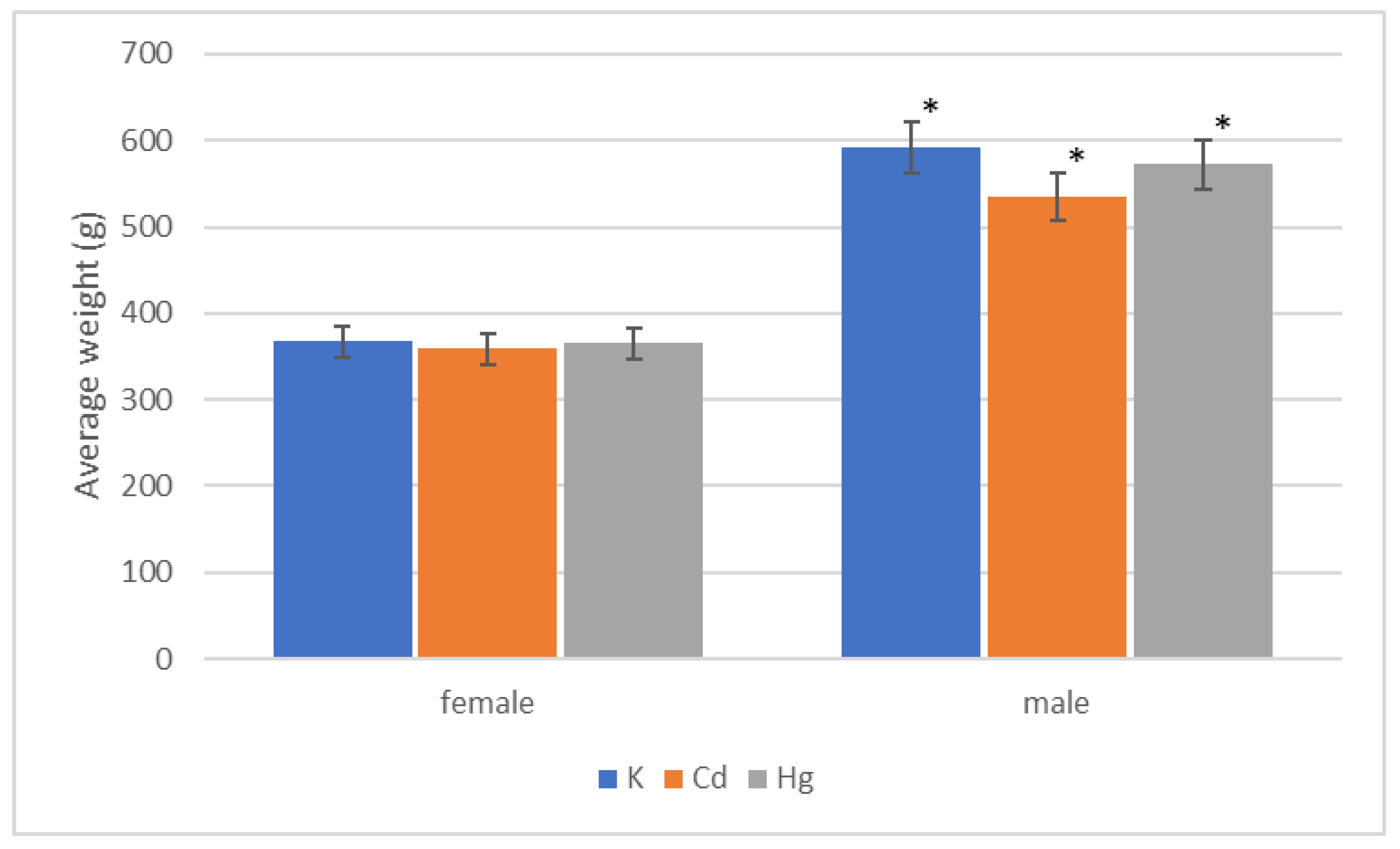
Figure 1. A comparison of weights after 52 weeks of the experiment. * p < 0.001, for all male groups compared to corresponding female groups; n = 8 per group.
Applsci 10 08066 g001
Figure 2. A comparison of water intake after 52 weeks of the experiment. ** p < 0.001, for K/Cd groups compared to corresponding female groups; *** p < 0.05, for Cd male group compared to Hg male group; n = 8 per group.
Applsci 10 08066 g002
Figure 3. A comparison of food intake after 52 weeks of the experiment. # p < 0.05, for K female group compared to Cd female group; ## p < 0.001, for Cd female group compared to Cd male group; ### p < 0.05, for Hg female group compared to Hg male group; n = 8 per group.
Applsci 10 08066 g003
Figure 4. A comparison of litters. n = 8 per group.
Applsci 10 08066 g004
Figure 5. A comparison of average number of pups born and raised during the experiment and the percentage of raised pups out of the total number of pups. n = 8 per group.
Applsci 10 08066 g005
Figure 6. A comparison of average number of pups born and raised per litter during the experiment and the percentage of pups raised/litter out of the born/litter. n = 8 per group.
Applsci 10 08066 g006
Table 1. Average weight of female rats ± SD.
GroupStart End of 1st QuarterEnd of 2nd QuarterEnd of 3rd QuarterEnd of 4th Quarter
K (g)70.62 ± 1.47272.50 ± 9.77327.50 ± 6.39333.75 ± 8.85367.14 ± 12.09
Cd (g)72.50 ± 1.33258.75 ± 8.11306.25 ± 13.75307.50 ± 15.32358.75 ± 21.50
Hg (g)81.25 ± 3.98277.50 ± 6.19331.25 ± 11.56335.00 ± 6.81365.00 ± 14.01
Note: n = 8 per group.
Table 2. Average weight of male rats ± SD.
GroupStartEnd of 1st QuarterEnd of 2nd QuarterEnd of 3rd QuarterEnd of 4th Quarter
K (g)73.12 ± 1.87401.25 ± 6.92488.57 ± 8.84512.85 ± 17.00592.85 ± 10.84
Cd (g)77.5 ± 5.34396.25 ± 9.62492.50 ± 9.77492.550 ± 9.77535.00 ± 14.63
Hg (g)86.87 ± 3.12395.00 ± 13.75471.25 ± 13.28491.25 ± 16.84572.50 ± 23.43
Note: n = 8 per group.
Table 3. Average water intake of female rats ± SD.
Group1st Quarter2nd Quarter3rd Quarter4th QuarterWhole
K (mL·day−1)36.18 ± 1.1638.31 ± 1.0839.40 ± 0.5038.84 ± 0.8337.76 ± 0.76
Cd (mL·day−1)34.51 ± 1.4832.79 ± 0.9936.01 ± 1.2841.94 ± 1.4535.86 ± 1.12
Hg (mL·day−1)38.54 ± 1.3235.67 ± 1.9738.44 ± 2.7842.32 ± 2.9438.42 ± 1.82
Note: n = 8 per group.
Table 4. Average water intake of male rats ± SD.
Group1st Quarter2nd Quarter3rd Quarter4th QuarterWhole
K (mL·day−1)44.97 ± 0.5850.85 ± 4.15250.17 ± 5.0148.85 ± 5.1948.35 ± 3.49
Cd (mL·day−1)51.98 ± 1.9153.25 ± 0.75054.04 ± 0.6960.04 ± 1.9354.45 ± 0.95
Hg (mL·day−1)45.91 ± 1.8641.25 ± 1.02546.80 ± 1.9248.55 ± 3.6145.39 ± 2.22
Note: n = 8 per group.
Table 5. Average food intake of female rats ± SD.
Group1st Quarter2nd Quarter3rd Quarter4th QuarterWhole
K (g·day−1)27.51 ± 1.3226.49 ± 2.8730.16 ± 4.36 §16.82 ± 0.5425.06 ± 2.14
Cd (g·day−1)24.45 ± 0.5219.36 ± 0.6220.71 ± 0.56 §17.95 ± 0.5520.28 ± 0.52
Hg (g·day−1)27.17 ± 0.6219.01 ± 0.3622.75 ± 0.5517.92 ± 0.4621.5 ± 0.39
Note: § p = 0.037, for K/female group compared to Cd/female group; n = 8 per group.
Table 6. Average food intake of male rats ± SD.
Group1st Quarter2nd Quarter3rd Quarter4th QuarterWhole
K (g·day−1)30.57 ± 0.2527.17 ± 0.9328.72 ± 0.5422.92 ± 0.6227.22 ± 0.28
Cd (g·day−1)32.26 ± 0.2227.81 ± 0.5331.24 ± 0.6224.72 ± 0.5428.86 ± 0.18
Hg (g·day−1)31.24 ± 0.9624.11 ± 0.4228.19 ± 0.9822.18 ± 0.8226.25 ± 0.72
Note: n = 8 per group.
Publisher’s Note: MDPI stays neutral with regard to jurisdictional claims in published maps and institutional affiliations.