Article

Predicting Intestinal and Hepatic First-Pass Metabolism of Orally Administered Testosterone Undecanoate

1
College of Pharmacy, University of Anbar, Ramadi 31001, Iraq
2
School of Pharmacy, University of Nottingham, Nottingham NG7 2RD, UK
3
Ernest Mario School of Pharmacy, Rutgers, The State University of New Jersey, Piscataway, NJ 08854, USA
*
Author to whom correspondence should be addressed.
Appl. Sci. 2020, 10(20), 7283; https://doi.org/10.3390/app10207283
Submission received: 2 October 2020 / Revised: 13 October 2020 / Accepted: 15 October 2020 / Published: 18 October 2020
(This article belongs to the Special Issue Towards a Systems Biology Approach)

Abstract

:
The bioavailability of orally administered drugs could be impacted by intestinal and hepatic first-pass metabolism. Testosterone undecanoate (TU), an orally administered ester prodrug of testosterone, is significantly subjected to first-pass metabolism. However, the individual contribution of intestinal and hepatic first-pass metabolism is not well determined. Therefore, the aim of the current study was to predict the metabolic contribution of each site. The hydrolysis–time profiles of TU incubation in human liver microsomes and Caco-2 cell homogenate were used to predict hepatic and intestinal first-pass metabolism, respectively. The in vitro half-life (t1/2 inv) for the hydrolysis of TU in microsomal mixtures was 28.31 ± 3.51 min. By applying the “well-stirred” model, the fraction of TU that could escape hepatic first-pass metabolism (FH) was predicted as 0.915 ± 0.009. The incubation of TU in Caco-2 cell homogenate yielded t1/2 inv of 109.28 ± 21.42 min, which was applied in a “Q gut” model to estimate the fraction of TU that would escape intestinal first-pass metabolism (FG) as 0.114 ± 0.02. Accordingly, only 11% of the absorbed fraction of TU could escape intestinal metabolism, while 91% can pass through hepatic metabolism. Hence, compared to the liver, the intestinal wall is the main site where TU is significantly metabolised during first-pass effect.

1. Introduction

Testosterone undecanoate (TU, Figure 1) is an ester prodrug of the anabolic steroid, testosterone. TU was introduced in the late 1970s as an effective oral testosterone replacement therapy [1,2]. In fact, testosterone is well absorbed from the intestinal lumen. However, therapeutic plasma levels following oral administration of testosterone cannot be achieved due to extensive first-pass metabolism [3]. Therefore, TU and other testosterone prodrugs have been proposed to reach effective levels of testosterone by avoiding pre-systemic metabolism and/or to provide more flexible therapeutic options in terms of the route of administration, and stability of plasma levels attained [4]. Once absorbed to the systemic circulation, the undecanoate side chain of TU is cleaved by the action of non-specific plasma esterase to release testosterone [5]. Currently, TU is commercially available as oral capsules, Andriol Testocaps® and Jatenzo®, and intramuscular injections, Aveed® and Nebido®. These formulations are commonly used in the treatment of male hypogonadal disorders characterized by low serum levels of testosterone [6].
Several animal studies have explored the mechanisms involved in the oral bioavailability of TU. Coert and colleagues have demonstrated that the metabolism of TU starts in the intestinal wall of rats, and part of the fraction that escapes intestinal metabolism is absorbed via the intestinal lymphatics [7]. Geelen et al. have also shown that TU is metabolised in the intestinal wall, as metabolites of TU could be detected in the portal vein of rats [8]. In lymph duct-cannulated dogs, it was shown that the bioavailability of orally administered TU was as low as 3%, almost all of which was absorbed through intestinal lymphatics [9]. In humans, Horst et al. have placed thoracic duct catheters in four participants following neck dissection for the resection of left mouth tumours [10]. In that study, following the oral administration of radiolabelled TU, the thoracic lymph was rich with TU and its main metabolite 5α-dihydrotestosterone undecanoate (5α-DHTU). However, both were undetectable in the systemic circulation. It should be mentioned that 5α-DHTU is a product of the metabolic activity of 5α-reductase on TU. Several other human studies have also suggested that lymphatically transported TU is the main source of testosterone in the systemic circulation [11,12,13], and the bioavailability of testosterone increases as the fat-content of the co-administered food increases [14]. To note, fatty-food increases intestinal lymphatic delivery of lipophilic drugs that have high lymphatic transport potential [15]. However, the systemic bioavailability of testosterone following the oral administration of TU in conditions that facilitate intestinal lymphatic transport was still as low as 6.8% in humans [1].
Taking together the above-mentioned observations, it is now well established that the intestinal wall contributes to the metabolism of TU. The fraction that survives intestinal first-pass metabolism is partly absorbed through the lymphatic system while the remaining part is delivered via the portal vein to the liver where it is substantially metabolised [16]. Both intestinal and hepatic first-pass metabolism contribute to the low bioavailability of TU. However, the relative contribution of each metabolic site (intestinal wall versus liver) is still unclear. Once the major site of TU metabolism is revealed, a clear insight for an approach to improve the bioavailability of TU could be achieved. Therefore, the aim of the current study was to predict the individual impact of intestinal and hepatic first-pass metabolism on the bioavailability of orally administered TU.

2. Materials and Methods

2.1. Materials

Testosterone undecanoate (TU; CAS: 5949-44-0; purity: 99%) was bought from Beijing Sjar Technology Development Co., Ltd. (Beijing, China). Vitamin D3 (CAS: 67-97-0; purity: 99%) was purchased from Alfa Aesar (Lancashire, UK). KH2PO4, K2HPO4, MgCl2, and NH₄CH₃CO₂ were purchased from Sigma (Dorset, UK). Nicotinamide adenine dinucleotide phosphate tetrasodium salt hydrate (NADPH) was bought from Fisher Scientific (Leicestershire, UK). Liver microsomes from human donors were obtained from Gibco Invitrogen (Paisley, UK). Caco-2 cells (passage number 47) were obtained from Cell Culture Collections, Public Health England (Salisbury, UK). Dulbecco’s modified eagle medium (DMEM) supplemented with GlutaMAX™, 4.5 g/L D-glucose and 25 mM 4-(2-hydroxyethyl)-1-piperazineethanesulfonic acid (HEPES) buffer was obtained from Gibco (Paisley, UK). Hank’s balanced salt solution (HBSS), HEPES buffer, fetal bovine serum (FBS), and all compounds used in the study were obtained from Sigma (Gillingham, UK). All other reagents were of analytical grade or higher.

2.2. Prediction of Hepatic First-Pass Metabolism

Microsomal metabolism stability studies were conducted to predict the fraction of TU that could escape hepatic first-pass metabolism (FH). Stability studies were performed using human liver microsomes. The in vitro intrinsic clearance (Clint, mL/min/mg protein) was calculated using the in vitro half-life (t1/2 inv) of TU depletion in human liver microsomes according to Equation (1) [17,18].
C l int = 0.693 i n   v i t r o   t 1 / 2 · mL ,   incubation   medium mg ,   microsomes
Microsomal incubations were performed as previously described [17]. Briefly, the reaction mixture consisted of human microsomal proteins and MgCl2 dissolved in solution a of potassium phosphate buffer (KH2PO4/K2HPO4, pH 7.4). TU was accurately weighed and dissolved in aqueous acetonitrile (50% v/v). Reaction tubes containing the reaction mixture were placed in a temperature-controlled orbital shaker at 37 °C and shaken at 200 rpm (Thermo Scientific MaxQ4000, Waltham, MA, USA). Following a pre-incubation period of 3 min, the reaction was initiated by the addition of TU and the NADPH solution in the potassium phosphate buffer. TU solution in potassium phosphate buffer without NADPH was added to another set of reaction tubes as a control. The concentration of acetonitrile in the final mixture was less than 1%. The final concentrations of TU, human liver microsomes, MgCl2, KH2PO4/K2HPO4, and NADHP were 1 µM, 0.5 mg/mL, 10 mM, 84.7 mM, and 1 mM, respectively. Two-hundred microlitre aliquots were withdrawn at 0 min, 10 min, 20 min, and 30 min and placed in a test tube that contained 300 µL ice-cold acetonitrile to stop the reaction. Samples were then processed on the same day for high-performance liquid chromatography (HPLC) analysis as described below. All experiments were performed in triplicates.
Clint was then corrected by the fraction of drug unbound in the incubation medium (fu,mic) according to Equation (2) to find the unbound intrinsic clearance (Clu,int) [19].
C l u , int = C l int f u , mic
The experimental determination of fu,mic for highly lipophilic drugs is quite challenging due to significant non-specific binding [20]. In addition, predictive tools for fu,mic, such as those suggested by Austin et al. [21] and Hallifax and Houston [22], were demonstrated to be unreliable for drugs with a logP value ≥ 5 such as TU (logP = 9.15 [23]) [20]. Therefore, the binding of TU to microsomal proteins was assumed to be similar to that of plasma proteins, corrected by protein concentration [18]. Based on this, fu,mic was calculated by Equation (3) [24].
f u , mic = 1 ( C mic C pp   ×   ( 1   f u , p ) f u , p ) + 1
where Cmic is the microsomal protein concentration (0.5 mg/mL), Cpp is the concentration of human plasma proteins (70 mg/mL [25]), and fu,p is the fraction of TU unbound in human plasma. The value of fu,p for TU was estimated by the online prediction tool (https://drumap.nibiohn.go.jp/) [26].
The calculated value of Clu,inc was then scaled, based on human physiological factors of 32 mg microsomes/g liver and 22 g liver/kg body weight [18,27,28], to find the hepatic intrinsic clearance (ClH,inc, mL/min/kg). Hepatic clearance (ClH) was then calculated assuming a well-stirred model using Equation (4)
C l H = Q   ×   f u , p   ×   C l H ,   inc Q +   f u , p   ×   C l H ,   inc
where Q is liver blood flow (21 mL/min/kg) [29].
Finally, FH was calculated from ClH as indicated in Equation (5)
F H = ( 1   C l H / Q )

2.3. Prediction of Intestinal First-Pass Metabolism

2.3.1. Preparation of Caco-2 Cell Homogenate

Caco-2 cells (passage numbers 53) were seeded in 75 cm2 cell culture flasks (Corning Inc, Corning, NY, USA) at 3.75 × 10⁴ cells/cm2 and cultured in DMEM supplemented with 10% FBS and 1% penicillin-streptomycin. Cells were kept at 37 °C, 95% relative humidity, and 5% CO2. The medium was replaced three times a week for 21 days. Confluent cells were then scraped and washed twice with phosphate buffer saline. Cells were lysed by ultrasonication on ice followed by centrifugation at 10,000× g for 5 min at 4 °C. Bradford assay was used to determine the protein content of the supernatant according to the manufacturer protocol (Alfa Aesar, Lancashire, UK). Protein concentration was diluted to 1 mg/mL for the hydrolysis study.

2.3.2. Hydrolysis Experiments

Human intestinal Caco-2 cell homogenates were used to assess the stability of TU; Caco-2 cell homogenate was previously shown to be an equivalent alternative to human small intestinal homogenate for ester prodrug stability studies [30]. Clint (mL/min/mg protein) was calculated from the in vitro half-life (t1/2 inv), which was measured from the first-order depletion constant of TU incubation in human intestinal Caco-2 cell homogenate. Hydrolysis experiments were performed as previously described [31]. Briefly, cell homogenates were spiked with TU to give a final concentration of 10 μM. The reaction mixtures were incubated at 37 °C and shaken at 200 rpm for 1 h. Samples were withdrawn at 0 min, 15 min, 30 min, 45 min, and 60 min time points. The reaction was then immediately terminated by the addition of ice-cold acetonitrile. Samples were processed on the same day for HPLC analysis, as described below. Hydrolysis experiments were performed in quadruplets.
Clint was then corrected by fu,mic to find Clu,int (mL/min/mg protein) as described for the microsomal stability studies using Equations (2) and (3). The calculated value of Clu,inc was then scaled, based on human physiological factors for the total weight of enterocytes in the small intestine, to calculate gut intrinsic clearance (ClG,int, L/h). It was previously determined that average regional weights of enterocytes in the duodenal, jejunal, and ileal parts of the small intestine are 18.2 g, 65.8 g, and 38.3 g, respectively [32].
The fraction of TU that could escape intestinal first-pass metabolism (FG) was estimated using the “Q gut” model as detailed in Equation (6) [33]:
F G = Q villi Q villi +   f u , G   .   C l G ,   int · ( 1 + Q v i l l i / C l Perm )
where Qvilli is the villous blood flow (18 L/h) [34], Clperm is the permeability across enterocytes, and fu,G is the fraction of drug unbound to enterocytes, which was assumed to be 1 as this value has been shown to give the most accurate prediction [33].
Clperm was calculated from the effective intestinal permeability (Peff) of TU and net cylindrical surface area of the small intestine (S, 0.66 m2) using Equation (7) [33].
C l   perm = P eff · S

2.4. Analytical Methods

The concentrations of TU in microsomal mixtures and Caco-2 cell homogenates were determined by Waters Alliance 2695 HPLC system equipped with Waters 996 photodiode-array detector (Waters Corporation, Milford, MA, USA).

2.4.1. Sample Preparation

Samples for stability studies were processed for HPLC analysis by a liquid–liquid extraction method. Briefly, a sample volume of 200 µL from microsomal reactions and 100 µL from Caco-2 homogenates was placed in 300 µL of ice-cold acetonitrile to stop the reaction as well as precipitating protein content [35]. Twenty microlitres of the IS (50 µm, vit D3) was added to the samples and then vortexed for 1 min. HPLC-grade water (200 µL) was then added and the samples were vortexed again for 1 min. n-Hexane (3 mL) was added to each tube and vortexed for 10 min. Samples were then centrifuged at 1160× g for 10 min at room temperature (Harrier 18/80 centrifuge, MSE, London, UK). The upper organic layer was then decanted and evaporated under N2 gas at 35 °C (DRI-Block type DB-3D, Techne, Cambridge, UK). Samples were then reconstituted in 200 µL (hepatic microsomes samples) or 100 µL (Caco-2 homogenates samples) of acetonitrile and 20 µL of the solution was then injected into the HPLC system.

2.4.2. Chromatographic Conditions

Separation of analytes was carried out with an ACE C18 column (100 × 4.6 mm, 5 µm particle size; Hichrom Ltd., Reading, UK), coupled with an ACE C18 3 µm guard column. Samples and column temperatures were maintained at 5 °C and 50 °C, respectively. The mobile phase, consisting of acetonitrile and water (96:04: v/v), was run at a flow rate of 0.5 mL/min for 20 min. The detector wavelength was set at 240 nm. HPLC data were integrated by EmpowerTM 2 software. The peak ratios of analyte/internal standard were normalised to the value obtained at time zero.

2.4.3. In silico Prediction of Human Effective Permeability

In vitro estimation of human effective intestinal permeability (Peff) of highly lipophilic compounds from Caco-2 monolayers is quite challenging due to poor solubility and non-specific binding to the in vitro system [36]. Therefore, Peff of TU was predicted using GastroPlus® software version 9.7 as described previously [17]. Physicochemical properties of the compound were predicted by ADMET PredictorTM. The LogD model was specified as the Structure-based version 9.5, and other settings were set to the default settings.

2.5. Statistical Analysis

Data are presented as mean ± standard deviation (SD). An unpaired two-tailed Student’s t-test was used to assess statistical differences between the data sets. A p value < 0.05 was considered to represent a significant difference.

3. Results

3.1. Hepatic First-Pass Metabolism

The depletion profiles of TU in microsomal mixtures in the presence of NADPH and without NADPH are presented in Figure 2. The in vitro half-life for the hydrolysis of TU over 30 min of incubation in the presence of NADPH was 28.31 ± 3.51 min, and 46.8 ± 12.55 min in the absence of NADPH (p < 0.05). The in vitro intrinsic clearance (Clint) calculated from depletion t1/2 inv in the presence of NADPH (Equation (1)) was 0.0497 ± 0.006 mL/min/mg protein.
The predicted unbound fraction of TU in human plasma (fu,p) was 0.0485. This value was used to estimate the fraction of unbound TU in the microsomal mixture (Equation (3), fu,mic = 0.877). The correction of Clint by fu,mic (Equation (2)) gave Clu,int value of 0.0566 ± 0.006 mL/min/mg protein. Scaling of Clu,int based on human physiological factors revealed a hepatic intrinsic clearance (ClH,inc) of 39.89 ± 4.81 mL/min/kg. Assuming a well-stirred model, the hepatic clearance (ClH, Equation (4)) of TU was 1.77 ± 0.19 mL/min/kg. Accordingly, it was estimated that 91% of TU would escape hepatic first-pass metabolism (Equation (5): FH = 0.915 ± 0.009).

3.2. Intestinal First-Pass Metabolism

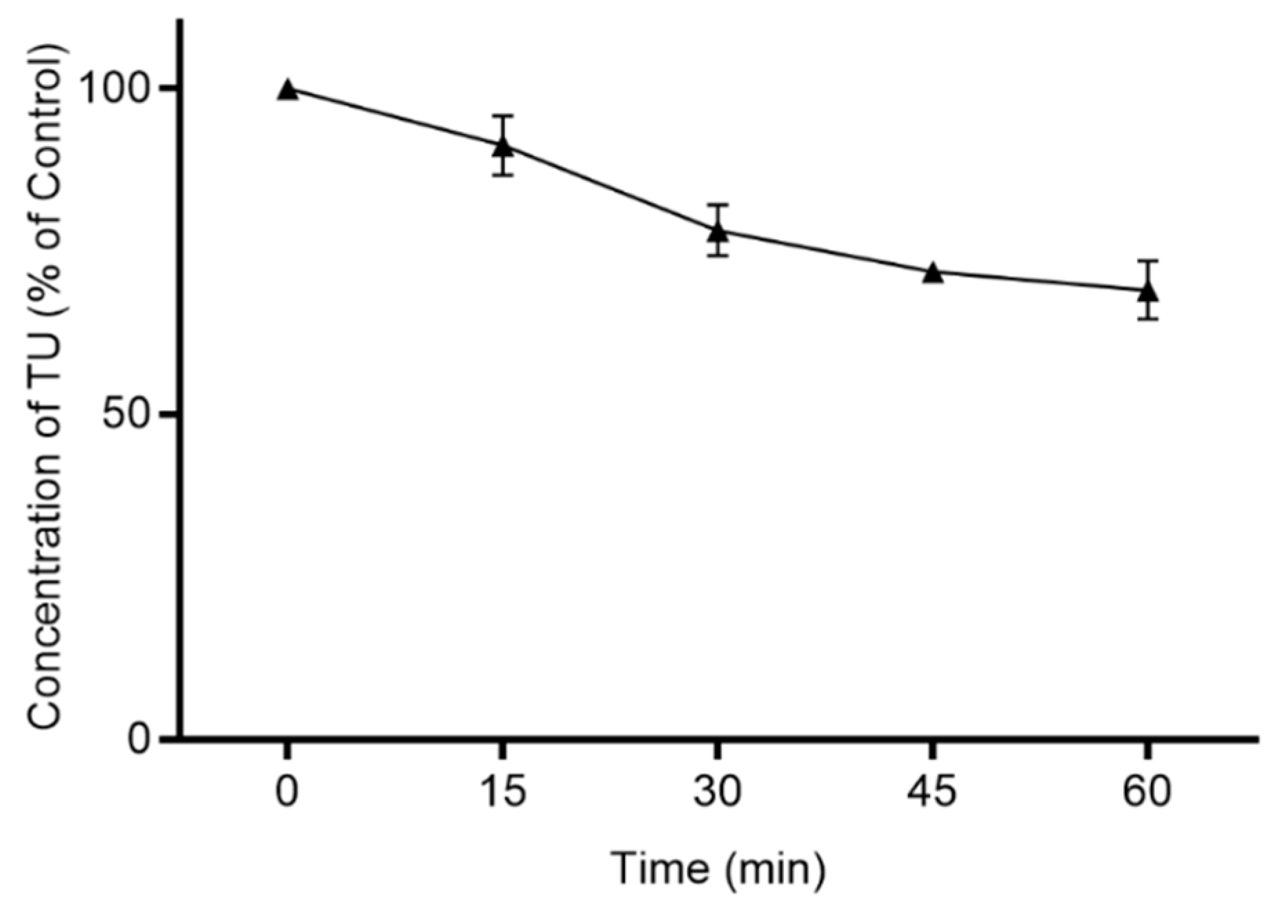
Caco-2 cell homogenate was used to assess the hydrolysis of TU. The hydrolysis–time profile of TU in cell homogenate is shown in Figure 3. The in vitro half-life (t1/2 inv) for the hydrolysis of TU was 109.28 ± 21.42 min. The value for the calculation of Clint form t1/2 inv (Equation (1)) was 0.0066 ± 0.001 mL/min/mg protein. Subsequently, Clint was corrected by fu,mic (0.781, Equation (2)) to give Clu,int value of 0.0084 ± 0.001 mL/min/mg protein. Scaling of Clu,int to physiological factors for the total protein weight of enterocytes in adult human (122.3 g [32]) resulted in a gut intrinsic clearance (ClG,int) of 61.94 ± 12.07 L/h.
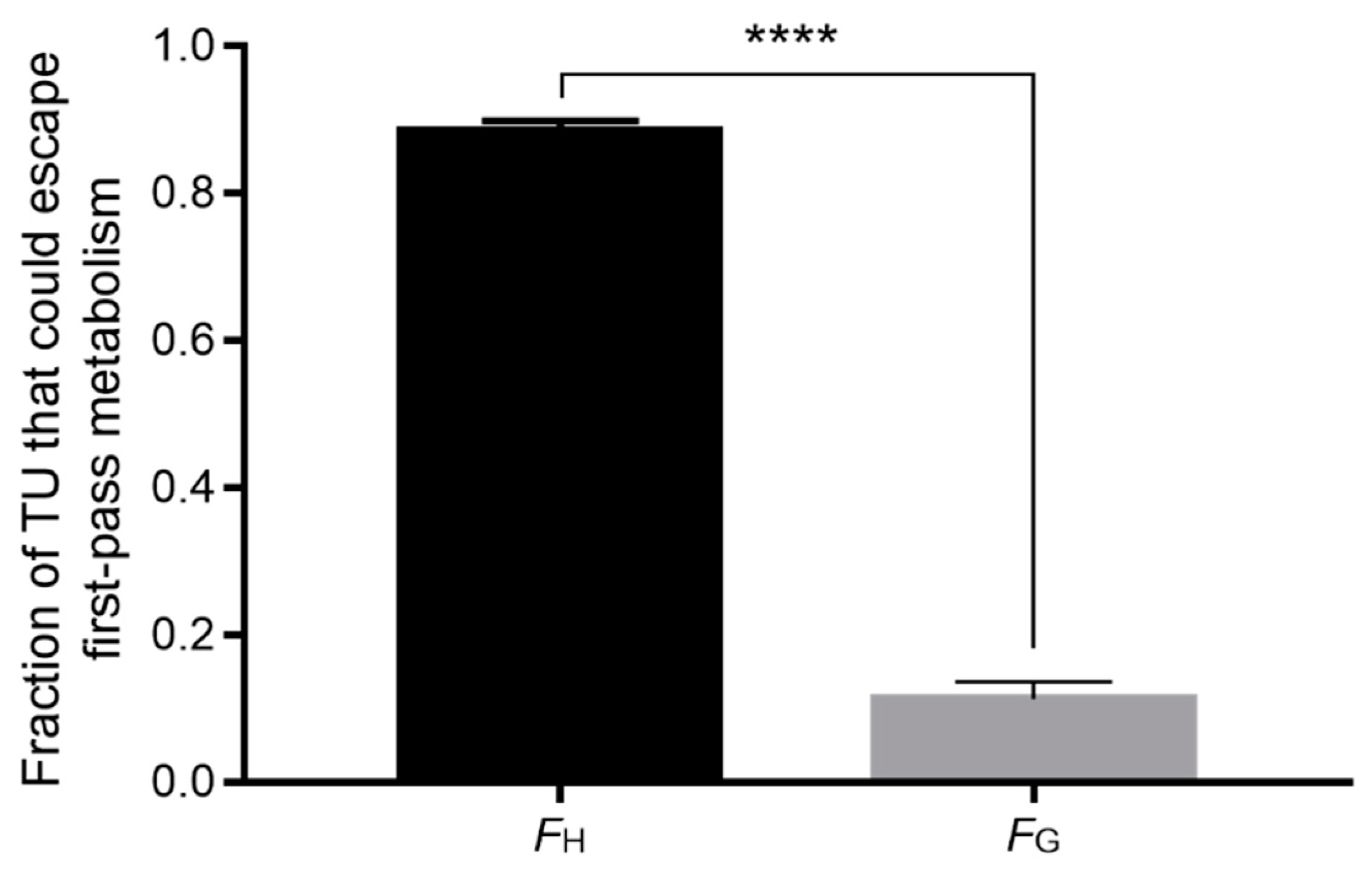
The effective intestinal permeability (Peff) of TU was 5.67 × 10⁻⁴ cm/s, as predicted by GastroPlus® software. Permeability across enterocytes (Clperm) was measured based on Equation (7) to yield 13.47 L/h. These values were applied in Equation (6) of the “Q gut” model to estimate the fraction of TU that would escape intestinal first-pass metabolism (FG), which was 0.114 ± 0.02. Therefore, it was estimated that only 11% of the absorbable fraction of TU could escape intestinal metabolism.
As presented in Figure 4, it is predicted that a significant fraction of TU could escape hepatic metabolism compared with intestinal metabolism.

4. Discussion

It is generally accepted that first-pass metabolism is a significant impediment to the bioavailability of many orally administered drugs. For these drugs, first-pass metabolism is not limited to the liver, the major site of metabolism, but other organs and tissues are involved, such as the gastrointestinal tract, blood, and vascular endothelium [37]. TU is an ester prodrug and is subject to significant first-pass metabolism [12]. Both intestinal and hepatic first-pass metabolism can markedly decrease the oral bioavailability of TU [9,10]. Herein, we have discriminated the contribution of hepatic and intestinal tissues to the first-pass metabolism of oral TU, which represents the main obstacle for the bioavailability of TU. This can thereby support the determination of an appropriate strategy to enhance the bioavailability of TU.
In the current study, it was estimated that 91% of TU can escape hepatic first-pass metabolism (FH = 0.915 ± 0.009), as predicted from the hydrolysis profile in hepatic microsomal fraction in the presence of NADPH (Figure 2). To note, the hydrolysis of TU in the presence of NADPH is a summation of NADPH-independent (carboxylesterase) and NADPH-dependant (5α-reductase) enzymatic activity [38]. In addition, it should be noted that esterase activity is not limited to the microsomal fraction. In fact, it has been depicted that microsomal fraction encompasses around 60% of total esterase activity in human liver homogenate, while cytosolic fraction accounts for approximately 20% of esterase activity [39]. The incorporation of cytosolic esterase activity to the observed hydrolysis profile of TU in liver microsomes can further decrease the fraction that could escape hepatic metabolism to 89%. Nevertheless, it seems that hepatic first-pass metabolism is not the main determinant for the low reported oral bioavailability of TU, since 91% was estimated to escape this metabolism.
In contrast, based on the hydrolysis profile of TU in Caco-2 cell homogenate (Figure 3), it can be derived that a high percentage of the orally absorbed TU would be hydrolysed in the intestinal wall. The fraction of TU that can escape intestinal metabolism was predicted to be 11.4% (FG = 0.114 ± 0.02). Accordingly, the fraction that could escape intestinal metabolism is significantly lower than fraction that could escape hepatic metabolism (p < 0.0001, Figure 4).
Taking together the predicted profiles of hepatic and intestinal metabolism of TU, it can be estimated that the oral bioavailability of TU would not exceed 10%, assuming complete absorption from the intestinal lumen. However, it is generally accepted that ester prodrugs are exposed to extensive hydrolysis in the intestinal lumen [40]. This can substantially decrease the oral bioavailability of TU below 10%, thereby delivering low levels of testosterone to the systemic circulation. Indeed, following the oral administration of TU in humans, the reported absolute bioavailability of testosterone was as low as 6.8% [1]. In that study, as well as in others [11,12,13], it was suggested that the fraction of TU that escaped first-pass metabolism was delivered to the systemic circulation via the intestinal lymphatic system. Through this pathway, the fraction of TU that could be metabolised during the first pass in the liver is saved. Therefore, the bioavailability is improved, as previously demonstrated for several other lipophilic ester prodrugs [31]. However, as predicted in the current study, hepatic metabolism has a minor contribution to the metabolic loss of TU compared to intestinal metabolism (Figure 4). In fact, it seems possible that the incorporation of TU within chylomicrons in intestinal enterocytes could preserve TU, at least partly, from hydrolysis in human intestines as well as in the liver. A similar mechanism was suggested for TU absorption from the intestinal wall of rats [7]. Hence, it can be suggested that co-administering TU with an intestinal carboxylesterase enzyme-inhibitor could significantly increase the fraction of TU available for intestinal lymphatic transport, thereby increasing the systemic bioavailability.
In addition, Caco-2 cell homogenates were previously proposed as a valid tool to assess the stability of ester prodrugs [30,41]. In the current study, the hydrolysis profile of TU in Caco-2 cell homogenate was successfully applied as a novel approach to predict the fraction of TU that could escape intestinal metabolism. The predicted values fit very well with the bioavailability reported in in vivo and clinical studies [9,11,12,13]. While the use of Caco-2 cell homogenate has provided a new insight into the prediction of intestinal first-pass metabolism, further research should be undertaken to investigate the application of this approach to ester prodrugs attached to chemically-divert promoieties. Furthermore, it is important to bear in mind that Caco-2 cells have physiological differences compared to the absorptive enterocytes in the human small intestine. Imai et al. have demonstrated differences in the expression of ester-hydrolysing carboxylesterase isozymes, carboxylesterase-1 (CE-1) and carboxylesterase-2 (CE-2). CE-1 is the main hydrolysing enzyme in Caco-2 cells, while the human small intestine primarily expresses CE-2 [42]. Substrates with small alcohol groups and large acyl groups are preferably hydrolysed by CE-1. In contrast, CE-2 favours substrates with small acyl group [43]. Nevertheless, CE-1 has a large flexible pocket that allows the hydrolysis of structurally-divers substrates, including those with large alcohol groups [44]. This might explain the hydrolytic activity of Caco-2 cell homogenate on TU in the current study, despite being a substrate with a relatively large alcohol group (Figure 1).

5. Conclusions

In conclusion, compared to the liver, the intestinal wall is the major site where the absorbed fraction of TU is extensively metabolised. It seems that intestinal lymphatic transport can decrease the exposure of TU to intestinal hydrolysing enzymes as well as circumventing hepatic first-pass metabolism. However, further studies are required to explore the metabolism of TU in vivo. Furthermore, the authors recommend investigating the impact of using an intestinal enzyme-inhibitor on the bioavailability of oral TU. Moreover, it appears that Caco-2 cell homogenate can be used to predict intestinal first-pass metabolism of particular ester prodrugs.

Author Contributions

Conceptualization, A.Z., L.K., P.G. and J.B.L.; methodology, A.Z. and J.B.L.; software, A.Z., H.-m.B. and J.B.L.; formal analysis, A.Z., L.K., P.G. and J.B.L.; investigation, A.Z., Y.D., S.M.I., H.-m.B. and J.B.L.; data curation, A.Z., Y.D., S.M.I., H.-m.B. and J.B.L.; writing—original draft preparation, A.Z.; writing—review and editing, Y.D., S.M.I., H.-m.B., L.K., P.G. and J.B.L.; project administration, A.Z., and P.G.; All authors have read and agreed to the published version of the manuscript.

Funding

This research received no external funding.

Acknowledgments

The authors thank The University of Anbar College of Pharmacy and The University of Nottingham School of Pharmacy for supporting this work.

Conflicts of Interest

The authors declare no conflict of interest.

References

  1. Täuber, U.; Schröder, K.; Düsterberg, B.; Matthes, H. Absolute bioavailability of testosterone after oral administration of testosterone-undecanoate and testosterone. Eur. J. Drug Metab. Pharmacokinet. 1986. [Google Scholar] [CrossRef]
  2. Hoberman, J. Testosterone Dreams: Rejuvenation, Aphrodisia, Doping; University of California Press: Berkeley, CA, USA, 2005; ISBN 9780520939783. [Google Scholar]
  3. Frey, H.; Aakvaag, A.; Saanum, D.; Falch, J. Bioavailability of oral testosterone in males. Eur. J. Clin. Pharmacol. 1979. [Google Scholar] [CrossRef]
  4. Stella, V.; Borchardt, R.; Hageman, M.; Oliyai, R.; Maag, H.; Tilley, J. Prodrugs: Challenges and Rewards; Springer Science & Business Media: Berlin/Heidelberg, Germany, 2007; ISBN 0387497854. [Google Scholar]
  5. Lachance, S.; Dhingra, O.; Bernstein, J.; Gagnon, S.; Savard, C.; Pelletier, N.; Boudreau, N.; Lévesque, A. Importance of measuring testosterone in enzyme-inhibited plasma for oral testosterone undecanoate androgen replacement therapy clinical trials. Future Sci. OA 2015. [Google Scholar] [CrossRef] [Green Version]
  6. Kempegowda, P.; Quinn, L.M.; Chandan, J.S.; Shepherd, L.; Kauser, S.; Rahim, A.; Bates, A. Long-term testosterone undecanoate replacement therapy: Impact of ethnicity. Clin. Endocrinol. 2020. [Google Scholar] [CrossRef]
  7. Coert, A.; Geelen, J.; De Visser, J.; Van Der Vies, J. The pharmacology and metabolism of testosterone undecanoate (TU), a new orally active androgen. Acta Endocrinol. 1975. [Google Scholar] [CrossRef] [PubMed]
  8. Geelen, J.; Coert, A.; Meijer, R.; Van Der Vies, J. Comparison of the metabolism of testosterone undecanoate and testosterone in the gastrointestinal wall of the rat in vitro and in vivo. Acta Endocrinol. 1977. [Google Scholar] [CrossRef]
  9. Shackleford, D.M.; Faassen, W.A.; Houwing, N.; Lass, H.; Edwards, G.A.; Porter, C.J.H.; Charman, W.N. Contribution of lymphatically transported testosterone undecanoate to the systemic exposure of testosterone after oral administration of two andriol formulations in conscious lymph duct-cannulated dogs. J. Pharmacol. Exp. Ther. 2003. [Google Scholar] [CrossRef]
  10. Horst, H.J.; Höltje, W.J.; Dennis, M.; Coert, A.; Geelen, J.; Voigt, K.D. Lymphatic absorption and metabolism of orally administered testosterone undecanoate in man. Klin. Wochenschr. 1976. [Google Scholar] [CrossRef]
  11. Nieschlag, E.; Mauss, J.; Coert, A.; Kićović, P. Plasma androgen levels in men after oral administration of testosterone or testosterone undecanoate. Eur. J. Endocrinol. 1975, 79, 366–374. [Google Scholar] [CrossRef] [PubMed]
  12. Bagchus, W.M.; Hust, R.; Maris, F.; Schnabel, P.G.; Houwing, N.S. Important effect of food on the bioavailability of oral testosterone undecanoate. Pharmacotherapy 2003. [Google Scholar] [CrossRef] [PubMed]
  13. Roth, M.Y.; Dudley, R.E.; Hull, L.; Leung, A.; Christenson, P.; Wang, C.; Swerdloff, R.; Amory, J.K. Steady-state pharmacokinetics of oral testosterone undecanoate with concomitant inhibition of 5α-reductase by finasteride. Int. J. Androl. 2011. [Google Scholar] [CrossRef] [PubMed] [Green Version]
  14. Yin, A.; Alfadhli, E.; Htun, M.; Dudley, R.; Faulkner, S.; Hull, L.; Leung, A.; Bross, R.; Longstreth, J.; Swerdloff, R.; et al. Dietary fat modulates the testosterone pharmacokinetics of a new self-emulsifying formulation of oral testosterone undecanoate in hypogonadal men. J. Androl. 2012. [Google Scholar] [CrossRef] [Green Version]
  15. Zgair, A.; Wong, J.C.M.; Lee, J.B.; Mistry, J.; Sivak, O.; Wasan, K.M.; Hennig, I.M.; Barrett, D.A.; Constantinescu, C.S.; Fischer, P.M.; et al. Dietary fats and pharmaceutical lipid excipients increase systemic exposure to orally administered cannabis and cannabis-based medicines. Am. J. Transl. Res. 2016, 8, 3448. [Google Scholar] [PubMed]
  16. Brocks, D.R.; Davies, N.M. Lymphatic drug absorption via the enterocytes: Pharmacokinetic simulation, modeling, and considerations for optimal drug development. J. Pharm. Pharm. Sci. 2018. [Google Scholar] [CrossRef] [Green Version]
  17. Lee, J.B.; Kim, T.H.; Feng, W.; Choi, H.G.; Zgair, A.; Shin, S.; Yoo, S.D.; Gershkovich, P.; Shin, B.S. Quantitative Prediction of Oral Bioavailability of a Lipophilic Antineoplastic Drug Bexarotene Administered in Lipidic Formulation Using a Combined In Vitro Lipolysis/Microsomal Metabolism Approach. J. Pharm. Sci. 2018. [Google Scholar] [CrossRef] [PubMed]
  18. Benito-Gallo, P.; Marlow, M.; Zann, V.; Scholes, P.; Gershkovich, P. Linking in Vitro Lipolysis and Microsomal Metabolism for the Quantitative Prediction of Oral Bioavailability of BCS II Drugs Administered in Lipidic Formulations. Mol. Pharm. 2016. [Google Scholar] [CrossRef] [PubMed] [Green Version]
  19. Obach, R.S. The importance of nonspecific binding in in vitro matrices, its impact on enzyme kinetic studies of drug metabolism reactions, and implications for in vitro-in vivo correlations. Drug Metab. Dispos. 1996, 24, 1047–1049. [Google Scholar]
  20. Gertz, M.; Kilford, P.J.; Houston, J.B.; Galetin, A. Drug Lipophilicity and Microsomal Protein Concentration as Determinants in the Prediction of the Fraction Unbound in Microsomal Incubations. Drug Metab. Dispos. 2008, 36, 535–542. [Google Scholar] [CrossRef] [Green Version]
  21. Austin, R.P.; Barton, P.; Cockroft, S.L.; Wenlock, M.C.; Riley, R.J. The influence of nonspecific microsomal binding on apparent intrinsic clearance, and its prediction from physicochemical properties. Drug Metab. Dispos. 2002. [Google Scholar] [CrossRef] [Green Version]
  22. Hallifax, D.; Houston, J.B. Binding of drugs to hepatic microsomes: Comment and assessment of current prediction methodology with recommendation for improvement. Drug Metab. Dispos. 2006, 34, 724–726. [Google Scholar] [CrossRef] [Green Version]
  23. Advanced Chemistry Developmen Inc. ACD/I-Lab Version 12.1.0.50375. 2014. Available online: www.acdlabs.com (accessed on 10 June 2020).
  24. Gao, H.; Yao, L.; Mathieu, H.W.; Zhang, Y.; Maurer, T.S.; Troutman, M.D.; Scott, D.O.; Ruggeri, R.B.; Lin, J. In silico modeling of nonspecific binding to human liver microsomes. Drug Metab. Dispos. 2008. [Google Scholar] [CrossRef] [Green Version]
  25. Yang, C.X.; Li, Y.F.; Huang, C.Z. Determination of Total Protein Content in Human Serum Samples with Fast Red Vr by Resonance Light Scattering Technique. Anal. Lett. 2002, 35, 1945–1957. [Google Scholar] [CrossRef]
  26. Watanabe, R.; Esaki, T.; Kawashima, H.; Natsume-Kitatani, Y.; Nagao, C.; Ohashi, R.; Mizuguchi, K. Predicting Fraction Unbound in Human Plasma from Chemical Structure: Improved Accuracy in the Low Value Ranges. Mol. Pharm. 2018. [Google Scholar] [CrossRef] [PubMed] [Green Version]
  27. Barter, Z.; Bayliss, M.; Beaune, P.; Boobis, A.; Carlile, D.; Edwards, R.; Brian Houston, J.; Lake, B.; Lipscomb, J.; Pelkonen, O.; et al. Scaling Factors for the Extrapolation of In Vivo Metabolic Drug Clearance from In Vitro Data: Reaching a Consensus on Values of Human Micro-somal Protein and Hepatocellularity Per Gram of Liver. Curr. Drug Metab. 2006. [Google Scholar] [CrossRef] [PubMed]
  28. McGinnity, D.F.; Soars, M.G.; Urbanowicz, R.A.; Riley, R.J. Evaluation of fresh and cryopreserved hepatocytes as in vitro drug metabolism tools for the prediction of metabolic clearance. Drug Metab. Dispos. 2004. [Google Scholar] [CrossRef] [Green Version]
  29. Davies, B.; Morris, T. Physiological Parameters in Laboratory Animals and Humans. Pharm. Res. 1993, 10, 1093–1095. [Google Scholar] [CrossRef]
  30. Tsume, Y.; Amidon, G.L. Selection of Suitable Prodrug Candidates for in vivo Studies via in vitro Studies; The correlation of Prodrug Stability in Between Cell Culture Homogenates and Human Tissue Homogenates. J. Pharm. Pharm. Sci. 2012. [Google Scholar] [CrossRef] [Green Version]
  31. Lee, J.B.; Zgair, A.; Malec, J.; Kim, T.H.; Kim, M.G.; Ali, J.; Qin, C.; Feng, W.; Chiang, M.; Gao, X.; et al. Lipophilic activated ester prodrug approach for drug delivery to the intestinal lymphatic system. J. Control. Release 2018, 286. [Google Scholar] [CrossRef]
  32. Gertz, M.; Houston, J.B.; Galetin, A. Physiologically based pharmacokinetic modeling of intestinal first-pass metabolism of CYP3A substrates with high intestinal extraction. Drug Metab. Dispos. 2011. [Google Scholar] [CrossRef] [Green Version]
  33. Yang, J.; Jamei, M.; Yeo, K.; Tucker, G.; Rostami-Hodjegan, A. Prediction of Intestinal First-Pass Drug Metabolism. Curr. Drug Metab. 2007. [Google Scholar] [CrossRef]
  34. Granger, D.N.; Richardson, P.D.I.; Kvietys, P.R.; Mortillaro, N.A. Intestinal blood flow. Gastroenterology 1980, 78, 837–863. [Google Scholar] [CrossRef]
  35. Zgair, A.; Wong, J.C.M.; Sabri, A.; Fischer, P.M.; Barrett, D.A.; Constantinescu, C.S.; Gershkovich, P. Development of a simple and sensitive HPLC-UV method for the simultaneous determination of cannabidiol and δ9-tetrahydrocannabinol in rat plasma. J. Pharm. Biomed. Anal. 2015. [Google Scholar] [CrossRef] [PubMed]
  36. Krishna, G.; Chen, K.J.; Lin, C.C.; Nomeir, A.A. Permeability of lipophilic compounds in drug discovery using in-vitro human absorption model, Caco-2. Int. J. Pharm. 2001. [Google Scholar] [CrossRef]
  37. Pond, S.M.; Tozer, T.N. First-Pass Elimination Basic Concepts and Clinical Consequences. Clin. Pharmacokinet. 1984, 9, 1–25. [Google Scholar] [CrossRef] [PubMed]
  38. Azzouni, F.; Godoy, A.; Li, Y.; Mohler, J. The 5 alpha-reductase isozyme family: A review of basic biology and their role in human diseases. Adv. Urol. 2012. [Google Scholar] [CrossRef] [Green Version]
  39. Junge, W.; Krisch, K.; Conney, A. The carboxylesterases/amidases of mammalian liver and their possible significance. Crit. Rev. Toxicol. 1975. [Google Scholar] [CrossRef]
  40. Rautio, J.; Mannhold, R.; Kubinyi, H. Prodrugs and Targeted Delivery: Towards Better ADME Properties. Methods Princ. Med. Chem. 2011, 47, 292. [Google Scholar]
  41. Gupta, D.; Gupta, S.V.; Lee, K.D.; Amidon, G.L. Chemical and enzymatic stability of amino acid prodrugs containing methoxy, ethoxy and propylene glycol linkers. Mol. Pharm. 2009, 6, 1604–1611. [Google Scholar] [CrossRef] [Green Version]
  42. Imai, T.; Imoto, M.; Sakamoto, H.; Hashimoto, M. Identification of esterases expressed in Caco-2 cells and effects of their hydrolyzing activity in predicting human intestinal absorption. Drug Metab. Dispos. 2005. [Google Scholar] [CrossRef] [Green Version]
  43. Hosokawa, M. Structure and catalytic properties of carboxylesterase isozymes involved in metabolic activation of prodrugs. Molecules 2008, 13, 412–431. [Google Scholar] [CrossRef] [Green Version]
  44. Satoh, T.; Hosokawa, M. Structure, function and regulation of carboxylesterases. Chem. Biol. Interact. 2006, 162, 195–211. [Google Scholar] [CrossRef] [PubMed]
Figure 1. Chemical structure of testosterone undecanoate (TU).
Applsci 10 07283 g001
Figure 2. The hydrolysis–time profiles of testosterone undecanoate (TU) in human hepatic microsomes in the presence of nicotinamide adenine dinucleotide phosphate tetrasodium salt hydrate (NADPH, open symbol) and without NADPH (closed symbol). Values are expressed as mean ± SD (n = 3).
Applsci 10 07283 g002
Figure 3. The hydrolysis–time profile of testosterone undecanoate (TU) in Caco-2 cell homogenate. Values are expressed as mean ± SD (n = 4).
Applsci 10 07283 g003
Figure 4. Predicted fractions of testosterone undecanoate (TU) that could escape hepatic (FH, n = 3) and intestinal (FG, n = 4) first-pass metabolism. Values are presented as mean ± SD. An unpaired two-tailed Student’s t-test was used to assess statistical differences between data sets. **** p < 0.0001.
Applsci 10 07283 g004
Publisher’s Note: MDPI stays neutral with regard to jurisdictional claims in published maps and institutional affiliations.