Depression and Microbiome—Study on the Relation and Contiguity between Dogs and Humans
Abstract
1. Introduction
2. Materials and Methods
2.1. Animals
2.2. DNA Extraction from Stool Samples
2.3. Library Preparation and Sequencing
2.4. Statistical Analysis
3. Results
4. Discussion
5. Conclusions
Supplementary Materials
Author Contributions
Funding
Acknowledgments
Conflicts of Interest
References
- Cocchi, M.; Tonello, L.; Gabrielli, F. The Molecular and Quantum Approach to Psychopathology and Consciousness—From Theory to Experimental Practice. In Major Depression Disorder- Cognitive and Neurobiological Mechanisms; Intechopen: London, UK, 2015; Chapter 3. [Google Scholar]
- Accorsi, P.A.; Mondo, E.; Cocchi, M. Did you know that your animals have consciousness? J. Integr. Neurosci. 2017, 16, S3–S11. [Google Scholar] [CrossRef]
- Cryan, J.F.; Dinan, T.G. Mind-altering microorganisms: The impact of the gut microbiota on brain and behavior. Nat. Rev. Neurosci. 2012, 13, 701–712. [Google Scholar] [CrossRef] [PubMed]
- Sudo, N.; Chida, Y.; Aiba, Y.; Sonoda, J.; Oyama, N.; Yu, X.; Kubo, C.; Koga, Y. Postnatal microbial colonization programs the hypothalamic-pituitary-adrenal system for stress response in mice. J. Physiol. 2004, 558, 263–275. [Google Scholar] [CrossRef] [PubMed]
- Bravo, J.A.; Forsythe, P.; Chew, M.V.; Escaravage, E.; Savignac, H.M.; Dinan, T.G.; Bienenstock, J.; Cryan, J.F. Ingestion of Lactobacillus strain regulates emotional behavior and central GABA receptor expression in a mouse via the vagus nerve. Proc. Natl. Acad. Sci. USA 2011, 108, 16050–16055. [Google Scholar] [CrossRef] [PubMed]
- Hsiao, E.Y.; McBride, S.W.; Hsien, S.; Sharon, G.; Hyde, E.R.; McCue, T.; Codelli, J.A.; Chow, J.; Reisman, S.E.; Petrosino, J.F.; et al. The microbiota modulates gut physiology and behavioral abnormalities associated with autism. Cell 2013, 155, 1451–1463. [Google Scholar] [CrossRef] [PubMed]
- Carabotti, M. The Gut-Brain Axis: Interactions Between Enteric Microbiota, Central and Enteric Nervous Systems. Ann. Gastroenterol. 2014, 28, 203–209. [Google Scholar]
- Lovelace, M.D.; Varney, B.; Sundaram, G.; Lennon, M.J.; Lim, C.K.; Jacobs, K.; Guillemin, G.J.; Brew, B.J. Recent evidence for an expanded role of the Kynurenine pathway of tryptophan metabolism in neurological diseases. Neuropharmacology 2017, 112, 373–388. [Google Scholar] [CrossRef] [PubMed]
- Cocchi, M.; Tonello, L.; Cappello, G.; Nabacino, L.; Passi, S.; Baldini, N.; Soreca, I.; Paffetti, I.; Castrogiovanni, P.; Tarozzi, G. Biochemical Markers in Major Depression as interface between Neuronal Network and Artificial Neural Network (ANN). J. Biol. Res. 2006, 77–81. [Google Scholar] [CrossRef]
- Benedetti, S.; Bucciarelli, S.; Canestrari, F.; Catalani, S.; Mandolini, S.; Marconi, V.; Mastrogiacomo, A.; Silvestri, R.; Tagliamonte, M.; Venanzini, R.; et al. Platelet’s Fatty Acids and Differential Diagnosis of Major Depression and Bipolar Disorder through the Use of an Unsupervised Competitive-Learning Network Algorithm (SOM). OJD 2014, 3, 52–73. [Google Scholar] [CrossRef][Green Version]
- Cocchi, M.; Tonello, L.; Rasenick Mark, M. Human depression: A new approach in quantitative psychiatry. Ann. Gen. Psychiatr. 2010, 9, 25. [Google Scholar] [CrossRef]
- Cocchi, M.; Tonello, L.; De Lucia, A.; Amato, P. Platelet and Brain Fatty Acids: A Model for the Classification of the Animals? Part 1. Int. J. Anthropol. 2009, 24, 69–76. [Google Scholar]
- Cocchi, M.; Gabrielli, F.; Tonello, L.; Delogu, M.; Beghelli, V.; Mattioli, M.; Accorsi, P.A. Molecular Contiguity between Human and Animal Consciousness through Evolution: Some Considerations. J. Phylogen Evol. Biol. 2013, 1, 119. [Google Scholar] [CrossRef]
- De Cesare, A.; Sirri, F.; Manfreda, G.; Moniaci, P.; Giardini, A.; Zampigna, M.; Meluzzi, A. Effect of dietary supplementation with Lactobacillus acidophilus D2/CSL (CECT 4529) on caecum microbioma and productive performance in broiler chickens. PLoS ONE 2017, 12, e0176309. [Google Scholar] [CrossRef] [PubMed]
- De Cesare, A.; Caselli, E.; Lucchi, A.; Sala, C.; Parisi, A.; Manfreda, G.; Mazzacane, S. Impact of a probiotic-based cleaning product on the microbiological profile of broiler litters and chicken caeca microbiota. Poult 2019, 98, 3602–3610. [Google Scholar] [CrossRef]
- Chesselet, M.F.; Carmichael, S.T. Animal Models of Neurological Disorders. Neurotherapeutics 2012, 9, 241–244. [Google Scholar] [CrossRef]
- Jiang, H.; Ling, Z.; Mao, H.; Ma, Z.; Yin, Y.; Wang, W.; Tang, W.; Tan, Z.; Shi, J.; Li, L.; et al. Altered fecal microbiota composition in patients with major depressive disorder. Brain Behav. Immun. 2015, 48, 186–194. [Google Scholar] [CrossRef]
- Zheng, P.; Zeng, B.; Zhou, C.; Liu, M.; Fang, Z.; Xu, X.; Zeng, L.; Chen, J.; Fan, S.; Du, X.; et al. Gut microbiome remodeling induces depressive-like behaviors through a pathway mediated by the host’s metabolism. Mol. Psychiatry 2016, 21, 786–796. [Google Scholar] [CrossRef]
- Valles-Colomer, M.; Faolony, G.; Darzi, Y.; Tigchelaar, E.F.; Wang, J.; Tito, R.L.; Schiweck, C.; Kurilshikov, A.; Joossens, M.; Vieira-Silva, S.; et al. The neuroactive potential of the human gut microbiota in quality of life and depression. Nat. Microbiol. 2019, 4, 623–632. [Google Scholar] [CrossRef]
- Sandri, M.; Del Monego, S.; Conte, G.; Sgorlon, S.; Stefanon, B. Raw meat based diet influences faecal microbiome and end products of fermentation in healthy dogs. BMC Vet. Res. 2017, 13, 65. [Google Scholar] [CrossRef]
- Omatsu, T.; Omura, M.; Katayama, Y.; Kimura, T.; Okumura, M.; Okumura, A.; Murata, Y.; Mizutani, T. Molecular diversity of the faecal microbiota of Toy Poodles in Japan. J. Vet. Med. Sci. 2018, 80, 749–754. [Google Scholar] [CrossRef]
- Winter, G.; Hart, R.A.; Charlesworth, R.P.G.; Sharpley, C.F. Gut microbiome and depression: What we know and what we need to know. Rev. Neurosci. 2018, 29, 629–643. [Google Scholar] [CrossRef] [PubMed]
- Yunes, R.A.; Poluektova, E.U.; Dyachkova, M.S.; Klimina, K.M.; Kovtun, A.S.; Averina, O.V.; Orlova, V.S.; Danilenko, V.N. GABA production and structure of gadB/gadC genes in Lactobacillus and Bifidobacterium strains from human microbiota. Anaerob 2016, 42, 197–204. [Google Scholar] [CrossRef] [PubMed]
- Evrensel, A.; Ceylan, M.E. The Gut-Brain Axis: The Missing Link in Depression. Clin. Psychopharmacol. Neurosci. 2015, 13, 239–244. [Google Scholar] [CrossRef]
- Butnoriene, J.; Bunevicius, A.; Norkus, A.; Bunevicius, R. Depression but not anxiety is associated with metabolic syndrome in primary care based community sample. Psychoneuroendocrinology 2014, 40, 269–276. [Google Scholar] [CrossRef] [PubMed]
- Bangsgaard Bendtsen, K.M.; Krych, L.; Sorensen, D.B.; Pang, W.; Nielsen, D.S.; Josefsen, K.; Hansen, L.H.; Sorensen, S.J.; Hansen, A.K. Gut microbiota composition is correlated to grid floor induced stress and behavior in the BALB/c mouse. PLoS ONE 2012, 7, e46231. [Google Scholar] [CrossRef]
- O’Malley, D.; Julio-Pieper, M.; Gibney, S.M.; Dinan, T.G.; Cryan, J.F. Distinct alterations in colonic morphology and physiology in two rat models of enhanced stress-induced anxiety and depression-like behavior. Stress 2010, 13, 114–122. [Google Scholar] [CrossRef]
- Maes, M.; Kubera, M.; Leunis, J.C.; Berk, M. Increased IgA and IgM responses against gut commensals in chronic depression: Further evidence for increased bacterial translocation or leaky gut. J. Affect. Disord. 2012, 141, 55–62. [Google Scholar] [CrossRef]
- Nobis, G. Der älteste Haushund lebte vor 14,000 Jahren. Umschau 1979, 79, 610. [Google Scholar]
- Sarviaho, R.; Hakosalo, O.; Tiira, K.; Sulkama, S.; Salmela, E.; Hytönen, M.K.; Sillanpää, M.J.; Lohi, H. Two novel genomic regions associated with fearfulness in dogs overlap human neuropsychiatric loci. Nature 2019, 9, 18. [Google Scholar] [CrossRef]
- Udell, M.A.; Wynne, C.D. A review of domestic dogs’ (Canis familiaris) human-like behaviors: Or why behavior analysts should stop worrying and love their dogs. J. Exp. Anal. Behav. 2008, 89, 247–261. [Google Scholar] [CrossRef]
- Shafquat, A.; Joice, R.; Simmons, S.; Huttenhower, C. Functional and phylogenetic assembly of microbial communities in the human microbiome. Trends Microbiol. 2014, 22, 261–266. [Google Scholar] [CrossRef] [PubMed]


| MB | GS | ||||
|---|---|---|---|---|---|
| Phylum | Mean (%) | std. dev. (%) | Mean (%) | PT std. dev. (%) | p Values |
| Firmicutes | 55.247 | 13.880 | 55.695 | 14.042 | 0.937 |
| Fusobacteria | 16.599 | 9.218 | 11.296 | 6.569 | 0.106 |
| Bacteroidetes | 21.306 | 11.162 | 10.742 | 10.611 | 0.024 |
| Actinobacteria | 3.608 | 2.868 | 3.453 | 1.827 | 0.871 |
| Proteobacteria | 0.540 | 0.293 | 1.916 | 0.756 | 0.000 |
| MB | GS | ||||
|---|---|---|---|---|---|
| Class | Mean (%) | std. dev. (%) | Mean (%) | std. dev. (%) | p Values |
| Actinobacteria | 3.608 | 2.868 | 3.453 | 1.827 | 0.871 |
| Bacteroidia | 21.298 | 11.157 | 10.724 | 10.603 | 0.023 |
| Clostridia | 39.318 | 11.190 | 33.425 | 8.456 | 0.146 |
| Bacilli | 2.019 | 1.476 | 14.761 | 7.947 | 0.000 |
| Erysipelotrichi | 8.471 | 7.689 | 4.664 | 3.423 | 0.113 |
| Negativicutes | 5.439 | 3.881 | 2.846 | 1.723 | 0.037 |
| Fusobacteria | 16.599 | 9.218 | 11.296 | 6.569 | 0.106 |
| Deltaproteobacteria | 0.158 | 0.217 | 0.973 | 0.504 | 0.000 |
| Gammaproteobacteria | 0.161 | 0.169 | 0.825 | 0.681 | 0.008 |
| Epsilonproteobacteria | 0.161 | 0.225 | 0.022 | 0.016 | 0.037 |
| MB | GS | ||||
|---|---|---|---|---|---|
| Order | Mean (%) | std. dev. (%) | Mean (%) | std. dev. (%) | p Values |
| Coriobacteriales | 3.502 | 2.839 | 2.471 | 0.991 | 0.222 |
| Actinomycetales | 0.081 | 0.044 | 0.946 | 1.758 | 0.131 |
| Bacteroidales | 21.298 | 11.157 | 10.724 | 10.603 | 0.023 |
| Clostridiales | 39.302 | 11.187 | 33.404 | 8.453 | 0.146 |
| Lactobacillales | 1.182 | 1.403 | 13.530 | 7.905 | 0.000 |
| Erysipelotrichales | 8.471 | 7.689 | 4.664 | 3.423 | 0.113 |
| Selenomonadales | 5.439 | 3.881 | 2.846 | 1.723 | 0.037 |
| Bacillales | 0.837 | 0.635 | 1.231 | 0.682 | 0.153 |
| Fusobacteriales | 16.599 | 9.218 | 11.296 | 6.569 | 0.106 |
| Desulfovibrionales | 0.111 | 0.147 | 0.952 | 0.503 | 0.000 |
| Enterobacteriales | 0.028 | 0.035 | 0.395 | 0.289 | 0.001 |
| Aeromonadales | 0.121 | 0.164 | 0.226 | 0.273 | 0.273 |
| MB | GS | ||||
|---|---|---|---|---|---|
| Family | Mean (%) | std. dev. (%) | Mean (%) | std. dev. (%) | p Values |
| Coriobacteriaceae | 3.502 | 2.839 | 2.471 | 0.991 | 0.222 |
| Microbacteriaceae | 0.030 | 0.029 | 0.587 | 0.907 | 0.067 |
| Micrococcaceae | 0.002 | 0.002 | 0.117 | 0.377 | 0.333 |
| Corynebacteriaceae | 0.002 | 0.001 | 0.042 | 0.104 | 0.222 |
| Prevotellaceae | 11.113 | 9.461 | 6.368 | 6.892 | 0.160 |
| Bacteroidaceae | 10.010 | 7.919 | 3.891 | 5.036 | 0.027 |
| Porphyromonadaceae | 0.165 | 0.209 | 0.448 | 0.315 | 0.019 |
| Clostridiaceae | 15.448 | 7.344 | 14.324 | 4.907 | 0.651 |
| Ruminococcaceae | 8.874 | 2.640 | 6.695 | 2.128 | 0.031 |
| Erysipelotrichaceae | 8.471 | 7.689 | 4.664 | 3.423 | 0.113 |
| Veillonellaceae | 3.811 | 3.828 | 0.908 | 0.560 | 0.014 |
| Lachnospiraceae | 3.810 | 2.160 | 2.799 | 1.241 | 0.155 |
| Eubacteriaceae | 1.990 | 1.349 | 1.766 | 1.604 | 0.714 |
| Acidaminococcaceae | 1.628 | 2.117 | 1.938 | 1.537 | 0.675 |
| Streptococcaceae | 0.608 | 1.407 | 3.440 | 2.936 | 0.010 |
| Paenibacillaceae | 0.525 | 0.472 | 0.640 | 0.371 | 0.501 |
| Lactobacillaceae | 0.417 | 0.442 | 8.616 | 7.893 | 0.005 |
| Bacillaceae | 0.281 | 0.383 | 0.222 | 0.085 | 0.584 |
| Peptostreptococcaceae | 0.215 | 0.127 | 0.148 | 0.063 | 0.097 |
| Aerococcaceae | 0.132 | 0.182 | 0.640 | 0.344 | 0.000 |
| Peptococcaceae | 0.091 | 0.069 | 0.334 | 0.201 | 0.002 |
| Enterococcaceae | 0.015 | 0.035 | 0.661 | 0.368 | 0.000 |
| Thermoactinomycetaceae | 0.010 | 0.010 | 0.136 | 0.248 | 0.120 |
| Leuconostocaceae | 0.008 | 0.017 | 0.131 | 0.152 | 0.021 |
| Clostridiales Family XII. Incertae Sedis | 0.008 | 0.020 | 0.177 | 0.116 | 0.001 |
| Listeriaceae | 0.001 | 0.001 | 0.180 | 0.577 | 0.324 |
| Fusobacteriaceae | 16.599 | 9.218 | 11.296 | 6.569 | 0.106 |
| Helicobacteraceae | 0.124 | 0.217 | 0.003 | 0.002 | 0.056 |
| Desulfohalobiaceae | 0.110 | 0.147 | 0.950 | 0.503 | 0.000 |
| Succinivibrionaceae | 0.090 | 0.155 | 0.223 | 0.272 | 0.168 |
| Enterobacteriaceae | 0.028 | 0.035 | 0.395 | 0.289 | 0.001 |
| MB | GS | ||||
|---|---|---|---|---|---|
| Genus | Mean (%) | std. dev. (%) | Mean (%) | std. dev. (%) | p Values |
| Microbacterium | 0.008 | 0.019 | 0.497 | 0.788 | 0.064 |
| Anaerobiospirillum | 0.090 | 0.155 | 0.223 | 0.272 | 0.168 |
| Paenibacillus | 0.521 | 0.471 | 0.634 | 0.374 | 0.512 |
| Bacillus | 0.200 | 0.390 | 0.160 | 0.088 | 0.718 |
| Thermoactinomyces | 0.009 | 0.010 | 0.133 | 0.246 | 0.124 |
| Prevotella | 11.078 | 9.463 | 6.317 | 6.814 | 0.157 |
| Bacteroides | 10.010 | 7.919 | 3.891 | 5.036 | 0.027 |
| Parabacteroides | 0.080 | 0.199 | 0.119 | 0.138 | 0.570 |
| Porphyromonas | 0.046 | 0.049 | 0.157 | 0.118 | 0.011 |
| Barnesiella | 0.023 | 0.033 | 0.118 | 0.111 | 0.017 |
| Helicobacter | 0.124 | 0.217 | 0.003 | 0.002 | 0.056 |
| Clostridium | 15.007 | 7.362 | 13.934 | 4.942 | 0.668 |
| Blautia | 6.746 | 3.580 | 5.307 | 2.441 | 0.245 |
| Ruminococcus | 5.411 | 2.917 | 3.892 | 0.998 | 0.086 |
| Faecalibacterium | 3.290 | 3.013 | 2.432 | 2.158 | 0.415 |
| Eubacterium | 1.987 | 1.350 | 1.753 | 1.586 | 0.699 |
| Hespellia | 1.141 | 0.728 | 0.778 | 0.265 | 0.101 |
| Robinsoniella | 0.519 | 0.513 | 0.442 | 0.501 | 0.707 |
| Coprococcus | 0.514 | 0.846 | 0.222 | 0.625 | 0.331 |
| Roseburia | 0.479 | 0.527 | 0.133 | 0.127 | 0.031 |
| Butyrivibrio | 0.369 | 0.485 | 0.369 | 0.132 | 0.999 |
| Lachnospira | 0.246 | 0.517 | 0.098 | 0.119 | 0.316 |
| Peptostreptococcus | 0.215 | 0.127 | 0.148 | 0.063 | 0.097 |
| Alkaliphilus | 0.201 | 0.427 | 0.093 | 0.097 | 0.372 |
| Syntrophococcus | 0.105 | 0.326 | 0.001 | 0.001 | 0.253 |
| Ethanoligenens | 0.099 | 0.139 | 0.231 | 0.220 | 0.101 |
| Butyricicoccus | 0.096 | 0.063 | 0.122 | 0.064 | 0.337 |
| Sarcina | 0.082 | 0.305 | 0.162 | 0.364 | 0.569 |
| Peptococcus | 0.037 | 0.064 | 0.222 | 0.102 | 0.000 |
| Fusibacter | 0.008 | 0.020 | 0.177 | 0.116 | 0.001 |
| Collinsella | 2.176 | 1.793 | 1.329 | 0.544 | 0.113 |
| Slackia | 0.926 | 0.759 | 0.726 | 0.365 | 0.394 |
| Enterorhabdus | 0.233 | 0.197 | 0.222 | 0.068 | 0.845 |
| Atopobium | 0.140 | 0.115 | 0.130 | 0.065 | 0.797 |
| Desulfonauticus | 0.102 | 0.147 | 0.923 | 0.495 | 0.000 |
| Escherichia | 0.009 | 0.023 | 0.249 | 0.156 | 0.000 |
| Catenibacterium | 1.503 | 2.894 | 0.680 | 0.972 | 0.333 |
| Erysipelothrix | 0.481 | 0.827 | 0.436 | 0.334 | 0.853 |
| Holdemania | 0.100 | 0.293 | 0.139 | 0.128 | 0.667 |
| Fusobacterium | 16.573 | 9.213 | 11.284 | 6.562 | 0.107 |
| Streptococcus | 0.600 | 1.406 | 3.403 | 2.930 | 0.011 |
| Lactobacillus | 0.417 | 0.442 | 8.611 | 7.888 | 0.005 |
| Aerococcus | 0.129 | 0.181 | 0.627 | 0.344 | 0.001 |
| Enterococcus | 0.014 | 0.035 | 0.651 | 0.362 | 0.000 |
| Megamonas | 2.671 | 3.011 | 0.432 | 0.503 | 0.015 |
| Phascolarctobacterium | 1.176 | 1.325 | 1.365 | 1.042 | 0.692 |
| Selenomonas | 1.125 | 0.903 | 0.463 | 0.387 | 0.023 |
| Acidaminococcus | 0.452 | 0.848 | 0.572 | 0.529 | 0.668 |
| MB | GS | ||||
|---|---|---|---|---|---|
| Species | Mean (%) | std. dev. (%) | Mean (%) | std. dev. (%) | p Values |
| Phascolarctobacterium sp. YIT 12067 | 1.176 | 1.325 | 1.365 | 1.042 | 0.692 |
| Acidaminococcus fermentans | 0.448 | 0.833 | 0.572 | 0.529 | 0.654 |
| Aerococcus viridans | 0.118 | 0.174 | 0.612 | 0.333 | 0.000 |
| Bacteroides plebeius | 2.619 | 2.635 | 0.370 | 0.456 | 0.007 |
| Bacteroides fragilis | 1.689 | 2.246 | 0.628 | 0.938 | 0.126 |
| Bacteroides stercoris | 0.984 | 0.883 | 0.741 | 1.515 | 0.643 |
| Bacteroides coprocola | 0.963 | 0.790 | 0.390 | 0.460 | 0.033 |
| Bacteroides vulgatus | 0.907 | 2.099 | 0.055 | 0.121 | 0.152 |
| Bacteroides uniformis | 0.785 | 0.844 | 0.184 | 0.204 | 0.021 |
| Bacteroides ovatus | 0.506 | 0.464 | 0.360 | 0.565 | 0.497 |
| Clostridium bifermentans | 5.080 | 3.341 | 3.625 | 1.436 | 0.158 |
| Clostridium sordellii | 3.167 | 3.022 | 3.073 | 1.121 | 0.916 |
| Clostridium bartlettii | 1.769 | 1.213 | 0.904 | 0.366 | 0.022 |
| Clostridium scindens | 1.142 | 1.020 | 0.230 | 0.120 | 0.005 |
| Clostridium hiranonis | 0.979 | 1.494 | 0.329 | 0.980 | 0.203 |
| Clostridium perfringens | 0.226 | 0.370 | 1.738 | 1.664 | 0.012 |
| Clostridium aminobutyricum | 0.044 | 0.120 | 0.813 | 0.551 | 0.001 |
| Collinsella intestinalis | 1.840 | 1.534 | 1.121 | 0.482 | 0.116 |
| Slackia heliotrinireducens | 0.890 | 0.734 | 0.684 | 0.363 | 0.370 |
| Desulfonauticus autotrophicus | 0.101 | 0.147 | 0.923 | 0.495 | 0.000 |
| Clostridium ramosum | 1.684 | 2.043 | 0.499 | 0.608 | 0.055 |
| Catenibacterium mitsuokai | 1.503 | 2.894 | 0.680 | 0.972 | 0.333 |
| Eubacterium biforme | 1.060 | 1.492 | 0.879 | 0.730 | 0.695 |
| Lactobacillus vitulinus | 0.823 | 1.583 | 0.348 | 0.688 | 0.326 |
| Clostridium spiroforme | 0.790 | 0.914 | 0.390 | 0.239 | 0.135 |
| Eubacterium cylindroides | 0.650 | 0.643 | 0.563 | 0.351 | 0.670 |
| Streptococcus pleomorphus | 0.620 | 0.956 | 0.481 | 0.417 | 0.630 |
| Eubacterium fissicatena | 1.079 | 1.098 | 0.397 | 0.176 | 0.037 |
| Fusobacterium nucleatum | 6.030 | 3.870 | 4.852 | 2.987 | 0.398 |
| Fusobacterium mortiferum | 2.156 | 1.684 | 1.167 | 0.892 | 0.072 |
| Fusobacterium varium | 2.109 | 1.455 | 1.945 | 1.408 | 0.779 |
| Fusobacterium ulcerans | 2.002 | 1.457 | 1.547 | 0.961 | 0.359 |
| Fusobacterium equinum | 1.698 | 1.015 | 0.702 | 0.539 | 0.005 |
| Fusobacterium perfoetens | 1.628 | 1.375 | 0.602 | 0.592 | 0.021 |
| Fusobacterium periodonticum | 0.794 | 0.851 | 0.418 | 0.612 | 0.211 |
| Hespellia porcina | 0.682 | 0.472 | 0.451 | 0.191 | 0.112 |
| Robinsoniella peoriensis | 0.519 | 0.513 | 0.442 | 0.501 | 0.707 |
| Coprococcus comes | 0.509 | 0.845 | 0.219 | 0.624 | 0.332 |
| Lactobacillus murinus | 0.014 | 0.015 | 5.202 | 5.009 | 0.006 |
| Lactobacillus reuteri | 0.004 | 0.003 | 1.193 | 1.600 | 0.031 |
| Prevotella copri | 5.976 | 5.824 | 2.289 | 2.473 | 0.045 |
| Prevotella intermedia | 1.434 | 1.868 | 1.018 | 1.008 | 0.483 |
| Prevotella oris | 1.024 | 1.700 | 0.325 | 0.556 | 0.166 |
| Prevotella ruminicola | 0.863 | 1.073 | 0.479 | 0.449 | 0.240 |
| Prevotella falsenii | 0.658 | 0.888 | 0.457 | 0.473 | 0.474 |
| Prevotella nigrescens | 0.599 | 0.811 | 1.281 | 2.520 | 0.404 |
| Faecalibacterium prausnitzii | 3.290 | 3.013 | 2.432 | 2.158 | 0.415 |
| Ruminococcus gnavus | 2.838 | 3.070 | 0.955 | 0.322 | 0.038 |
| Ruminococcus sp. 5_1_39BFAA | 0.885 | 1.151 | 0.835 | 0.282 | 0.878 |
| Ruminococcus obeum | 0.815 | 0.997 | 0.649 | 0.309 | 0.565 |
| Ruminococcus torques | 0.542 | 0.663 | 0.566 | 0.163 | 0.897 |
| Ruminococcus gauvreauii | 0.254 | 0.148 | 0.687 | 0.252 | 0.000 |
| Streptococcus agalactiae | 0.247 | 0.633 | 1.630 | 2.194 | 0.065 |
| Blautia sp. Ser8 | 6.484 | 3.417 | 5.052 | 2.480 | 0.236 |
| butyrate-producing bacterium SM4/1 | 1.262 | 1.041 | 1.201 | 0.538 | 0.852 |
| Megamonas hypermegale | 2.671 | 3.011 | 0.432 | 0.503 | 0.015 |
| Selenomonas ruminantium | 0.957 | 0.804 | 0.449 | 0.391 | 0.050 |
| Phylum | Class | Order | Family | Genus | Species | |
|---|---|---|---|---|---|---|
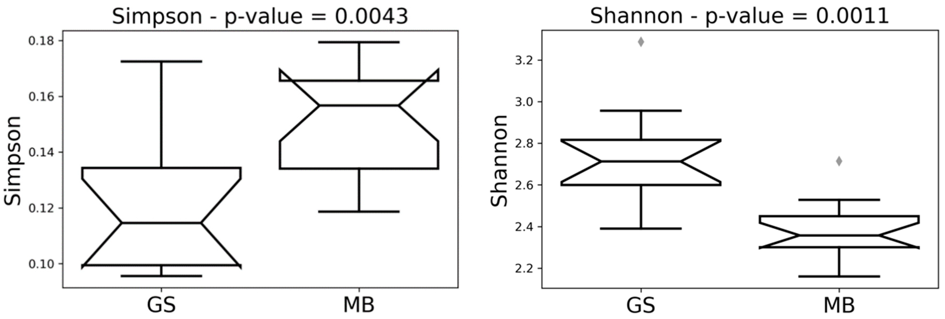
| Simpson | 0.139 | 0.326 | 0.231 | 0.003 | 0.004 | 0.142 |
| Shannon | 0.356 | 0.024 | 0.002 | 0.000 | 0.000 | 0.117 |
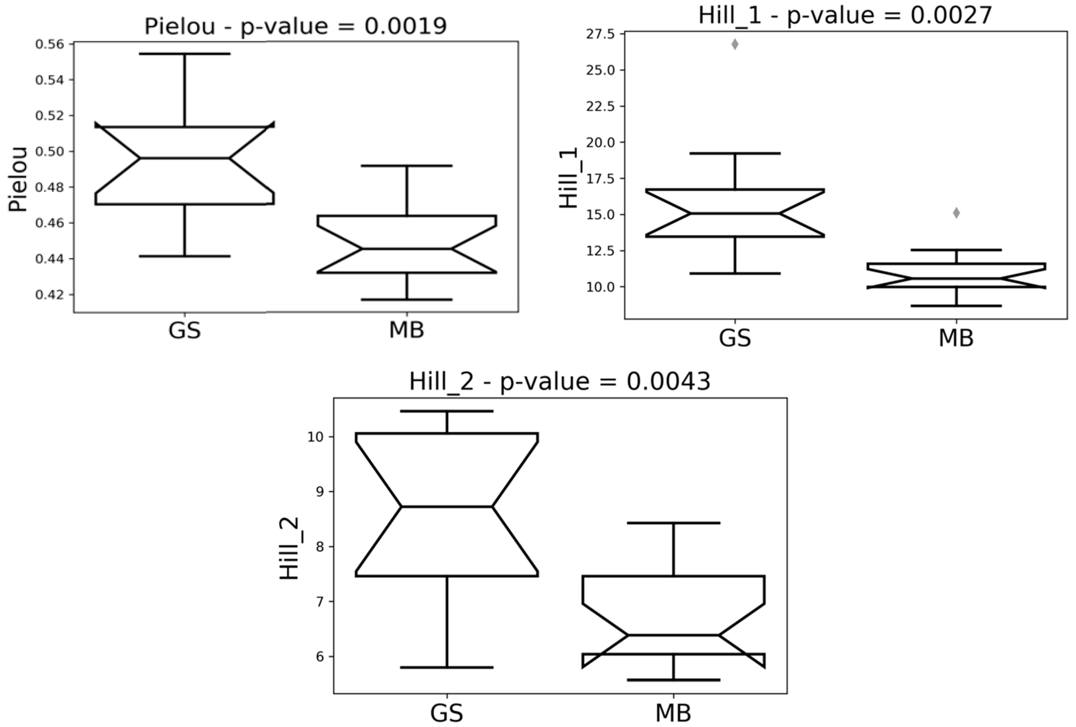
| Pielou | 0.174 | 0.156 | 0.015 | 0.000 | 0.001 | 0.931 |
| Hill_1 | 0.356 | 0.023 | 0.003 | 0.000 | 0.002 | 0.068 |
| Hill_2 | 0.121 | 0.270 | 0.194 | 0.002 | 0.004 | 0.476 |
| Simpson | Shannon | Pielou | Hill_1 | Hill_2 | |
|---|---|---|---|---|---|
| Phylum | |||||
| MB | 0.446 | 1.010 | 0.420 | 2.792 | 2.382 |
| GS | 0.531 | 0.927 | 0.366 | 2.594 | 2.032 |
| Class | |||||
| MB | 0.296 | 1.467 | 0.486 | 4.365 | 3.454 |
| GS | 0.277 | 1.592 | 0.514 | 4.956 | 3.742 |
| Order | |||||
| MB | 0.296 | 1.482 | 0.388 | 4.434 | 3.456 |
| GS | 0.272 | 1.660 | 0.421 | 5.308 | 3.809 |
| Family | |||||
| MB | 0.171 | 2.090 | 0.462 | 8.148 | 6.021 |
| GS | 0.135 | 2.445 | 0.523 | 11.752 | 7.618 |
| Genus | |||||
| MB | 0.150 | 2.379 | 0.449 | 10.895 | 6.771 |
| GS | 0.122 | 2.736 | 0.496 | 15.835 | 8.511 |
| Species | |||||
| MB | 0.045 | 3.712 | 0.589 | 41.635 | 22.712 |
| GS | 0.065 | 3.881 | 0.590 | 50.536 | 20.544 |
© 2020 by the authors. Licensee MDPI, Basel, Switzerland. This article is an open access article distributed under the terms and conditions of the Creative Commons Attribution (CC BY) license (http://creativecommons.org/licenses/by/4.0/).
Share and Cite
Mondo, E.; De Cesare, A.; Manfreda, G.; Sala, C.; Cascio, G.; Accorsi, P.A.; Marliani, G.; Cocchi, M. Depression and Microbiome—Study on the Relation and Contiguity between Dogs and Humans. Appl. Sci. 2020, 10, 573. https://doi.org/10.3390/app10020573
Mondo E, De Cesare A, Manfreda G, Sala C, Cascio G, Accorsi PA, Marliani G, Cocchi M. Depression and Microbiome—Study on the Relation and Contiguity between Dogs and Humans. Applied Sciences. 2020; 10(2):573. https://doi.org/10.3390/app10020573
Chicago/Turabian StyleMondo, Elisabetta, Alessandra De Cesare, Gerardo Manfreda, Claudia Sala, Giuseppe Cascio, Pier Attilio Accorsi, Giovanna Marliani, and Massimo Cocchi. 2020. "Depression and Microbiome—Study on the Relation and Contiguity between Dogs and Humans" Applied Sciences 10, no. 2: 573. https://doi.org/10.3390/app10020573
APA StyleMondo, E., De Cesare, A., Manfreda, G., Sala, C., Cascio, G., Accorsi, P. A., Marliani, G., & Cocchi, M. (2020). Depression and Microbiome—Study on the Relation and Contiguity between Dogs and Humans. Applied Sciences, 10(2), 573. https://doi.org/10.3390/app10020573


