Review

The Link between Oral and Gut Microbiota in Inflammatory Bowel Disease and a Synopsis of Potential Salivary Biomarkers

1
Department of Physiology & Cell Biology, School of Medicine, University of Nevada, Reno, NV 89557, USA
2
Department of Nutrition, University of Nevada, Reno, NV 89557, USA
3
School of Medicine, University of Nevada, Reno, NV 89557, USA
4
Environmental Sciences Graduate Program, University of Nevada, Reno, NV 89557, USA
*
Author to whom correspondence should be addressed.
Appl. Sci. 2020, 10(18), 6421; https://doi.org/10.3390/app10186421
Submission received: 28 August 2020 / Revised: 10 September 2020 / Accepted: 10 September 2020 / Published: 15 September 2020
(This article belongs to the Special Issue Salivary Biomarkers: Future Diagnostic and Clinical Utilities)

Abstract

:
The objective of this review is to provide recent evidence for the oral–gut axis connection and to discuss gastrointestinal (GI) immune response, inflammatory bowel disease (IBD) pathogenesis, and potential salivary biomarkers for determining GI health. IBD affects an estimated 1.3% of the US adult population. While genetic predisposition and environment play a role, abnormal immune activity and microbiota dysbiosis within the gastrointestinal tract are also linked in IBD pathogenesis. It has been inferred that a reduced overall richness of bacterial species as well as colonization of opportunistic bacteria induce systemic inflammation in the GI tract. Currently, there is supporting evidence that both oral and gut microbiota may be related to the development of IBD. Despite this, there are currently no curative therapies for IBD, and diagnosis requires samples of blood, stool, and invasive diagnostic imaging techniques. Considering the relative ease of collection, emerging evidence of association with non-oral diseases may imply that saliva microbiome research may have the potential for gut diagnostic or prognostic value. This review demonstrates a link between saliva and intestinal profiles in IBD patients, suggesting that saliva sampling has the potential to serve as a non-invasive biomarker for gut diseases such as IBD in the oral–gut axis.

Graphical Abstract

1. Introduction

The purpose of this review is to update the status of current research on the oral–gut connection as well as the relationship between oral and gut microbiomes. This review will also provide evidence for using salivary biomarkers as a diagnostic tool to monitor physiological changes in gut health for GI related diseases and disorders; particularly in inflammatory bowel disease. University of Nevada Reno’s “first search” was used to identify studies and articles matching key words. Keywords used were “IBD”; “oral microbiota”; “gut microbiota”; “oral and gut microbiota”; “saliva”; “salivary biomarkers and IBD”; “IBD and gut microbiome”; “IBD and oral microbiome”.

2. Inflammatory Bowel Disease

Inflammatory bowel disease (IBD) is a chronic idiopathic disease of the GI tract, which encompasses several diagnoses such as Ulcerative colitis (UC), Crohn’s disease (CD), and Indeterminate colitis (IC) [1]. IBD typically manifests as ulcerations in varying regions of the GI tract; UC is primarily found to affect the mucosal layers of the colon in a continuous pattern, while CD may affect any part of the gastrointestinal (GI) tract in non-continuous form and IC patients are those in which neither a diagnosis of UC or CD can be determined but still present with characteristics of IBD [2]. Cardinal symptoms of IBD include diarrhea, rectal bleeding, fatigue, abdominal pain, unintended weight loss, decreased appetite, and vomiting [3]. However, disease presentations can vary extensively from patient-to-patient and overlaps with other GI conditions, such as irritable bowel syndrome [1]. The pathogenesis of IBD remains fundamentally indeterminate; however, it is believed to be a delicate interplay between a multitude of factors including GI immune dysregulation, genetic predisposition, gut microbiota dysbiosis, and environmental factors [4,5]. For example, in a large-sample based genome-wide association study (GWAS) published in 2016, several gene variant associations were found in the chronic immune-mediated diseases CD, UC, psoriasis, and primary sclerosing cholangitis [6]. This study presented evidence for overlap of genetic susceptibility loci of these diseases, which may be contributed to biological pleiotropy [6]. Thus, pleiotropy and gene variants could be a factor in understanding IBD pathogenesis and identifying genetically susceptible individuals. Although genetic susceptibility has been shown to contribute to IBD pathogenesis, environmental risk factors at play seem to be a major contributor to the majority of diagnosed IBD patients [7]. Environmental risk factors have the ability to impact gut microbiota in IBD pathogenesis as well, and include antibiotics and other medication use, tobacco use, diet, socioeconomic status [8], and psychological stressors [8] however it is difficult to determine whether microbiota shifts are the cause or consequence of these environmental factors.
IBD affects multiple ethnic and racial groups, with onset of disease occurring throughout early adulthood between the ages of 20–40 [9,10,11] with approximately 0.4–0.6% of cases being reported globally [12,13]. While data is still lacking for many developing countries, increases in IBD prevalence appears to be paralleled with increasing development index [13] as the incidence of UC is highest in North America with 16.4 new cases per 100,000 people annually and 16.7 per 1000 people in Canada [8]. The Western diet is considered a major contributor to the obesity epidemic and is concurrently a major risk factor in IBD pathogenesis [14]. Therefore, not surprisingly, the incidence of IBD is highest in Westernized countries [8]. Additionally, diet has been associated with which gut microbes will thrive and those that will not [15]. Microbial dysbiosis has been implicated as a major pathophysiological mechanism in IBD contributing to a perpetuating feedback loop of dysregulated host-microbiota shifts [16]. As the incidence of IBD continues to rise globally, and across new populations, such as in children [1], the need for effective treatment options, diagnostics, and understanding of the pathophysiology mechanisms become more prudent.
Treatments for IBD include several classes of drug therapy, i.e., immunosuppressants, corticosteroids, aminosalicylates, and, more recently, monoclonal antibodies, in addition to palliative treatments like antibiotics, analgesics, anticholinergics, and antidiarrheal agents [3,9,17]. Holistic treatments and therapies emphasizing the importance of diet as well as intentional microbial shifts through the use of probiotics and heterologous and autologous fecal microbiota transplantation (FMT) have also gained momentum in recent years [18,19,20], although currently FMT is only FDA-approved for use in qualified recurrent Clostridium difficile associated infection in patients. Proper treatment and diagnosis of IBD are important as they have the potential to significantly improve patients’ daily lives as IBD can become a crippling disease. One population-based cohort study showed that IBD patients had a lower survival rate from colorectal cancer compared to non-IBD patients [21]. In another recent, 6-year prospective, multicenter, nested, case–control study published in 2019, perforating CD, extensive UC, and abdominal surgery for UC were major risk factors identified for the incidence of cancer overall as well as in extracolonic cancer [22]. With the greater risk of cancers and mortality, research on effective IBD treatment and accurate diagnoses using non-invasive methods have been increasing alongside growing cases worldwide.

3. Inflammatory Bowel Disease Pathogenesis

3.1. IBD Immune Dysregulation

Although IBD development is of unknown etiology, several factors including genetics and environmental risk factors have been examined in great depth. Interestingly, the host immune response is interlinked with commensal microorganisms in properly mediating inflammatory response cytokines [23]. Intestinal macrophages are largely responsible for immune homeostasis between commensal organisms and produce cytokines such as interleukin-10 (IL-10) to support the expansion of T regulatory cells (Tregs). In genetically susceptible individuals with IBD, intestinal macrophage polarization occurs from anti-inflammatory macrophage cells (M2) to the more reactive, classically activated, (M1) macrophages [24] which reduces production of antigen-specific Tregs and causes excessive inflammation especially against commensal organisms and food antigens [25]. Additionally, macrophage polarization leads to an overreactive response to toll-like receptor (TLR) ligands, which are greeted with a production of proinflammatory cytokine cascades and overproduction of reactive oxygen species (ROS) in M1-type responses leading to intestinal barrier dysfunction and tissue damage indicative of IBD ulceration [26]. In homeostasis, the intestinal barrier protects the epithelium and prevents permeability from the outside environment passing through the intestinal lumen. Without functioning structural reinforcement through tight junction proteins (TJs), located between epithelial cells, or the secretion of antimicrobial peptides (AMPs), the intestinal epithelial barrier is left susceptible to invading pathogens and subsequent ulceration development [27]. Additionally, the proliferation and differentiation of interleukin-17 (IL-17)-secreting Th17 CD4+ memory T cells has been shown to be a key factor in autoimmune diseases and chronic inflammation [28]. IL-23, a member of the IL-12 family, is characterized as a proinflammatory cytokine stimulating the production of IL-17 through the differentiation of naïve T cells to T-helper 17 cells (Th17). Interleukin 23 (IL-23) can also stimulate the expression of other IL-17-secreting immune cells expressing the IL-23 receptor (IL-23R), particularly in chronic inflammatory conditions of the intestines, and recently an IL-23 monoclonal antibody has been approved for phase III clinical trials for use in UC [29] although mechanisms leading to the development of chronic inflammatory diseases of the GI tract through the IL-23/Th17 pathway remain elusive [28].

3.2. IBD Genetic Predisposition

In addition to immune dysregulation by environmental risk factors, several genetic mutations in susceptible hosts can lead to the exacerbation of intestinal epithelial barrier function failure and IBD development. Frameshift mutations in the nucleotide-binding oligomerization domain-containing protein 2 (NOD2, also called CARD15) is associated with increased risk for the development of CD [30]. The NOD2-associated protein recognizes a signature component of bacterial peptidoglycan, muramyl dipeptide (MDP), to elicit an appropriate immune response through the nuclear factor-kappa beta (NF-kβ) pathway [31]. In CD-associated NOD2 mutants, binding abilities are weaker and therefore have an altered and irregular immune response to bacteria contributing to dysbiosis and disease pathogenesis [31]. Additionally, myeloid differentiation primary response 88 protein (MyD88)-dependent TLR signaling is an important signaling cascade regulating commensal microbiota–host symbiosis at the transcriptional level [32]. In MyD88 loss-of-function mutant zebrafish in the presence of commensal bacterial colonization in the gut, researchers Bjørn et al. found a significant downregulation of host genes downstream of MyD88 through TLR-MyD88-microbiota signaling [33]. These results indicate the significance of commensal microbiota in mediating the host transcriptome via TLR-MyD88 cascades. Toll-like receptor 4 (TLR4) is a key receptor for initiation of the innate immune system in response to binding of ligands, such as lipopolysaccharide (LPS) from Gram-negative bacteria [34]. TLR-receptor signaling, specifically toll-like receptor 2 (TLR2) and TLR4, play a fundamental role in intestinal barrier integrity via the gut microbiome and dysregulation could contribute to the pathogenesis of IBD [35].

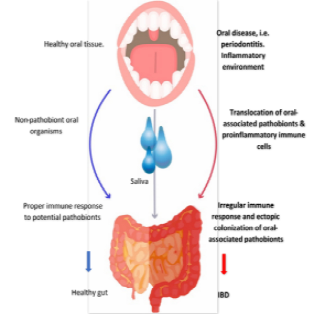
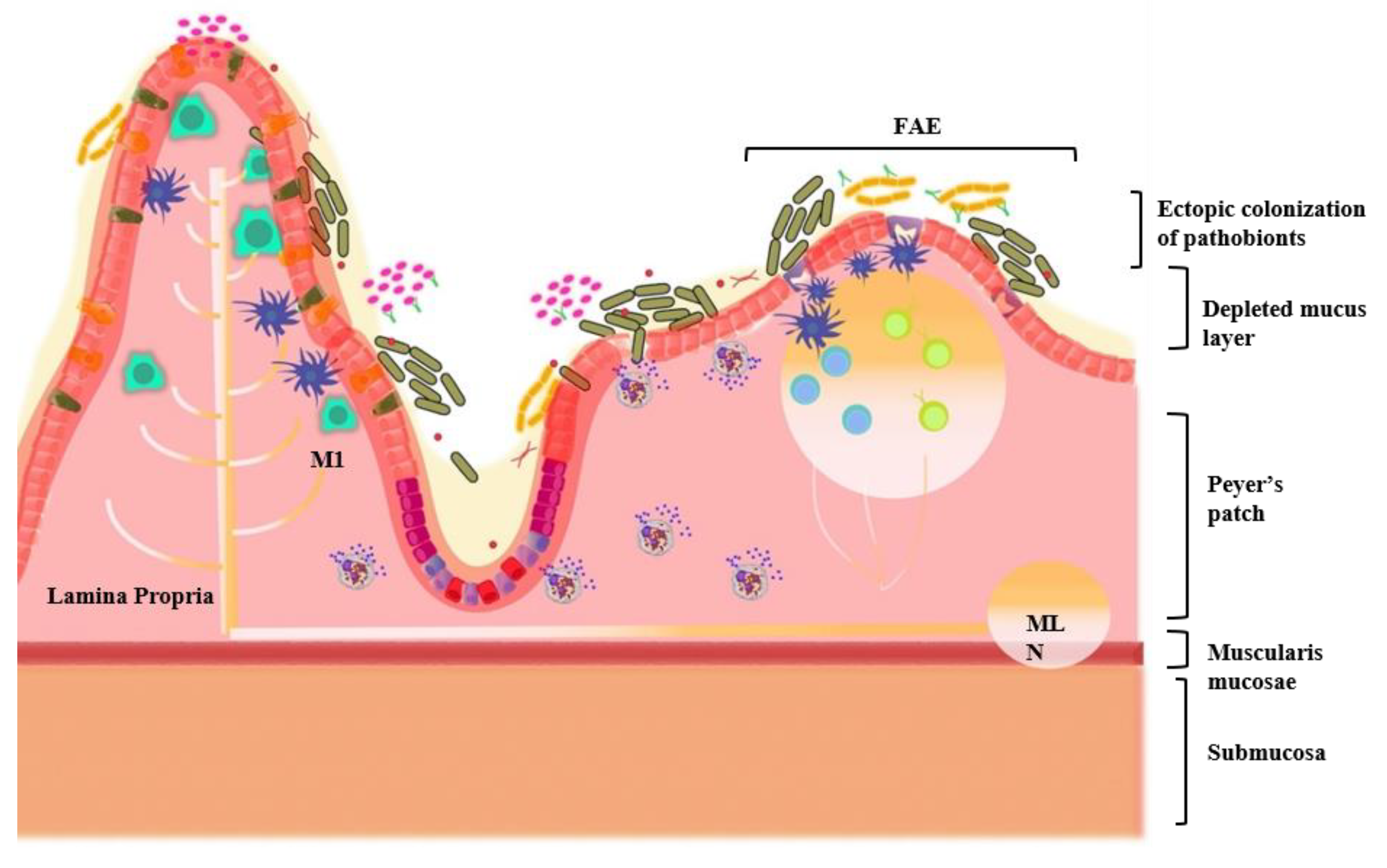
3.3. Oral–Gut Relationship in IBD Pathogenesis

The role of heritable genetics in microbial composition and regulation of the intestinal inflammatory response is unclear, and it is said that microbial profiles are largely due to environmental factors [36,37]. Some environmental factors that may contribute to intestinal inflammatory dysfunction include a diet lacking key nutrients required for commensal survival [38], drug and antibiotic use, physical activity, smoking, and psychological and emotional stress [39,40]. Phyla associated with oral-derived pathobionts have also been found to be associated with idiopathic inflammatory diseases, such as IBD [41,42,43,44]. While there is evidence for the oral–gut axis in disease models, using gut-targeted probiotics to improve oral health was recently reviewed in depth and may be helpful in the prevention of dental carries [45]. This suggests there may be a relationship between improving the GI tract through beneficial commensal bacteria while simultaneously improving bacteria OTUs residing in the oral cavity [45]. While we cannot assume that saliva mirrors the human gut microbiome entirely, some studies suggest a shift in saliva microbial profiles may be reflective of a shift in gut microbiota profiles, especially with the supplementation of probiotics [46,47,48]. Studies show increased inflammatory responses due to oral cavity diseases that cause pernicious shifts in bacterial colony composition in both the oral cavity and the intestines. Interestingly, the oral–gut connection in inflammation and IBD pathogenesis has recently been explored in an article published by Kitamoto et al. [49]. In this article, oral-originating inflammatory Th17 cells and oral pathobiont bacterial species present in periodontitis mouse models were able to ectopically colonize and translocate to the intestines causing IBD [49]. This study emphasizes the oral–gut connection in IBD and dysregulated inflammatory responses originating in the oral cavity and migrating systemically.

4. The Gastrointestinal System

The gastrointestinal system includes everything starting from the oral cavity to the rectum and is essentially a continuous pipeline connecting the external environment to the internal. This amazing system is related and connected yet can be distinguished depending on location. The oral cavity is important for the initiation of digestion and conducts initial immune responses. The oral cavity comprises the tongue, teeth, gingiva, buccal mucosa, salivary glands, hard and soft palate, and gingival crevicular fluid (GCF), which is found in the gingival sulcus between the teeth and gums [50,51]. Surfaces of the oral cavity contain diverse microbial communities, with any one site hosting approximately 50 different species [51]. From the oral cavity, the GI tract continues to the pharynx, esophagus, stomach, and intestine. The small intestine consists of villi comprised of mature epithelial tissue that protrude towards the lumen and Crypts of Lieberkühn that scope downward into the mesenchymal stromal cell layer. Transit amplifying progenitor (TAP) cells or intestinal stem cells (ISCs) can differentiate into either enterocytes or secretory cells [52]. Secretory cells further differentiate into either goblet cells, responsible for the production of mucus and protection of epithelial barrier; enteroendocrine cells, which are primarily dedicated to nutrient sensing and release of over 20 different peptide hormones [53]; Paneth cells, which are almost exclusively located in the distal ilium, secrete antimicrobial peptides, called α-defensins that are responsible for innate immunity; or chemo-sensing tuft cells that link the microbiome of the intestinal lumen to the host immune system [52,54,55]. At the base of the crypts of Lieberkühn are the ISCs, Paneth cells, and TAP cells; these cells are responsible for immunity, barrier protection, replication, and differentiation. More superficially, enterocyte and goblet cells emerge and migrate up to the mature villi to eventually be sloughed off. ISCs are important as they allow for rapid cellular turnover (i.e., cell proliferation) after exposure to toxins, cellular damage, inflammation, and changes in the luminal microenvironment [56]. The environment within the crypts of Lieberkühn also exhibits high plasticity to remain resilient to the effects of the external environment [57]. Indeed, most cells of the small intestine exhibit rapid turnover, with proliferation occurring approximately every 3–5 days [56]. Near the epithelial layer, AMPs are secreted by Paneth cells [58] and IECs to provide further control and separation from the intestinal epithelium [59]. Generally, the small intestine has a relatively low density and diversity of microbiota colonizers due to frequent disruption of digestive secretions such as bile and vary depending on oxygen and nutrient availability [60]. The major functions of the large intestine are to absorb and synthesize vitamins, absorb electrolytes and water via osmosis and electrochemical gradients, and to aid in the elimination of indigestible material [61]. Like much of the GI tract, the large intestine is made up of several layers including the mucosa layer, submucosa, muscle layer, and serosa, from the lumen out. However, in contrast to the small intestine, the large intestine is void of villi and its walls contain a larger amount of goblet cells and enterocytes and contains two relatively distinct mucus layers; the inner layer proximal to the epithelium and an outer mucus layer [59]. Goblet cells secrete protective, highly glycosylated proteins such as Mucin 2 (MUC2), which form the majority of the two mucus layers and are protective against colitis and colorectal cancers [62]. Goblet cells also contribute to gut immune homeostasis by secreting anti-microbial peptides, cytokines, and chemokines [63] for further protection and prevention of bacteria and other organism adherence to the epithelium. In the large intestine, the inner mucus layer is largely devoid of microorganisms and acts as a substantial line of defense from potential epithelial barrier breach. The inner mucus layer is a frontline for protection largely harboring secretory IgA (sIgA), AMPs, and other goblet cell products, for example, zymogen granule protein 16 (ZG16) [63], MUC2 [64], and resistin-like molecule beta (RELMβ) [65] which possess antimicrobial activity.

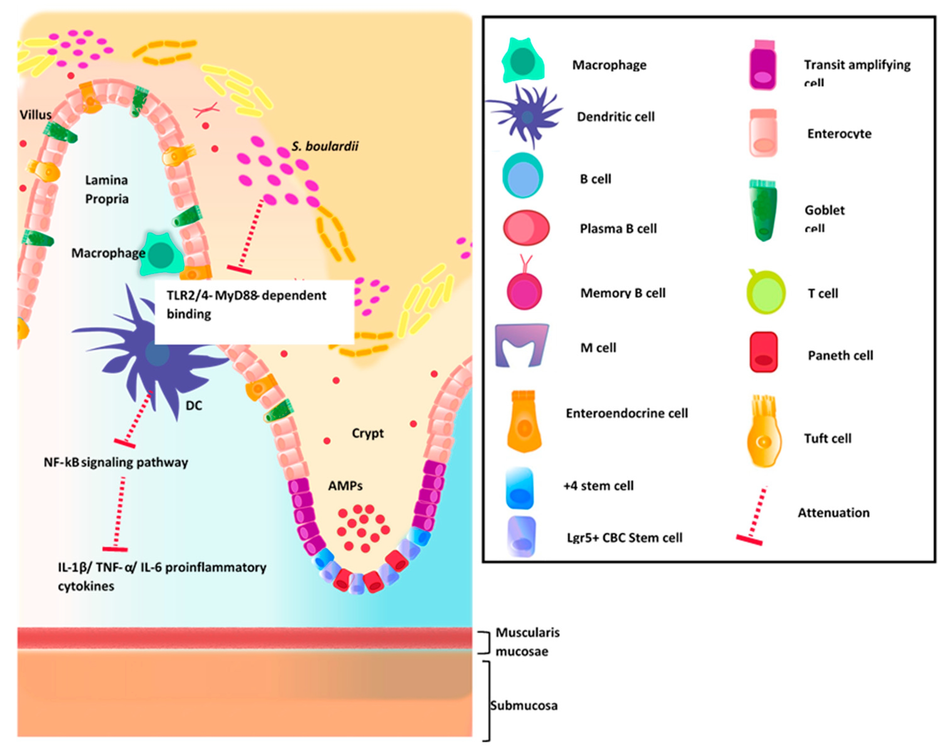
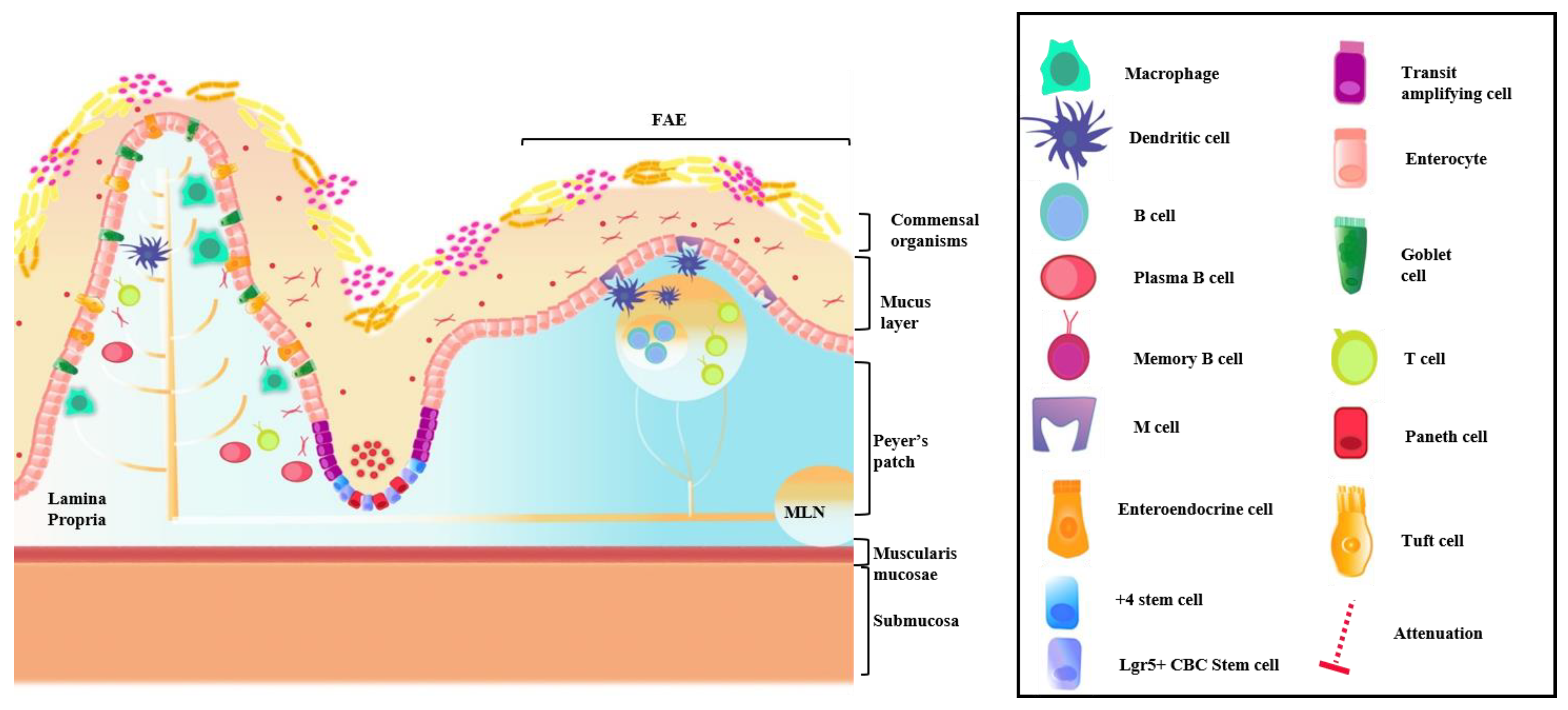
4.1. Intestinal Immune System

Inflammation is a term widely used to describe a state in which there is an acute or chronic response to cellular injury, toxic compounds, or infection which trigger inflammatory signaling pathways and allow immune cells to infiltrate tissue and eliminate pathogenic or damaged materials [66]. Therefore, the need for an effective and appropriate immune response that maintains proper physical defenses and inflammatory pathway is vital to maintaining a healthy GI tract [67]. GI natural physical defenses are often categorized into two groups: (1) the extrinsic barrier and (2) the intrinsic barrier [67]. The epithelial layers, which form the mucosa and include the tight junctions (TJs) in between individual cells, act to maintain epithelial cell layer integrity. The extrinsic barrier at the intestinal epithelia includes mucus secretions, antibiotic peptides as well as various hormones, cytokines, chemokines, and commensal resident microbes [68]. Underlying the extrinsic barrier and epithelia is specialized gut-associated lymphoid tissue (GALT) [69]. GALT is the largest immune organ in the body and is the intestinal immune inductive site composed of scattered and isolated lymphoid follicles (ILFs), Peyer’s patches [68], and mesenteric lymph nodes (MLNs) [69], containing immune cells including antigen presenting cells, macrophages, and B and T lymphocytes [70]. The lamina propria is a secretory effector site where differentiated B cells from germinal centers residing under Peyer’s patches are funneled into via lymphatics [71]. TJs regulate the paracellular passages of ions, solutes, vesicles, and immune cells between adjacent cells and intracellularly [72]. Luminal noxious macromolecules cannot penetrate the epithelium because of TJ proteins occludin, transmembrane claudins, scaffold proteins zonula occludens 1, 2, and 3 and junctional adhesion molecules [72]. However, TJ impairment allows the passage of noxious molecules and organisms which can stimulate the activation of mucosal immune cells and inflammation. As mentioned above, epithelial barrier function can be reinforced or protected from perturbations by commensal organisms through the TLR-MyD88 inflammatory pathway and intestinal barrier dysfunction is associated with the initiation and development of various intestinal and systemic diseases [73]. For example, mice were given the chemotherapy drug 5-fluorouracil (5FU) to induce intestinal mucosal disturbances via the TLR4/TLR2/MyD88-NF-kB/mitogen-activated protein kinase (MAPK) pathways and then given the beneficial organism Saccharomyces boulardii (S. boulardii) [74]. Data indicated that through modulation of these inflammatory pathways, S. boulardii was able to attenuate the otherwise rampant inflammatory response in test mice, improving intestinal barrier permeability and overall intestinal health status, as summarized in Figure 1 [74].

4.2. GI Host Defense

GI homeostasis is characterized by the ability to maintain important physiological functions that include the ability to protect the host from potential pathogens and at the same time, maintain a balanced relationship with commensal bacterial populations. Host defense pathways serve to maintain GI homeostasis, including immune responses and physical defenses, such as the epithelial barrier in the GI tract. Protection and homeostasis of the gut mucosal barrier are also achieved with the help of the enteric nervous system (ENS) [75]. In response to a pathogen at the intestinal barrier, immune cells are signaled to secrete the proinflammatory cytokine, interleukin-18 (IL-18), and in homeostasis, goblet cells produce AMPs via IL-18 cytokine release via enteric neurons [75]. While the signaling mechanisms of mature IL-18 from epithelial cells and immune cells in the intestinal tract are well understood [76], modulation of IL-18 stimulation via enteric neurons remains elusive [77]. In patients with IBD, specifically in colitis [78], these immune system regulatory mechanisms are disrupted and cause further progression of the disease. In mice with DSS-induced colitis, pretreatment of IL-18 attenuated severity of IBD by reducing inflammatory infiltration, promoting MUC2 expression and goblet cell functionality and overall quantity [78]. As mentioned above, factors including host genetics and commensal microbial immune modulation are key in determining pathogenesis of IBD.
Peyer’s patches and ILFs are a component of specialized follicle associated epithelium (FAE) [69], a subset of intestinal epithelial cells (IECs), residing in the region of the epithelium covering GALT lymphoid follicles [79]. Peyer’s patches can directly sample contents from the lumen and are considered lymphoid organs [80]. Integrated into the FAE are Microfold (M) cells, which are distinctively different in morphology as well as in function from epithelial cells in that they are highly specialized to take up intestinal microbial antigens and deliver them to GALT [79,81]. M cells fundamentally function to communicate between the external and internal GI environment as they possess the ability to recognize several different features of microbes via distinct surface recognition proteins. Glycoprotein 2 (GP2) is a glycosylphosphatidylinositol-anchored glycoprotein exclusively present on the apical surface of M cells capable of binding fimbrin D-mannose specific adhesion (FimH), a component of type I pili present on the outer membrane of some enteric species of Gram-negative bacteria, such as Escherichia coli (E. coli) and Salmonella enterica [82]. The ability for M cells to recognize, bind, and uptake specific bacteria is imperative for developing specialized antigen class switching and adaptive immunity [82]. Although intestinal epithelia functions to absorb nutrients from the intestine and some gut antigens, Peyer’s patches and ILF of the FAE are primary sites for adaptive immunity from pathogens as well as gut-microbiota homeostasis [80,83].
In FAE, antigens from the lumen are met by dendritic cells (DCs) and macrophages in the inductive sites of Peyer’s patches, also coined as the subepithelial dome site, along with B cells and CD4+ T cells, to present in germinal centers to undergo class-switching and somatic hypermutation of IgA+ memory B cells and IgA-secreting plasma cells. The differentiation of B cells into IgA-secreting plasma cells is in part due to the crosstalk with gut microbiota, B cells, and the intestinal epithelium [69]. Immunoglobulin class switching occurs in the mesenteric lymph nodes, Peyer’s patches, ILFs, and lamina propria [84]. The intestinal immune system is a dynamic, multifactorial process which has the ability to confer adaptive and innate immunity, combat pathogenic antigens, as well as maintain synergy with commensal organisms. The host GI immune system works in harmony with the commensal resident intestinal microbiota which largely contributes to mucosal IgA production and protection and is summarized in Figure 2 [84]. In a study published by Beller et al., specific intestinal microbiota were found to influence the production and protection of the gut by increasing expression of transforming growth factor beta-1 (TGF-β1) and therefore mediating mucosal IgA levels [84]. TGF-β1 is a cytokine involved in intestinal immune homeostasis, preventing overreaction of pro-inflammatory cytokine production and Th1 and Th2 cell differentiation while encouraging T cell polarization of Tregs [84]. Beller et al., used fecal microbiota transplantation (FMT) to transplant stool from high-IgA producing BALB/c mice and low-IgA producing eosinophil knockout mice (ΔdblGATA-1) into specific pathogen free (SPF) BALB/c mice. Results indicated that SPF mice which received the high-IgA FMT had significantly higher levels of serum and fecal IgA levels as well as an approximately two-fold higher absolute count of CD45+IgA+ B plasma cells in the small intestinal lamina propria [84]. These results show that specific populations of the gut microbiota, namely Anaeroplasma and Erysipelotrichaceae incertae sedis, are capable of promoting IgA expression despite lack of eosinophil help, contrary to popular belief [84] and emancipates the microbiota, showing independent control over immune responses.
In IEC, antigens from the lumen are also capable of being taken up by DCs through epithelial cells such as goblet cells [63], and other subepithelial mononuclear phagocytes such as macrophages [84], in the lamina propria which are then presented to T cells for B cell IgA class switching. T cells, macrophages, and other antigen presenting cells are also able to sample and respond [69,79,84,85] by conferring specific immunity with IgA [81]. Antigen-specific mucosal effector cells such as memory B cells, CD4+ Th1, CD4+ Th2, and plasma cells producing IgA all reside within mucosal effector sites [59,84]. Once specialized immunoglobulins are made by mucosal plasma cells in the lamina propria, they are transported across the epithelial barrier. First, the pentameric IgA (pIgA) and J chain complex are secreted and bind to the basolateral membrane of IECs by the polymeric immunoglobulin receptor (pIgR). Next, these complexes are transcytosed into the intestinal mucus layers, driven by cleavage of pIgR, yielding secretory (sIgA) [86] which further confers mucosal surface protection. sIgA’s innate ability to distinguish commensal organisms from overt pathogens has been debated and discussed in detail [86]; it has been suggested that sIgA antibodies may have greater affinity in the detection of pathogens due to various opportunistic and established virulence mechanisms of the pathogens [86].

5. The Gut Microbiota in Health and IBD

The human bacterial microbiota is diverse consisting of 5–6 primary phyla in varying ratios that are dependent on both environment and genetics. Among these phyla, Firmicutes, Actinobacteria, Proteobacteria, Bacteroidetes, Fusobacteria, and Verrucomicrobia are highly expressed under normal physiological conditions in the human gut [87]. Contrary to popular belief, baseline microbial populations remain relatively stable and unchanging long term, even with diet interventions. The study published by Fragiadakis et al. found that after initial microbe population changes in the first three months of diet interventions, participant microbiota reverted to, or close to, its original composition after the year-long study was complete, suggesting a resilience, which might be harder to alter than originally thought. While commensals are generally considered helpful, in disease or conditions suboptimal to the organism, malevolent changes can occur. These organisms are known as “pathobionts,” and under normal conditions remain symbiotic, yet in response to environmental exposures or genetic predisposition become pathogenic to the host [88]. Unlike externally acquired pathogens, pathobionts remain symbiotic and only cause an acute inflammatory response when environmental cues reduce resident protective microbiota allowing pathogenic bacteria to colonize [89].
UC and CD are two distinct subgroups of IBD including several distinguishing symptoms and genetic variant predispositions. Interestingly, gut microbiota sequencing from UC and CD patients also show population differences [90]. Overall, reduced diversity of microorganisms and disproportionate phyla are associated with both UC and CD pathogenesis, although there have been studies indicating differences in phylum distribution in UC and CD independently [91].

Commensal Organisms and Epithelial Barrier Function

The adaptive immunity at the epithelial barrier is what helps to maintain barrier protection. Commensal bacteria help this process by producing secondary metabolites such as aryl hydrocarbon receptor (AhR) ligands and short-chain fatty acids (SCFAs), as well as TLR/NOD ligands [92]. Commensal bacteria, metabolites, and ligands can bind to the corresponding receptors on immune cells and enterocytes, protecting them from targeted immune responses [92]. In addition to targeted mucosal immune cell responses to pathogens, IECs are also adapted to protect against excessive intestinal inflammation, maintain the balance of commensal organisms, and repair epithelial damage. IECs sustain this balance through several mechanisms. Pattern recognition receptors (PRRs), as well as specific family members of TLRs, NOD-like receptors (NODRs), and RIG-I-like receptors (RLR) are integrated to aid in recognition of microbes and cause further signal cascades to respond appropriately in determining friend or foe [93].

6. The Oral Microbiota in Health and IBD

While most studies have focused on the microbiota of the large intestine, the oral cavity also contains diverse microbiota. To date, over 700 oral bacterial species have been identified in the human oral microbiome database (HOMD) [94]. Saliva, shedding of soft surfaces, buccal mucosa, palatal mucosa, tongue dorsum, and supragingival plaque contribute to the oral microbiome [95,96]. Likewise, hard and mucosal surfaces within the oral cavity maintain environments suitable for commensal bacteria to flourish and protect the integrity of the gingival and epithelial cells [96]. Similar to other regions of the digestive tract, the oral cavity is subject to constant environmental exposures that may impact pH, temperature, oxygen, and available nutrients due to continuous exposure to environmental insults such as food, beverage, and potential pathogens. Throughout all of this, microbial communities within resident biofilms remain relatively stable and diverse in healthy individuals [97].
Periodontal disease and dental carries contain distinct microbial profiles compared to healthy oral cavity patients [51,98]. Community shifts can occur as a result of syntrophic metabolism between microbes, microbial byproducts, and competition for nutrients [50]. For example, low molecular weight fermentable carbohydrates, namely glucose, shift microbial communities that have been linked to oral diseases such as periodontal disease as well as non-oral diseases, such as cardiovascular disease [99,100,101,102]. A recent study evaluating over 200 children also showed oral microbiota may be a determining factor in predicting weight gain trajectories [103]. The Ecological Plaque Hypothesis (EPH) and Keystone-Pathogen Hypothesis (KPH) propose mechanisms for the switch from a healthy to diseased oral state. These hypotheses agree that cariogenic, acidogenic, and aciduric bacteria primarily contribute to cavity production; fermentation from these bacteria produce acidic metabolites that promote oral diseases [104]. Indeed, research supporting the KPH suggests that Porphyromonas gingivalis (P. gingivalis), a bacterium which originates in the oral cavity where it typically resides as a symbiont, is highly expressed in oral diseases where it has the ability to over-activate the immune system, leading to periodontal disease, poor gum health, and dental carries [98,105,106]. Further studies also suggest that pathogenic periodontal bacteria contribute to the development of metabolic, autoimmune, and inflammatory diseases, such as in IBD [107] by entering the bloodstream through lesions in the oral cavity [50]. P. gingivalis is an anaerobic, asaccharolytic, Gram negative rod and an oral-originating pathobiont that ferments protein as opposed to carbohydrates. P. gingivalis utilizes virulence factor cysteine proteases called gingipain to break down proteins, such as hemin acquired from hemoglobin. However, when homeostasis is lost, P. gingivalis causes periodontal disease and can cause further disease progression in distant tissues [108,109]. Together, these studies suggest that the oral cavity microbiota plays an important role in regulating human health and may also serve as a mechanism for the diagnosis and/or prognosis of patient health outcomes, as in IBD.

7. The Oral Microbiota and Gut Microbiota Axis

Literature linking the microbiota of the human oral to the gut microbiota is gaining momentum, and the “oral–gut axis” has been recently proposed [49]. In the study by Kitamoto et al., a group of gastroenterologists, dentists, and researchers studied the role of the intramucosal connections between the gut and oral cavity of patients with IBD. SPF mice mimicking a periodontitis phenotype (SPF-ligature) and normal healthy controls (HC) were treated with dextran sodium sulfate (DSS) to induce colitis. Before treatment with DSS, SPF-ligature mice showed oral dysbiosis compared to controls as well as promotion of a Th1 response into the lamina propria via increased immune infiltration of Th17 cells, B cells, and T cells. After DSS treatment, greater inflammation in the colonic mucosa showed increased myeloid and lymphoid infiltration and further isolation of CD3+CD4+ T cells from SPF-ligature DSS mice as well as higher production of IL-17A and interferon gamma (IFN-γ) compared to control mice [49]. The oral and gut microbiota of SPF-ligature DSS mice showed that specific oral-originating bacteria were found to colonize both environments. Using linear discriminant analysis effect size (LEfSe), Klebsiella spp. and Enterobacter spp. from the bacterial family Enterobacteriaceae, were predominately found in the oral cavity of disease models as well as in accumulation in the gut and were not found in non-ligature DSS controls [49]. In general, Enterobacteriaceae is the most predominant bacterial taxon found in the oral cavity during periodontitis infections, and these results indicate that non-periodontitis DSS mice were protected from ectopic colonization of oral-derived pathobionts while the periodontitis-DSS mice were greatly susceptible. Increased inflammation and infiltration of gut immune cells initiated by periodontitis ligatures showed that DSS-treated mice were more likely to have oral-derived pathobiont colonization in the gut, thus supporting the “oral–gut axis” paradigm. In another study, Rautava et al. showed that colitis-induced mouse models have a significant association between saliva microbial profiles and proximal gut profiles reflective to the proportion of inflammation [110]. Additionally, a recent study published by Atarashi et al., reported evidence supporting the relationship between oral and gut microbiota [111] and a more expanded overview of relative oral-gut bacteria composition is summarized in Table 1. Saliva samples from two patients with IBD were transplanted into C57BL/6 (B6) GF mice and examined for immunological changes in colonic and small intestinal lamina propria tissue; B6 GF mouse saliva was also examined before and after IBD saliva transplantation. Tissue taken from small intestinal lamina propria showed a significant increase in the relative amount of IFN-γ-producing CD4+ Th1 helper cells as well as fecal microbiota composed of many oral-originating bacterial species, particularly Klebsiella pneumoniae [111]. When administered orally, isolated K. pneumonia was also found to contribute significantly to the induction of Th1 cells in the oral cavity and colon [111] and significantly attributed to greater expression of colonic lamina propria CD4+ T cells producing IL-17 (Th17 cells) and IFN-γ in colitis-prone IL-10 knockout mice [111]. These results infer that in susceptible patients, the effect of oral-originating K. pneumoniae pathobiont could contribute to the induction of significant immune responses and IBD pathogenesis. It should also be noted that increased production of IFN-γ by memory CD4+ T cells in addition to increased production of IL-17-secreting Th17 cells leads to further production of proinflammatory cytokines IL-6, IL-1, and TNF-α and may be related to the IL-23/Th17 axis as mentioned in earlier sections. In a study published by Santos et al., oral infection with the parasite Toxoplasma gondii in microbiota-depleted mice treated with antibiotics were shown to have higher susceptibility to infection and dysregulated immune responses in the gut [112], further supporting the interplay not only in bacteria but other organisms and pathogens.
Oral-derived pathobionts, such as Fusobacteriaceae, Enterobacteriaceae, Pasteurellaceae, and Vellonellaceae, have been found in the mucosal tissues of IBD patients, as illustrated in Figure 3, in addition to depletion of Clostridiales [97,113]. In a recent study, Shirmer et al., found significant associations of increased operational taxonomic units (OTUs) from oral-typical bacteria and increased IBD disease severity, summarized in the graphical abstract [113]. This study also revealed a 75% decreased abundance of OTUs with increased IBD disease severity from the order Clostridiales, Lachnospiriaceae, and Ruminococcaceae, suggesting a significant decrease in SCFA production and intestinal epithelium barrier protection by commensal organisms [113]. These results indicate oral-derived pathobiont bacteria are found predominately in patients with increasing IBD severity and demonstrate that healthy patients are fairly resistant to microbial shifts of oral-derived bacterial colonization in the gut associated with IBD.

7.1. Beneficial Gut Commensal Organisms

Many studies examining probiotic supplementation for the treatment of IBD have shown improvement in patient symptoms [116], however, more research examining the diverse relationships of multiple system environments, such as the oral cavity and digestive tract, must be investigated [117,118]. In a paramount study by Sokol et al., the commensal organism Faecalibacterium prausnitzii (F. prausnitzii) was delivered orally to 2,4,6-trinitrobenzenesulfonic acid (TNBS)-treated colitis mouse models and controls. Results showed F. prausnitzii and its supernatant were able to successfully improve the fecal microbiota of colitis-induced mice. In patients with CD, as well as in vitro with Caco-2-cells, F. prausnitzii attenuated NF-kB activation and pro-inflammatory IL-8 secretion resulting in anti-inflammatory effects along with improved shifts in the microbiota composition [119].
While it is difficult to pinpoint the range of IBD symptom-ameliorating bacteria, there are several phyla commonly found in healthy participants, such as F. prausnitzii [119] and Roseburia spp. [120], which may be lacking in patients with IBD. Several studies indicate the positive effects of specific probiotic bacteria for humans, such as strains belonging to the genera Lactobacillus and Bifidobacterium [121]. Lactobacillus casei (L. casei) and Lactobacillus rhamnosus (L. rhamnosus) are among the most studied strains of probiotics for their wide-range of notable health benefits [122]. In a study administering fermented milk products containing Bifidobacterium lactis in murine models with colitis, researchers found a decrease in inflammation due to metabolic shifts stemming from a change in gut microbial profiles. This evidence suggests the shift to a more robust commensal bacterial gut environment may protect the lining of the gut and create a non-permissive environment for opportunistic strains [123]. Along with gut barrier protection, metabolites of intestinal bacterial species may be associated with overall human health [124,125]. For example, the well-studied probiotic strain L. casei subspecies Shirota (LcS) is a relatively stable probiotic, resistant to stomach acid and other gastric enzymes, and is most commonly known for its ability to ferment dairy products. LcS is a rod-shaped lactic acid bacterium linked to improved immune responses and gut barrier function. In a study by Ou et al., symptoms of constipation were ameliorated, and gastric emptying improved after adults were given a LcS probiotic drink [126]. Interestingly, levels of acetylcholine (Ach) and serotonin (5-HT) also increased when compared to controls [126]. There is also some evidence suggesting probiotic supplementation of LcS may help to ameliorate DSS-induced colitis in mice [127], improve fasting plasma glucose as well as prevent weight gain in humans fed a high fat diet [128].

7.2. Beneficial Oral Commensal Organisms

The oral cavity is the docking point for the outside world, constantly being assaulted by incoming elements. Oral health can be defined similarly to that of the gut: microbial eubiosis, appropriate immune regulation, and physiological function [129]. The use of probiotics to maintain oral homeostasis has recently been gaining momentum in dentistry as more clinical practitioners are gaining a better understanding of the importance of beneficial microbes in oral health. The study published by Moman R. and colleagues utilized probiotics strains Lactobacillus reuteri, L. rhamnosus GG, and Streptococcus salivarius to combat periodontal infection with commonly-causing periodontitis bacteria, including P. gingivalis, in wax moth larvae [130]. All pathogenic strains showed significant lethality when administered by themselves while, when administered together, probiotics significantly improved viability providing evidence for host defense against periodontitis-causing pathogens [130]. In a randomized, clinical, double-blind, placebo-controlled study published by Shimauchi et al., 6.7 × 10⁸ colony forming units (CFUs) was administered of Lactobacillus salivarius, related to S. salivarius, or a placebo control to 66 healthy human volunteers for eight weeks [131]. Saliva and plaque samples were collected at baseline and after the intervention. The authors reported improved clinically defined parameters of periodontal health, more so in smokers, and from past studies suggests protection from oral cavity development of periodontopathic organisms [131]. In addition to diet and proper oral hygiene, introduction of beneficial microbes into the oral cavity may be helpful in prophylaxis treatments preventing gingivitis, dental carries, and periodontitis. Although there is burgeoning research on these topics, additional data is needed to accept any concrete conclusions.

8. Salivary Biomarkers for Monitoring Health Status in the Gut

The exploitation of the oral–gut axis has been used to determine health status in the gut by using oral samples. Individual saliva production is between 1 and 2 L per day [132]; therefore, being an appealing biospecimen for sampling. Saliva is composed mostly of water but also contains a variety of organic and inorganic components such as proteins, enzymes electrolytes, immunoglobulins, hormones, and micronutrients [133,134,135,136]. These components are essentially blood-based and permeate into the saliva due to different capillaries, acinar cells, and ductal cells [137]. As sampling saliva is noninvasive and requires minimal safety-training, saliva represents an attractive alternative to blood and serum. Advantages of using saliva as a biofluid include a fast, easy, and inexpensive collection process that is suitable outside of the laboratory without the need of medical personnel [138]. Saliva fluid is easy to store and ship, particularly in that it does not clot, and is reflective of the current physiological state of an individual. While some systemic biomarkers in health correlate well between saliva and blood in diagnosis (e.g., HIV; hepatitis; oral cancer; periodontal disease; obesity), saliva is not the best biospecimen in that it does not always accurately represent systemic concentrations such as with amylase, some proteomes, and phosphate [139]. Some diagnostic applications of saliva include oral cancer, pancreatic cancer, breast cancer, lung cancer, or gastric cancers [138]. Saliva is also a promising biofluid for use in infectious bacterial diseases, infectious viral diseases and even infectious fungal diseases [140,141,142,143,144,145,146]. Salivary diagnostics can also play an important role in cardiology, [147,148] for acute myocardial infarction [149]. In addition, we have previously noted that saliva analysis enables researchers to monitor the metabolic response of individuals during physical training [139,150]. Therefore, sampling saliva, plaque, and other surfaces in the oral cavity have the potential to conjure insight into distant, yet relative, organ systems, i.e., the gut.
Controversy and uncertainty lie in the accuracy of using the oral cavity microbiota, or other parameters of biomarker testing, for specific disease states in the GI tract. As we have discussed previously, diseases such as UC and CD have shown specific oral microbiota changes parallel to gut populations. Studies evaluating salivary biomarkers and microbiota composition in connection to IBD pathology may be useful for the future development of more conservative IBD diagnostics [114]. Said et al. evaluated salivary inflammatory biomarkers and microbiota of patients with diagnosed IBD, which revealed significantly elevated inflammatory cytokine levels, IL-6, IL-8, TNFα, and IL-1β as well as elevated secretory IgA [114]. These observations in increased cytokine levels were also correlated with relative abundance of IBD-disease associated genera, Streptococcus, Prevotella, Haemophilus, and Veillonella. Recently, oral anti-TNFα has been used in experimental trials for treatment of IBD, validating the oral–gut axis while providing a blueprint for non-invasive therapeutics and diagnostics alike [151]. Additionally, research in salivary exosomes as biomarkers for IBD and other inflammatory diseases has gained momentum as well [152]. Exosomes are cellular nanovesicles present in a range of biofluids including saliva and intestinal luminal fluid aspirates, and transport proteins, nucleic acids, and cellular information. Exosomes are involved in cell–cell communication and gene regulation and the information they carry has potential to act as disease biomarkers, especially in IBD [152,153]. Newer microbiome-based technologies (the study of microbial sequences using RNA or DNA sequencing) are making it possible to unravel complex interactions between the oral and gut microbiomes. While still in its infancy, the future of oral–gut axis research holds promise. By using the oral cavity as an aperture of enriched information into the conditions of the gut, options for better diagnostics and targeted treatments for IBD and related diseases may be attainable.

9. Conclusions

In recent years, burgeoning evidence for an oral–gut axis has been underway as there has been more interest in the gut and oral microbiota in IBD. While there is much room for growth on this topic, this review emphasizes the immune-related and potential pathophysiological mechanisms involved in IBD development and how the oral-gut axis can be used as a powerful tool in determining gut health. Gut and oral environments are infinitely complex, and the microbiota of these environments act as a fulcrum in maintaining the homeostatic physiological function of the host. Although the relationship between the mouth and gut may seem unconventional, optimism remains as an option for improved, cost-effective and less invasive diagnostic tools for people suffering from IBD. Therefore, saliva should be considered as a means to surveille the gut for future avenues of IBD research.

Author Contributions

S.T.O. conceived the concept and wrote the paper. A.B., R.G.G., R.S. and S.R. wrote the paper and contributed to substantial edits and revisions. All authors have read and agreed to the published version of the manuscript.

Funding

This work did not receive any specific grant from funding agencies in the public, commercial, or non-profit sectors.

Acknowledgments

The authors would like to thank Brad Ferguson and Seungil Ro for their edits and counsel.

Conflicts of Interest

The authors declare no conflict of interest.

Abbreviations

IBDinflammatory bowel disease
UCulcerative colitis
CDCrohn’s disease
ICindeterminate colitis
GIgastrointestinal
GWASgenome-wide association study
NOD2nucleotide-binding oligomerization domain containing 2
MDPmuramyl dipeptide
NF-kβnuclear factor-kappa beta
TLRtoll-like receptor
LPSlipopolysaccharide
IL-17interleukin-17
IL-23Interleukin 23
IL-23RIL-23 receptor
Th17T-helper 17 cells
FMTfecal microbiota transplantation
FOSfructo-oligosaccharides
R. bromiiRuminococcus bromii
ROSreactive oxygen species
S. TyphimuriumSalmonella enterica serotype Typhimurium
SHIMEsimulator of the human intestinal microbial ecosystem
B. infantisBifidobacterium longum subspecies infantis
GCFgingival crevicular fluid
TAPTransit amplifying progenitor cells
ISCsintestinal stem cells
AMPsantimicrobial peptides
TGF-β1transforming growth factor beta-1
TregsT-regulatory cells
SPFspecific pathogen free
pIgApentameric IgA
pIgRpolymeric immunoglobulin receptor
sIgAsecretory IgA
SCFAsshort-chain fatty acids
PRRsPattern recognition receptors
NODRsNOD-like receptors
RLRsRIG-I-like receptors
AhRaryl hydrocarbon receptor
C. diffClostridium difficile
OTUsoperational taxonomic units
HOMDhuman oral microbiome database
EPHEcological Plaque Hypothesis
KPHKeystone-Pathogen Hypothesis
P. gingivalisPorphyromonas gingivalis
ADAlzheimer’s disease
HChealthy controls
DSSdextran sodium sulfate
LEfSelinear discriminant analysis effect size
IFN-γinterferon gamma
B6C57BL/6
F. prausnitziiFaecalibacterium prausnitzii
L. caseiLactobacillus casei
L. rhamnosusLactobacillus rhamnosus
LcSL. casei subspecies Shirota
CFUscolony forming units
MUC2Mucin 2
ZG16zymogen granule protein 16
RELMβresistin-like molecule beta
ENSenteric nervous system
IL-18interleukin-18
ILFsisolated lymphoid follicles
MLNsmesenteric lymph nodes
TJstight junctions
GALTgut-associated lymphoid tissue
5FU5-fluorouracil
MAPKmitogen-activated protein kinase
S. boulardiiSaccharomyces boulardii
FAEfollicle associated epithelium
IECsintestinal epithelial cells
MMicrofold
GP2Glycoprotein 2
FimHfimbrin D-mannose specific adhesion
DCsdendritic cells

References

  1. Yu, Y.R.; Rodriguez, R.J. Clinical Presentation of Crohn’s, Ulcerative Colitis, and Indeterminate Colitis: Symptoms, Extraintestinal Manifestations, and Disease Phenotypes. Semin. Pediatric Surg. 2017, 26, 349–355. [Google Scholar] [CrossRef] [PubMed]
  2. Guindi, M.; Riddell, R.H. Indeterminate colitis. J. Clin. Pathol. 2004, 57, 1233–1244. [Google Scholar] [CrossRef] [PubMed]
  3. Fakhoury, M.; Negrulj, R.; Mooranian, A.; Al-Salami, H. Inflammatory bowel disease: Clinical aspects and treatments. J. Inflamm. Res. 2014, 7, 113–120. [Google Scholar] [CrossRef] [PubMed] [Green Version]
  4. Eom, T.; Kim, Y.S.; Choi, C.H.; Sadowsky, M.J.; Unno, T. Current understanding of microbiota- and dietary-therapies for treating inflammatory bowel disease. J. Microbiol. 2018, 56, 189–198. [Google Scholar] [CrossRef]
  5. Cohen, L.J.; Cho, J.H.; Gevers, D.; Chu, H. Genetic Factors and the Intestinal Microbiome Guide Development of Microbe-Based Therapies for Inflammatory Bowel Diseases. Gastroenterology 2019, 156, 2174–2189. [Google Scholar] [CrossRef] [PubMed] [Green Version]
  6. Ellinghaus, D.; The International IBD Genetics Consortium (IIBDGC); Jostins, L.; Spain, S.L.; Cortes, A.; Bethune, J.; Han, B.; Park, Y.R.; Raychaudhuri, S.; Pouget, J.G.; et al. Analysis of five chronic inflammatory diseases identifies 27 new associations and highlights disease-specific patterns at shared loci. Nat. Genet. 2016, 48, 510–518. [Google Scholar] [CrossRef]
  7. Annese, V. Genetics and epigenetics of IBD. Pharmacol. Res. 2020, 159, 104892. [Google Scholar] [CrossRef]
  8. Piovani, D.; Danese, S.; Peyrin-Biroulet, L.; Bonovas, S. Environmental, Nutritional, and Socioeconomic Determinants of IBD Incidence: A Global Ecological Study. J. Crohn’s Colitis 2020, 14, 323–331. [Google Scholar] [CrossRef]
  9. Sairenji, T.; Collins, K.L.; Evans, D.V. An Update on Inflammatory Bowel Disease. Prim Care Clin. Off. Pract. 2017, 44, 673–692. [Google Scholar] [CrossRef]
  10. Cury, D.; Oliveira, R.; Cury, M. Inflammatory bowel diseases: Time of diagnosis, environmental factors, clinical course, and management – a follow-up study in a private inflammatory bowel disease center (2003–2017). J. Inflamm. Res. 2019, 12, 127–135. [Google Scholar] [CrossRef] [Green Version]
  11. Cosnes, J.; Gower-Rousseau, C.; Seksik, P.; Cortot, A. Epidemiology and natural history of inflammatory bowel diseases. Gastroenterology 2011, 140, 1785–1794. [Google Scholar] [CrossRef] [PubMed]
  12. Ng, S.C.; Shi, H.Y.; Hamidi, N.; Underwood, F.E.; Tang, W.; Benchimol, E.I.; Panaccione, R.; Ghosh, S.; Wu, J.C.Y.; Chan, F.K.L.; et al. Worldwide incidence and prevalence of inflammatory bowel disease in the 21st century: A systematic review of population-based studies. Lancet 2018, 390, 2769–2778. [Google Scholar] [CrossRef]
  13. Takahashi, L.A.R.; Cardial, D.T.; Argani, I.L.; Arnoni, L.R.R.; Cury, V.N.; Silva, L.F.A.C.C.; Júnior, A.O.; Borato, S.D.F. Human Development Index and Inflammatory Bowel Diseases. J. Adv. Med. Med Res. 2018, 26, 1–8. [Google Scholar] [CrossRef]
  14. Statovci, D.; Aguilera, M.; MacSharry, J.; Melgar, S. The Impact of Western Diet and Nutrients on the Microbiota and Immune Response at Mucosal Interfaces. Front. Immunol. 2017, 8. [Google Scholar] [CrossRef] [Green Version]
  15. Round, J.L.; Mazmanian, S.K. The gut microbiome shapes intestinal immune responses during health and disease. Nat. Rev. Immunol. 2009, 9, 313–323. [Google Scholar] [CrossRef]
  16. Rigottier-Gois, L. Dysbiosis in inflammatory bowel diseases: The oxygen hypothesis. ISME J. 2013, 7, 1256–1261. [Google Scholar] [CrossRef] [PubMed]
  17. Pithadia, A.B.; Jain, S. Treatment of Inflammatory Bowel Disease (IBD). Pharmacol. Rep. 2011, 63, 294–295. [Google Scholar] [CrossRef]
  18. Celiberto, L.S.; Vallance, B.A.; Cavallini, D.C. Lactic Acid Bacteria: A Functional Approach; Taylor & Francis Group LLC: Boca Raton, FL, USA, 2020. [Google Scholar]
  19. Dang, X.; Xu, M.; Liu, D. Assessing the efficacy and safety of fecal microbiota transplantation and probiotic VSL#3 for active ulcerative colitis: A systematic review and meta-analysis. PLoS ONE 2020, 15, e0228846. [Google Scholar]
  20. Imdad, A.; Nicholson, M.R.; Tanner-Smith, E.E.; Zackular, J.P.; Gómez-Duarte, O.G.; Beaulieu, D.B.; Acra, S. Fecal transplantation for treatment of inflammatory bowel disease. Cochrane Database Syst. Rev. 2018, 11, CD012774. [Google Scholar] [CrossRef]
  21. Bogach, J.; Pond, G.; Eskicioglu, C.; Seow, H. Age-Related Survival Differences in Patients With Inflammatory Bowel Disease-Associated Colorectal Cancer: A Population-Based Cohort Study. Inflamm. Bowel Dis. 2019, 25, 1957–1965. [Google Scholar] [CrossRef]
  22. Biancone, L.; Armuzzi, A.; Scribano, M.L.; Castiglione, F.; D’Incà, R.; Orlando, A.; Papi, C.; Daperno, M.; Vecchi, M.; Riegler, G.; et al. Cancer Risk in Inflammatory Bowel Disease: A 6-Year Prospective Multicenter Nested Case–Control IG-IBD Study. Inflamm. Bowel Dis. 2020, 26, 450–459. [Google Scholar] [CrossRef]
  23. Leone, V.; Chang, E.B.; Devkota, S. Diet, Microbes, and Host Genetics: The Perfect Storm in Inflammatory Bowel Diseases. J. Gastroenterol. 2013, 48, 315–321. [Google Scholar] [CrossRef] [PubMed] [Green Version]
  24. Zhang, Y.; Li, X.; Luo, Z.; Ma, L.; Zhu, S.; Wang, Z.; Wen, J.; Cheng, S.; Gu, W.; Lian, Q.; et al. ECM1 is an essential factor for the determination of M1 macrophage polarization in IBD in response to LPS stimulation. Proc. Natl. Acad. Sci. USA 2020, 117, 3083–3092. [Google Scholar] [CrossRef] [PubMed]
  25. Na, Y.R.; Stakenborg, M.; Seok, S.H.; Matteoli, G. Macrophages in intestinal inflammation and resolution: A potential therapeutic target in IBD. Nat. Rev. Gastroenterol. Hepatol. 2019, 16, 531–543. [Google Scholar] [CrossRef] [PubMed]
  26. Tan, H.Y.; Wang, N.; Li, S.; Hong, M.; Wang, X.; Feng, Y. The Reactive Oxygen Species in Macrophage Polarization: Reflecting Its Dual Role in Progression and Treatment of Human Diseases. Oxidative Med. Cell. Longev. 2016, 2016, 2795090. [Google Scholar] [CrossRef] [PubMed] [Green Version]
  27. McGuckin, M.A.; Eri, R.; Simms, L.A.; Florin, T.H.; Radford-Smith, G. Intestinal barrier dysfunction in inflammatory bowel diseases. Inflamm. Bowel Dis. 2009, 15, 100–113. [Google Scholar] [CrossRef] [PubMed] [Green Version]
  28. Bianchi, E.; Rogge, L. The IL-23/IL-17 pathway in human chronic inflammatory diseases—New insight from genetics and targeted therapies. Genes Immun. 2019, 20, 415–425. [Google Scholar] [CrossRef]
  29. Sandborn, W.J.; Ferrante, M.; Bhandari, B.R.; Berliba, E.; Feagan, B.G.; Hibi, T.; Tuttle, J.L.; Klekotka, P.; Friedrich, S.; Durante, M.; et al. Efficacy and Safety of Mirikizumab in a Randomized Phase 2 Study of Patients With Ulcerative Colitis. Gastroenterology 2020, 158, 537–549.e10. [Google Scholar] [CrossRef] [Green Version]
  30. Kitamoto, S.; Nagao-Kitamoto, H.; Jiao, Y.; Gillilland, M.G. The Intermucosal Connection between the Mouth and Gut in Commensal Pathobiont-Driven Colitis. Cell 2020, 182, 447–462.e14. [Google Scholar] [CrossRef]
  31. Ogura, Y.; Bonen, D.K.; Inohara, N.; Nicolae, D.L.; Chen, F.F.; Ramos, R.; Britton, H.; Moran, T.; Karaliuskas, R.; Duerr, R.H.; et al. A Frameshift Mutation in NOD2 Associated With Susceptibility to Crohn’s Disease. Nature 2001, 411, 603–606. [Google Scholar] [CrossRef]
  32. Lauro, M.L.; D’Ambrosio, E.A.; Bahnson, B.J.; Grimes, C.L. The Molecular Recognition of Muramyl Dipeptide Occurs in the Leucine-rich Repeat Domain of Nod2. ACS Infect. Dis. 2017, 3, 264–270. [Google Scholar] [CrossRef] [PubMed]
  33. Round, J.L.; Lee, S.M.; Li, J.; Tran, G.; Jabri, B.; Chatila, T.A.; Mazmanian, S.K. The Toll-Like Receptor 2 Pathway Establishes Colonization by a Commensal of the Human Microbiota. Science 2011, 332, 974–977. [Google Scholar] [CrossRef] [Green Version]
  34. Koch, B.E.V.; Yang, S.; Lamers, G.; Stougaard, J.; Spaink, H.P. Intestinal microbiome adjusts the innate immune setpoint during colonization through negative regulation of MyD88. Nature Commun. 2018, 9, 1–11. [Google Scholar] [CrossRef] [PubMed]
  35. Lu, Y.; Yeh, W.; Ohashi, P. LPS/TLR4 Signal Transduction Pathway. Cytokine 2008, 42, 145–151. [Google Scholar] [CrossRef] [PubMed]
  36. Guo, S.; Nighot, M.; Al-Sadi, R.; Alhmoud, T.; Nighot, P.; Ma, T.Y. Lipopolysaccharide Regulation of Intestinal Tight Junction Permeability Is Mediated by TLR4 Signal Transduction Pathway Activation of FAK and MyD88. J. Immunol. 2015, 195, 4999–5010. [Google Scholar] [CrossRef] [PubMed]
  37. Chen, L.; Deng, H.; Cui, H.; Fang, J.; Zuo, Z.; Deng, J.; Li, Y.; Wang, X.; Zhao, L. Inflammatory responses and inflammation-associated diseases in organs. Oncotarget 2018, 9, 7204–7218. [Google Scholar] [CrossRef] [Green Version]
  38. Allaire, J.M.; Crowley, S.M.; Law, H.T.; Chang, S.Y.; Ko, H.J.; Vallance, B.A. The Intestinal Epithelium: Central Coordinator of Mucosal Immunity. Trends Immunol. 2018, 39, 677–696. [Google Scholar] [CrossRef]
  39. Granger, D.A.; Taylor, M.K. Salivary Bioscience—Foundations of Interdisciplinary Saliva Research and Applications; Granger, D.A., Ed.; Springer: Berlin/Heidelberg, Germany, 2020. [Google Scholar]
  40. Lamont, R.J.; Koo, H.; Hajishengallis, G. The oral microbiota: Dynamic communities and host interactions. Nat. Rev. Microbiol. 2018, 16, 745–759. [Google Scholar] [CrossRef]
  41. Umar, S. Intestinal Stem Cells. Curr. Gastroenterol. Rep. 2010, 12, 340–348. [Google Scholar] [CrossRef]
  42. Krasteva-Christ, G. Reference Module in Neuroscience and Biobehavioral Psychology. In Chapter: Extraoral Taste Receptors; Elsevier Inc.: Amsterdam, The Netherlands, 2020. [Google Scholar]
  43. Troll, J.V.; Hamilton, M.K.; Abel, M.L.; Ganz, J.; Bates, J.M.; Stephens, W.Z.; Melancon, E.; Van Der Vaart, M.; Meijer, A.H.; Distel, M.; et al. Microbiota promote secretory cell determination in the intestinal epithelium by modulating host Notch signaling. Development 2018, 145, dev155317. [Google Scholar] [CrossRef] [Green Version]
  44. Banerjee, A.; McKinley, E.T.; von Moltke, J.; Coffey, R.J.; Lau, K.S. Interpreting heterogeneity in intestinal tuft cell structure and function. J. Clin. Investig. 2018, 128, 1711–1719. [Google Scholar] [CrossRef] [PubMed]
  45. Peck, B.C.E.; Shanahan, M.T.; Singh, A.P.; Sethupathy, P. Gut Microbial Influences on the Mammalian Intestinal Stem Cell Niche. Stem Cells Int. 2017, 2017, 1–17. [Google Scholar] [CrossRef] [PubMed]
  46. Clevers, H. The Intestinal Crypt, a Prototype Stem Cell Compartment. Cell 2013, 154, 274–284. [Google Scholar] [CrossRef] [PubMed] [Green Version]
  47. Lueschow, S.R.; McElroy, S.J. The Paneth Cell: The Curator and Defender of the Immature Small Intestine. Front. Immunol. 2020, 11, 587. [Google Scholar] [CrossRef]
  48. Caruso, R.; Lo, B.C.; Núñez, G. Host–microbiota interactions in inflammatory bowel disease. Nat. Rev. Immunol. 2020, 20, 411–426. [Google Scholar] [CrossRef]
  49. Kastl, A.J., Jr.; Terry, N.A.; Wu, G.D.; Albenberg, L.G. The Structure and Function of the Human Small Intestinal Microbiota: Current Understanding and Future Directions. Cell. Mol. Gastroenterol. Hepatol. 2020, 9, 33–45. [Google Scholar] [CrossRef] [Green Version]
  50. Azzouz, L.L.; Sharma, S. Physiology, Large Intestine; StatPearls Publishing: Treasure Island, FL, USA, 2020. [Google Scholar]
  51. Hiroto, K. Roles of the Gel-Forming MUC2 Mucin and Its O-glycosylation in the Protection Against Colitis and Colorectal Cancer. Biol. Pharm. Bull. 2012, 35, 1637–1641. [Google Scholar]
  52. Knoop, K.A.; Newberry, R.D. Goblet cells: Multifaceted players in immunity at mucosal surfaces. Mucosal Immunol. 2018, 11, 1551–1557. [Google Scholar] [CrossRef]
  53. Birchenough, G.M.H.; Nyström, E.E.L.; Johansson, M.E.V.; Hansson, G.C. A sentinel goblet cell guards the colonic crypt by triggering Nlrp6-dependent Muc2 secretion. Science 2016, 352, 1535–1542. [Google Scholar] [CrossRef] [Green Version]
  54. Watanabe, K.; Itoh, K.; Park, S.-H.; Kaku, M.; Ishii, K.; Sasano, H.; Naitoh, T.; Unno, M.; Fukushima, K. Resistin-like Molecule Beta, a Colonic Epithelial Protein, Exhibits Antimicrobial Activity Against Staphylococcus Aureus Including Methicillin-Resistant Strains. Surg. Today 2020. [Google Scholar] [CrossRef]
  55. Jarret, A.; Jackson, R.; Duizer, C.; Healy, M.E.; Zhao, J.; Rone, J.M.; Bielecki, P.; Sefik, E.; Roulis, M.; Rice, T.; et al. Enteric Nervous System-Derived IL-18 Orchestrates Mucosal Barrier Immunity. Cell 2020, 180, 50–63.e12. [Google Scholar] [CrossRef] [PubMed]
  56. Nakanishi, K. Unique Action of Interleukin-18 on T Cells and Other Immune Cells. Front. Immunol. 2018. [Google Scholar] [CrossRef] [Green Version]
  57. Strowig, T.; Henao-Mejia, J.; Elinav, E.; Flavell, R. Inflammasomes in health and disease. Nature 2012, 481, 278–286. [Google Scholar] [CrossRef]
  58. Pu, Z.; Che, Y.; Zhang, W.; Sun, H.; Meng, T.; Xie, H.; Cao, L.-J.; Hao, H. Dual roles of IL-18 in colitis through regulation of the function and quantity of goblet cells. Int. J. Mol. Med. 2019, 43, 2291–2302. [Google Scholar] [CrossRef]
  59. Neu, J. Gastroenterology and Nutrition: Neonatology Questions and Controversies; Elsevier: Amsterdam, The Netherlands, 2008. [Google Scholar]
  60. Johansen, F.-E.; Yen, E.H.; Dickinson, B. Physiology of the Gastrointestinal Tract (Fourth Edition). In Chapter 43—Biology of Gut Immunoglobulins; Academic Press: Cambridge, MA, USA, 2006. [Google Scholar]
  61. Slifer, Z.M.; Blikslager, A.T. The Integral Role of Tight Junction Proteins in the Repair of Injured Intestinal Epithelium. Int. J. Mol. Sci. 2020, 21, 972. [Google Scholar] [CrossRef] [PubMed] [Green Version]
  62. Tulkens, J.; Vergauwen, G.; Van Deun, J.; Geeurickx, E.; Dhondt, B.; Lippens, L.; De Scheerder, M.-A.; Miinalainen, I.; Rappu, P.; De Geest, B.G.; et al. Increased levels of systemic LPS-positive bacterial extracellular vesicles in patients with intestinal barrier dysfunction. Gut 2020, 69, 191–193. [Google Scholar] [CrossRef] [PubMed] [Green Version]
  63. Justino, P.C.; Franco, A.X.; Pontier-Bres, R. Modulation of 5-fluorouracil Activation of toll-like/MyD88/NF-κB/MAPK Pathway by Saccharomyces Boulardii CNCM I-745 Probiotic. Cytokine 2020, 125, 154791. [Google Scholar] [CrossRef] [PubMed]
  64. Fagarasan, S.; Honjo, T. Intestinal IgA synthesis: Regulation of front-line body defences. Nat. Rev. Immunol. 2020, 3, 63–72. [Google Scholar] [CrossRef]
  65. Chang, J.E.; Buechler, M.B.; Gressier, E.; Turley, S.J.; Carroll, M.C. Mechanosensing by Peyer’s patch stroma regulates lymphocyte migration and mucosal antibody responses. Nat. Immunol. 2019, 20, 1506–1516. [Google Scholar] [CrossRef]
  66. McGhee, J.R.; Fujihashi, K. Inside the Mucosal Immune System. PLoS Biol. 2012, 10, e1001397. [Google Scholar] [CrossRef] [Green Version]
  67. Kanaya, T.; Williams, I.; Ohno, H. Intestinal M Cells: Tireless Samplers of Enteric Microbiota. Traffic (Copenhagen, Denmark) 2020, 21, 617. [Google Scholar] [CrossRef] [PubMed]
  68. Gebert, A.; Rothkotter, H.J.; Pabst, R. M cells in Peyer’s patches of the intestine. Int. Rev. Cytol. 1996, 167, 91–159. [Google Scholar]
  69. Beller, A.; Kruglov, A.; Durek, P.; von Goetze, V.; Werner, K.; Heinz, G.; et al. Specific Microbiota Enhances Intestinal IgA Levels by Inducing TGF-β in T Follicular Helper Cells of Peyer’s Patches in Mice. Eur. J. Immunol. 2020, 50, 783–794. [Google Scholar] [CrossRef] [Green Version]
  70. Ohno, H. Intestinal M cells. J. Biochem. 2016, 159, 151–160. [Google Scholar] [CrossRef] [PubMed] [Green Version]
  71. Brandtzaeg, P. Secretory IgA: Designed for Anti-Microbial Defense. Front. Immunol. 2013, 4. [Google Scholar] [CrossRef] [Green Version]
  72. Zheng, D.; Liwinski, T.; Elinav, E. Interaction between microbiota and immunity in health and disease. Cell Res. 2020, 30, 492–506. [Google Scholar] [CrossRef] [PubMed]
  73. Peterson, L.W.; Artis, D. Intestinal epithelial cells: Regulators of barrier function and immune homeostasis. Nat. Rev. Immunol. 2014, 14, 141–153. [Google Scholar] [CrossRef]
  74. Rinninella, E.; Raoul, P.; Cintoni, M.; Franceschi, F.; Miggiano, G.A.D.; Gasbarrini, A.; Mele, M.C. What is the Healthy Gut Microbiota Composition? A Changing Ecosystem across Age, Environment, Diet, and Diseases. Microorganisms 2019, 7, 14. [Google Scholar] [CrossRef] [PubMed] [Green Version]
  75. Kamada, N.; Chen, G.Y.; Inohara, N.; Núñez, G. Control of Pathogens and Pathobionts by the Gut Microbiota. Nat. Immunol. 2013, 14, 685–690. [Google Scholar] [CrossRef]
  76. Chow, J.; Tang, H.; Mazmanian, S.K. Pathobionts of the Gastrointestinal Microbiota and Inflammatory Disease. Curr. Opin. Immunol. 2011, 23, 473–480. [Google Scholar] [CrossRef] [PubMed] [Green Version]
  77. David, L.A.; Maurice, C.F.; Carmody, R.N.; Gootenberg, D.; Button, J.E.; Wolfe, B.E.; Ling, A.V.; Devlin, A.S.; Varma, Y.; Fischbach, M.A.; et al. Diet rapidly and reproducibly alters the human gut microbiome. Nature 2014, 505, 559–563. [Google Scholar] [CrossRef] [PubMed] [Green Version]
  78. Rupnik, M.; Wilcox, M.H.; Gerding, D.N. Clostridium difficile infection: New developments in epidemiology and pathogenesis. Nat. Rev. Microbiol. 2009, 7, 526–536. [Google Scholar] [CrossRef]
  79. van Nood, E.; Vrieze, A.; Nieuwdorp, M.; Fuentes, S.; Zoetendal, E.G.; De Vos, W.M.; Visser, C.E.; Kuijper, E.J.; Bartelsman, J.F.; Tijssen, J.G.; et al. Duodenal infusion of donor feces for recurrent Clostridium difficile. N. Engl. J. Med. 2013, 368, 407–415. [Google Scholar] [CrossRef] [Green Version]
  80. Pascal, V.; Pozuelo, M.; Borruel, N.; Casellas, F.; Campos, D.; Santiago, A.; Martinez, X.; Varela, E.; Sarrabayrouse, G.; Machiels, K.; et al. A microbial signature for Crohn’s disease. Gut 2017, 66, 813–822. [Google Scholar] [CrossRef] [PubMed]
  81. Vieira-Silva, S.; Sabino, J.; Valles-Colomer, M.; Falony, G.; Kathagen, G.; Caenepeel, C.; Cleynen, I.; Van Der Merwe, S.; Vermeire, S.; Raes, J. Quantitative microbiome profiling disentangles inflammation- and bile duct obstruction-associated microbiota alterations across PSC/IBD diagnoses. Nat. Microbiol. 2019, 4, 1826–1831. [Google Scholar] [CrossRef]
  82. Gao, L.; Xu, T.; Huang, G.; Jiang, S.; Gu, Y.; Chen, F. Oral microbiomes: More and more importance in oral cavity and whole body. Protein Cell 2018, 9, 488–500. [Google Scholar] [CrossRef] [Green Version]
  83. Dewhirst, F.E.; Chen, T.; Izard, J.; Paster, B.J.; Tanner, A.C.R.; Yu, W.-H.; Lakshmanan, A.; Wade, W.G. The human oral microbiome. J. Bacteriol. 2010, 192, 5002–5017. [Google Scholar] [CrossRef] [PubMed] [Green Version]
  84. Avila, M.; Ojcius, D.M.; Yilmaz, O. The Oral Microbiota: Living with a Permanent Guest. DNA Cell Biol. 2009, 28, 405–411. [Google Scholar] [CrossRef] [PubMed]
  85. Palmer, R.J.; Shah, N.; Valm, A.; Paster, B.; Dewhirst, F.; Inui, T.; Cisar, J.O. Interbacterial Adhesion Networks within Early Oral Biofilms of Single Human Hosts. Appl. Environ. Microbiol. 2017, 83, e00407-17. [Google Scholar] [CrossRef] [PubMed] [Green Version]
  86. Colombo, A.P.V.; Boches, S.K.; Cotton, S.L.; Goodson, J.M.; Kent, R.; Haffajee, A.D.; Socransky, S.S.; Hasturk, H.; Van Dyke, T.E.; Dewhirst, F.; et al. Comparisons of subgingival microbial profiles of refractory periodontitis, severe periodontitis, and periodontal health using the human oral microbe identification microarray. J. Periodontol. 2009, 80, 1421–1432. [Google Scholar] [CrossRef]
  87. Zarco, M.F.; Vess, T.J.; Ginsburg, G.S. The oral microbiome in health and disease and the potential impact on personalized dental medicine. Oral Dis. 2012, 18, 109–120. [Google Scholar] [CrossRef] [PubMed]
  88. de Aquino, S.G.; Abdollahi-Roodsaz, S.; Koenders, M.I.; Van De Loo, F.A.J.; Pruijn, G.J.M.; Marijnissen, R.J.; Walgreen, B.; Helsen, M.M.; Bersselaar, L.A.V.D.; De Molon, R.S.; et al. Periodontal pathogens directly promote autoimmune experimental arthritis by inducing a TLR2- and IL-1-driven Th17 response. J. Immunol. 2014, 192, 4103–4111. [Google Scholar] [CrossRef] [PubMed] [Green Version]
  89. Arimatsu, K.; Yamada, H.; Miyazawa, H.; Minagawa, T.; Nakajima, M.; Ryder, M.I.; Gotoh, K.; Motooka, D.; Nakamura, S.; Iida, T.; et al. Oral pathobiont induces systemic inflammation and metabolic changes associated with alteration of gut microbiota. Sci. Rep. 2014, 4, 4828. [Google Scholar] [CrossRef] [PubMed] [Green Version]
  90. Flemer, B.; Warren, R.D.; Barrett, M.P.; Cisek, K.; Das, A.; Jeffery, I.B.; Hurley, E.; O’Riordain, M.; Shanahan, F.; O’Toole, P.W. The oral microbiota in colorectal cancer is distinctive and predictive. Gut 2018, 67, 1454–1463. [Google Scholar] [CrossRef] [Green Version]
  91. Craig, S.; Blankenberg, D.; Parodi, A.C.L.; Paul, I.M.; Birch, L.L.; Savage, J.S.; Marini, M.E.; Stokes, J.L.; Nekrutenko, A.; Reimherr, M.L.; et al. Child Weight Gain Trajectories Linked To Oral Microbiota Composition. Sci. Rep. 2018, 8, 14030. [Google Scholar] [CrossRef] [Green Version]
  92. Rosier, B.T.; De Jager, M.; Zaura, E.; Krom, B.P. Historical and contemporary hypotheses on the development of oral diseases: Are we there yet? Front. Cell Infect. Microbiol. 2014, 4. [Google Scholar] [CrossRef] [Green Version]
  93. Cantarel, B.L.; Waubant, E.; Chehoud, C.; Kuczynski, J.; DeSantis, T.Z.; Warrington, J.; Venkatesan, A.; Fraser, C.M.; Mowry, E.M. Gut microbiota in multiple sclerosis: Possible influence of immunomodulators. J. Investig. Med. 2015, 63, 729–734. [Google Scholar] [CrossRef]
  94. Aas, J.A.; Paster, B.J.; Stokes, L.N.; Olsen, I.; Dewhirst, F.E. Defining the normal bacterial flora of the oral cavity. J. Clin. Microbiol. 2005, 43, 5721–5732. [Google Scholar] [CrossRef] [Green Version]
  95. Mikuls, T.R.; Payne, J.B.; Yu, F.; Thiele, G.M.; Reynolds, R.J.; Cannon, G.W.; Markt, J.; McGowan, D.; Kerr, G.S.; Redman, R.S.; et al. Periodontitis and Porphyromonas gingivalis in patients with rheumatoid arthritis. Arthritis Rheumatol. 2014, 66, 1090–1100. [Google Scholar] [CrossRef] [PubMed] [Green Version]
  96. Seo, Y.; Oh, S.J.; Ahn, J.S.; Shin, Y.Y.; Yang, J.W.; Kim, H.S. Implication of Porphyromonas gingivalis in colitis and homeostasis of intestinal epithelium. Lab. Anim. Res. 2019, 35, 26. [Google Scholar] [CrossRef]
  97. Nakajima, M.; Arimatsu, K.; Kato, T.; Matsuda, Y.; Minagawa, T.; Takahashi, N.; Ohno, H.; Yamazaki, K. Oral Administration of P. gingivalis Induces Dysbiosis of Gut Microbiota and Impaired Barrier Function Leading to Dissemination of Enterobacteria to the Liver. PLoS ONE 2015, 10, e0134234. [Google Scholar] [CrossRef] [PubMed] [Green Version]
  98. Rautava, J.; Pinnell, L.J.; Vong, L.; Akseer, N.; Assa, A.; Sherman, P.M. Oral microbiome composition changes in mouse models of colitis. J. Gastroenterol. Hepatol. 2015, 30, 521–527. [Google Scholar] [CrossRef]
  99. Atarashi, K.; Suda, W.; Luo, C.; Kawaguchi, T.; Motoo, I.; Narushima, S.; Kiguchi, Y.; Yasuma, K.; Watanabe, E.; Tanoue, T.; et al. Ectopic colonization of oral bacteria in the intestine drives TH1 cell induction and inflammation. Science 2017, 358, 359–365. [Google Scholar] [CrossRef] [PubMed]
  100. Santos, L. Intestinal microbiota regulates tryptophan metabolism following oral infection with Toxoplasma gondii—Santos—Parasite Immunology—Wiley Online Library. Parasite Immunol. 2020. [Google Scholar] [CrossRef] [PubMed]
  101. Schirmer, M.; Denson, L.; Vlamakis, H.; Franzosa, E.; Thomas, S.; Gotman, N.M.; Rufo, P.; Baker, S.S.; Sauer, C.; Markowitz, J.; et al. Compositional and Temporal Changes in the Gut Microbiome of Pediatric Ulcerative Colitis Patients Are Linked to Disease Course. Cell Host Microbe 2018, 24, 600–610.e4. [Google Scholar] [CrossRef] [PubMed] [Green Version]
  102. Rothschild, D.; Weissbrod, O.; Barkan, E.; Kurilshikov, A.; Korem, T.; Zeevi, D.; Costea, P.I.; Godneva, A.; Kalka, I.N.; Bar, N.; et al. Environment dominates over host genetics in shaping human gut microbiota. Nature 2018, 555, 210–215. [Google Scholar] [CrossRef]
  103. Freire, M.O.; Moustafa, A.; Harkins, D.M.; Torralba, M.G.; Zhang, Y.; Leong, P.; Saffery, R.; Bockmann, M.; Kuelbs, C.; Hughes, T.; et al. Longitudinal Study of Oral Microbiome Variation in Twins. Sci. Rep. 2020, 10, 7954. [Google Scholar] [CrossRef]
  104. Bailey, M.T.; Dowd, S.E.; Galley, J.D.; Hufnagle, A.R.; Allen, R.G.; Lyte, M. Exposure to a social stressor alters the structure of the intestinal microbiota: Implications for stressor-induced immunomodulation. Brain Behav. Immun. 2011, 25, 397–407. [Google Scholar] [CrossRef] [Green Version]
  105. Liu, R.T. The microbiome as a novel paradigm in studying stress and mental health. Am. Psychol. 2017, 72, 655–667. [Google Scholar] [CrossRef]
  106. Docktor, M.J.; Paster, B.J.; Abramowicz, S.; Ingram, J.; Wang, Y.E.; Correll, M.; Jiang, H.; Cotton, S.L.; Kokaras, A.S.; Bousvaros, A. Alterations in diversity of the oral microbiome in pediatric inflammatory bowel disease. Inflamm. Bowel Dis. 2012, 18, 935–942. [Google Scholar] [CrossRef]
  107. Frank, D.N.; St Amand, A.L.; Feldman, R.A.; Boedeker, E.C.; Harpaz, N.; Pace, N.R. Molecular-phylogenetic characterization of microbial community imbalances in human inflammatory bowel diseases. Proc. Natl. Acad. Sci. USA 2007, 18, 13780–13785. [Google Scholar] [CrossRef] [Green Version]
  108. Lucas Lopez, R.; Grande Burgos, M.J.; Galvez, A.; Perez Pulido, R. The human gastrointestinal tract and oral microbiota in inflammatory bowel disease: A state of the science review. Apmis 2017, 125, 3–10. [Google Scholar] [CrossRef]
  109. Salim, S.Y.; Soderholm, J.D. Importance of disrupted intestinal barrier in inflammatory bowel diseases. Inflamm. Bowel Dis. 2011, 17, 362–381. [Google Scholar] [CrossRef]
  110. Hasslöf, P.; Stecksén-Blicks, C. Chapter 10, Probiotic Bacteria and Dental Caries. Fluoride Oral Environ. 2020, 28, 99–107. [Google Scholar]
  111. Derrien, M.; van Passel, M.W.; van de Bovenkamp, J.H.; Schipper, R.G.; de Vos, W.M.; Dekker, J. Mucin-bacterial interactions in the human oral cavity and digestive tract. Gut Microbes 2010, 1, 254–268. [Google Scholar] [CrossRef] [Green Version]
  112. Fourie, N.H.; Wang, D.; Abey, S.K.; Sherwin, L.B.; Joseph, P.V.; Rahim-Williams, B.; Ferguson, E.G.; Henderson, W.A. The microbiome of the oral mucosa in irritable bowel syndrome. Gut Microbes 2016, 7, 286–301. [Google Scholar] [CrossRef] [Green Version]
  113. Zhang, X.; Zhang, D.; Jia, H.; Feng, Q.; Wang, N.; Liang, D.; Wu, X.; Li, J.; Tang, L.; Li, Y.; et al. The oral and gut microbiomes are perturbed in rheumatoid arthritis and partly normalized after treatment. Nat. Med. 2015, 21, 895–905. [Google Scholar] [CrossRef] [PubMed]
  114. Said, H.; Suda, W.; Nakagome, S.; Chinen, H.; Oshima, K.; Kim, S.; Kimura, R.; Iraha, A.; Ishida, H.; Fujita, J.; et al. Dysbiosis of salivary microbiota in inflammatory bowel disease and its association with oral immunological biomarkers. DNA Res. 2014, 21, 15–25. [Google Scholar] [CrossRef] [PubMed] [Green Version]
  115. Yost, S.; Duran-Pinedo, A.E.; Krishnan, K.; Frias-Lopez, J. Potassium is a key signal in host-microbiome dysbiosis in periodontitis. PLoS Pathog. 2017, 13, e1006457. [Google Scholar] [CrossRef] [PubMed] [Green Version]
  116. Salonen, A.; de Vos, W.M. Impact of Diet on Human Intestinal Microbiota and Health. Annu. Rev. 2014, 5, 239–262. [Google Scholar] [CrossRef]
  117. Derwa, Y.; Gracie, D.J.; Hamlin, P.J.; Ford, A.C. Systematic review with meta-analysis: The efficacy of probiotics in inflammatory bowel disease. Aliment. Pharmacol. Ther. 2017, 46, 389–400. [Google Scholar] [CrossRef] [PubMed]
  118. Dickson, I. Gut microbiota: Oral bacteria: A cause of IBD? Nat. Rev. Gastroenterol. Hepatol. 2018, 15, 4–5. [Google Scholar] [PubMed]
  119. Sokol, H.; Pigneur, B.; Watterlot, L.; Lakhdari, O.; Bermúdez-Humarán, L.G.; Gratadoux, J.-J.; Blugeon, S.; Bridonneau, C.; Furet, J.-P.; Corthier, G.; et al. Faecalibacterium prausnitzii is an anti-inflammatory commensal bacterium identified by gut microbiota analysis of Crohn disease patients. Proc. Natl. Acad. Sci. USA 2008, 105, 16731–16736. [Google Scholar] [CrossRef] [PubMed] [Green Version]
  120. Machiels, K.; Joossens, M.; Sabino, J.; Preter, V.D.; Arijs, I.; Eeckhaut, V.; Ballet, V.; Claes, K.; Van Immerseel, F.; Verbeke, K.; et al. A decrease of the butyrate-producing species Roseburia hominis and Faecalibacterium prausnitzii defines dysbiosis in patients with ulcerative colitis. Gut 2014, 63, 1275–1283. [Google Scholar] [CrossRef]
  121. Borchers, A.T.; Selmi, C.; Meyers, F.J.; Keen, C.L.; Gershwin, M.E. Probiotics and immunity. J. Gastroenterol. 2009, 44, 26–46. [Google Scholar] [CrossRef] [Green Version]
  122. Hill, D.; Sugrue, I.; Tobin, C.; Hill, C.; Stanton, C.; Ross, R.P. The Lactobacillus casei Group: History and Health Related Applications. Front. Microbiol. 2018, 9, 2107. [Google Scholar] [CrossRef] [Green Version]
  123. Veiga, P.; Gallini, C.A.; Beal, C.; Michaud, M.; Delaney, M.L.; Dubois, A.; Khlebnikov, A.; Vlieg, J.E.V.H.; Punit, S.; Glickman, J.N.; et al. Bifidobacterium animalis subsp. lactis fermented milk product reduces inflammation by altering a niche for colitogenic microbes. Proc. Natl. Acad. Sci. USA 2010, 107, 18132–18137. [Google Scholar] [CrossRef] [Green Version]
  124. Venkatesh, M.; Mukherjee, S.; Wang, H.; Li, H.; Sun, K.; Benechet, A.P.; Qiu, Z.; Maher, L.; Redinbo, M.R.; Phillips, R.S.; et al. Symbiotic bacterial metabolites regulate gastrointestinal barrier function via the xenobiotic sensor PXR and Toll-like receptor 4. Immunity 2014, 41, 296–310. [Google Scholar] [CrossRef] [Green Version]
  125. Reigstad, C.S.; Salmonson, C.E.; Rainey, J.F., 3rd; Szurszewski, J.H.; Linden, D.R.; Sonnenburg, J.L.; Farrugia, G.; Kashyap, P.C. Gut microbes promote colonic serotonin production through an effect of short-chain fatty acids on enterochromaffin cells. FASEB J. 2015, 29, 1395–1403. [Google Scholar] [CrossRef] [Green Version]
  126. Ou, Y.; Chen, S.; Ren, F.; Zhang, M.; Ge, S.; Guo, H.; Zhang, H.; Zhao, L. Lactobacillus casei Strain Shirota Alleviates Constipation in Adults by Increasing the Pipecolinic Acid Level in the Gut. Front. Microbiol. 2019, 10, 324. [Google Scholar] [CrossRef] [Green Version]
  127. Liu, M.-L.; Ding, J.; Zhang, H.; Shen, J.; Hao, Y.; Zhang, X.; Qi, W.; Luo, X.; Zhang, T.; Wang, N. Lactobacillus casei LH23 modulates the immune response and ameliorates DSS-induced colitis via suppressing JNK/p-38 signal pathways and enhancing histone H3K9 acetylation. Food Funct. 2020, 11, 5473–5485. [Google Scholar] [CrossRef] [PubMed]
  128. Hulston, C.J.; Churnside, A.A.; Venables, M.C. Probiotic supplementation prevents high-fat, overfeeding-induced insulin resistance in human subjects. Br. J. Nutr. 2015, 113, 596–602. [Google Scholar] [CrossRef] [PubMed] [Green Version]
  129. Haukioja, A. Probiotics and Oral Health. Eur. J. Dent. 2010, 4, 348–355. [Google Scholar] [CrossRef] [PubMed] [Green Version]
  130. Moman, R.; O’Neill, C.A.; Ledder, R. Mitigation of the Toxic Effects of Periodontal Pathogens by Candidate Probiotics in Oral Keratinocytes, and in an Invertebrate Model. Front. Microbiol. 2020, 11. [Google Scholar] [CrossRef] [PubMed]
  131. Shimauchi, H.; Mayanagi, G.; Nakaya, S.; Minamibuchi, M.; Ito, Y.; Yamaki, K.; Hirata, H. Improvement of Periodontal Condition by Probiotics With Lactobacillus Salivarius WB21, A Randomized, Double-Blind, Placebo-Controlled Study. J. Clin. Periodontol. 2008, 35, 897–905. [Google Scholar] [CrossRef]
  132. Edgar, W. Saliva: Its secretion, composition and functions. Br. Dent. J. 1992, 172, 305–312. [Google Scholar] [CrossRef]
  133. Ahmadi-Motamayel, F.; Falsafi, P.; Goodarzi, M.; Poorolaja, L. Evaluation of Salivary Catalase, Vitamin C, and Alpha-Amylase in Smokers and Non-Smokers: A Retrospective Cohort Study. J. Oral Pathol. Med. 2017, 46, 377–380. [Google Scholar] [CrossRef]
  134. Van Nieuw Amerongen, A.; Bolscher, J.; Veerman, E. Salivary Proteins: Protective and Diagnostic Value in Cariology? Caries Res. 2004, 38, 247–253. [Google Scholar] [CrossRef]
  135. Humphrey, S.; Williamson, R. A Review of Saliva: Normal Composition, Flow, and Function. J. Prosthet. Dent. 2001, 85, 162–169. [Google Scholar] [CrossRef]
  136. Zalewska, A.; Zwierz, K.; Zółkowski, K.; Gindzieński, A. Structure and Biosynthesis of Human Salivary Mucins. Acta Biochim. Pol. 2000, 47, 1067–1079. [Google Scholar] [CrossRef] [Green Version]
  137. Holsinger, C.F.; Bui, D.T. Anatomy, Function, and Evaluation of the Salivary Glands. In Salivary Gland Disorders; SpringerLink: Berlin/Heidelberg, Germany, 2007; pp. 1–16. [Google Scholar]
  138. Kaczor-Urbanowicz, K.; Martin Carreras-Presas, C.; Aro, K.; Tu, M.; Garcia-Godoy, F.; Wong, D. Saliva Diagnostics—Current Views and Directions. Exp. Biol. Med. (Maywood, NJ) 2017, 242, 459–472. [Google Scholar] [CrossRef] [PubMed] [Green Version]
  139. Evans, L.W.; Omaye, S.T. Use of Saliva Biomarkers to Monitor Efficacy of Vitamin C in Exercise-Induced Oxidative Stress. Antioxidants (Basel) 2017, 6, 5. [Google Scholar] [CrossRef] [PubMed] [Green Version]
  140. Gisbert, J.P. Helicobacter Pylori-Associated Diseases. Gastroenterol. Hepatol. 2015, 38 (Suppl. 1), 15–26. [Google Scholar]
  141. Gholami Parizad, E.; Khosravi, A.; Amraei, M.; Valizadeh, A.; Davoudian, A. Comparing HBV Viral Load in Serum, Cerumen, and Saliva and Correlation With HBeAg Serum Status in Patients With Chronic Hepatitis B Infection. Zahedan J. Res. Med. Sci. 2016, 16. [Google Scholar] [CrossRef] [Green Version]
  142. Khadse, S.V.; Bajaj, G.; Vibhakar, P.; Nainani, P.; Ahuja, R.; Deep, G. Evaluation of Specificity and Sensitivity of Oral Fluid for Diagnosis of Hepatitis, B.J. Clin. Diagn. Res. 2016, 10, BC12–BC14. [Google Scholar]
  143. Leon, L.A.A.; De Almeida, A.J.; De Paula, V.S.; Tourinho, R.S.; Villela, D.A.M.; Gaspar, A.M.C.; Lewis-Ximenez, L.L.; Pinto, M.A. Longitudinal Study of Hepatitis A Infection by Saliva Sampling: The Kinetics of HAV Markers in Saliva Revealed the Application of Saliva Tests for Hepatitis A Study. PLoS ONE 2015, 10, e0145454. [Google Scholar]
  144. Ghosh, M.; Nandi, S.; Dutta, S.; Saha, M. Detection of Hepatitis B Virus Infection: A Systematic Review. World J. Hepatol. 2015, 7, 2482–2491. [Google Scholar] [CrossRef]
  145. Bonaldo, M.C.; Ribeiro, I.P.; Lima, N.S.; Dos Santos, A.A.C.; Menezes, L.S.R.; Da Cruz, S.O.D.; Da Mello, I.S.; Furtado, N.D.; De Moura, E.E.; Damasceno, L.; et al. Isolation of Infective Zika Virus From Urine and Saliva of Patients in Brazil. PLoS Negl. Trop. Dis. 2016, 10, 045443. [Google Scholar] [CrossRef] [Green Version]
  146. Weerasekera, M.; Sissons, C.; Wong, L.; Anderson, S.; Holmes, A.; Cannon, R. Use of Denaturing Gradient Gel Electrophoresis for the Identification of Mixed Oral Yeasts in Human Saliva. J. Med. Microbiol. 2013, 62, 319–330. [Google Scholar] [CrossRef] [Green Version]
  147. Ozbay, Y.; Aydin, S.; Dagli, A.F.; Akbulut, M.; Dagli, N.; Kilic, N.; Rahman, A.; Sahin, I.; Polat, V.; Ozercan, H.I.; et al. Obestatin is present in saliva: Alterations in obestatin and ghrelin levels of saliva and serum in ischemic heart disease. BMB Rep. 2008, 41, 55–61. [Google Scholar] [CrossRef] [Green Version]
  148. Chatterton, R.; Vogelsong, K.; Lu, Y.; Ellman, A.; Hudgens, G. Salivary Alpha-Amylase as a Measure of Endogenous Adrenergic Activity. Clin. Physiol. (Oxf., Engl.) 1996, 16, 433–448. [Google Scholar] [CrossRef]
  149. Indira, M.; Chandrashekar, P.; Kattappagari, K.; Chandra, L.; Chitturi, R.; Bv, R. Evaluation of Salivary Glucose, Amylase, and Total Protein in Type 2 Diabetes Mellitus Patients. Indian J. Dent. Res. 2015, 26, 271. [Google Scholar] [PubMed]
  150. Evans, L.W.; Zhang, F.; Omaye, S.T. Vitamin C Supplementation Reduces Exercise-Induced Oxidative Stress and Increases Peak Muscular Force. Food Nutr. Sci. 2017, 8, 812. [Google Scholar] [CrossRef] [Green Version]
  151. Owen, R.; Griffiths, J.L.; Ruth, E.; Coxon, K.M.; Philip James, R.A. Chapter Five—Inflammatory Bowel Disease and Targeted Oral Anti-TNFα Therapy: Advances in Protein Chemistry and Structural Biology; Academic Press: Cambridge, MA, USA, 2020. [Google Scholar]
  152. Console, L.; Scalise, M.; Indiveri, C. Exosomes in inflammation and role as biomarkers. Clin. Chim. Acta 2019, 488, 165–171. [Google Scholar] [CrossRef] [PubMed]
  153. Zheng, X.; Chen, F.; Zhang, Q.; Liu, Y.; You, P.; Sun, S.; Lin, J.; Chen, N. Salivary exosomal PSMA7, A promising biomarker of inflammatory bowel disease. Protein Cell 2017, 8, 686–695. [Google Scholar] [CrossRef] [Green Version]
Figure 1. Attenuation of nuclear factor kappa beta (NF-kB) signaling pathway via commensal organism. In the presence of inflammation, the introduction of Saccharomyces boulardii as a probiotic modulates toll-like receptor TLR, MyD88-dependent signaling. S. boulardii attenuates and normalizes the overexpression of TLR-MyD88 pathway, further attenuating NF-kB signaling and downstream proinflammatory cytokine production in the presence of existing inflammation. Abbreviations: TLR 2/4, Toll-like receptor 2 and toll-like receptor 4; AMPs, antimicrobial peptides; DC, dendritic cell; MyD88, myeloid differentiation primary response 88 protein; NF-kB, nuclear factor kappa beta; S. boulardii, Saccharomyces boulardii; IL-1β, interleukin-1 beta; TNF-α, tumor necrosis factor-alpha; IL-6, interleukin-6.
Applsci 10 06421 g001
Figure 2. Immune regulation by the gut microbiota in healthy small intestinal epithelium. Follicle associated epithelium (FAE) shown by a Peyer’s patch and germinal center including naïve B and T cells maturing into memory B cells and plasma B cells. Plasma cells are shown secreting pentameric IgA and J chain complex and being funneled through the epithelium as secretory IgA. Healthy small intestinal epithelium shows homeostasis of pro-and anti-inflammatory pathways as well as an adequate mucus layer including antimicrobial peptides secreted by Paneth cells and commensal organisms. Abbreviations: sIgA, secretory immunoglobulin A; FAE, follicle associated epithelium; DC, dendritic cells; AMPs, antimicrobial peptides; M cells, microfold cells.
Applsci 10 06421 g002
Figure 3. Immune dysregulation mediated by the gut microbiota dysbiosis in an inflammatory bowel disease intestinal epithelium after ectopic colonization by oral pathobionts. Follicle associated epithelium (FAE) shown by a Peyer’s patch and germinal center including naïve B and T cells maturing into memory B cells and plasma B cells. Ectopic colonization of pathobionts at the intestinal epithelium shows depleted mucus layers and inadequate intestinal barrier protection by tight junctions as well as reduced diversity of gut microbiota and abundance of pathogenic organisms. Pictured is also infiltration of immune cells as well as polarization of M1 macrophage cells. Abbreviations: MLN, mesenteric lymph node; FAE, follicle associated epithelium; DC, dendritic cells; M1, M1 macrophages.
Applsci 10 06421 g003
Table 1. Bacteria species found in both periodontitis infection and inflammatory bowel disease (IBD).
PhylumGenusSpeciesMorphologyReference
ProteobacteriaKlebsiellaKlebsiella pneumoniaeNonmotile, encapsulated, rod-shaped bacilli, facultative anaerobic[111]
ProteobacteriaEnterobacterEnterobacter spp.Rod-shaped bacilli, non-spore-forming, motile, facultative anaerobic[49]
ProteobacteriaNeisseriaNeisseria spp.Lipooligosaccharide (LOS), strict aerobe[114]
BacteroidetesPrevotellaPrevotella nigrescensNonmotile, rod-shaped bacilli, anaerobic[115]
FusobacteriaFusobacteriumFusobacterium spp.Non-spore forming, rod-shaped bacilli, potent LPS, anaerobic[115]
BacteroidetesPorphyromonasPorphyromonas gingivalisNonmotile, Gram-negative, anaerobic, rod-shaped[108]

