Hypothesis

Angiotensin-Converting Enzyme 2 as a Possible Correlation between COVID-19 and Periodontal Disease

Department of Life, Health and Environmental Sciences, University of L’Aquila, Piazzale Salvatore Tommasi 1, L’Aquila 67100 Coppito, Italy
*
Author to whom correspondence should be addressed.
Appl. Sci. 2020, 10(18), 6224; https://doi.org/10.3390/app10186224
Submission received: 8 July 2020 / Revised: 4 September 2020 / Accepted: 7 September 2020 / Published: 8 September 2020

Abstract

:
SARS-CoV-2 propagation in the world has led to rapid growth and an acceleration in the discoveries and publications of various interests. The main focus of a consistent number of studies has been the role of angiotensin-converting enzyme 2 (ACE2) in binding the virus and its role in expression of the inflammatory response after transmission. ACE2 is an enzyme involved in the renin–angiotensin system (RAS), whose key role is to regulate and counter angiotensin-converting enzyme (ACE), reducing the amount of angiotensin II and increasing angiotensin 1–7 (Ang1–7), making it a promising drug target for treating cardiovascular diseases. The classical RAS axis, formed by ACE, angiotensin II (Ang II), and angiotensin receptor type 1 (AT1), activates several cell functions and molecular signalling pathways related to tissue injury and inflammation. In contrast, the RAS axis composed of ACE2, Ang1–7, and Mas receptor (MasR) exerts the opposite effect concerning the inflammatory response and tissue fibrosis. Recent studies have shown the presence of the RAS system in periodontal sites where osteoblasts, fibroblasts, and osteoclasts are involved in bone remodelling, suggesting that the role of ACE2 might have a fundamental function in the under- or overexpression of cytokines such as interleukin-6 (IL-6), interleukin-7 (IL-7), tumour necrosis factor alpha (TNF-α), interleukin-2 (IL-2), interleukin-1 beta (IL-1β), monocyte chemoattractant protein-1 (MCP-1), and transforming growth factor-beta (TGF-β), associated with a periodontal disorder, mainly during coinfection with SARS-CoV-2, where ACE2 is underexpressed and cannot form the ACE2–Ang1–7–MasR axis. This renders the patient unresponsive to an inflammatory process, facilitating periodontal loss.

1. Introduction

SARS-CoV-2 is a novel coronavirus [1]. The spike proteins of this RNA virus bind to the cellular receptors of cells to mediate infection of their target cells, after which viral replication begins in the cytoplasm. The main targets of SARS-CoV-2 are the lungs, immune organs, heart, arteries, and, last but not least, the oral cavity, with involvement of the first upper airways (nasal cavity, paranasal sinuses, oral cavity, pharynx, epiglottis, and larynx.) [2]. The virus transmission path is linked to contact or faecal–oral transmission, coughing, and sneezing, as well as to respiratory droplets [3,4]. The pivotal effect is that patients’ symptoms are related to the onset of the disease [5] and contagiousness can persist for up to 2 or 3 weeks after convalescence [6,7]. Moreover, patients’ symptoms and disease manifestations are diversified for each person affected; indeed, mild or subclinical symptoms have been associated with infective patients [8]. Several aspects might facilitate the outspread of COVID-19. Predisposition, genetics, systematic diseases, population, gender, and age are key factors for the onset and progression of the viral infection, and patients suffering from asthma or pulmonary deficiency are more exposed to a high risk of mortality [9,10].
Patients’ origin might affect the infection response; indeed, recent studies showed a higher susceptibility in Asia, mainly in males, than in other continents such as Europe and America [6,11].
Recently, a metallopeptidase, angiotensin-converting enzyme 2 (ACE2), was isolated from a patient with COVID-19 [12], which is, convincingly, the functional receptor for SARS-CoV-2. Another study, before this discovery, showed the presence of ACE2 in various organs and tissues [13]. Indeed, from a sample of 93 patients with a mean age of 52 ± 22 years and a gender ratio of 50/43, ACE2 was identified in type I and type II alveolar epithelial cells residing in both the nasal and oral mucosa, in the nasopharynx, in the smooth muscle cells and endothelium of vessels in the stomach and the skin, precisely in the basal cell layer of the epidermis extending to the basal cell layer of hair follicles, and in the basal layer of the non-keratinizing squamous epithelium [13]. The inflammatory manifestation can be dissimilar according to the key factors mentioned before and, consequently, ACE2 levels might also be different in each population. Thus, the onset and progression of the disease might show acute and aggressive sequel or subclinical symptoms [6,11].
ACE2 is also an enzyme identified in COVID-free patients with periodontal disease in different cell types, such as fibroblasts, osteoblasts, and osteoclasts, that are involved in the bone and soft tissue remodelling around teeth and implants [14]. Furthermore, the role of this enzyme in response to the onset and progression of periodontal disease is clear [14].
The possible coexistence of COVID-19 and periodontitis could be a critical situation to observe and treat. The viral exacerbation associated with periodontal bacteria might facilitate cross-infections and mutual empowerment. This situation in patients with special needs might be fatal and a monitoring phase needs to be planned [15,16].
ACE2 is related to the renin–angiotensin system (RAS) in which it has an indispensable role in counteracting ACE function. There are enhancers and diminishers in the regulation of functional activities and vital responses such as blood pressure, organ protection, inflammation response, oxidative stress, and diuresis. Their role is associated with other enzymes and receptors forming two opposite axes: the angiotensin-converting enzyme 2–angiotensin 1–7–Mas receptor (ACE2–Ang1–7–MasR) axis and the angiotensin-converting enzyme (ACE)e–angiotensin II(Ang II) –angiotensin receptor type 1(AT1) axis. The first one has a downregulating function, blocking oxidative stress, cell proliferation, hypertension, and inflammatory response, while the second is aimed at initiating the inflammatory process, inducing chemotaxis of inflammatory cells and mediators. (Figure 1).
In the first axis, Ang1–7 has a key role in the oxidative response and in the inflammation process, with the release of prostanoids and nitric oxide which have an important function in vasodilatation and the reduction in oxidative stress. Mas receptor (MasR) has the same functions, inducing downregulation of the ACE–Ang II–AT1 axis. On the other hand, Ang II enhances the inflammation process and blood pressure with the release of vasopressin and catecholamine.
This hypothesis aimed to open a new field of interest concerning the role of ACE2, and consequently of the RAS system, in periodontal disease, and at the same time examine how the presence of a viral infection such as SARS-CoV-2 might play a key role in the onset or progression of periodontal disease.

2. Data Collection

A simple analysis of the literature was carried out focusing on several factors:
  • The outbreak of COVID-19;
  • ACE2 as a key factor in the SARS-CoV-2 infection pathway;
  • Cytokine overexpression in a patient affected by COVID-19;
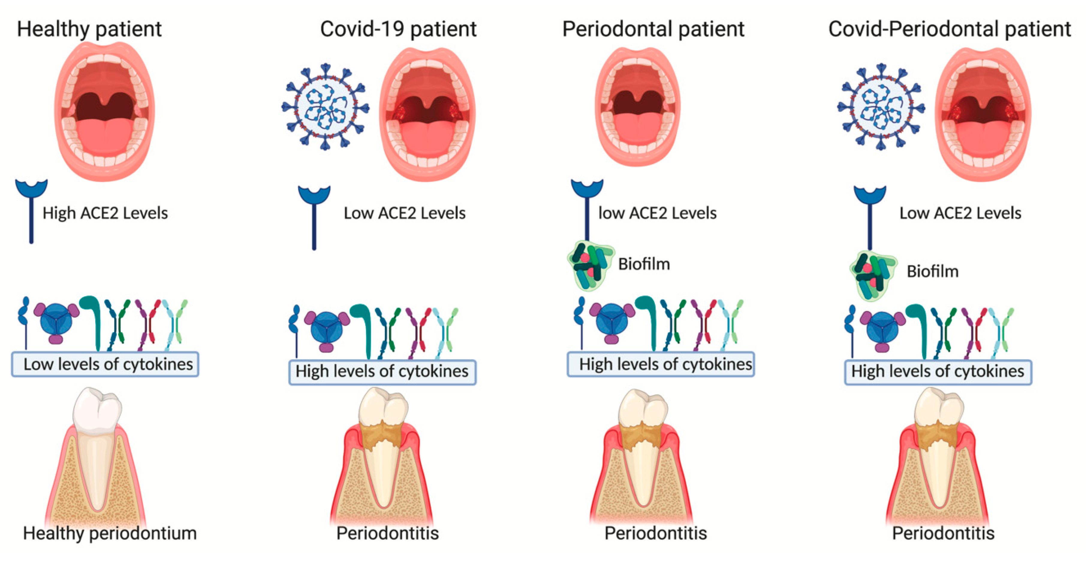
  • ACE2 and cytokines in a periodontal patient;
  • Possible correlation between COVID-19 and periodontitis.
Manual research was conducted in relevant databases (MEDLINE, Scopus, Cochrane Library, and Web of Science) with the following words: ‘SARS-CoV-2’, or ‘COVID-19’, and ‘ACE2’ and ‘Periodontal disease’ or ‘Periodontitis’. The hypothesis was extracted through the analysis of different studies of various interest, aiming to explain each of the topics mentioned above. The inclusion criteria were clinical and in vitro studies concerning correlations between ACE2 and periodontal disease or COVID-19 and ACE2. Unfortunately, this hypothesis is not related to a clinical study in which the correlation between the two diseases is underlined and clearly explained, due to the fact that the COVID-19 pandemic provoked a real rush to publication since a lot of authors considered the hot topic as an interesting element for editors and a simple method to speed up the normal editorial process. This resulted in a large number of retracted articles about the COVID-19 pandemic [17].
Thus, after collecting information and data from original articles, and clinical and in vitro studies, this postulation was performed.

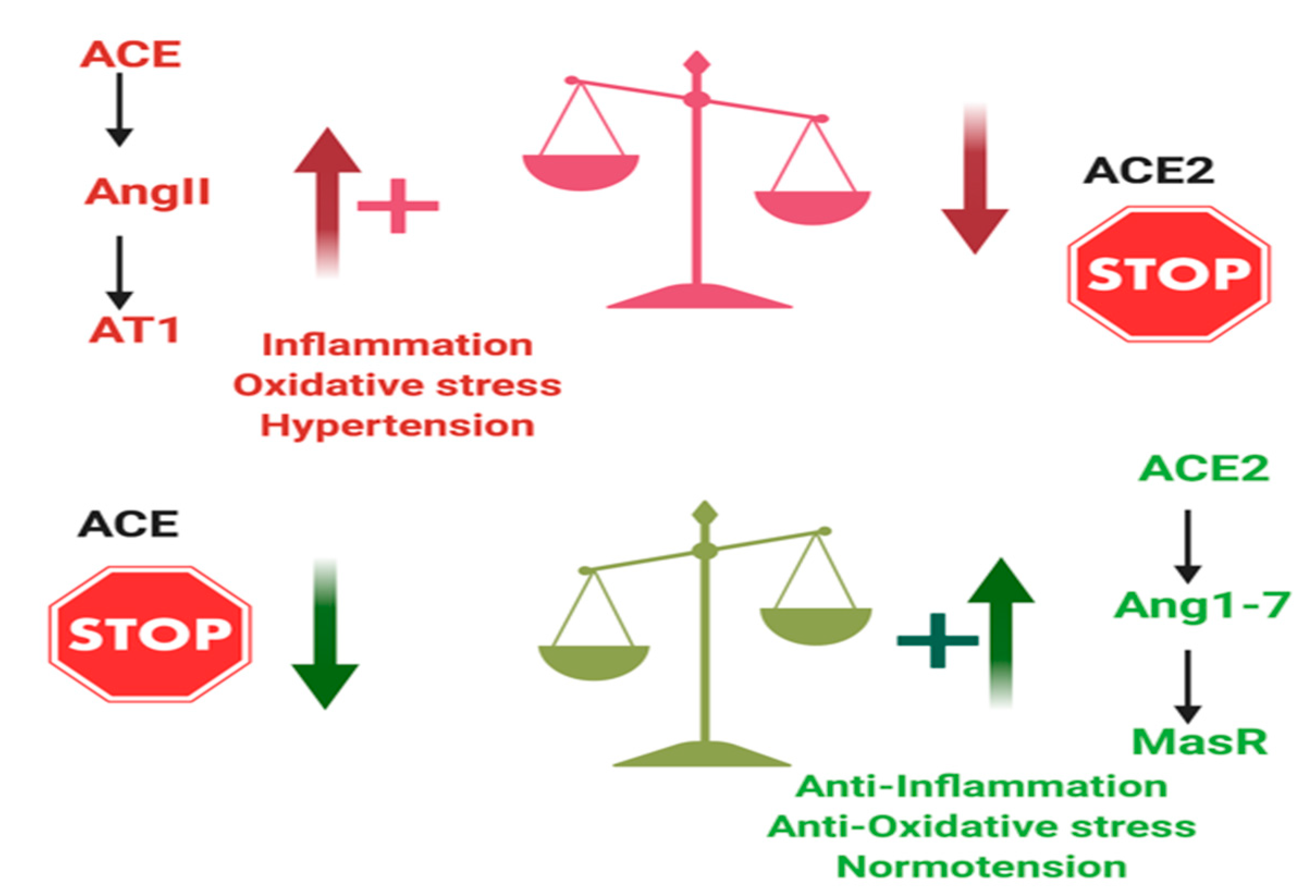
3. ACE2 and Inflammation

ACE2 is an important component of the RAS and, as mentioned formerly, is linked to the entry of SARS-CoV-2 into cells. The mechanism related to the entry of SARS-CoV-2 into cells is correlated with endocytosis and an internalization of ACE2; consequently, a lower level of ACE2 leads to low degradation of Ang II [13]. AT1 associated with Ang II increases neutrophil accumulation, enhancing the inflammation process [18]. Recent data from COVID-19 patients have shown an increase in Ang II levels, which was correlated with viral load due to the binding between ACE2 and SARS-CoV-2 [19]. Ang II has a key role in the activation of functions and signalling pathways correlated with inflammation, fibrosis, and tissue injury, such as the generation of free radicals, activation of protein kinases and recruitment of inflammatory cells, and the synthesis and release of cytokines and chemokines [20,21]. ACE2 is a regulator of the RAS system, modulating the damaging actions of Ang II and AT1, decreasing the inflammatory response [22]. ACE and ACE2 are similar enzymes related to contrasting functions. While ACE increases the production of Ang II, ACE2 reduces it. Regulation is achieved by several mechanisms:
  • ACE2 acts as a mediator in the conversion of Ang I to Angiotensin 1–9 (Ang1–9) instead of Ang II inducing an anti-inflammatory response [23,24]. This result is related to the hydrolyzation of AngI in Ang1–9 and not in AngII, which acts as an inflammatory enhancer;
  • ACE2 directly acts on Ang II and converts it to the vasodilator Ang1–7.
As a result, ACE2 increases Ang1–7 production at the expense of Ang II. Ang1–7 mediates its effects via MasR, having vasodilatory and antiproliferative effects [25,26]. Other protective effects of the ACE2/Ang1–7/MasR axis include reductions in the release of pro-inflammatory cytokines [27].

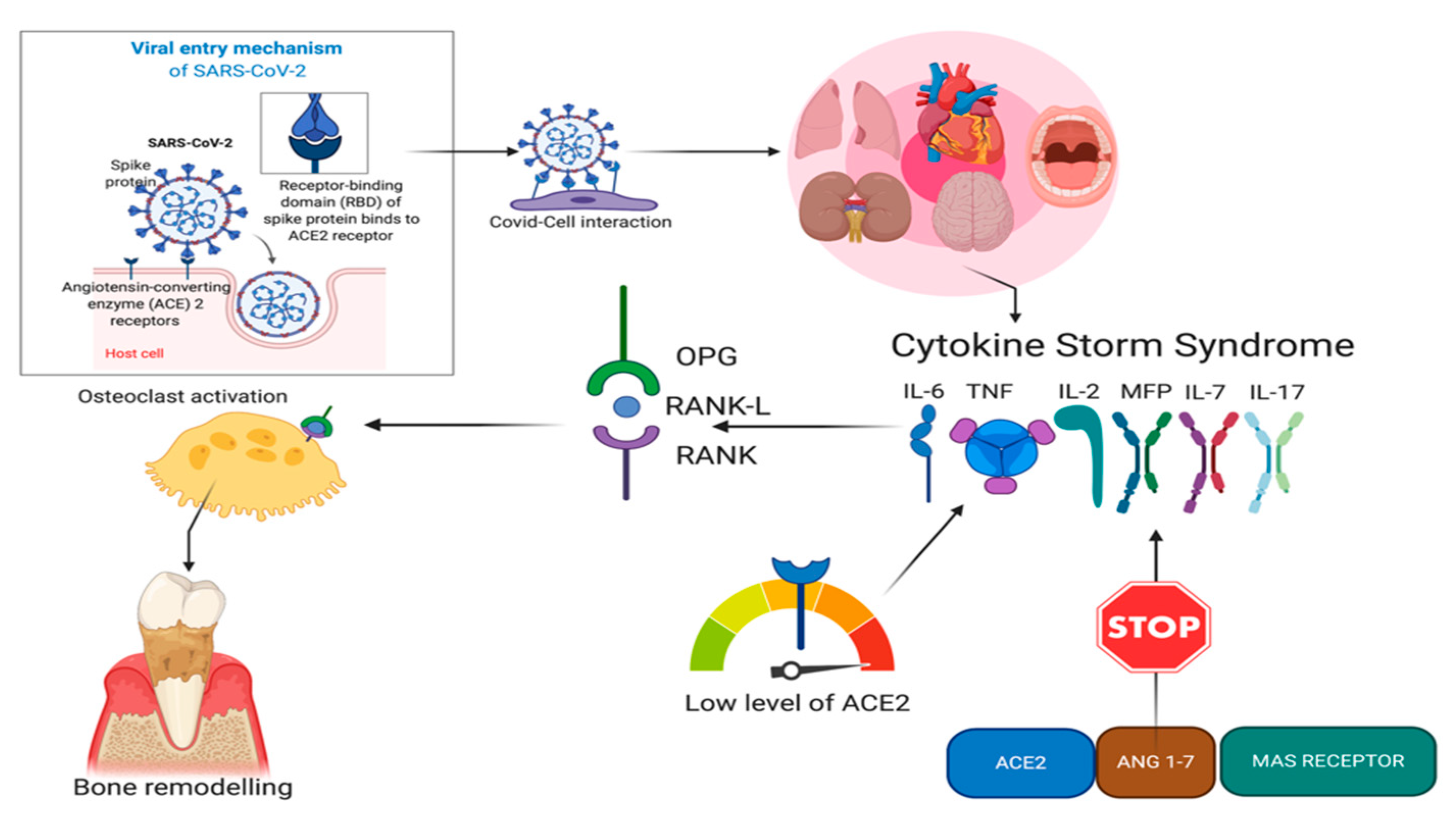
4. ACE2 and COVID-19

ACE2 is a primary receptor for SARS-CoV-2 infections [28]. Viral replication leads the immune system to downregulate ACE2 expression, giving rise to several acute inflammatory injuries [29,30]. An ACE2–SARS-COV-2 connection could potentially result in reduced ACE2 levels at the cell surface, decreasing the degradation of Ang II, and generating Ang1–7 [31,32,33]. Moreover, a reduction in ACE2 through its internalization may increase the Ang II/Ang1–7 ratio, which could exacerbate the inflammatory pattern of the SARS-CoV-2 infection. Dysregulation could aggravate the progression of inflammatory responses that are subordinated to the local overactivity of ACE and Ang II, which promotes dysfunction through COX-2 activation, generating vasoactive prostaglandins and reactive oxygen species (ROS). Moreover, Ang II favours the recruitment of infiltrating inflammatory cells into tissues by stimulating the production of specific cytokines/chemokines such as the potent monocyte chemoattractant protein 1 (MCP-1). Other cytokines involved in the onset and progression of the viral inflammatory condition are tumour necrosis factor alpha (TNF-α), interleukin-6 (IL-6), and interleukin-1 beta (IL-1β), inducing apoptosis of endothelial cells and the activation of macrophages in the inflamed tissue or organ.
During a SARS-CoV-2 infection, Ang II is inefficiently counterbalanced by Ang1–7 [28]. The oral cavity and lungs are primary targets for viruses, mainly SARS-CoV-2. According to the literature, the oral mucosa and the epithelial cells of the lungs are involved in entrance of the virus, facilitating its replication and inducing an inflammatory response. Indeed, in vivo data on mice infected by SARS-CoV-2 revealed lung failure related to the downregulation of ACE2 [32].

5. ACE2 and Periodontitis

Recent studies have shown the presence of ACE2 and, consequently, of the RAS system in the oral cavity, mainly in cells associated with the periodontal structure [33]. Indeed, osteoblasts, osteoclasts, and fibroblasts are involved in the conservation of bone and soft tissues, which play a key role in maintaining and guaranteeing the stability and functionality of the teeth. As mentioned before, regulation of the inflammatory pattern by ACE2 might be connected to the response in periodontal disease (Figure 2).
Thus, the system formed with Ang1–7 and MasR has a positive effect on reducing cytokine levels in periodontal patients [34]. On the other hand, several factors can improve or impair the progression of the disease. The initiation pathway is completely different from that of SARS-CoV-2; indeed, bacteria are involved in facilitating the initiation of an inflammatory response inside the periodontal pocket. During a SARS-CoV-2 coinfection, the periodontal pattern might be exacerbated due to the downregulation of ACE2 and an increase in ACE and Ang II, with the consequent involvement of several pro-inflammatory factors. The cytokines involved are numerous: IL-6, IL-7, IL-2, TNF-α, IL-1β, and MCP-1. IL-6 has a crucial role in the onset and acute phase of periodontal disease [35]. It is also associated with bone homeostasis. Indeed, the activation of Receptor activator of nuclear factor kappa-Β ligand (RANKL)in osteoblasts might be upregulated by IL-6, leading to the activation of osteoclasts and bone resorption [36,37]. Elevated IL-17 levels have been determined in the crevicular fluid of chronic periodontitis patients and correlated with disease severity [38,39,40]. IL-17 and a dysbiotic microbiome might promote each other, leading to the enhancement of both microbiome pathogenicity and mucosal immunopathology [40,41]. TNF-α is associated with bone metabolism, upregulating RANKL expression in gingival epithelial cells, T cells, and osteoblasts [42,43]. TNF-α also mediates the apoptosis of gingival fibroblasts and epithelial cells and inhibits extracellular matrix production in gingival fibroblasts [44], thus TNF-α might be involved in the initiation of periodontitis by damaging the gingiva and the oral mucosa. IL-1β has been shown to induce the expansion and activation of both T helper 1 and T helper 2 cells as a result of the host–microbiota interaction [45]. MCP-1 is expressed as the predominant cytokine of gingival tissues and it has a main role in the chemotaxis of monocytes in inflamed tissues, enhancing the release of IL and TNFs [46,47]. Thus, ACE2 can regulate cytokine expression through the Ang1–7 and MasR axis and this might have a fundamental role in all inflammatory diseases, such as periodontitis [33]. Downregulation of IL-6, TNF-α, IL-1β, MCP-1, and IL-17 might improve the survival rate of hopeless teeth, reducing inflammation and consequently facilitating the healing phase.

6. Hypothesis of Correlation

ACE2 can regulate the pro-inflammatory and anti-inflammatory processes, balancing cytokine expression through the formation of the ACE2–Ang1–7–MasR axis, which has a primary role in the downregulation of the following cytokines: IL-6, IL-7, IL-2, TNF-α, IL-1β and MCP-1 [33]. In a recent study, COVID-19 was defined as a cytokine storm syndrome, where the cytokine levels are very high, inducing an exaggerated general inflammation [48]. The same cytokines are involved in periodontal disease, but they have a different initiation path related to biofilm and bacteria [49]. Indeed, bacteria are necessary for the progression of the disease, but they are not sufficient. The presence of other factors, such as genetics, family history, systematic disease, and bad habits, can aggravate and accentuate the progression of the pathology [50,51,52]. In this situation, in association with these factors, ACE2 can solve different functions, and its expression might have a role in tissue response [53]. High levels of ACE2 can facilitate anti-inflammatory feedback, binding Ang1–7 and activating MasR [54]. On the other hand, higher levels of this protein can facilitate the entry of SARS-CoV-2 into the oral cavity [55]. The complex formed between the virus and ACE2 proteins leads to a reduction in ACE2 levels in infected tissue [56], and this might increase the expression of cytokines, with the consequent activation of the osteoprotegerin (OPG)– the receptor for RANK-Ligand (RANK)–RANKL axis, stimulating osteoclast function and improving bone cell phenotype modulation (Figure 3) [57].
RANKL expression in periodontal tissues is a very complicated process that involves many cells, such as T helper 1, T helper 2, B, and T lymphocytes. RANKL is identified in several cell types concerning periodontal tissues and it plays an important role in direct or indirect regulatory roles. Cytokines such as IL-1β and TNF-α can upregulate RANKL expression in periodontal cells and increase osteoclast formation. ACE2 associated with the Ang1–7–MasR axis has the main function in cytokine regulation. Moreover, the use of ACE inhibitors might increase ACE2 levels, facilitating the anti-inflammatory response [58]. Another aspect is the anti-inflammatory potential of scaling and root planing, which induce a reduction in IL-17 levels [59,60,61]. Thus, the combination of ACE inhibitors and periodontal therapy might have better outcomes in the progression of periodontal disease.

7. Future and Limitations

The potential of this hypothesis regarding the possible correlation between COVID-19 and periodontal disease might open a new field of interest concerning periodontitis and other factors such as ACE2 in the onset and progression of the disorder. Indeed, if verified, it could suggest a new way of treating periodontal disease according to alternative drugs, ACE inhibitors, used for other pathologies, such as hypertension or cardiovascular disease, confirming the implication of systematic involvement [62,63,64]. Another aspect that might be examined is the possible predisposition of periodontal patients to COVID-19, and how a periodontal supportive therapy, which may be associated with ACE inhibitors, could reduce the progression of periodontal disease but conversely accentuate susceptibility to COVID-19. On the other hand, this hypothesis has some limitations due to the novelty of SARS-CoV-2, and consequently, the limited literature, and there are few studies on ACE2 and periodontal disease. Moreover, for future studies, the in vivo analysis of ACE2 levels in COVID patients could be challenging and carry a high risk for examiners. The oral cavity should be a critical site for the study and prevention of COVID-19 infection; indeed, according to Wyllie et al. [65], saliva is a viable and more sensitive alternative to swabs, facilitating SARS-CoV-2 testing.

8. Conclusions

This study aimed to introduce and share a primary hypothesis of the correlation between COVID-19 and periodontitis. The data extracted and summarized are from different fields of study. This might have reinforced the hypothesized concept. Nevertheless, clinical studies with the principal aim of showing correlations between these diseases are needed. The presence and implications of ACE2 in both disorders points towards a possible association between periodontitis and COVID-19, where down- or upregulation of this enzyme might reduce or activate several cytokines which are expressed in both diseases. Therefore, further investigations related to the comparison of ACE2 levels in periodontal COVID-free patients and periodontal patients affected by COVID-19 are indispensable for the validation of this work.

Author Contributions

Conceptualization, L.M. and E.M.; methodology, G.M.; software, V.Q.; validation, S.M., E.M. and V.Q.; formal analysis, L.M.; investigation, L.M.; resources, E.M.; data curation, L.M.; writing—original draft preparation, L.M.; writing—review and editing, E.M.; visualization, V.Q.; supervision, S.M.; project administration, G.M.; funding acquisition, G.M. All authors have read and agreed to the published version of the manuscript.

Funding

No funding was received for carrying out the present study.

Conflicts of Interest

The authors declare no conflict of interest.

References

  1. Rabaan, A.A.; Al-Ahmed, S.H.; Haque, S.; Sah, R.; Tiwari, R.; Malik, Y.S.; Dhama, K.; Yatoo, M.I.; Bonilla-Aldana, D.K.; Rodriguez-Morales, A.J. SARS-CoV-2, SARS-CoV, and MERS-COV: A comparative overview. Infez. Med. 2020, 28, 174–184. [Google Scholar] [PubMed]
  2. Sardu, C.; Gambardella, J.; Morelli, M.B.; Wang, X.; Marfella, R.; Santulli, G. Hypertension, thrombosis, kidney failure, and diabetes: Is COVID-19 an endothelial disease? A comprehensive evaluation of clinical and basic evidence. J. Clin. Med. 2020, 9, E1417. [Google Scholar] [CrossRef] [PubMed]
  3. Chan, J.F.; Yuan, S.; Kok, K.H.; To, K.K.; Chu, H.; Yang, J.; Xing, F.; Liu, J.; Yip, C.C.; Poon, R.W.; et al. A familial cluster of pneumonia associated with the 2019 novel coronavirus indicating person-to-person transmission: A study of a family cluster. Lancet 2020, 395, 514–523. [Google Scholar] [CrossRef] [Green Version]
  4. Li, Q.; Guan, X.; Wu, P.; Wang, X.; Zhou, L.; Tong, Y.; Ren, R.; Leung, K.S.M.; Lau, E.H.Y.; Wong, J.Y.; et al. Early transmission dynamics in Wuhan, China, of novel coronavirus-infected pneumonia. N. Engl. J. Med. 2020, 382, 1199–1207. [Google Scholar] [CrossRef] [PubMed]
  5. Woelfel, R.; Corman, V.M.; Guggemos, W.; Seilmaier, M.; Zange, S.; Mueller, M.A.; Niemeyer, D.; Vollmar, P.; Rothe, C.; Hoelscher, M.; et al. Clinical presentation and virological assessment of hospitalized cases of coronavirus disease 2019 in a travel-associated transmission cluster. MedRxiv 2020, in press. [Google Scholar]
  6. Rothe, C.; Schunk, M.; Sothmann, P.; Bretzel, G.; Froeschl, G.; Wallrauch, C.; Zimmer, T.; Thiel, V.; Janke, C.; Guggemos, W.; et al. Transmission of 2019-nCoV infection from an asymptomatic contact in Germany. N. Engl. J. Med. 2020, 382, 970–971. [Google Scholar] [CrossRef] [Green Version]
  7. Hoehl, S.; Rabenau, H.; Berger, A.; Kortenbusch, M.; Cinatl, J.; Bojkova, D.; Behrens, P.; Boddinghaus, B.; Gotsch, U.; Naujoks, F.; et al. Evidence of SARS-CoV-2 infection in returning travelers from Wuhan, China. N. Engl. J. Med. 2020, 382, 1278–1280. [Google Scholar] [CrossRef]
  8. Qian, G.; Yang, N.; Ma, A.H.Y.; Wang, L.; Li, G.; Chen, X.; Chen, X. A COVID-19 transmission within a family cluster by presymptomatic infectors in China. Clin. Infect. Dis. 2020, 71, 861–862. [Google Scholar] [CrossRef]
  9. Li, L.Q.; Huang, T.; Wang, Y.Q.; Wang, Z.P.; Liang, Y.; Huang, T.B.; Zhang, H.Y.; Sun, W.; Wang, Y. COVID-19 patients’ clinical characteristics, discharge rate, and fatality rate of meta-analysis. J. Med. Virol. 2020, 92, 577–583. [Google Scholar] [CrossRef]
  10. Halpin, D.M.G.; Singh, D.; Hadfield, R.M. Inhaled corticosteroids and COVID-19: A systematic review and clinical perspective. Eur. Respir. J. 2020, 55, 2001009. [Google Scholar] [CrossRef]
  11. Zhao, Y.; Zhao, Z.; Wang, Y.; Zhou, Y.; Ma, Y.; Zuo, W. Single-cell RNA expression profiling of ACE2, the putative receptor of Wuhan 2019-nCoV. bioRxiv 2020, in press. [Google Scholar]
  12. Lan, J.; Ge, J.; Yu, J.; Shan, S.; Zhou, H.; Fan, S.; Zhang, Q.; Shi, X.; Wang, Q.; Zhang, L.; et al. Structure of the SARS-CoV-2 spike receptor-binding domain bound to the ACE2 receptor. Nature 2020, 581, 215–220. [Google Scholar] [CrossRef] [Green Version]
  13. Hamming, I.; Timens, W.; Bulthuis, M.L.; Lely, A.T.; Navis, G.; van Goor, H. Tissue distribution of ACE2 protein, the functional receptor for SARS coronavirus. A first step in understanding SARS pathogenesis. J. Pathol. 2004, 203, 631–637. [Google Scholar] [CrossRef]
  14. Gabriele, L.G.; Morandini, A.C.; Dionísio, T.J.; Santos, C.F. Angiotensin II type 1 receptor knockdown impairs interleukin-1β-induced cytokines in human periodontal fibroblasts. J. Periodontol. 2017, 88, e1–e11. [Google Scholar] [CrossRef] [PubMed]
  15. Patini, R. How to Face the Post-SARS-CoV-2 Outbreak Era in Private Dental Practice: Current Evidence for Avoiding Cross-infections. J. Int. Soc. Prev. Community Dent. 2020, 10, 237–239. [Google Scholar] [CrossRef] [PubMed]
  16. Saccomanno, S.; Quinzi, V.; Sarhan, S.; Laganà, D.; Marzo, G. Perspectives of tele-orthodontics in the COVID-19 emergency and as a future tool in daily practice. Eur J Paediatr Dent. 2020, 21, 157–162. [Google Scholar] [CrossRef] [PubMed]
  17. Soltani, P.; Patini, R. Retracted COVID-19 articles: A side-effect of the hot race to publication. Scientometrics 2020, 1–4. [Google Scholar] [CrossRef]
  18. Guan, B.; Yang, P.; Sarao, R.; Wada, T.; Leong-Poi, H.; Crackower, M.A.; Fukamizu, A.; Hui, C.C.; Hein, L.; Uhlig, S.; et al. Angiotensin-converting enzyme 2 protects from severe acute lung failure. Nature 2005, 436, 112–116. [Google Scholar]
  19. Liu, Y.; Yang, Y.; Zhang, C.; Huang, F.; Wang, F.; Yuan, J.; Wang, Z.; Li, J.; Li, J.; Feng, C.; et al. Clinical and biochemical indexes from 2019-nCoV infected patients linked to viral loads and lung injury. Sci. China Life Sci. 2020, 63, 364–374. [Google Scholar] [CrossRef] [Green Version]
  20. Capettini, L.S.; Montecucco, F.; Mach, F.; Stergiopulos, N.; Santos, R.A.; da Silva, R.F. Role of renin-angiotensin system in inflammation, immunity and aging. Curr. Pharm. Des. 2012, 18, 963–970. [Google Scholar] [CrossRef]
  21. Sukumaran, V.; Watanabe, K.; Veeraveedu, P.T.; Veeraveedu, P.T.; Gurusamy, N.; Ma, M.; Thandavarayan, R.A.; Lakshmanan, A.P.; Yamaguchi, K.; Suzuki, K.; et al. Olmesartan, an AT1 antagonist, attenuates oxidative stress, endoplasmic reticulum stress and cardiac inflammatory mediators in rats with heart failure induced by experimental autoimmune myocarditis. Int. J. Biol. Sci. 2011, 7, 154–167. [Google Scholar] [CrossRef] [PubMed] [Green Version]
  22. Burrell, L.M.; Johnston, C.I.; Tikellis, C.; Cooper, M.E. ACE2, a new regulator of the renin-angiotensin system. Trends Endocrinol. Metab. 2004, 15, 166–169. [Google Scholar] [CrossRef]
  23. Donoghue, M.; Hsieh, F.; Baronas, E.; Godbout, K.; Gosselin, M.; Stagliano, N.; Donovan, M.; Woolf, B.; Robison, K.; Jeyaseelan, R.; et al. A novel angiotensin-converting enzyme-related carboxypeptidase (ACE2) converts angiotensin I to angiotensin 1–9. Circ. Res. 2000, 87, E1–E9. [Google Scholar] [CrossRef] [PubMed]
  24. Santos, R.A.S.; Sampaio, W.O.; Alzamora, A.C.; Motta-Santos, D.; Alenina, N.; Bader, M.; Campagnole-Santos, M.J. The ACE2/angiotensin-(1-7)/MAS axis of the renin-angiotensin system: Focus on angiotensin-(1-7). Physiol. Rev. 2018, 98, 505–553. [Google Scholar] [CrossRef] [PubMed] [Green Version]
  25. Fraga-Silva, R.A.; Costa-Fraga, F.P.; Murça, T.M.; Moraes, P.L.; Martins Lima, A.; Lautner, R.Q.; Castro, C.H.; Soares, C.M.; Borges, C.L.; Nadu, A.P.; et al. Angiotensin-converting enzyme 2 activation improves endothelial function. Hypertension 2013, 61, 1233–1238. [Google Scholar] [CrossRef] [PubMed]
  26. Rodrigues Prestes, T.R.; Rocha, N.P.; Miranda, A.S.; Teixeira, A.L.; Simoes-E-Silva, A.C. The anti-inflammatory potential of ACE2/angiotensin-(1-7)/Mas receptor axis: Evidence from basic and clinical research. Curr. Drug Targets 2017, 18, 1301–1313. [Google Scholar] [CrossRef]
  27. Marshall, R.P. The pulmonary renin-angiotensin system. Curr. Pharm. Des. 2003, 9, 715–722. [Google Scholar] [CrossRef]
  28. Carson, S.D. Receptor for the group B coxsackieviruses and adenoviruses: CAR. Rev. Med. Virol. 2001, 11, 219–226. [Google Scholar] [CrossRef]
  29. Staunton, D.E.; Merluzzi, V.J.; Rothlein, R.; Barton, R.; Marlin, S.D.; Springer, T.A. A cell adhesion molecule, ICAM-1, is the major surface receptor for rhinoviruses. Cell 1989, 56, 849–853. [Google Scholar] [CrossRef]
  30. Belouzard, S.; Chu, V.C.; Whittaker, G.R. Activation of the SARS coronavirus spike protein via sequential proteolytic cleavage at two distinct sites. Proc. Natl. Acad. Sci. USA 2009, 106, 5871–5876. [Google Scholar] [CrossRef] [Green Version]
  31. Yamada, Y.; Liu, D.X. Proteolytic activation of the spike protein at a novel RRRR/S motif is implicated infurin-dependent entry, syncytium formation, and infectivity of coronavirus infectious bronchitis virus in cultured cells. J. Virol. 2009, 83, 8744–8758. [Google Scholar] [CrossRef] [Green Version]
  32. Barton, E.S.; Forrest, J.C.; Connolly, J.L.; Chappell, J.D.; Liu, Y.; Schnell, F.J.; Nusrat, A.; Parkos, C.A.; Dermody, T.S. Junction adhesion molecule is a receptor for reovirus. Cell 2001, 104, 441–451. [Google Scholar] [CrossRef] [Green Version]
  33. Santos, C.F.; Morandini, A.C.; Dionísio, T.J.; Faria, F.A.; Lima, M.C.; Figueiredo, C.M.; Colombini-Ishikiriama, B.L.; Sipert, C.R.; Maciel, R.P.; Akashi, A.P.; et al. Functional local renin-angiotensin system in human and rat periodontal tissue. PLoS ONE 2015, 10, e0134601. [Google Scholar] [CrossRef] [PubMed]
  34. Villalobos, L.A.; San Hipólito-Luengo, Á.; Ramos-González, M.; Cercas, E.; Vallejo, S.; Romero, A.; Romacho, T.; Carraro, R.; Sánchez-Ferrer, C.F.; Peiró, C. The Angiotensin-(1-7)/Mas Axis Counteracts Angiotensin II-Dependent and -Independent Pro-inflammatory Signaling in Human VascularSmooth Muscle Cells. Front. Pharmacol. 2016, 7, 482. [Google Scholar] [CrossRef]
  35. Ebersole, J.L.; Kirakodu, S.; Novak, M.J.; Stromberg, A.J.; Shen, S.; Orraca, L.; Gonzalez-Martinez, J.; Burgos, A.; Gonzalez, O.A. Cytokine gene expression profiles during initiation, progression and resolution of periodontitis. J. Clin. Periodontol. 2014, 41, 853–861. [Google Scholar] [CrossRef] [PubMed]
  36. De Benedetti, F.; Rucci, N.; Del Fattore, A.; Peruzzi, B.; Paro, R.; Longo, M.; Vivarelli, M.; Muratori, F.; Berni, S.; Ballanti, P.; et al. Impaired skeletal development in interleukin-6–transgenic mice: A model for the impact of chronic inflammation on the growing skeletal system. Arthritis Rheum. 2004, 54, 3551–3563. [Google Scholar] [CrossRef]
  37. Wu, Q.; Zhou, X.; Huang, D.; Yingchen, J.; Kang, F. IL-6 enhances osteocyte-mediated osteoclastogenesis by promoting JAK2 and RANKL activity in vitro. Cell. Physiol. Biochem. 2017, 41, 1360–1369. [Google Scholar]
  38. Zenobia, C.; Hajishengallis, G. Basic biology and role of interleukin-17 in immunity and inflammation. Periodontol 2000 2015, 69, 142–159. [Google Scholar] [CrossRef]
  39. Moutsopoulos, N.M.; Kling, H.M.; Angelov, N.; Jin, W.; Palmer, R.J.; Nares, S.; Osorio, M.; Wahl, S.M. Porphyromonas gingivalis promotes Th17 inducing pathways in chronic periodontitis. J. Autoimmun. 2012, 39, 294–303. [Google Scholar] [CrossRef] [Green Version]
  40. Okui, T.; Aoki, Y.; Ito, H.; Honda, T.; Yamazaki, K. The presence of IL-17+/FOXP3 + double-positive cells in periodontitis. J. Dent. Res. 2012, 91, 574–579. [Google Scholar] [CrossRef]
  41. Dutzan, N.; Kajikawa, T.; Abusleme, L.; Greenwell-Wild, T.; Zuazo, C.E.; Ikeuchi, T.; Brenchley, L.; Abe, T.; Hurabielle, C.; Martin, D.; et al. A dysbiotic microbiome triggers TH17 cells to mediate oral mucosal immunopathology in mice and humans. Sci. Transl. Med. 2018, 10, eaat0797. [Google Scholar] [CrossRef] [PubMed] [Green Version]
  42. Fujihara, R.; Usui, M.; Yamamoto, G.; Nishii, K.; Tsukamoto, Y.; Okamatsu, Y.; Sato, T.; Asou, Y.; Nakashima, K.; Yamamoto, M. Tumor necrosis factor-α enhances RANKL expression in gingival epithelial cells via protein kinase A signaling. J. Periodontal Res. 2014, 49, 508–517. [Google Scholar] [CrossRef] [PubMed]
  43. Kawai, T.; Matsuyama, T.; Hosokawa, Y.; Makihira, S.; Seki, M.; Karimbux, N.Y.; Goncalves, R.B.; Valverde, P.; Dibart, S.; Li, Y.P.; et al. B and T lymphocytes are the primary sources of RANKL in the bone resorptive lesion of periodontal disease. Am. J. Pathol. 2006, 169, 987–998. [Google Scholar] [CrossRef] [PubMed] [Green Version]
  44. Basso, F.G.; Pansani, T.N.; Turrioni, A.P.; Soares, D.G.; de Souza Costa, C.A.; Hebling, J. Tumor necrosis factor-α and interleukin (IL)-1β, IL-6, and IL-8 impair in vitro migration and induce apoptosis of gingival fibroblasts and epithelial cells, delaying wound healing. J. Periodontol. 2016, 87, 990–996. [Google Scholar] [CrossRef] [PubMed]
  45. Ben-Sasson, S.Z.; Hu-Li, J.; Quiel, J.; Cauchetaux, S.; Ratner, M.; Shapira, I.; Dinarello, C.A.; Paul, W.E. IL-1 acts directly on CD4 T cells to enhance their antigendriven expansion and differentiation. Proc. Natl. Acad. Sci. USA 2009, 106, 7119–7124. [Google Scholar] [CrossRef] [PubMed] [Green Version]
  46. Hanazawa, S.; Kawata, Y.; Takeshita, A.; Kumada, H.; Okithu, M.; Tanaka, S.; Yamamoto, Y.; Masuda, T.; Umemoto, T.; Kitano, S. Expression of monocyte chemoattractant protein 1 (MCP-1) in adult periodontal disease: Increased monocyte chemotactic activity in crevicular fluids and induction of MCP-1 expression in gingival tissues. Infect. Immun. 1993, 61, 5219–5224. [Google Scholar] [CrossRef] [Green Version]
  47. Mark, L.L.; Haffajee, A.D.; Socransky, S.S.; Kent, R.L., Jr.; Guerrero, D.; Kornman, K.; Newman, M.; Stashenko, P. Effect of the interleukin-1 genotype on monocyte IL-1beta expression in subjects with adult periodontitis. J. Periodontal Res. 2000, 35, 172–177. [Google Scholar] [CrossRef]
  48. Mehta, P.; McAuley, D.F.; Brown, M.; Sanchez, E.; Tattersall, R.S.; Manson, J.J. HLH Across speciality collaboration. COVID-19: Consider cytokine storm syndromes and immunosuppression. Lancet 2020, 395, 1033–1034. [Google Scholar] [CrossRef]
  49. Sahni, V.; Gupta, S. COVID-19 & periodontitis: The cytokine connection. MedHypotheses 2020, in press. [Google Scholar]
  50. Lang, N.P.; Tonetti, M.S. Periodontal risk assessment (PRA) for patients in supportive periodontal therapy (SPT). Oral Health Prev. Dent. 2003, 1, 7–16. [Google Scholar]
  51. Ebersole, J.L.; Graves, C.L.; Gonzalez, O.A.; Dawson, D.; Morford, L.A.; Huja, P.E.; Hartsfield, J.K., Jr.; Huja, S.S.; Pandruvada, S.; Wallet, S.M. Aging, inflammation, immunity and periodontal disease. Periodontol 2000 2016, 72, 54–75. [Google Scholar] [CrossRef]
  52. Meyle, J.; Chapple, I. Molecular aspects of the pathogenesis of periodontitis. Periodontol 2000 2015, 69, 7–17. [Google Scholar] [CrossRef] [PubMed]
  53. Queiroz-Junior, C.M.; Santos, A.C.P.M.; Galvão, I.; Souto, G.R.; Mesquita, R.A.; Sá, M.A.; Ferreira, A.J. The angiotensin converting enzyme 2/angiotensin-(1-7)/Mas receptor axis as a key player in alveolar bone remodeling. Bone 2019, 128, 115041. [Google Scholar] [CrossRef]
  54. Simões e Silva, A.C.M.; Silveira, K.D.; Ferreira, A.J.; Teixeira, M.M. ACE2, angiotensin-(1–7) and Mas receptor axis in inflammation and fibrosis. Br. J. Pharmacol. 2013, 169, 477–492. [Google Scholar] [CrossRef] [PubMed] [Green Version]
  55. Xu, H.; Zhong, L.; Deng, J.; Peng, J.; Dan, H.; Zeng, X.; Li, T.; Chen, Q. High expression of ACE2 receptor of 2019-nCoV on the epithelial cells of oral mucosa. Int. J. Oral Sci. 2020, 12, 8. [Google Scholar] [CrossRef] [PubMed]
  56. Silhol, F.; Sarlon, G.; Deharo, J.C.; Vaïsse, B. Downregulation of ACE2 induces overstimulation of the renin-angiotensin system in COVID-19: Should we block the renin-angiotensin system? Hypertens Res. 2020, in press. [Google Scholar] [CrossRef] [PubMed]
  57. Hienz, S.A.; Paliwal, S.; Ivanovski, S. Mechanisms of bone resorption in periodontitis. J. Immunol. Res. 2015, 2015, 615486. [Google Scholar] [CrossRef] [PubMed] [Green Version]
  58. Kortekaas, K.E.; Meijer, C.A.; Hinnen, J.W.; Dalman, R.L.; Xu, B.; Hamming, J.F.; Lindeman, J.H. ACE inhibitors potently reduce vascular inflammation, results of an open proof-of-concept study in the abdominal aortic aneurysm. PLoS ONE 2014, 9, e111952. [Google Scholar] [CrossRef]
  59. Cheng, W.C.; Hughes, F.J.; Taams, L.S. The presence, function and regulation of IL-17 and Th17 cells in periodontitis. J. Clin. Periodontol. 2014, 41, 541–549. [Google Scholar] [CrossRef]
  60. Marchetti, E.; Tecco, S.; Caterini, E.; Casalena, F.; Quinzi, V.; Mattei, A.; Marzo, G. Alcohol-free essential oils containing mouthrinse efficacy on three-day supragingival plaque regrowth: A randomized crossover clinical trial. Trials 2017, 18, 154. [Google Scholar] [CrossRef] [Green Version]
  61. Isola, G.; Polizzi, A.; Iorio-Siciliano, V.; Alibrandi, A.; Ramaglia, L.; Leonardi, R. Effectiveness of a nutraceutical agent in the non-surgical periodontal therapy: A randomized, controlled clinical trial. Clin. Oral Investig. 2020. [Google Scholar] [CrossRef] [PubMed]
  62. Pietropaoli, D.; Del Pinto, R.; Ferri, C.; Wright, J.T.; Giannoni, M.; Ortu, E.; Monaco, A. Poor oral health and blood pressure control among US hypertensive adults. Hypertension 2018, 72, 1365–1373. [Google Scholar] [CrossRef] [PubMed]
  63. Türkoğlu, O.; Barış, N.; Tervahartiala, T.; Şenarslan, Ö.; Sorsa, T.; Atilla, G. Evaluation of systemic levels of neutrophilic enzymes in patients with hypertension and chronic periodontitis. J. Periodontol. 2014, 85, 908–916. [Google Scholar] [CrossRef] [PubMed]
  64. Isola, G.; Alibrandi, A.; Currò, M.; Matarese, M.; Ricca, S.; Matarese, G.; Ientile, R.; Kocher, T. Evaluation of salivary and serum ADMA levels in patients with periodontal and cardiovascular disease as subclinical marker of cardiovascular risk. J. Periodontol. 2020. [Google Scholar] [CrossRef]
  65. Wyllie, A.L.; Fournier, J.; Casanovas-Massana, A.; Campbell, M.; Tokuyama, M.; Vijayakumar, P.; Geng, B.; Muenker, M.C.; Moore, A.J.; Vogels, C.B.F.; et al. Saliva is more sensitive for SARS-CoV-2 detection in COVID-19 patients than nasopharyngeal swabs. medRxiv 2020. [Google Scholar] [CrossRef] [Green Version]
Figure 1. The balance between angiotensin-converting enzyme (ACE) and angiotensin-converting enzyme 2 (ACE2) regulating several functions.
Applsci 10 06224 g001
Figure 2. ACE2 levels and biofilm as enhancers in periodontal disease in periodontal and COVID-19 patients.
Applsci 10 06224 g002
Figure 3. Role of ACE2 in binding COVID-19 and up- or downregulating levels and expression of cytokines inducing bone remodelling.
Applsci 10 06224 g003

