Article

Post-Effect on the Centre of Feet Pressure during Stance by Continuous Asymmetric Mediolateral Translations of a Supporting Platform—A Preliminary Study in Healthy Young Adults

1
Centro Studi Attività Motorie (CSAM), Istituti Clinici Scientifici Maugeri, IRCCS, 27100 Pavia, Italy
2
Department of Clinical-Surgical, Diagnostic and Paediatric Sciences, University of Pavia, 27100 Pavia, Italy
3
Division of Physical Medicine and Rehabilitation, ICS Maugeri, IRCCS, Institute of Veruno, 28013 Veruno, Italy
4
Istituti Clinici Scientifici Maugeri, IRCCS, 27100 Pavia, Italy
*
Author to whom correspondence should be addressed.
Appl. Sci. 2020, 10(17), 5969; https://doi.org/10.3390/app10175969
Submission received: 31 July 2020 / Revised: 22 August 2020 / Accepted: 27 August 2020 / Published: 28 August 2020
(This article belongs to the Special Issue Assistive Technology: Biomechanics in Rehabilitation Engineering)

Abstract

:

Featured Application

A platform continuously translating in the horizontal plane in an asymmetric mode can be employed as a means for normalizing the position of the centre of feet pressure in patients standing on it.

Abstract

Various diseases are associated with the impaired control of the medio-lateral (ML) position of the centre of feet pressure (CoP), and several manoeuvres have been proposed for enhancing the CoP symmetry. Here, we assessed in healthy standing subjects the feasibility and outcome of a novel protocol entailing a reaction to a continuous asymmetric ML displacement (10 cm) of the support base. The periodic perturbation consisted of a fast half-cycle (0.5 Hz) followed by a slow half-cycle (0.18 Hz). One hundred successive horizontal translation cycles were delivered in sequence. Eyes were open or closed. CoP was recorded before, after, and during the stimulation by a dynamometric platform fixed onto the translating platform. We found that the post-stimulation CoP was displaced towards the direction of the fast half-cycles. The displacement lasted several tens of seconds. Vision did not affect the amplitude or duration of the post-stimulation effect. The magnitude of post-stimulation CoP displacement was related to the perturbation-induced ML motion of CoP recorded during the stimulation. Over the successive perturbation cycles, the time-course of this motion revealed an adaptation phenomenon. Vision moderately reduced the adaptation rate. The findings support the feasibility of the administration of a simple asymmetric balance perturbation protocol in clinical settings to help patients recover the symmetry of the CoP. This protocol needs to be further validated in older populations and in patients.

1. Introduction

Numerous studies on the control of quiet stance have focused on balance and balance control in the anterior–posterior (AP) plane [1,2], likely because of the analogy of the body profile in the sagittal plane with an inverted pendulum that facilitates a formal treatment of AP sway [3]. Besides, a plethora of studies examining dynamic balancing behaviour in standing have mostly considered the responses to AP perturbations, also because of the elegant theoretical framework of the control pattern proposed at earlier times [4,5].
The postural control in the medio-lateral (ML) direction has received less attention. Under normal standing condition, the weight of the body is symmetrically distributed, so that the centre of feet pressure (CoP) remains halfway the distance between the feet [6]. This is at variance with the position of the CoP in the sagittal plane, where it lies ahead of the ankle joint, requiring the tonic activity of the foot, leg and axial postural muscles to avoid falling forwards [7,8]. Distinctive control modes and muscle activities appear to regulate body sway in the frontal compared to the sagittal plane [6,9,10,11,12]. The modulation of both amplitude and frequency in the EEG trace is more pronounced during ML compared to AP sway, implying that AP and ML sway are supervised by independent strategies and that ML control may require greater neural resources [13,14].
The clinical relevance of the ML control of the CoP is emphasized by the need of good medio-lateral stability during turning [15] and by the strong dependence of gait initiation on the capacity of transferring the body along the frontal plane for launching the first step [16]. While healthy ageing hardly challenges the postural control during quiet stance [17], there is now accumulating evidence that aging effects on balance may be accentuated in the medio-lateral direction. Measures of lateral stability are related to fall risk [18] and excessive movements of CoP in the ML direction predict falls in older adults [19]. The abnormal control of the CoP may depend on asymmetries in muscle force [20], sensation [21] or altered central control. Patients with stroke typically show an asymmetric distribution of the body weight between the feet [22,23] and problems in voluntary shifting the body toward the paretic extremity [24].
Several old and recent investigations have brought to light the effects of diverse sensory manipulations on the body’s postural orientation. For example, muscle spindle activation by tendon vibration produces body forward or backward leaning depending on the site of the application [24], and the alternate vibration of trunk muscles produces the displacement of the CoP of standing individuals in the frontal plane synchronous with the vibration trains [25]. Furthermore, several manoeuvres can produce striking postural post-effects [26]. An intense voluntary isometric contraction of a muscle or group of muscles produces a sustained involuntary contraction of the same muscles lasting several seconds or minutes [27]. Neck unilateral vibration induces persistent directional post-effects on the subjective straight ahead and on the self-motion perception of vestibular origin [28]. Another example of a postural post-effect occurs during the podokinetic adaptation induced by prolonged stepping with concurrent body yaw rotation [29,30]. On a different note, a six-minute period of treadmill walking induces a post-effect consisting in an antero–posterior tilt of the body when standing quietly afterwards, both eyes open (EO) and eyes closed (EC) [31,32]. These cases show that the continuous activation/stimulation of the proprioceptive system may not be necessary and can be substituted by conditions eliciting prolonged rhythmic muscle activities. Hence, these post-effects are not necessarily connected to a prolonged voluntary muscle tonic contraction but can also emerge when the muscle contractions (and their sensory feedback) are repeated for a certain period. They depend on the intensity, duration, side of the conditioning stimulation, visual conditions and muscle status. These sensory inputs and motor activities would be centrally integrated and interpreted with respect to a stable frame of reference [33].
Here, we leveraged the aforementioned knowledge to design a protocol able to modify the control of CoP during stance in the frontal plane. This study tested the hypothesis that the position of the CoP can be modified (thereby opening the possibility for patient treatment) by teaching the nervous centres an ‘adapted’ postural condition through the implicit learning of a new equilibrium reference. In particular, the purpose of this preliminary study was to investigate involuntary weight shifting behaviour in the frontal plane during quiet stance as a consequence of a novel stimulation pattern consisting in a repetitive periodic asymmetric ML translation of the support base upon which healthy subjects stand. The possible effect of vision was assessed, in the hypothesis that the visual reference to a stationary scene during the platform displacements [34] would rescind any proprioceptive effect elicited by the continuous perturbation through processes similar to those put in place during head stabilization in space [35]. Preliminary data have been published in abstract form [36].

2. Materials and Methods

Twenty (10 males and 10 females) healthy young individuals (mean ± SD: 27.9 years ± 3.25, height of 171.8 cm ± 8.24, weight 67.3 kg ± 11.07) volunteered to participate in this study. All were right-handed except three. Participants were naïve to the experimental procedures and all succeeded in performing the trials. All gave written informed consent to participate in the experiments. This translational study was approved by the institutional ethics committee (Ethics Committee, Istituti Clinici Scientifici Maugeri, approval number #2399 CE) and was performed in accordance with the Declaration of Helsinki.

2.1. Task and Procedures

Participants stood with bare feet apart at pelvis width and with the arms by their side on a force platform firmly fixed on the top of a motorized platform (Officina Lomazzi, Legnano, Italy). The location of the feet of each participant was marked on a thin (0.5 mm) rubber sheet of the same size as the force platform (see below), onto which it was firmly attached, to assure consistent placement between testing trials. Both at rest and during the platform translations, the mean distance between the parallel feet was 16.8 cm ± 3.4 (intermalleolar distance). The motorized platform moved periodically from side to side for a 10 cm distance in the horizontal plane. The profile of the continuous translation cycles was asymmetric, each cycle being made of a half cycle with a fast phase (0.5 Hz) followed by a half cycle with a slow return phase (0.18 Hz). The profile was switched from fast right to fast left in different trials. A trial consisted in one hundred continuous perturbation cycles (henceforth, the stimulation). All trials always started with the fast half-cycle and ended with the slow half-cycle. These translations produced a perturbation of the standing body in the frontal plane and a medio-lateral displacement of CoP from right to left and vice versa. There were no still periods between the cycles but the stimulation was preceded and followed by two extended periods of quiet stance. Each trial lasted 14 min and was composed by a quiet-stance period (3 min), the stimulation (5 min) and another stance period (6 min). The first 2 min of the trial were considered a familiarization period and not analysed.
Participants performed one trial in each of these five different conditions: (1) eyes closed (EC) during the entire trial, with the fast platform translation phase toward the right (i.e., fast right, FR–EC): the platform made a fast half-cycle to the right side of the participant, followed by a slow half-cycle to the left side; (2) EC, with the fast phase towards the left (fast left, FL–EC), followed by a slow half-cycle to the right; (3) eyes open (EO) with the fast phase to the right side (FR–EO); (4) EO with the fast phase to the left side (FL–EO); and (5) like 3, but subjects kept their eyes closed during the 6 min post-stimulation period of quiet stance (FR–EO/EC). At the very end of the first trial, participants were invited to slowly tilt their body to the right and to the left as much as possible, without moving their feet, and keep those positions for 1–2 s. When vision was allowed, participants were invited not to focus on one particular point of the patterned laboratory wall (at 8 m distance) and were allowed to freely explore the scene in front of them, without moving the head. Thus, no precise visual task was required [37]. The control stimulation was a symmetric translation sequence (10 cm, 0.5 Hz) performed with EC, again preceded and followed by quiet stance periods.
The various trials with different vision conditions and asymmetry profiles were randomized across participants and conditions and separated by one or more days. During all trials, participants wore earphones with music to screen the noise produced by the moving platform. They were instructed to stand in a relaxed mode but not to move their feet during the trial. They were aware of the onset of the acquisition and were warned of the onset of the platform perturbation sequence in order to minimize any startle reaction [38]. Nausea and other unpleasant feelings were never reported by any participant either during or after the recording sessions. Some subjects (5/20) spontaneously reported a mild sense of heaviness of the thigh muscles of the side toward which the platform translation moved slowly.

2.2. Data Acquisition and Treatment

The position of the CoP was recorded for the entire duration of the trials by the force platform (Kistler 9286BA, Winterthur, Switzerland) at a sampling frequency of 140 Hz, and was expressed in platform coordinates. No artefacts were produced on the force-platform output by the translations of the motorized platform. The force-platform data were stored in a PC for offline analysis. For each participant and condition, the mean ML and AP CoP positions were calculated on a time window of 1 min duration, just before the onset of platform motion (these mean CoP values were the reference for further analysis) and at the end of the last platform translation cycle. For the same time windows, the standard deviation (SD) of the ML and AP CoP traces over time were also calculated and accepted as an index of the CoP sway around its mean position. The pre-/post-stimulation difference in the SD of the ML CoP trace in the quiet stance periods (visual conditions collapsed) was plotted against the extent of the pre-/post-stimulation ML CoP displacement. This distribution was studied by a linear regression model and the coefficient of determination (R2) was calculated to assess the goodness of the fit.
The mean ML position of the CoP was also calculated cycle by cycle, for each of the successive perturbation cycles, during both the fast and the slow phases of the translations using a software developed in LabVIEW (National Instruments, Austin, TX, USA). These mean positions, averaged across all subjects, changed over time featuring an exponential trend from the early to the late trials. The data were therefore fitted with an exponential model (y = A + Be−t/τ) for each participant and for each condition by means of the iterative conjugate gradient method of the Excel® Solver Utility [39]. A, B and τ (tau) parameters were computed by using the minimum sum squared algorithm. Tau is the time-constant of the exponential function expressed in cycles, where the duration of an entire translation cycle is 3.8 s (the fast phase lasted 1 s and the slow phase lasted 2.8 s). A is the value at steady state of the function (asymptote), and A + B the value of the intercept with the ordinate at the first cycle [40]. The difference between the asymptotic value of the ML CoP position during the fast and the slow phases of the translation cycles was calculated and plotted against the ML CoP position post-stimulation. This relationship was studied by a linear regression model (asymmetric translation profile and visual conditions collapsed) and the coefficient of determination (R2) was calculated.
The duration of the post-effect produced by the stimulation on the ML CoP position was estimated for each participant according to the following procedure. Each successive sample of the CoP position acquired in the post-stimulation epoch was compared by the one-sample Student’s t-test to the mean position of the CoP computed during 1 min pre-stimulation. The time-period during which the t-value of the above comparison bypassed the critical t-value corresponding to a probability of α = 0.05 and remained above that value for at least 500 ms was taken as the duration of the post-effect [41]. The relationship between the amplitude of the ML CoP position post-stimulation and the duration of the post-effect was studied by a linear regression model and the R2 was calculated.

2.3. Statistical Analysis

The following analyses were conducted: (1) the mean displacement of the ML and AP CoP positions post- with respect to pre-stimulation and the mean asymptotic value of the ML CoP position during the fast and during the slow phases of the perturbation cycles were compared to zero (the case in which the post-stimulation CoP position is equal to the pre-stimulation CoP position) by means of the Student’s t-test; (2) a 2 × 2 repeated-measure ANOVA (factors: FL and FR conditions; visual conditions EO and EC) was used to compare the mean ML and AP CoP positions post-stimulation; (3) the duration of the post-effects of the ML CoP position were compared between the conditions using a 2 × 2 repeated-measure ANOVA (factors: FL and FR condition; visual conditions EO and EC); (4) two separate 2 × 2 × 2 repeated measure ANOVAs (factors: FL and FR conditions; visual conditions; pre- and post-stimulation periods) were used to compare the SD of the ML and AP CoP traces; (5) a 2 × 2 × 2 repeated-measure ANOVA (FL and FR condition; visual conditions; fast and slow phase of perturbation cycles) was used to compare the asymptotic values of the ML CoP position of the fast and the slow phase of the perturbation cycles and the time-constants obtained under the four stimulation conditions. For all ANOVAs, the post hoc test analysis was done with the Fisher’s LSD test and the differences were considered statistically significant at p < 0.05. Where the differences were significant, the Cohen’s d effect sizes highlighted the strength of the difference (with d = 0.2, 0.5, 0.8 considered as a small, medium and large effect size, respectively), and the power of the test (π) [42] was also reported. The software package used for the analysis was Statistica (StatSoft, Tulsa, OK, USA).

3. Results

3.1. The CoP Position Pre- and Post-Stimulation

The CoP was recorded by the force platform firmly mounted onto the motorized platform translating in the frontal plane with two asymmetric cycle profiles (fast left, FL or fast right, FR). The mean ML position of the CoP during the initial quiet stance period was close to zero in platform coordinates (i.e., the centre of the force platform rested halfway between the feet) (mean ± SD: −0.028 cm ± 0.76). The CoP position of a representative participant during the entire trial epoch (standing quietly EC, platform stimulation EC, standing quietly EC) is shown in Figure 1A. Here, the fast half-cycle of the perturbation was directed toward the participant’s right side (FR–EC). The panel B shows two trial segments with a large medio-lateral peak-to-peak CoP displacement during the fast phase followed by a less ample and slower shift during the slow phase. The corresponding profiles of the platform translations are shown in panel C, where the fast half-cycles are directed upwards (to the right side of the body). In all the cycles, the fast right translation produced a clear rapid displacement of the CoP to the opposite left side. This was followed by a displacement to the right during the subsequent slow half cycle. The red area in panel B indicates the medio-lateral shift to the right direction (upward in the Figure) of the ML CoP post-stimulation with respect to its pre-stimulation mean position (dotted red line). The opposite perturbation profile (FL, not shown) produced a very similar but opposite CoP displacement pattern.
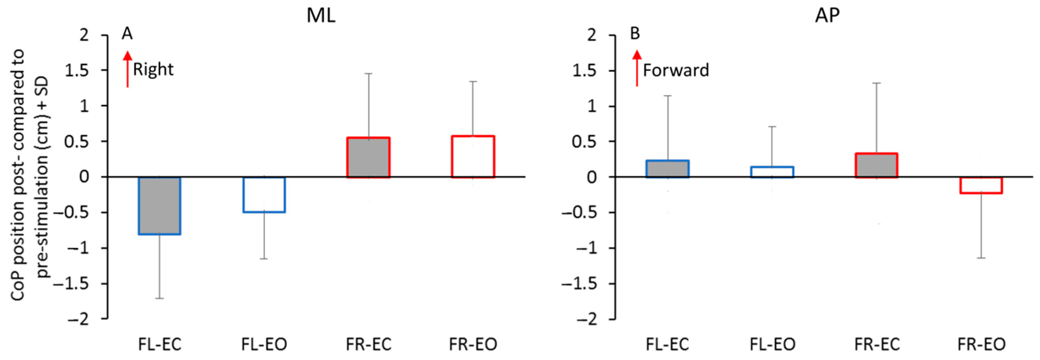
Across all participants, the ML CoP position during the quiet-stance period following the end of the stimulation sequence was normally different from the pre-stimulation position, constituting a post-effect of the continuous asymmetric platform translation cycles. The difference was significant, as assessed by the comparison of the mean difference between these positions to zero by means of the Student’s t test, both under the EC and EO condition, for both profiles of the asymmetric platform translation (p < 0.05, d > 0.85 and π > 0.83 for the four conditions). The post-stimulation displacement (mean ± SD: FL–EC −0.81 cm ± 0.89, FL–EO −0.49 cm ± 0.66, FR–EC 0.55 cm ± 0.9, FR–EO 0.57 cm ± 0.77) depended on the profile of the asymmetric platform translation cycles. On average, the CoP was displaced in the same direction as the fast phase of the cycles, i.e., to the left when the fast phase of the cycle was directed to the left (FL) and vice versa (FR). The bar graph in Figure 2A shows the mean displacement of the ML CoP, with respect to the pre-stimulation positions, during the post-effects in the four conditions (FL–EC, FL–EO; FR–EC, FR–EO). The opposite displacement after the stimulation with the reverse profile is obvious. These displacements were of moderate amplitude: for comparison, the mean absolute displacement of the CoP during the maximal voluntary body tilts to the right and to the left on the still platform was 9.85 cm ± 2.06 (right) and 10.45 cm ± 1.47 (left), respectively (right vs. left: paired t-test, p = 0.19). ANOVA showed that the main effect of the translation profile asymmetry (FR vs. FL) on the ML CoP post-stimulation position was significant (F(1,19) = 26.5, p < 0.001, d = 2.35, π = 0.99). A difference between the vision conditions was observable but did not reach significance (EO vs. EC, F(1,19) = 1.87, p = 0.19). There was no interaction between the profile asymmetry (i.e., fast half-cycle to the left, FL, or to the right, FR) and vision (F(1,19) = 1.27, p = 0.27).

3.2. Antero–Posterior Position of the CoP

The CoP in the sagittal plane showed minor changes with respect to its pre-stimulation position (mean ± SD pre-stimulation: FL–EC = −1.22 cm ± 1.66; FL–EO = −1.72 cm ± 1.9; FR–EC = −1.46 cm ± 2.02; FR–EO = −1.0 cm ± 2.16). The difference in the post-stimulation positions (mean ± SD: FL–EC = −1.01 cm ± 1.6; FL–EO = −1.58 cm ± 1.9; FR–EC = −1.13 cm ± 2.0; FR–EO = −1.22 cm ± 2.19) compared to the initial positions (see Figure 2B) were not significant (t-test, p > 0.13 for all four comparisons). There was no main effect of cycle profiles (F(1,19) = 1.11, p = 0.3), no difference between the visual conditions (main effect, F(1,19) = 1.46, p = 0.24) and no interaction between the cycle profiles and visual conditions (F(1,19) = 2.35, p = 0.14).

3.3. Body Sway around the Mean CoP Position

The mean variances (assessed by the SD) of the CoP traces along the frontal and the sagittal plane during the 1 min quiet-stance periods pre- and post-stimulation were calculated to evaluate the possible effect of vision and stimulation on the body sway around its mean CoP position (Table 1). Although the mean SDs of the CoP traces on the frontal and sagittal planes were larger with EC than with EO (by about 4% for ML and about 13% for AP, all conditions collapsed), the main vision effect was not significant for the ML (F(1,19) = 0.0017, p = 0.97) and just significant for the AP direction (F(1,19) = 4.59, p = 0.045, d = 0.98, π = 0.53). The main pre-/post stimulation effect was significant, both for the ML (F(1,19) = 11.5, p < 0.01, d = 1.54, π = 0.89) and the AP direction (F(1,19) = 13.0, p < 0.05, d = 1.65, π = 0.93), but there were no interactions with either vision (ML: F(1,19) = 0.82, p = 0.37; AP: F(1,19) = 3.41, p = 0.08) or stimulation profiles (ML: F(1,19) = 0.0004, p = 0.98; AP: F(1,19) = 0.24, p = 0.63). The post-hoc test showed no pre-post differences in the AP SDs except for the FR and FL condition under EC (p < 0.01). Instead, for the ML direction, the difference in the SDs between the pre- and post-stimulation was always significant (post-hoc, p < 0.05 for all comparisons), likely because the variance of the ML sway incorporated the slow changes in the CoP displacement determined by the asymmetric stimulation. There was no main effect of translation cycle profile, either for the ML (F(1,19) = 0.8, p = 0.38) or the AP direction (F(1,19) = 2.82, p = 0.11). Across the subjects, there was no relationship between the pre-/post-stimulation difference of ML sway and the CoP positions, either for the FL or FR condition (EC and EO collapsed) (regression lines, FL: y = −0.05x + 0.066, R2 = 0.036, p = 0.24, FR: y = 0.03x + 0.045, R2 = 0.013, p = 0.48).

3.4. Closing the Eyes at the End of the Stimulation with EO did not Modify the Post-Effect on the CoP Position

All participants also performed an additional trial (condition FR–EO/EC) in which they were instructed to close their eyes at the end of the asymmetric perturbation sequence administered with vision (FR–EO) (this trial was not systematically repeated for the FL–EO condition). The mean ML CoP displacement in the post-stimulation condition FR–EO/EC (mean ± SD, 0.54 cm ± 1.28) was similar to the displacement recorded in the EO condition (0.57 cm ± 0.77) (FR–EO vs. FR–EO/EC, paired t-test, p = 0.88).

3.5. Symmetric Perturbation Cycles

When the platform translation profile was made symmetric (as driven by a sinusoidal 0.5 Hz function, administered with EC only), there were almost no changes in the ML CoP in the post- compared to the pre-stimulation period across all participants (mean difference, 0.04 cm ± 0.47; paired t-test, p = 0.88).

3.6. Duration of the Post-Effect

The duration of the time-interval, during which the adapted ML CoP position post-stimulation remained significantly different from the pre-stimulation value, was variable across participants and visual conditions. It fluctuated in an ample range, between 9 s and 360 s with FL–EC (mean 144.2 s ± 105.4) and between 10 s and 343 s with FL–EO (mean 118.99 s ± 89.4) (Figure 3). When the asymmetry of the profile of the platform translation cycle was reversed (FR–EC), the duration of the post-effect fluctuated in a similar range, between 40 s and 314 s (mean 162.2 s ± 76.7) and between 29 s and 371 s with FR–EO (mean 182.3 s ± 85.4). ANOVA showed no significant difference in the duration of the post-effect between the two cycle profiles (FL vs. FR, F(1,19) = 3.4, p = 0.08) or between the visual conditions (EC vs. EO, F(1,19) = 0.1, p = 0.75).
In addition, there was no significant interaction between the cycle profiles and visual conditions (F(1,19) = 0.72, p = 0.4). Across the subjects, there was no significant relationship between the amplitude and duration of the post-effect (FL–EC, y = −21.6x + 126.7, R2 = 0.03, p = 0.44; FL–EO, y = 19.6x + 133.7, R2 = 0.02, p = 0.54; FR–EC, y = 12.2x + 155.5, R2 = 0.02, p = 0.54; FR–EO, y = −16.7x + 178.1, R2 = 0.03, p = 0.44).

3.7. Habituation to the Series of Perturbations

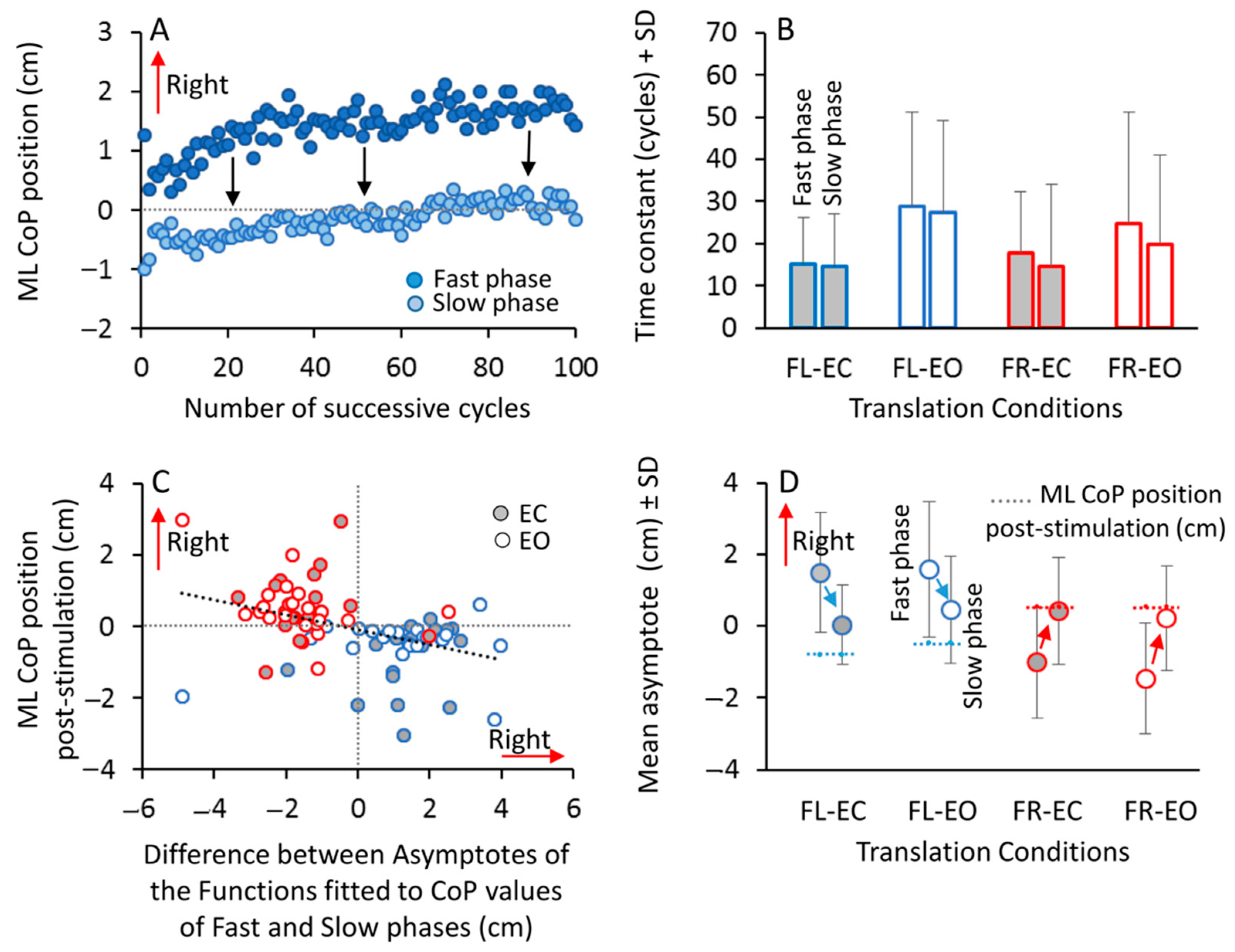
During the translation cycles, the CoP moved rapidly on the force platform over a large distance in the direction opposite to the fast-phase motion and returned toward a more central position during the slow phase (see Figure 1B,C). Opposite translation profiles produced very similar but reverse CoP motions. The displacements of the CoP during the sequence of the translation cycles were not constant (compare the first and last cycle in Figure 1) but exhibited a progressive decrease in peak-to-peak amplitude. Therefore, the hypothesis that the CoP position post-stimulation may be specified by the continuous postural adjustments occurring during the stimulation period was tested. The top panel (A) of Figure 4 shows the average value across the participants of the ML CoP positions (condition FL–EC) plotted over time, cycle-to-cycle, during the fast left (dark blue) and the slow right phases (light blue) of the translation cycles. At the first cycle, the fast translation to the left moved the CoP to the right of the force platform (upwards in the Figure). During the next slow phase, the CoP showed an ample shift to the left (downwards) to counteract the large perturbation. In the successive cycles, the CoP featured a progressive shift during the fast phases and a recovery towards the initial position during the slow phases (points near zero in the ordinate). Toward the end of the stimulation sequence, the CoP mean position was close to the pre-stimulation position during the slow phases and distant during the fast phases. The exponential fits through the data points (separately for the fast and the slow phases, and separately for all participants) allowed to identify a time constant and an asymptote, the value of which was assumed to indicate the steady state. When the stimulation was administered in the opposite direction (fast phase to the right, FR) a similar picture emerged (not shown in the Figure). Owing to the opposite profile of platform translation and CoP motion condition, the trend in the average ML CoP positions during the fast and the slow phases of the perturbation cycles was increasing during FL (Figure 4A) but decaying during FR.
Across all participants, the mean time course of this adaptation (Figure 4B) had a long time constant under all the tested conditions for both the fast and the slow phases (mean ± SD; fast phase: FL–EC, 15.3 cycles ± 10.9; FL–EO, 28.7 ± 22.5; FR–EC, 17.7 ± 14.6; FR–EO, 24.7 ± 26.4; slow phase, FL–EC, 14.3 cycles ± 12.8; FL–EO, 27.5 cycles ± 21.6; FR–EC, 14.6 cycles ± 19.6; FR–EO, 19.7 cycles ± 21.2). Considering the duration of a complete translation cycle (3.8 s), these time constants range from about 1 to 2 min on average, and this variability explains the large SDs of the bars. ANOVA showed no differences between the time constants of the fast and of the slow phases (main effect, F(1.19) = 1.17, p = 0.29) and between the two translation profile conditions (FL vs. FR, main effect, F(1,19) = 0.22, p = 0.64). However, there was a significant difference in the time constants between the visual conditions (main effect, F(1,19) = 10.3, p < 0.01, d = 1.47, π = 0.86) due to the longer time constants with EO compared to with EC. This effect was present for all the cycle phases and profiles and in most of the participants (65% for FL, 55% for FR), revealing a robust effect of vision, whereby no-vision speeded up the achievement of the plateau.
The mean value of the ML CoP asymptotic position, calculated by the exponential fits for the fast phase of the cycles, was always different from the initial ML CoP position (t-test, p < 0.01, d > 0.93 and π > 0.8 for all comparisons). The mean asymptote for the slow half-cycles was instead close to the pre-stimulation position (t-test, p > 0.20 for all comparisons). ANOVA on the asymptotic values showed a difference between cycle profiles (main effect, FR vs. FL, F(1.19) = 13.2, p < 0.01, d = 1.67, π = 0.94) and an interaction between the cycle profiles and fast or slow phase (F(1.19) = 46.15, p < 0.0001, d = 3.11, π = 0.99). ANOVA showed no main effect of visual conditions (F(1.19) = 0.038, p = 0.85). Figure 4D shows that the asymptotic values of the fast phase were opposite between the FL and FR conditions (post hoc, p < 0.0001 for all comparisons), but not different between FL and FR during the slow phase (post hoc, p > 0.2 for all comparisons) both under EC and EO condition. The post hoc test showed a significant difference between the asymptotic value of the fast and of the slow phase within each visual and perturbation profile condition (post hoc, p < 0.0001 for all comparisons). Moreover, there was an association between the ML CoP post-stimulation and the displacements of the CoP measured between the fast and the slow phases of the translation cycles. Panel C of Figure 4 shows that, across all participants (FR and FL conditions and EO and EC collapsed), there was a significant relationship between the CoP position post-stimulation and the difference in the asymptotic values of the functions fit the mean CoP positions during the fast and slow phases (regression line, y = − 0.2x − 0.09, R2 = 0.16, p < 0.001).
As a way of summarizing these findings, Figure 4D shows the mean values of the asymptote calculated by the exponential fit on the ML CoP position during the fast and the slow phases of the perturbation cycles. The shift (arrows) from the position imposed by the fast half-cycle to that recorded during the slow half-cycle brings the CoP very close to the post-stimulation position (the dotted horizontal segments).

4. Discussion

4.1. The CoP Displacement after the Sequence of Asymmetric Perturbations

The body weight shifting capacity during the stance can be restored in patients by different rehabilitation procedures, not directly focusing on the operation of proprioceptive processes [43,44]. Here, we tested a novel, simple and unpretentious protocol in healthy subjects; whereby asymmetric balance perturbations were administered with the purpose of inducing a post-effect consisting of a temporary shift in the position of the CoP during quiet stance. In keeping with our initial hypothesis, we found that the administration of a prolonged sequence of periodic asymmetric translations of a supporting platform in the horizontal plane changes the post-stimulation CoP position in the frontal plane of the participants standing on it. This pre-/post- CoP displacement has a small but significant amplitude and is consistent in direction across subjects. It takes place toward the direction of the fast half-cycle of the platform translations. The effect is unrecognized by the participants, and is immediate, being obvious at the very end of the sequence of perturbation cycles. The duration of the effect is variable within a limited range across participants and on average amounts to a couple of minutes, before the CoP recovers its initial pre-stimulation position.
Before the beginning of the sequence of translations, the mean CoP rested midway between the feet in all the participants. Its displacement after the continuous asymmetric translations amounted to about 4% of the distance between the heels (set equal to the distance between the anterior superior iliac spines) and corresponded to about 7% with EC and about 5% with the EO of the maximum functional CoP displacement produced by voluntarily leaning to the right or to the left. The post-stimulation mean displacement entails a non-negligible difference in the distribution of weight on each foot (about 5%, see [45,46]). Although less than 1 cm may seem negligible, one may note that such a distance during the random sway of the CoP is an uncommon occurrence in healthy subjects [1,47], even when standing in tandem condition [9]. Here, the mean SD of the ML CoP sway during the pre-stimulation period was about 0.2 cm and displacements of 0.5 cm from the mean were never observed. Moreover, we noted that the symmetric platform translations did not produce any consistent or significant post-effects on the average position of the CoP. These findings allow to trace the post-stimulation displacement to the asymmetric profile of the perturbation sequence. In spite of a considerable variability across subjects, there was a significant relationship between the amplitude of the displacement reached during the fast and the slow phases of the translations and the amplitude of the post-stimulation displacement. Furthermore, the ML CoP shift was not accompanied by a major increase in the ML sway around the mean position, and there was no relationship between the pre-/post-stimulation difference in the SD of the ML sway and the ML displacement of the mean CoP position. Hence, the post-stimulation displacement would not be a non-specific effect of an excessive body sway.
The duration of the post-effect was relatively short. It was just longer than a couple of minutes on the average. However, it is unexpected that the CoP can drift about 0.5–1 cm out of its normal standing position for this stretch of time. During clinical tests it is customary to record the CoP position during a quiet stance for 1 min or 30 s or even less (see [48,49]), assuming that no major drift occurs in this time-interval. In some cases, the effect vanished slowly, whilst in other cases it stopped in coincidence with a rapid redistribution of the body weight, resetting the postural attitude as often occurs during quiet stance [50,51]. The duration of the present post-effect is similar to that observed by Lee et al. [52] in subjects who stood on an inclined surface for 3 min. Whether the differences across subjects might be related to their attention to spatial orientation has not been addressed in the present study.

4.2. Relationship between the CoP Motion during the Platform Perturbation Cycles and the Post-Effect

When the supporting platform translates rapidly toward one side, say to the left, the inertia of the body produces an immediate displacement of the CoP relative to the force platform toward the right side. During the following slow return half-cycle of the platform translation to the right, i.e., to its initial position, the body tends to correct the rightward tilt and recover the original symmetric orientation. After the first few cycles, the CoP moves further right during the fast half cycle. During the slow half-cycle it shows an overshoot to the left that becomes more evident towards the end of the stimulation sequence. This configures an adaptation behaviour whereby the mean CoP displacements during the slow half-cycles tends to remain between the feet. At the end of the stimulation, across the participants, the average amplitude of the CoP shift bears some association with the amplitude of the shift of the CoP position from the fast to the slow phase of the perturbation sequence. The larger the mean value of the CoP during the slow return phase, i.e., the difference between its mean position during the fast and the slow half-cycle, the larger the amplitude of the post-stimulation displacement in the direction of the fast translation.
Therefore, the strength of the post-effect seems to be related to the unwitting effort of complying with the continuous medio-lateral translation of the support base, particularly with the recovery of the symmetric posture of the body during the slow return phase of the translating platform. This effort can be viewed as proportionate to the vertical distance (arrows in Figure 4) between the traces of the CoP mean position during the fast and during the slow half-cycle. As a consequence, when the perturbation cycles stop, the post-effect would be driven by the ‘tail’ of this motion (in search of the new vertical reference built up as to compensate the displacement produced by the fast translation). This process shifts the CoP position on the side of the fast cycles. This new position would be the most appropriate for counteracting a fast displacement of the support base by minimizing the destabilizing effect of the body inertia. This displacement toward the side of the fast platform movement understandably occurs in anticipation of the impending fast phase, in order to more easily counteract the perturbation delivered by the next fast cycle.
The behaviour would therefore configure a sequence of compensatory balance correcting reactions to the fast translation phase [53,54,55] and of anticipatory postural adjustments (APA) during the slow translation phases [56,57]. The balance-correcting reaction would be appropriate for halting the ML body tilt induced by the fast platform displacement, a braking action possibly produced by the reflex contraction of the abductor muscles of the side opposite to the fast body displacement; whereas the APA would attenuate the effects of the subsequent ML fast perturbation. Over time, this sequence would be constructed and adjusted by the continuous asymmetric proprioceptive input from the muscles stretched by the fast cycle of the platform translation, and by the effort to return to the initial position during the slow phase and prepare the postural adjustment in anticipation of the following fast translation. In this light, individuals would implicitly learn to anticipate any reflex response by enhancing the anticipatory adjustment (and the post-effect would attest to this phenomenon). At the end of the stimulation, the body would be kept in its new standing position (say to the left) by a tonic activity of the abductor muscles of the right side.
This adaptation phenomenon would be produced by a sensory reweighting mechanism, aimed to reduce the postural destabilization [58]. Contrary to many comparable adaptation behaviours described in the literature, the time-course of this phenomenon is longer (see [59]). Here, the vertical position during both the fast and the slow phase is fully recovered after about 60–80 cycles, after which the ‘overshoot’ becomes visible, and lasts until the end of the stimulation. During the stimulation, a crucial information would be the proprioceptive input from the muscles stretched and activated by the medio-lateral perturbations, rather than the visual flow from the stationary scene, as attested by the non-significant effect of vision on the post-stimulation position of the CoP. However, the time constant is longer with vision compared to without vision, regardless of the profile of the perturbation. We have no explanation for this consistent finding. Most likely, vision information from the fixed, structured environment cognitively encroached with, and delayed the process of the proprioceptive control of CoP rather than help the body reach a steady state sooner. Perhaps, vision produced continuous adjustments in the first part of the trial. Fransson et al. [60] showed that during vestibular and proprioceptive stimulation, the dynamics in the motion responses had higher complexity with vision, the responses induced by the body deviations being significantly larger compared to EC condition. Anyhow, we must point to the considerable individual variation in the adaptation time course and magnitude, testified by the large error bars in Figure 4B. A large variety of dynamic postures had been shown earlier [61] when subjects counteracted a fore-aft continuous perturbation, and largely variable time constants have also been found in the adaptation to such balancing behaviour [59]. This notwithstanding, the longer time constants EO than EC were observed in 13/20 subjects for the FL condition and in 11/20 for the FR condition, supporting the notion of a more complex behaviour with EO.

4.3. The Effect of Vision in the Post-Stimulation Period

Contrary to our initial hypothesis, we observed no major effect of vision on the occurrence, amplitude or duration of the post-stimulation effect on the ML position of the CoP. This consistent trend is supported by the qualitatively and quantitatively similar post-effects present when the stimulation was administered with both EO and EC. Incidentally, the well known decrement of ML body sway with vision was not remarkable in the present study, probably because of the wide support base with feet apart [62]. The ML sway about the mean CoP position post-stimulation showed a modest increment with both EO and EC. This can be explained by the post-stimulation displacement in the position of the CoP in the frontal plane and by its slow drift to the pre-stimulation values. The AP sway was greater with EC than with EO and greater in the post-stimulation period, as a non-specific vision-dependent adjustment to the termination of the perturbation cycles.
The absence of a significant effect of vision on the post-effect ML CoP displacement is supported by the trials in which the participants were invited to close their eyes simultaneously with the cessation of the stimulation. Clear vision of the stationary environment did not correct the displacement of the CoP ‘adapted’ during the EO translations. This was unexpected, not least because vision during tilts of a support base can reduce sway variability by reducing the contribution of vestibular noise and improving the disturbance compensation [35]. Furthermore, our feet-apart condition would have favoured a symmetric position of the CoP with EO [63], but this did not occur. The effect of vision was not observed at the steady state of the adapted behaviour to the sequence of perturbations either. Therefore, we would conjecture that the visual flow from the substantially stationary visual scene from the environment played no or a minor role in the process of adaptation to the continuous perturbations, except for the delay in reaching the steady state, as discussed above. This shows that the CoP displacement and the responsible process dependent on the reaction to the continuous platform translation and on the anticipatory reaction to it, is robust to the rectifying effect of vision.
Therefore, the post-effect is not prevented by either a continuous or a post-stimulation visual reference. In this light, the present post-effect would be related to that elicited on body AP orientation by a period of brisk walking on a treadmill, where the post-effect tilt is present when standing both with eyes open and eyes closed, but is larger without vision [31,64]. Another study has shown that the effect of sensory manipulation producing a sway of the body in the frontal plane (by vestibular or cutaneous foot stimulation or the vibration of hip abductors) is not affected by the absence of normal visual information [65]. On the contrary, after the podokinetic stimulation, the continuing unwitting yaw rotation with EC, after the prolonged stepping-in-place, is temporarily reduced or actually stopped when the eyes are opened [30], to start again with the closing the eyes [66]. Standing on an inclined surface produces a post-effect as well, whereby subjects show a post-incline leaning post-effect in which they maintain a similar trunk to support the surface orientation as during the period of standing upright on the inclined surface [67,68]. However, in that case, participants immediately return to upright when they open their eyes briefly during the post-incline period. Our findings are different from those obtained during continuous AP translations of the supporting platform, since in the latter case, the upper body was greatly stabilized by vision but goes through large oscillations with eyes closed or blindfolded [69,70,71].
On the other hand, a precision visual task (e.g., reading a text, as opposed to simply keeping the eyes open) can rescind the stabilizing effect of the vision of the environment when participants stand on an AP oscillating platform [72]. We can only remind that vision sensitivity to AP perturbations is more than two times higher than ML perturbations [73], and that direction-specific, visually-guided weight-shift training protocols in healthy elderly women on standing balance had a poorer effect on the stabilization of ML than of AP sway [74]. The lack of vision effect at the end of the present prolonged asymmetric stimulation gives a further indirect explanation of the implicit nature of the present post-effect and of its dependence on the proprioceptive input, since removing visual feedback normally alters the voluntary control of sway while standing [75].
Additional information on the control of posture against a ML perturbation is wanted [54], more so for continuous perturbations, which surely entail APA adjustments and adaptation processes, not explicitly addressed here or in the literature, contrary to what happens for the AP perturbations [59,76]. Most likely, the supplementary motor area [77] as well as several other brain structures [78,79] are active during the induction period and might contribute to the expression of the post-effects by collecting the stimulation-elicited inputs and modifying the body’s reference [33]. Still, future research should direct attention toward the powerful effect of asymmetric proprioceptive stimulation, which has been proven to strongly modify body orientation and self-motion perception [80,81].

4.4. Limitations

We acknowledge that the present study originates from a simple and straightforward hypothesis. This explains, but does not exempt, the absence of kinematic and EMG recordings. This limitation prevents framing the effect described in the context of the neuromechanical command for the control of the medio-lateral position of the CoP, and prevents a direct support to the assumption that the post-effect is constructed by a repetitive proprioceptive discharge from and by the activation of the muscles responsible for the active medio-lateral tilts of the body. In addition, we did not test a different distance between the feet, which might modify the postural responses compared to the pelvis-width condition [82,83] or different feet orientation, known to modulate the APA preceding perturbations [56]. Furthermore, considering the potential application of this protocol for stroke patients exhibiting an asymmetric position of the CoP during stance, information on the modulation of the post-effect by the numerosity of the perturbation cycles or by the amplitude of the translation distance appears relevant.

5. Conclusions and Perspectives

Upright standing requires an integration of visual, somatosensory and graviceptive inputs to adapt to changes in sensory conditions [84]. Older persons experience changes in the force of hip abductor–adductor muscles, leading to reduced balance performance when it comes to reacting to lateral perturbations [20]. Patients with stroke are often complicated with sensory and motor dysfunctions [85] and experience poor standing balance, asymmetric weight distribution and impaired weight shifting ability [86]. Deficits in lateral weight transfer and body stability could potentially be one of the limiting factors underlying comfortable walking speeds in these patients [87]. The present preliminary findings show that a novel and easy proprioceptive training by a prolonged asymmetric medio-lateral perturbation can be a tailored intervention able to specifically affect the CoP position toward the desired direction. This protocol gives a potentially useful answer to the recent encouragement to identify procedures for improving APAs in people with stroke as a result of targeted exercise [88]. It is important to remind that the appropriate and meaningful proprioceptive inflow is paramount in assuring appropriate brain plasticity in central lesions [89,90]. We believe that the findings of this study can be translated into the clinic. In particular, the repetition of this protocol can help rectify the postural vertical and equalizing the distribution of body weight in patients. Indeed, we plan to apply this protocol to elderlies first, in whom proprioception is impaired but can be enhanced by exercise [91], and to post-stroke patients, and conjecture that such regular proprioceptive training can induce the long-lasting symmetrisation of the CoP displaced by hemiparesis and poorer postural control. The protocol can be administered without vision, leveraging the report that balance rehabilitation in stroke patients is more effective with visual deprivation than with free vision [92].

Author Contributions

M.S. conceived the idea behind the study. S.S. and M.S. were responsible for conducting the study, data collection, management and data analysis and drafted the manuscript. S.S. and M.S. prepared the figures. A.N. and S.C. participated in the design of the study and revised the manuscript. All authors have read and agreed to the published version of the manuscript.

Funding

This research was supported by the Ricerca Corrente funding scheme of the Ministry of Health, Italy. It did not receive any specific grant from funding agencies in the commercial or for-profit sectors.

Conflicts of Interest

The authors declare no conflict of interest.

References

  1. Schieppati, M.; Hugon, M.; Grasso, M.; Nardone, A.; Galante, M. The limits of equilibrium in young and elderly normal subjects and in parkinsonians. Electroencephalogr. Clin. Neurophysiol. 1994, 93, 286–298. [Google Scholar] [CrossRef]
  2. Morasso, P.G.; Schieppati, M. Can muscle stiffness alone stabilize upright standing? J. Neurophysiol. 1999, 82, 1622–1626. [Google Scholar] [CrossRef] [Green Version]
  3. Masani, K.; Popovic, M.R.; Nakazawa, K.; Kouzaki, M.; Nozaki, D. Importance of body sway velocity information in controlling ankle extensor activities during quiet stance. J. Neurophysiol. 2003, 90, 3774–3782. [Google Scholar] [CrossRef] [PubMed] [Green Version]
  4. Horak, F.B.; Nashner, L.M. Central programming of postural movements: Adaptation to altered support-surface configurations. J. Neurophysiol. 1986, 55, 1369–1381. [Google Scholar] [CrossRef] [PubMed]
  5. Ghai, S.; Nardone, A.; Schieppati, M. Human Balance in Response to Continuous, Predictable Translations of the Support Base: Integration of Sensory Information, Adaptation to Perturbations, and the Effect of Age, Neuropathy and Parkinson’s Disease. Appl. Sci. 2019, 9, 5310. [Google Scholar] [CrossRef] [Green Version]
  6. Winter, D.A.; Prince, F.; Frank, J.S.; Powell, C.; Zabjek, K.F. Unified theory regarding A/P and M/L balance in quiet stance. J. Neurophysiol. 1996, 75, 2334–2343. [Google Scholar] [CrossRef]
  7. Soames, R.W.; Atha, J. The role of the antigravity musculature during quiet standing in man. Eur. J. Appl. Physiol. Occup. Physiol. 1981, 47, 159–167. [Google Scholar] [CrossRef]
  8. Ivanenko, Y.; Gurfinkel, V.S. Human Postural Control. Front. Neurosci. 2018, 12, 171. [Google Scholar] [CrossRef]
  9. Sozzi, S.; Honeine, J.L.; Do, M.C.; Schieppati, M. Leg muscle activity during tandem stance and the control of body balance in the frontal plane. Clin. Neurophysiol. 2013, 124, 1175–1186. [Google Scholar] [CrossRef]
  10. Rietdyk, S.; Patla, A.E.; Winter, D.A.; Ishac, M.G.; Little, C.E. NACOB presentation CSB New Investigator Award. Balance recovery from medio-lateral perturbations of the upper body during standing. North American Congress on Biomechanics. J. Biomech. 1999, 32, 1149–1158. [Google Scholar] [CrossRef]
  11. Bingham, J.T.; Choi, J.T.; Ting, L.H. Stability in a frontal plane model of balance requires coupled changes to postural configuration and neural feedback control. J. Neurophysiol. 2011, 106, 437–448. [Google Scholar] [CrossRef] [Green Version]
  12. Henry, S.M.; Fung, J.; Horak, F.B. Control of stance during lateral and anterior/posterior surface translations. IEEE Trans. Rehabil. Eng. 1998, 6, 32–42. [Google Scholar] [CrossRef] [PubMed] [Green Version]
  13. Slobounov, S.; Hallett, M.; Cao, C.; Newell, K. Modulation of cortical activity as a result of voluntary postural sway direction: An EEG study. Neurosci. Lett. 2008, 442, 309–313. [Google Scholar] [CrossRef] [PubMed] [Green Version]
  14. Nandi, T.; Fisher, B.E.; Hortobágyi, T.; Salem, G.J. Increasing mediolateral standing sway is associated with increasing corticospinal excitability, and decreasing M1 inhibition and facilitation. Gait Posture 2018, 60, 135–140. [Google Scholar] [CrossRef] [PubMed]
  15. Turcato, A.M.; Godi, M.; Giordano, A.; Schieppati, M.; Nardone, A. The generation of centripetal force when walking in a circle: Insight from the distribution of ground reaction forces recorded by plantar insoles. J. Neuroeng. Rehabil. 2015, 12, 4. [Google Scholar] [CrossRef] [Green Version]
  16. Honeine, J.L.; Schieppati, M.; Crisafulli, O.; Do, M.C. The Neuro-Mechanical Processes That Underlie Goal-Directed Medio-Lateral APA during Gait Initiation. Front. Hum. Neurosci. 2016, 10, 445. [Google Scholar] [CrossRef] [Green Version]
  17. Schieppati, M.; Grasso, M.; Siliotto, R.; Nardone, A. Effect of Age, Chronic Diseases and Parkinsonism on Postural Control. In Sensorimotor Impairment in the Elderly. NATO ASI Series; Stelmach, G.E., Hömberg, V., Eds.; Series D: Behavioural and Social Sciences; Springer: Dordrecht, The Netherlands, 1993; Volume 75, pp. 355–373. [Google Scholar] [CrossRef]
  18. Hilliard, M.J.; Martinez, K.M.; Janssen, I.; Edwards, B.; Mille, M.L.; Zhang, Y.; Rogers, M.W. Lateral balance factors predict future falls in community-living older adults. Arch. Phys. Med. Rehabil. 2008, 89, 1708–1713. [Google Scholar] [CrossRef] [PubMed] [Green Version]
  19. Piirtola, M.; Era, P. Force platform measurements as predictors of falls among older people—A review. Gerontology 2006, 52, 1–16. [Google Scholar] [CrossRef]
  20. Inacio, M.; Creath, R.; Rogers, M.W. Low-dose hip abductor-adductor power training improves neuromechanical weight-transfer control during lateral balance recovery in older adults. Clin. Biomech. 2018, 60, 127–133. [Google Scholar] [CrossRef]
  21. Nardone, A.; Schieppati, M. Group II spindle fibres and afferent control of stance. Clues from diabetic neuropathy. Clin. Neurophysiol. 2004, 115, 779–789. [Google Scholar] [CrossRef] [PubMed]
  22. Bensoussan, L.; Viton, J.M.; Schieppati, M.; Collado, H.; Milhe de Bovis, V.; Mesure, S.; Delarque, A. Changes in postural control in hemiplegic patients after stroke performing a dual task. Arch. Phys. Med. Rehabil. 2007, 88, 1009–1015. [Google Scholar] [CrossRef] [PubMed]
  23. Corriveau, H.; Hébert, R.; Raîche, M.; Dubois, M.F.; Prince, F. Postural stability in the elderly: Empirical confirmation of a theoretical model. Arch. Gerontol. Geriatr. 2004, 39, 163–177. [Google Scholar] [CrossRef] [PubMed]
  24. Eklund, G. General features of vibration-induced effects on balance. Ups. J. Med. Sci. 1972, 77, 112–124. [Google Scholar] [CrossRef]
  25. De Nunzio, A.M.; Nardone, A.; Picco, D.; Nilsson, J.; Schieppati, M. Alternate trains of postural muscle vibration promote cyclic body displacement in standing parkinsonian patients. Mov. Disord. 2008, 23, 2186–2193. [Google Scholar] [CrossRef] [PubMed]
  26. Wierzbicka, M.M.; Gilhodes, J.C.; Roll, J.P. Vibration-induced postural posteffects. J. Neurophysiol. 1998, 79, 143–150. [Google Scholar] [CrossRef]
  27. Ivanenko, Y.P.; Wright, W.G.; Gurfinkel, V.S.; Horak, F.; Cordo, P. Interaction of involuntary post-contraction activity with locomotor movements. Exp. Brain Res. 2006, 169, 255–260. [Google Scholar] [CrossRef] [Green Version]
  28. Pettorossi, V.E.; Panichi, R.; Botti, F.M.; Kyriakareli, A.; Ferraresi, A.; Faralli, M.; Schieppati, M.; Bronstein, A.M. Prolonged asymmetric vestibular stimulation induces opposite, long-term effects on self-motion perception and ocular responses. J. Physiol. 2013, 591, 1907–1920. [Google Scholar] [CrossRef]
  29. Weber, K.D.; Fletcher, W.A.; Gordon, C.R.; Melvill Jones, G.; Block, E.W. Motor learning in the “podokinetic” system and its role in spatial orientation during locomotion. Exp. Brain. Res. 1998, 120, 377–385. [Google Scholar] [CrossRef]
  30. Sozzi, S.; Schieppati, M. Stepping in Place While Voluntarily Turning Around Produces a Long-Lasting Posteffect Consisting in Inadvertent Turning While Stepping Eyes Closed. Neural. Plast. 2016, 2016, 7123609. [Google Scholar] [CrossRef]
  31. Zanetti, C.; Schieppati, M. Quiet stance control is affected by prior treadmill but not overground locomotion. Eur. J. Appl. Physiol. 2007, 100, 331–339. [Google Scholar] [CrossRef]
  32. Hirjaková, Z.; Bizovská, L.; Bzdúšková, D.; Hlavačka, F.; Janura, M. Postural stability after treadmill and overground walking in young and elderly. Gait Posture 2020, 80, 84–89. [Google Scholar] [CrossRef] [PubMed]
  33. Gurfinkel, V.S.; Ivanenko, Y.U.P.; Levik, Y.U.S.; Babakova, I.A. Kinesthetic reference for human orthograde posture. Neuroscience 1995, 68, 229–243. [Google Scholar] [CrossRef]
  34. Cappa, P.; Patanè, F.; Rossi, S.; Petrarca, M.; Castelli, E.; Berthoz, A. Effect of changing visual condition and frequency of horizontal oscillations on postural balance of standing healthy subjects. Gait Posture 2008, 28, 615–626. [Google Scholar] [CrossRef] [PubMed]
  35. Assländer, L.; Hettich, G.; Mergner, T. Visual contribution to human standing balance during support surface tilts. Hum. Mov. Sci. 2015, 41, 147–164. [Google Scholar] [CrossRef] [PubMed] [Green Version]
  36. Sozzi, S.; Nardone, A.; Schieppati, M. Orientation in space during quiet stance is affected by prolonged asymmetric translations of the supporting platform. In Proceedings of the RIN, 1st Annual Meeting, Milan, Italy, 24–25 March 2020; p. 183. [Google Scholar]
  37. Bonnet, C.T.; Davin, T.; Hoang, J.Y.; Baudry, S. Relations between Eye Movement, Postural Sway and Cognitive Involvement in Unprecise and Precise Visual Tasks. Neuroscience 2019, 416, 177–189. [Google Scholar] [CrossRef] [Green Version]
  38. Campbell, A.D.; Squair, J.W.; Chua, R.; Inglis, J.T.; Carpenter, M.G. First trial and StartReact effects induced by balance perturbations to upright stance. J. Neurophysiol. 2013, 110, 2236–2245. [Google Scholar] [CrossRef] [Green Version]
  39. Sozzi, S.; Monti, A.; De Nunzio, A.M.; Do, M.C.; Schieppati, M. Sensori-motor integration during stance: Time adaptation of control mechanisms on adding or removing vision. Hum. Mov. Sci. 2011, 30, 172–189. [Google Scholar] [CrossRef]
  40. Schmid, M.; Bottaro, A.; Sozzi, S.; Schieppati, M. Adaptation to continuous perturbation of balance: Progressive reduction of postural muscle activity with invariant or increasing oscillations of the center of mass depending on perturbation frequency and vision conditions. Hum. Mov. Sci. 2011, 30, 262–278. [Google Scholar] [CrossRef] [Green Version]
  41. Sozzi, S.; Do, M.C.; Monti, A.; Schieppati, M. Sensorimotor integration during stance: Processing time of active or passive addition or withdrawal of visual or haptic information. Neuroscience 2012, 212, 59–76. [Google Scholar] [CrossRef]
  42. O’Keefe, D.J. Brief Report: Post Hoc Power, Observed Power, A Priori Power, Retrospective Power, Prospective Power, Achieved Power: Sorting Out Appropriate Uses of Statistical Power Analyses. Commun. Methods Meas. 2007, 1, 291–299. [Google Scholar] [CrossRef]
  43. De Haart, M.; Geurts, A.C.; Dault, M.C.; Nienhuis, B.; Duysens, J. Restoration of weight-shifting capacity in patients with postacute stroke: A rehabilitation cohort study. Arch. Phys. Med. Rehabil. 2005, 86, 755–762. [Google Scholar] [CrossRef] [PubMed] [Green Version]
  44. De Nunzio, A.; Zucchella, C.; Spicciato, F.; Tortola, P.; Vecchione, C.; Pierelli, F.; Bartolo, M. Biofeedback rehabilitation of posture and weightbearing distribution in stroke: A center of foot pressure analysis. Funct. Neurol. 2014, 29, 127–134. [Google Scholar] [PubMed]
  45. Genthon, N.; Gissot, A.S.; Froger, J.; Rougier, P.; Pérennou, D. Posturography in patients with stroke: Estimating the percentage of body weight on each foot from a single force platform. Stroke 2008, 39, 489. [Google Scholar] [CrossRef] [Green Version]
  46. Anker, L.C.; Weerdesteyn, V.; van Nes, I.J.; Nienhuis, B.; Straatman, H.; Geurts, A.C. The relation between postural stability and weight distribution in healthy subjects. Gait Posture 2008, 27, 471–477. [Google Scholar] [CrossRef] [PubMed]
  47. Raymakers, J.A.; Samson, M.M.; Verhaar, H.J. The assessment of body sway and the choice of the stability parameter(s). Gait Posture 2005, 21, 48–58. [Google Scholar] [CrossRef]
  48. Benvenuti, F.; Mecacci, R.; Gineprari, I.; Bandinelli, S.; Benvenuti, E.; Ferrucci, L.; Baroni, A.; Rabuffetti, M.; Hallett, M.; Dambrosia, J.M.; et al. Kinematic characteristics of standing disequilibrium: Reliability and validity of a posturographic protocol. Arch. Phys. Med. Rehabil. 1999, 80, 278–287. [Google Scholar] [CrossRef]
  49. Paillard, T.; Noé, F. Techniques and Methods for Testing the Postural Function in Healthy and Pathological Subjects. Biomed. Res. Int. 2015, 2015, 891390. [Google Scholar] [CrossRef] [Green Version]
  50. Jacono, M.; Casadio, M.; Morasso, P.G.; Sanguineti, V. The sway-density curve and the underlying postural stabilization process. Motor Control 2004, 8, 292–311. [Google Scholar] [CrossRef] [Green Version]
  51. Yamamoto, T.; Smith, C.E.; Suzuki, Y.; Kiyono, K.; Tanahashi, T.; Sakoda, S.; Morasso, P.; Nomura, T. Universal and individual characteristics of postural sway during quiet standing in healthy young adults. Physiol. Rep. 2015, 3, e12329. [Google Scholar] [CrossRef]
  52. Lee, K.H.; Baksh, A.; Bryant, A.; McGowan, M.; McMillan, R.; Chong, R.K. Two Mechanisms of Sensorimotor Set Adaptation to Inclined Stance. Front. Hum. Neurosci. 2017, 11, 480. [Google Scholar] [CrossRef]
  53. Lee, Y.J.; Liang, J.N.; Chen, B.; Aruin, A.S. Characteristics of medial-lateral postural control while exposed to the external perturbation in step initiation. Sci. Rep. 2019, 9, 16817. [Google Scholar] [CrossRef] [PubMed]
  54. Hof, A.L.; Duysens, J. Responses of human hip abductor muscles to lateral balance perturbations during walking. Exp. Brain Res. 2013, 230, 301–310. [Google Scholar] [CrossRef] [PubMed]
  55. Mühlbeier, A.; Puta, C.; Boström, K.J.; Wagner, H. Monosynaptic Stretch Reflex Fails to Explain the Initial Postural Response to Sudden Lateral Perturbations. Front. Hum. Neurosci. 2017, 11, 296. [Google Scholar] [CrossRef] [PubMed] [Green Version]
  56. Aruin, A.S. The effect of asymmetry of posture on anticipatory postural adjustments. Neurosci. Lett. 2006, 401, 150–153. [Google Scholar] [CrossRef]
  57. Bouisset, S.; Do, M.C. Posture, dynamic stability, and voluntary movement. Neurophysiol. Clin. 2008, 38, 345–362. [Google Scholar] [CrossRef]
  58. Goodworth, A.D.; Peterka, R.J. Influence of stance width on frontal plane postural dynamics and coordination in human balance control. J. Neurophysiol. 2010, 104, 1103–1118. [Google Scholar] [CrossRef] [Green Version]
  59. Sozzi, S.; Nardone, A.; Schieppati, M. Adaptation of balancing behaviour during continuous perturbations of stance. Supra-postural visual tasks and platform translation frequency modulate adaptation rate. PLoS ONE 2020, 15, e0236702. [Google Scholar] [CrossRef]
  60. Fransson, P.A.; Hafström, A.; Karlberg, M.; Magnusson, M.; Tjäder, A.; Johansson, R. Postural control adaptation during galvanic vestibular and vibratory proprioceptive stimulation. IEEE Trans. Biomed. Eng. 2003, 50, 1310–1319. [Google Scholar] [CrossRef]
  61. Schieppati, M.; Giordano, A.; Nardone, A. Variability in a dynamic postural task attests ample flexibility in balance control mechanisms. Exp. Brain Res. 2002, 144, 200–210. [Google Scholar] [CrossRef]
  62. Berencsi, A.; Ishihara, M.; Imanaka, K. The functional role of central and peripheral vision in the control of posture. Hum. Mov. Sci. 2005, 24, 689–709. [Google Scholar] [CrossRef]
  63. Albertsen, I.M.; Ghédira, M.; Gracies, J.M.; Hutin, É. Postural stability in young healthy subjects—Impact of reduced base of support, visual deprivation, dual tasking. J. Electromyogr. Kinesiol. 2017, 33, 27–33. [Google Scholar] [CrossRef] [PubMed]
  64. De Nunzio, A.M.; Zanetti, C.; Schieppati, M. Post-effect of forward and backward locomotion on body orientation in space during quiet stance. Eur. J. Appl. Physiol. 2009, 105, 297–307. [Google Scholar] [CrossRef] [PubMed]
  65. Cofré Lizama, L.E.; Pijnappels, M.; Reeves, N.P.; Verschueren, S.M.; van Dieën, J.H. Can explicit visual feedback of postural sway efface the effects of sensory manipulations on mediolateral balance performance? J. Neurophysiol. 2016, 115, 907–914. [Google Scholar] [CrossRef] [PubMed] [Green Version]
  66. Falvo, M.J.; Schmidt, H.E.; Horak, F.B.; Earhart, G.M. Influence of visual and haptic cues on podokinetic after-rotation. J. Mot. Behav. 2009, 41, 553–560. [Google Scholar] [CrossRef]
  67. Kluzik, J.; Horak, F.B.; Peterka, R.J. Differences in preferred reference frames for postural orientation shown by after-effects of stance on an inclined surface. Exp. Brain Res. 2005, 162, 474–489. [Google Scholar] [CrossRef]
  68. Earhart, G.M.; Henckens, J.M.; Carlson-Kuhta, P.; Horak, F.B. Influence of vision on adaptive postural responses following standing on an incline. Exp. Brain Res. 2010, 203, 221–226. [Google Scholar] [CrossRef] [Green Version]
  69. Corna, S.; Tarantola, J.; Nardone, A.; Giordano, A.; Schieppati, M. Standing on a continuously moving platform: Is body inertia counteracted or exploited? Exp. Brain Res. 1999, 124, 331–341. [Google Scholar] [CrossRef]
  70. Nardone, A.; Grasso, M.; Tarantola, J.; Corna, S.; Schieppati, M. Postural coordination in elderly subjects standing on a periodically moving platform. Arch. Phys. Med. Rehabil. 2000, 81, 1217–1223. [Google Scholar] [CrossRef]
  71. De Nunzio, A.M.; Schieppati, M. Time to reconfigure balancing behaviour in man: Changing visual condition while riding a continuously moving platform. Exp. Brain Res. 2007, 178, 18–36. [Google Scholar] [CrossRef]
  72. Sozzi, S.; Nardone, A.; Schieppati, M. Vision Does Not Necessarily Stabilize the Head in Space during Continuous Postural Perturbations. Front. Neurol. 2019, 10, 748. [Google Scholar] [CrossRef] [Green Version]
  73. O’Connor, S.M.; Kuo, A.D. Direction-dependent control of balance during walking and standing. J. Neurophysiol. 2009, 102, 1411–1419. [Google Scholar] [CrossRef] [Green Version]
  74. Hatzitaki, V.; Amiridis, I.G.; Nikodelis, T.; Spiliopoulou, S. Direction-induced effects of visually guided weight-shifting training on standing balance in the elderly. Gerontology 2009, 55, 145–152. [Google Scholar] [CrossRef]
  75. Rasouli, O.; Solnik, S.; Furmanek, M.P.; Piscitelli, D.; Falaki, A.; Latash, M.L. Unintentional drifts during quiet stance and voluntary body sway. Exp. Brain Res. 2017, 235, 2301–2316. [Google Scholar] [CrossRef] [PubMed]
  76. Sozzi, S.; Nardone, A.; Schieppati, M. Calibration of the Leg Muscle Responses Elicited by Predictable Perturbations of Stance and the Effect of Vision. Front. Hum. Neurosci. 2016, 10, 419. [Google Scholar] [CrossRef] [PubMed] [Green Version]
  77. Duclos, C.; Roll, R.; Kavounoudias, A.; Roll, J.P. Cerebral correlates of the “Kohnstamm phenomenon”: An fMRI study. Neuroimage 2007, 34, 774–783. [Google Scholar] [CrossRef] [PubMed]
  78. Varghese, J.P.; Beyer, K.B.; Williams, L.; Miyasike-daSilva, V.; McIlroy, W.E. Standing still: Is there a role for the cortex? Neurosci. Lett. 2015, 590, 18–23. [Google Scholar] [CrossRef] [PubMed]
  79. Massa, R.E.; Rosso, A.; Metti, A.L.; Sparto, P.J.; Aizenstein, H.; Ferrucci, L.; Divecha, A.; Rosano, C. Health ABC Study. Neuroimaging correlates of lateral postural control in older ambulatory adults. Aging Clin. Exp. Res. 2019, 31, 611–619. [Google Scholar] [CrossRef]
  80. Sozzi, S.; Nardone, A.; Crisafulli, O.; Schieppati, M. Podokinetic After-Rotation Is Transiently Enhanced or Reversed by Unilateral Axial Muscle Proprioceptive Stimulation. Neural. Plast. 2019, 2019, 7129279. [Google Scholar] [CrossRef]
  81. Pettorossi, V.E.; Schieppati, M. Neck proprioception shapes body orientation and perception of motion. Front. Hum. Neurosci. 2014, 8, 895. [Google Scholar] [CrossRef] [Green Version]
  82. Day, B.L.; Steiger, M.J.; Thompson, P.D.; Marsden, C.D. Effect of vision and stance width on human body motion when standing: Implications for afferent control of lateral sway. J. Physiol. 1993, 469, 479–499. [Google Scholar] [CrossRef]
  83. Bonnet, C.T.; Cherraf, S.; Szaffarczyk, S.; Rougier, P.R. The contribution of body weight distribution and center of pressure location in the control of mediolateral stance. J. Biomech. 2014, 47, 1603–1608. [Google Scholar] [CrossRef] [PubMed]
  84. Peterka, R.J.; Loughlin, P.J. Dynamic regulation of sensorimotor integration in human postural control. J Neurophysiol. 2004, 91, 410–423. [Google Scholar] [CrossRef] [PubMed] [Green Version]
  85. Stinear, C.M.; Lang, C.E.; Zeiler, S.; Byblow, W.D. Advances and challenges in stroke rehabilitation. Lancet Neurol. 2020, 19, 348–360. [Google Scholar] [CrossRef]
  86. Barra, J.; Oujamaa, L.; Chauvineau, V.; Rougier, P.; Pérennou, D. Asymmetric standing posture after stroke is related to a biased egocentric coordinate system. Neurology 2009, 72, 1582–1587. [Google Scholar] [CrossRef]
  87. Hsiao, H.; Gray, V.L.; Creath, R.A.; Binder-Macleod, S.A.; Rogers, M.W. Control of lateral weight transfer is associated with walking speed in individuals post-stroke. J. Biomech. 2017, 60, 72–78. [Google Scholar] [CrossRef] [PubMed]
  88. Curuk, E.; Lee, Y.; Aruin, A.S. Individuals with stroke improve anticipatory postural adjustments after a single session of targeted exercises. Hum. Mov. Sci. 2020, 69, 102559. [Google Scholar] [CrossRef]
  89. Aman, J.E.; Elangovan, N.; Yeh, I.L.; Konczak, J. The effectiveness of proprioceptive training for improving motor function: A systematic review. Front. Hum. Neurosci. 2015, 8, 1075. [Google Scholar] [CrossRef] [Green Version]
  90. Frame, H.B.; Finetto, C.; Dean, J.C.; Neptune, R.R. The influence of lateral stabilization on walking performance and balance control in neurologically-intact and post-stroke individuals. Clin. Biomech. 2020, 73, 172–180. [Google Scholar] [CrossRef]
  91. Ribeiro, F.; Oliveira, J. Aging effects on joint proprioception: The role of physical activity in proprioception preservation. Eur. Rev. Aging Phys. Act. 2007, 4, 71–76. [Google Scholar] [CrossRef] [Green Version]
  92. Bonan, I.V.; Yelnik, A.P.; Colle, F.M.; Michaud, C.; Normand, E.; Panigot, B.; Roth, P.; Guichard, J.P.; Vicaut, E. Reliance on visual information after stroke. Part II: Effectiveness of a balance rehabilitation program with visual cue deprivation after stroke: A randomized controlled trial. Arch. Phys. Med. Rehabil. 2004, 85, 274–278. [Google Scholar] [CrossRef]
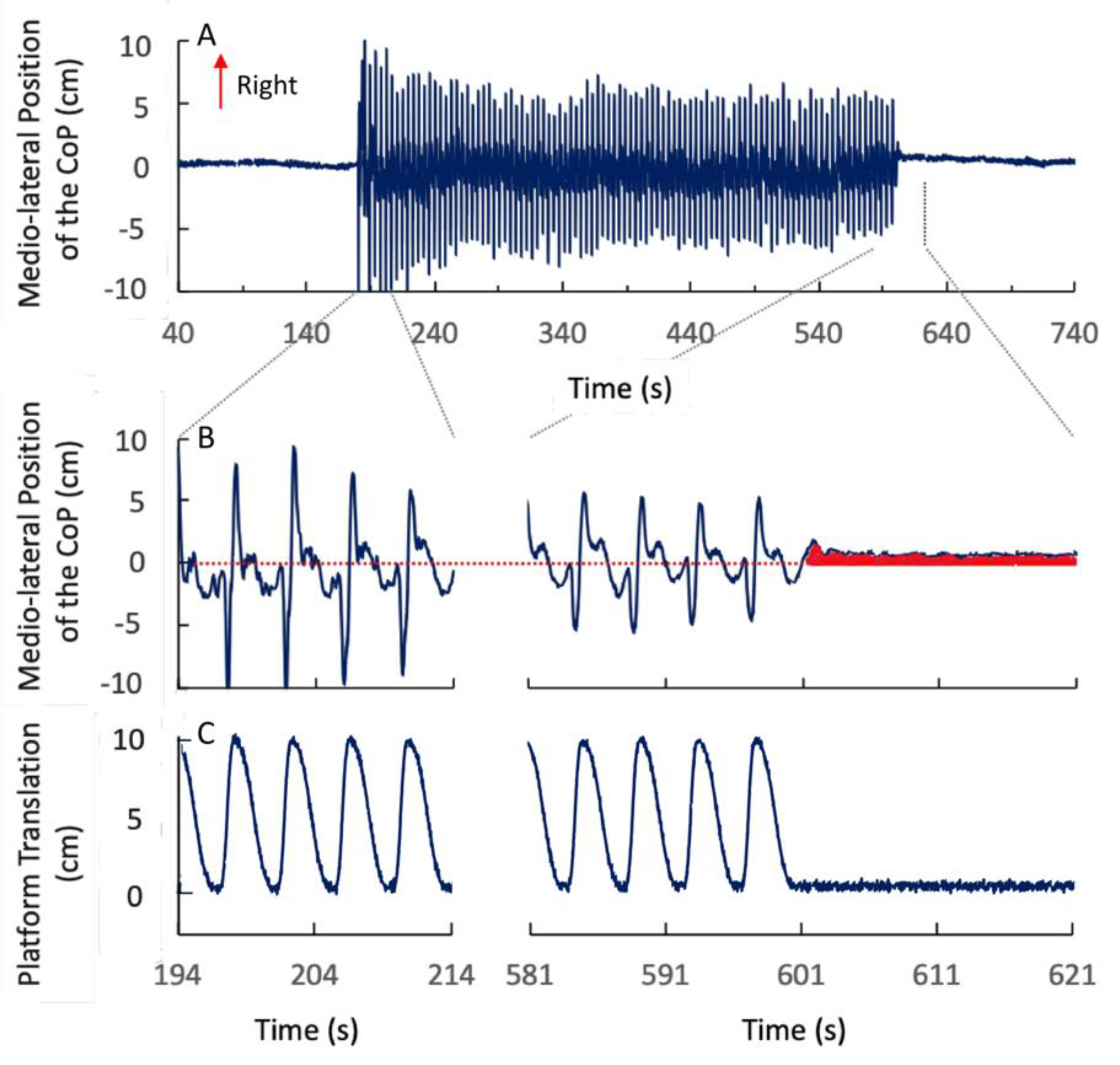
Figure 1. Medio-lateral (ML) centre of feet pressure (CoP) position in a representative participant during the fast right (FR) stimulation under the eyes closed condition (EC) (A,B) and platform translation trace (C). During the fast half-cycle to the right side, the CoP showed a large and fast ML displacement followed by a less ample and slower displacement during the slower half-cycle. In the central panel, the red area indicates the medio-lateral shift of the CoP post-stimulation, while the dotted red line indicates the pre-stimulation ML CoP position. Here and in the following Figures, right is upward.
Applsci 10 05969 g001
Figure 2. Mean post-stimulation ML (A) and anterior–posterior (AP) (B) CoP position. The bars with the blue outline refer to the fast left (FL) condition, the bars with the red outline to the fast right (FR) condition. Grey filling refers to EC, white filling to eyes open (EO). (A) At the end of the FL stimulation period, the mean ML CoP position moved to the left (negative values in the ordinate) with respect to its pre-stimulation position. At the end of the FR stimulation, the CoP position moved to the right (positive values in the ordinate). (B) The post-stimulation CoP position in the sagittal plane (AP) was not different from the pre-stimulation position.
Applsci 10 05969 g002
Figure 3. Duration of the post-stimulation effect on the ML CoP position. The bars with a blue outline refer to the fast left (FL) condition, and the bars with a red outline to the fast right (FR) condition. Grey filling refers to EC, white filling to EO. There was no difference in the duration of the post-effect regardless of the asymmetry of the stimulation-cycle profile or of the visual condition.
Applsci 10 05969 g003
Figure 4. Mean ML CoP positions during the fast and slow phases of the stimulation cycles (FL–EC condition) and their asymptotic values obtained by the exponential fit (ensemble averaged group means). (A) During the fast phase to the left (dark blue circles), the mean ML CoP position shifted to the right side of the force platform. During the slow phase (light blue circles) it moved to the left (arrows) with respect to the position reached during the fast phase. In the last cycles, the ML CoP position during the slow phase was close to the pre-stimulation position. (B) Mean values of the time constant of the exponential function fitted to the ML CoP position during the fast and during the slow phases. The bars with the blue outline refer to FL, and the bars with the red outline to FR. Grey filling denotes EC, and white filling EO. (C) For each subject, the mean post-stimulation ML CoP position was plotted against the difference in the asymptotic value of the ML CoP position between the fast and the slow phase for the FL-EC (grey filling with blue outlined circles), FL–EO (white filling with blue outline), FR–EC (grey filling with red outline) and FR–EO (white filling with red outline). (D) The mean values of the asymptotes of the fast and the slow phases were reported. Circles with blue outlines refer to FL and the circles with red outlines to FR condition. Grey filling refers to EC, and white filling to EO. The blue and red dotted lines are the mean ML post-stimulation CoP positions in the four conditions.
Applsci 10 05969 g004
Table 1. Mean Sway (Standard Deviation of the CoP Sway ± SD) in the Frontal (ML) and Sagittal (AP) Planes. P Refers to the Post-Hoc Test Results.
MLAP
Pre-StimulationPost-StimulationPre-StimulationPost-Stimulation
FL–EC0.20 cm ± 0.130.37 cm ± 0.32p < 0.010.39 cm ± 0.120.48 cm ± 0.15p < 0.01
FL–EO0.22 cm ± 0.150.30 cm ± 0.17p < 0.050.37 cm ± 0.130.39 cm ± 0.13p = 0.42
FR–EC0.22 cm ± 0.120.33 cm ± 0.15p < 0.010.42 cm ± 0.140.53 cm ± 0.18p < 0.01
FR–EO0.23 cm ± 0.150.31 cm ± 0.19p < 0.050.39 cm ± 0.140.45 cm ± 0.18p = 0.12