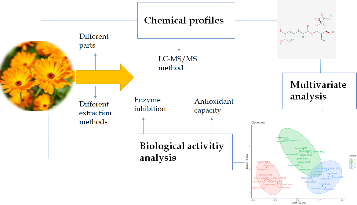
A Comparative Bio-Evaluation and Chemical Profiles of Calendula officinalis L. Extracts Prepared via Different Extraction Techniques


 , , ,
, , ,  ,
,  and
and 
Abstract
Featured Application
Abstract
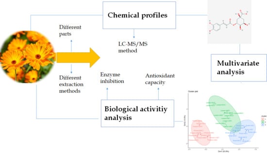
1. Introduction
2. Materials and Methods
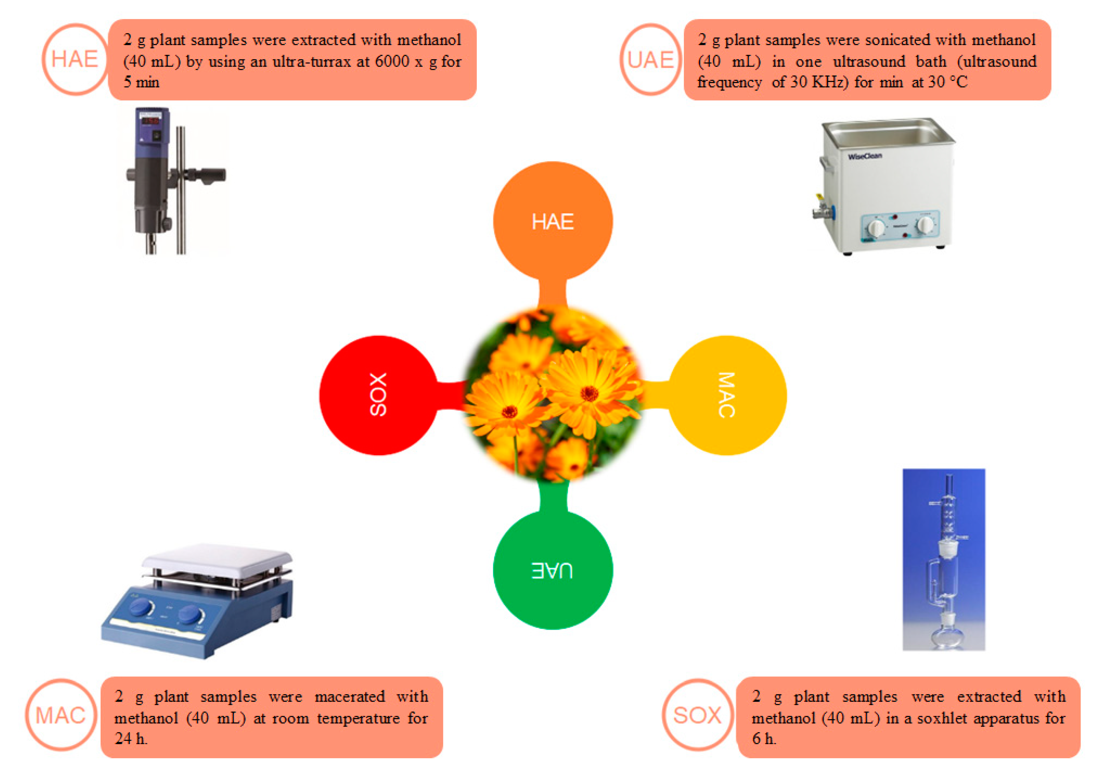
2.1. Plant Material and Extracts Preparation
2.2. Profile of Bioactive Compounds
2.3. Mass Spectrometer and Chromatograph Conditions
2.4. Antioxidant and Enzyme Inhibitory Assays
2.5. Data Analysis
3. Results and Discussion
3.1. Chemical Profile
3.2. Antioxidant Effects
3.3. Enzyme Inhibitory Properties
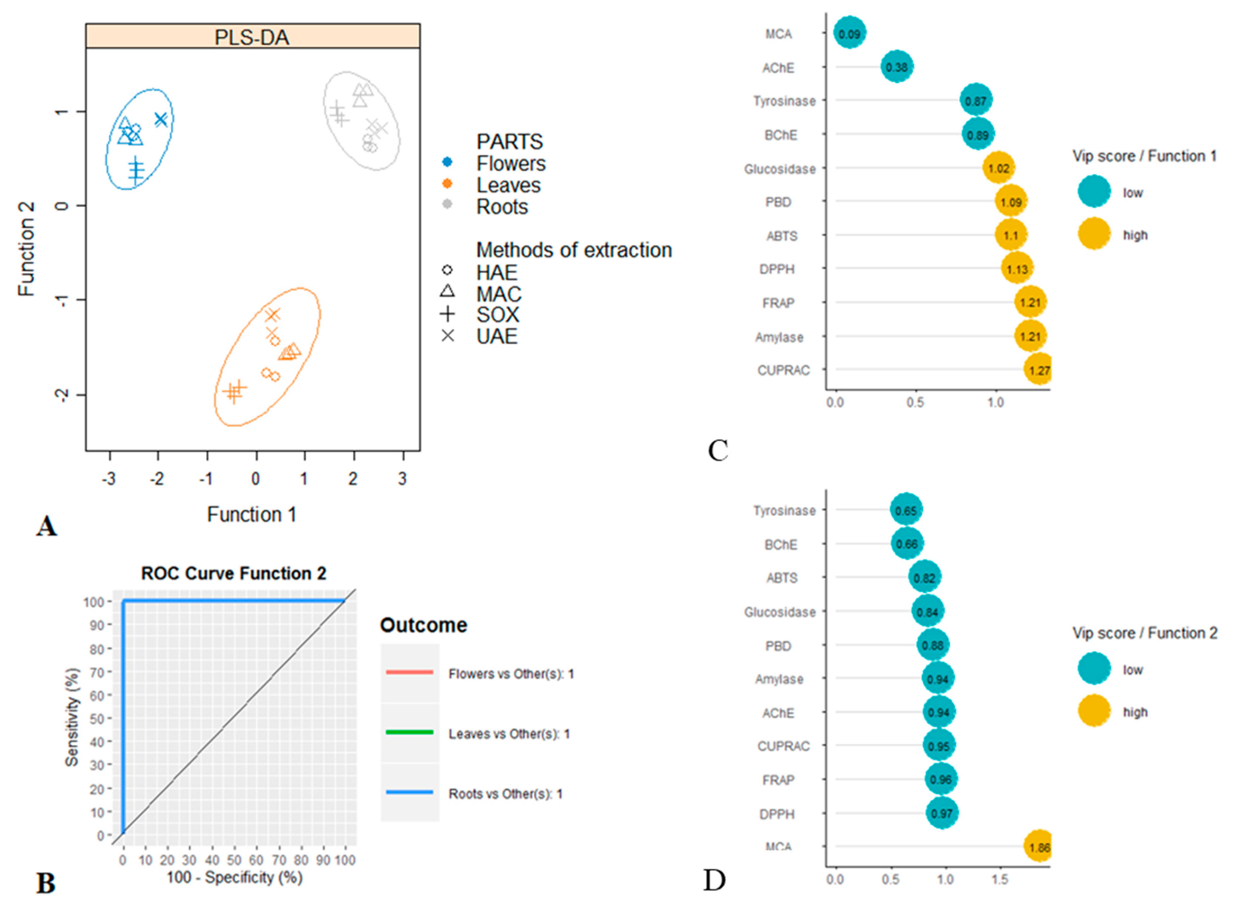
3.4. Multivariate Analysis
4. Conclusions
Supplementary Materials
Author Contributions
Funding
Conflicts of Interest
References
- Vieira da Silva, B.; Barreira, J.C.M.; Oliveira, M.B.P.P. Natural phytochemicals and probiotics as bioactive ingredients for functional foods: Extraction, biochemistry and protected-delivery technologies. Trends Food Sci. Technol. 2016, 50, 144–158. [Google Scholar] [CrossRef]
- Belwal, T.; Ezzat, S.M.; Rastrelli, L.; Bhatt, I.D.; Daglia, M.; Baldi, A.; Devkota, H.P.; Orhan, I.E.; Patra, J.K.; Das, G. A critical analysis of extraction techniques used for botanicals: Trends, priorities, industrial uses and optimization strategies. TrAC Trends Anal. Chem. 2018, 100, 82–102. [Google Scholar] [CrossRef]
- Mishra, A.K.; Mishra, A.; Pragya; Chattopadhyay, P. Screening of acute and sub-chronic dermal toxicity of Calendula officinalis L. essential oil. Regul. Toxicol. Pharmacol. 2018, 98, 184–189. [Google Scholar] [CrossRef]
- Efstratiou, E.; Hussain, A.I.; Nigam, P.S.; Moore, J.E.; Ayub, M.A.; Rao, J.R. Antimicrobial activity of Calendula officinalis petal extracts against fungi, as well as Gram-negative and Gram-positive clinical pathogens. Complement. Ther. Clin. Pract. 2012, 18, 173–176. [Google Scholar] [CrossRef]
- Cruceriu, D.; Diaconeasa, Z.; Socaci, S.; Socaciu, C.; Rakosy-Tican, E.; Balacescu, O. Biochemical profile, selective cytotoxicity and molecular effects of Calendula officinalis extracts on breast cancer cell lines. Not. Bot. Horti Agrobot. Cluj-Napoca 2020, 48, 24–39. [Google Scholar] [CrossRef][Green Version]
- Escher, G.B.; Borges, L.D.C.; Santos, J.S.; Cruz, T.M.; Marques, M.B.; do Carmo, M.A.V.; Azevedo, L.; Furtado, M.M.; Sant’Ana, A.S.; Wen, M.C.; et al. From the field to the pot: Phytochemical and functional analyses of Calendula officinalis L. flower for incorporation in an organic yogurt. Antioxidants 2019, 8, 20. [Google Scholar]
- Chitrakar, B.; Zhang, M.; Bhandari, B. Edible flowers with the common name “marigold”: Their therapeutic values and processing. Trends Food Sci. Technol. 2019, 89, 76–87. [Google Scholar] [CrossRef]
- Hamburger, M.; Adler, S.; Baumann, D.; Förg, A.; Weinreich, B. Preparative purification of the major anti-inflammatory triterpenoid esters from Marigold (Calendula officinalis). Fitoterapia 2003, 74, 328–338. [Google Scholar] [CrossRef]
- Neukirch, H.; D’Ambrosio, M.; Via, J.D.; Guerriero, A. Simultaneous quantitative determination of eight triterpenoid monoesters from flowers of 10 varieties of Calendula officinalis L. and characterisation of a new triterpenoid monoester. Phytochem. Anal. 2004, 15, 30–35. [Google Scholar] [CrossRef]
- Ohmiya, A. Diversity of carotenoid composition in flower petals. Jarq-Jpn. Ag. Res. Q. 2011, 45, 163–171. [Google Scholar] [CrossRef]
- Petrović, L.; Lepojevic, Z.; Sovilj, V.; Adamović, D.; Tešević, V. An investigation of CO2 extraction of marigold (Calendula officinalis L.). J. Serb. Chem. Soc. 2007, 72, 407–413. [Google Scholar] [CrossRef]
- Kozlowska, J.; Stachowiak, N.; Prus, W. Stability studies of collagen-based microspheres with Calendula officinalis flower extract. Polym. Degradation Stab. 2019, 163, 214–219. [Google Scholar] [CrossRef]
- Cioinac, S.E. Use of calendula cream balm to medicate the feet of diabetic patients: Case series. Int. J. Nurs. Stud. 2016, 3, 102–112. [Google Scholar] [CrossRef][Green Version]
- Pedram Rad, Z.; Mokhtari, J.; Abbasi, M. Calendula officinalis extract/PCL/Zein/Gum arabic nanofibrous bio-composite scaffolds via suspension, two-nozzle and multilayer electrospinning for skin tissue engineering. Int. J. Biol. Macromol. 2019, 135, 530–543. [Google Scholar] [CrossRef]
- Slavov, A.; Ognyanov, M.; Vasileva, I. Pectic polysaccharides extracted from pot marigold (Calendula officinalis) industrial waste. Food Hydrocoll. 2020, 101, 105545. [Google Scholar] [CrossRef]
- Pires, T.C.S.P.; Dias, M.I.; Barros, L.; Ferreira, I.C.F.R. Nutritional and chemical characterization of edible petals and corresponding infusions: Valorization as new food ingredients. Food Chem. 2017, 220, 337–343. [Google Scholar] [CrossRef]
- Hamzawy, M.A.; El-Denshary, E.S.M.; Hassan, N.S.; Mannaa, F.A.; Abdel-Wahhab, M.A. Dietary Supplementation of Calendula officinalis Counteracts the Oxidative Stress and Liver Damage Resulted from Aflatoxin. ISRN Nutr. 2013, 2013, 538427. [Google Scholar] [CrossRef]
- Vladimir-Knežević, S.; Blažeković, B.; Bival Štefan, M.; Alegro, A.; Kőszegi, T.; Petrik, J. Antioxidant activities and polyphenolic contents of three selected Micromeria species from Croatia. Molecules 2011, 16, 1454–1470. [Google Scholar] [CrossRef]
- Zengin, G.; Aktumsek, A. Investigation of antioxidant potentials of solvent extracts from different anatomical parts of Asphodeline anatolica E. Tuzlaci: An endemic plant to Turkey. Afr. J. Tradit. Complement. Altern. Med. 2014, 11, 481–488. [Google Scholar] [CrossRef]
- Yilmaz, M.A. Simultaneous quantitative screening of 53 phytochemicals in 33 species of medicinal and aromatic plants: A detailed, robust and comprehensive LC–MS/MS method validation. Ind. Crop. Prod. 2020, 149, 112347. [Google Scholar] [CrossRef]
- Grochowski, D.M.; Uysal, S.; Aktumsek, A.; Granica, S.; Zengin, G.; Ceylan, R.; Locatelli, M.; Tomczyk, M. In vitro enzyme inhibitory properties, antioxidant activities, and phytochemical profile of Potentilla thuringiaca. Phytochem. Lett. 2017, 20, 365–372. [Google Scholar] [CrossRef]
- Ercetin, T.; Senol, F.S.; Erdogan Orhan, I.; Toker, G. Comparative assessment of antioxidant and cholinesterase inhibitory properties of the marigold extracts from Calendula arvensis L. and Calendula officinalis L. Ind. Crop. Prod. 2012, 36, 203–208. [Google Scholar] [CrossRef]
- BEKDEŞER, B. Modeling and optimizing microwave-assisted extraction of antioxidant compounds from marigold Calendula officinalis L. using response surface methodology. Turk. J. Chem. 2019, 43, 1457–1471. [Google Scholar] [CrossRef]
- Ourabia, I.; Djebbar, R.; Tata, S.; Sabaou, N.; Fouial-Djebbar, D. Determination of essential oil composition, phenolic content, and antioxidant, antibacterial and antifungal activities of marigold (Calendula officinalis l.) cultivated in algeria. Carpath. J. Food Sci. Technol. 2019, 11, 93–110. [Google Scholar]
- Petkova, D.; Mihaylova, D.; Denev, P.; Krastanov, A. Antioxidant activity of some edible flowers water extracts from Bulgaria. Bull. UASVM Food Sci. Technol. 2020, 77, 1. [Google Scholar] [CrossRef]
- Sytar, O.; Hemmerich, I.; Zivcak, M.; Rauh, C.; Brestic, M. Comparative analysis of bioactive phenolic compounds composition from 26 medicinal plants. Saudi J. Biol. Sci. 2018, 25, 631–641. [Google Scholar] [CrossRef]
- Babaee, N.; Moslemi, D.; Khalilpour, M.; Vejdani, F.; Moghadamnia, Y.; Bijani, A.; Baradaran, M.; Kazemi, M.T.; Khalilpour, A.; Pouramir, M.; et al. Antioxidant capacity of calendula officinalis flowers extract and prevention of radiation induced oropharyngeal mucositis in patients with head and neck cancers: A randomized controlled clinical study. Daru 2013, 21, 18. [Google Scholar] [CrossRef]
- Preethi, K.C.; Kuttan, G.; Kuttan, R. Antioxidant potential of an extract of Calendula officinalis. flowers in vitro and in vivo. Pharm. Biol. 2006, 44, 691–697. [Google Scholar] [CrossRef]
- Soberón, J.R.; Sgariglia, M.A.; Sampietro, D.A.; Quiroga, E.N.; Vattuone, M.A. Free radical scavenging activities and inhibition of inflammatory enzymes of phenolics isolated from Tripodanthus acutifolius. J. Ethnopharmacol. 2010, 130, 329–333. [Google Scholar] [CrossRef]
- Catapano, M.C.; Tvrdý, V.; Karlíčková, J.; Migkos, T.; Valentová, K.; Křen, V.; Mladěnka, P. The stoichiometry of isoquercitrin complex with iron or copper is highly dependent on experimental conditions. Nutrients 2017, 9, 1193. [Google Scholar] [CrossRef]
- Pires, T.C.S.P.; Dias, M.I.; Barros, L.; Calhelha, R.C.; Alves, M.J.; Oliveira, M.B.P.P.; Santos-Buelga, C.; Ferreira, I.C.F.R. Edible flowers as sources of phenolic compounds with bioactive potential. Food Res. Int. 2018, 105, 580–588. [Google Scholar] [CrossRef] [PubMed]
- Marian, E.; Vicaș, L.G.; Jurca, T.; Mureasan, M.; Stan, R.L.; Sevastre, B.; Diaconeasa, Z.; Ionescu, C.; Hangan, A.C. A comparative study on the biologic activity of Centaurea cyanus versus Calendula officinalis. Farmacia 2017, 65, 940–946. [Google Scholar]
- Caratelli, V.; Ciampaglia, A.; Guiducci, J.; Sancesario, G.; Moscone, D.; Arduini, F. Precision medicine in Alzheimer’s disease: An origami paper-based electrochemical device for cholinesterase inhibitors. Biosens. Bioelectron. 2020, 28, 112411. [Google Scholar] [CrossRef] [PubMed]
- Erdogan-Orhan, I.; Altun, M.L.; Sever-Yilmaz, B.; Saltan, G. Anti-acetylcholinesterase and antioxidant assets of the major components (salicin, amentoflavone, and chlorogenic acid) and the extracts of Viburnum opulus and Viburnum lantana and their total phenol and flavonoid contents. J. Med. Food 2011, 14, 434–440. [Google Scholar] [CrossRef] [PubMed]
- Eroglu Ozkan, E.; Yilmaz Ozden, T.; Ozsoy, N.; Mat, A. Evaluation of chemical composition, antioxidant and anti-acetylcholinesterase activities of Hypericum neurocalycinum and Hypericum malatyanum. S. Afr. J. Bot. 2018, 114, 104–110. [Google Scholar] [CrossRef]
- Ishola, I.O.; Tota, S.; Adeyemi, O.O.; Agbaje, E.O.; Narender, T.; Shukla, R. Protective effect of Cnestis ferruginea and its active constituent on scopolamine-induced memory impairment in mice: A behavioral and biochemical study. Pharm. Biol. 2013, 51, 825–835. [Google Scholar] [CrossRef] [PubMed]
- Silva, L.; Rodrigues, A.M.; Ciriani, M.; Falé, P.L.V.; Teixeira, V.; Madeira, P.; Machuqueiro, M.; Pacheco, R.; Florêncio, M.H.; Ascensão, L.; et al. Antiacetylcholinesterase activity and docking studies with chlorogenic acid, cynarin and arzanol from Helichrysum stoechas (Lamiaceae). Med. Chem. Res. 2017, 26, 2942–2950. [Google Scholar] [CrossRef]
- Topal, M.; Gocer, H.; Topal, F.; Kalin, P.; Köse, L.P.; Gülçin, İ.; Çakmak, K.C.; Küçük, M.; Durmaz, L.; Gören, A.C.; et al. Antioxidant, antiradical, and anticholinergic properties of cynarin purified from the Illyrian thistle (Onopordum illyricum L.). J. Enzyme Inhib. Med. Chem. 2016, 31, 266–275. [Google Scholar] [CrossRef]
- Akhtar, N.; Khan, B.A.; Haji, M.; Khan, S.; Ahmad, M.; Rasool, F.; Mahmood, T.; Rasul, A. Evaluation of various functional skin parameters using a topical cream of Calendula officinalis extract. Afr. J. Pharm. Pharmaco. 2011, 5, 199–206. [Google Scholar] [CrossRef]
- Olennikov, D.N.; Kashchenko, N.I. Componential profile and amylase inhibiting activity of phenolic compounds from Calendula officinalis L. leaves. Sci. World J. 2014, 2014, 654193. [Google Scholar] [CrossRef]
- Iloki-Assanga, S.B.; Lewis-Luján, L.M.; Lara-Espinoza, C.L.; Gil-Salido, A.A.; Fernandez-Angulo, D.; Rubio-Pino, J.L.; Haines, D.D. Solvent effects on phytochemical constituent profiles and antioxidant activities, using four different extraction formulations for analysis of Bucida buceras L. and Phoradendron californicum. BMC Res. Notes 2015, 8, 396. [Google Scholar] [CrossRef] [PubMed]
- Del Baño, M.J.; Lorente, J.; Castillo, J.; Benavente-García, O.; del Río, J.A.; Ortuño, A.; Quirin, K.W.; Gerard, D. Phenolic diterpenes, flavones, and rosmarinic acid distribution during the development of leaves, flowers, stems, and roots of Rosmarinus officinalis. Antioxidant activity. J. Agric. Food Chem. 2003, 51, 4247–4253. [Google Scholar] [CrossRef] [PubMed]
- Rajan, M.; Rajkumar, G.; Farias Lima Guedes, T.J.; Chagas Barros, R.G.; Narain, N. Performance of different solvents on extraction of bioactive compounds, antioxidant and cytotoxic activities in Phoenix loureiroi Kunth leaves. J. Appl. Res. Med. Aromat. Plants 2020, 17, 100247. [Google Scholar] [CrossRef]
- Zhu, J.-J.; Yang, J.-J.; Wu, G.-J.; Jiang, J.-G. Comparative antioxidant, anticancer and antimicrobial activities of essential oils from Semen Platycladi by different extraction methods. Ind. Crop. Prod. 2020, 146, 112206. [Google Scholar] [CrossRef]
- Daud, M.N.H.; Fatanah, D.N.; Abdullah, N.; Ahmad, R. Evaluation of antioxidant potential of Artocarpus heterophyllus L. J33 variety fruit waste from different extraction methods and identification of phenolic constituents by LCMS. Food Chem. 2017, 232, 621–632. [Google Scholar] [CrossRef]
- Dall’Acqua, S.; Kumar, G.; Sinan, K.I.; Sut, S.; Ferrarese, I.; Mahomoodally, M.F.; Seebaluck-Sandoram, R.; Etienne, O.K.; Zengin, G. An insight into Cochlospermum planchonii extracts obtained by traditional and green extraction methods: Relation between chemical compositions and biological properties by multivariate analysis. Ind. Crop. Prod. 2020, 147, 112226. [Google Scholar] [CrossRef]


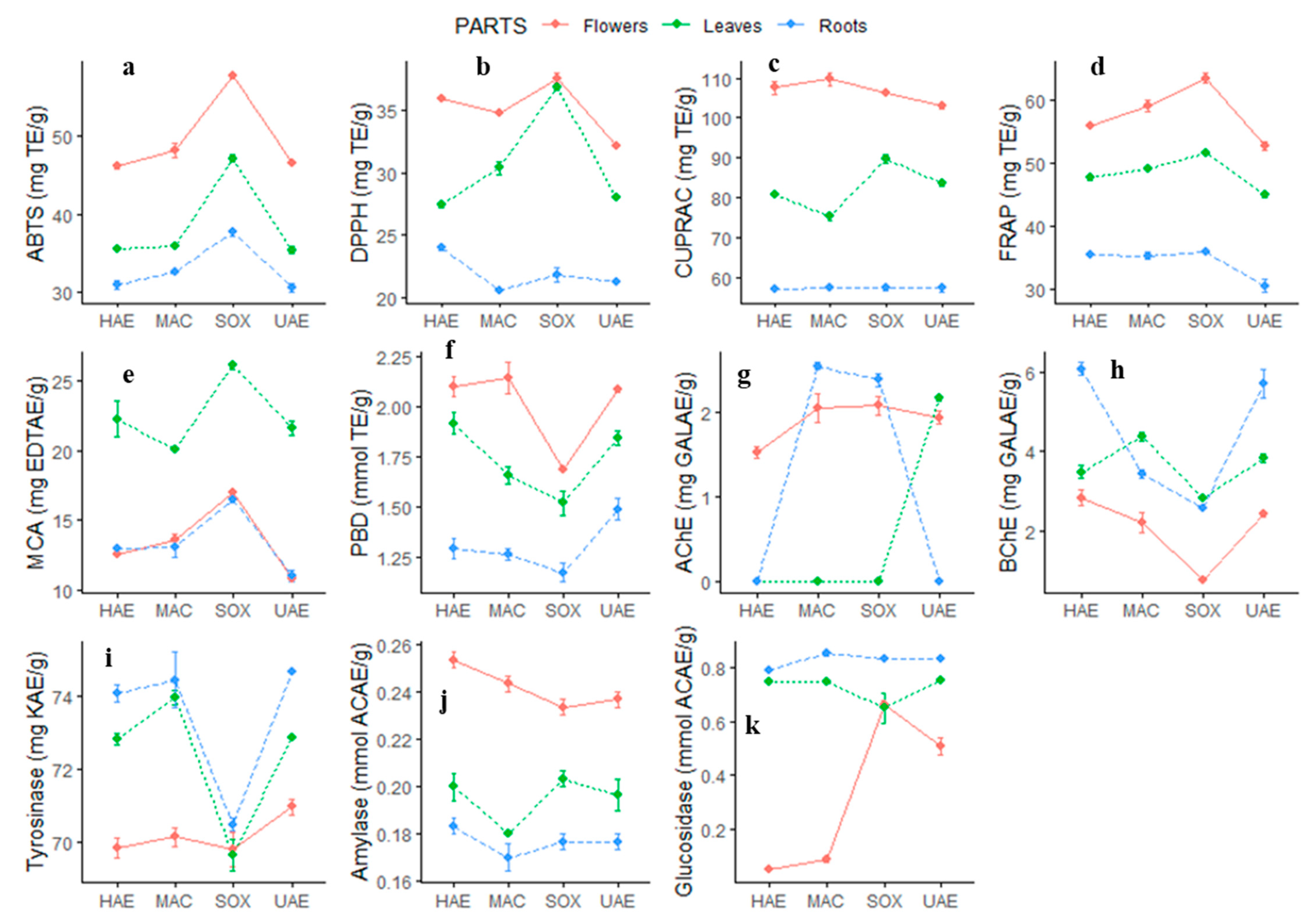
| Parts | Methods | TPC (mg GAE/g extract) | TFC (mg RE/g extract) | PBD ( mmol TE/g extract) |
|---|---|---|---|---|
| Flowers | HAE | 32.18 ± 0.40 b | 28.48 ± 0.40 d | 2.10 ± 0.08 a |
| MAC | 34.27 ± 0.40 a | 28.38 ± 0.45 d | 2.14 ± 0.13 a | |
| UAE | 32.32 ± 0.40 b | 28.41 ± 0.50 d | 2.08 ± 0.02 a | |
| SOX | 30.96 ± 0.17 c | 32.50 ± 0.67 b,c | 1.68 ± 0.02 b,c,d | |
| Leaves | HAE | 23.57 ± 0.30 f | 31.60 ± 0.74 c | 1.91 ± 0.09 a,b |
| MAC | 25.20 ± 0.26 e | 28.33 ± 0.14 d | 1.66 ± 0.07 c,d | |
| UAE | 24.39 ± 0.14 e,f | 32.95 ± 0.35 b | 1.84 ± 0.06 b,c | |
| SOX | 27.93 ± 0.25 d | 34.61 ± 0.29 a | 1.52 ± 0.11 d,e | |
| Roots | HAE | 18.40 ± 0.37 g | 2.54 ± 0.09 e | 1.29 ± 0.09 e,f,g |
| MAC | 19.27 ± 0.25 g | 2.45 ± 0.12 e | 1.26 ± 0.05 f,g | |
| UAE | 16.49 ± 0.39 h | 2.45 ± 0.07 e | 1.49 ± 0.09 d,e,f | |
| SOX | 17.36 ± 0.19 h | 2.62 ± 0.06 e | 1.17 ± 0.08 g |
| Analyte | 1 | 2 | 3 | 4 | 5 | 6 | 7 | 8 | 9 | 10 | 11 | 12 |
|---|---|---|---|---|---|---|---|---|---|---|---|---|
| Vanillin | N.D. | N.D. | N.D. | N.D. | N.D. | N.D. | N.D. | N.D. | N.D. | N.D. | N.D. | N.D. |
| Daidzin | N.D. | N.D. | N.D. | N.D. | N.D. | N.D. | N.D. | N.D. | N.D. | N.D. | N.D. | N.D. |
| Piceid | N.D. | N.D. | N.D. | N.D. | N.D. | N.D. | N.D. | N.D. | N.D. | N.D. | N.D. | N.D. |
| Coumarin | N.D. | N.D. | N.D. | N.D. | N.D. | N.D. | N.D. | N.D. | N.D. | N.D. | N.D. | N.D. |
| Hesperidin | 2.964 | 3.076 | 3.091 | 2.618 | 0.343 | 0.264 | 0.284 | 0.297 | N.D. | N.D. | N.D. | N.D. |
| Quinic acid | 8.757 | 6.362 | 6.322 | 13.426 | 4.847 | 4.889 | 4.262 | 6.804 | 4.329 | 4.646 | 4.072 | 5.896 |
| Fumaric aid | 3.739 | 3.758 | 3.141 | 10.322 | 5.122 | 5.077 | 4.154 | 1.145 | 1.129 | 5.236 | 3.255 | 8.746 |
| Aconitic acid | N.D. | N.D. | N.D. | N.D. | N.D. | N.D. | N.D. | N.D. | N.D. | N.D. | N.D. | N.D. |
| Gallic acid | N.D. | N.D. | N.D. | N.D. | N.D. | N.D. | N.D. | N.D. | N.D. | N.D. | N.D. | N.D. |
| Protocatechuic acid | 0.084 | 0.124 | 0.121 | 0.120 | 0.074 | 0.090 | 0.075 | 0.112 | 0.089 | 0.112 | 0.066 | 0.127 |
| Gentisic acid | N.D. | N.D. | N.D. | N.D. | N.D. | N.D. | N.D. | N.D. | N.D. | N.D. | N.D. | N.D. |
| Epigallocatechin | N.D. | N.D. | N.D. | N.D. | N.D. | N.D. | N.D. | N.D. | N.D. | N.D. | N.D. | N.D. |
| Protocatechuic aldehyde | N.D. | N.D. | 0.028 | 0.023 | 0.043 | 0.048 | 0.046 | 0.061 | 0.072 | 0.076 | 0.045 | 0.083 |
| Catechin | N.D. | N.D. | N.D. | 0.088 | N.D. | N.D. | N.D. | N.D. | N.D. | N.D. | N.D. | 0.025 |
| Chlorogenic acid | 8.555 | 9.214 | 8.532 | 8.313 | 8.748 | 8.575 | 7.036 | 10.118 | 4.901 | 4.835 | 3.767 | 4.869 |
| Tannic acid | N.D. | N.D. | N.D. | N.D. | N.D. | N.D. | N.D. | N.D. | N.D. | N.D. | N.D. | N.D. |
| 4-OH Benzoic acid | N.D. | N.D. | N.D. | N.D. | N.D. | N.D. | N.D. | N.D. | N.D. | N.D. | N.D. | N.D. |
| Epigallocatechin gallate | N.D. | N.D. | N.D. | N.D. | N.D. | N.D. | N.D. | N.D. | N.D. | N.D. | N.D. | N.D. |
| Cynarin | N.D. | N.D. | N.D. | N.D. | N.D. | N.D. | N.D. | N.D. | N.D. | 0.042 | N.D. | N.D. |
| Vanilic acid | N.D. | N.D. | N.D. | N.D. | N.D. | N.D. | N.D. | N.D. | N.D. | N.D. | N.D. | N.D. |
| Epicatechin | N.D. | N.D. | N.D. | N.D. | N.D. | N.D. | N.D. | N.D. | N.D. | N.D. | N.D. | N.D. |
| Caffeic acid | 0.405 | 0.535 | 0.477 | 0.635 | 0.571 | 0.610 | 0.472 | 0.963 | 0.422 | 0.572 | 0.357 | 0.734 |
| Syringic acid | N.D. | N.D. | N.D. | N.D. | N.D. | N.D. | N.D. | N.D. | N.D. | N.D. | N.D. | N.D. |
| Syringic aldehyde | N.D. | N.D. | N.D. | N.D. | N.D. | N.D. | N.D. | N.D. | N.D. | N.D. | N.D. | N.D. |
| Epicatechin gallate | N.D. | N.D. | N.D. | N.D. | N.D. | N.D. | N.D. | N.D. | N.D. | N.D. | N.D. | N.D. |
| p-Coumaric acid | N.D. | N.D. | N.D. | N.D. | N.D. | N.D. | N.D. | N.D. | N.D. | N.D. | N.D. | N.D. |
| Sinapic acid | N.D. | N.D. | N.D. | N.D. | N.D. | N.D. | N.D. | N.D. | N.D. | N.D. | N.D. | N.D. |
| Ferulic acid | 0.084 | N.D. | N.D. | N.D. | N.D. | N.D. | N.D. | N.D. | N.D. | N.D. | N.D. | N.D. |
| Salicylic acid | 0.398 | 0.649 | 0.561 | 0.570 | 0.463 | 0.670 | 0.393 | 0.724 | 0.192 | 0.302 | 0.229 | 0.350 |
| Cyranoside | N.D. | N.D. | N.D. | N.D. | N.D. | N.D. | N.D. | N.D. | N.D. | N.D. | N.D. | N.D. |
| Miquelianin | 0.015 | N.D. | 0.015 | 0.023 | 0.003 | 0.013 | 0.011 | 0.005 | 0.004 | 0.006 | 0.007 | 0.005 |
| isoquercitrin | 2.645 | 3.706 | 2.600 | 5.512 | 9.778 | 13.562 | 7.056 | 24.907 | 0.996 | 0.421 | 0.207 | 0.545 |
| Rutin | 3.844 | 4.351 | 4.810 | 3.656 | 0.557 | 0.365 | 0.458 | 0.518 | N.D. | N.D. | N.D. | N.D. |
| Genistin | N.D. | N.D. | N.D. | N.D. | N.D. | N.D. | N.D. | N.D. | N.D. | N.D. | N.D. | N.D. |
| o-Coumaric acid | N.D. | N.D. | N.D. | N.D. | N.D. | N.D. | N.D. | N.D. | N.D. | N.D. | N.D. | N.D. |
| Ellagic acid | N.D. | N.D. | N.D. | N.D. | N.D. | N.D. | N.D. | N.D. | N.D. | N.D. | N.D. | N.D. |
| Rosmarinic acid | N.D. | N.D. | N.D. | N.D. | N.D. | N.D. | N.D. | N.D. | N.D. | N.D. | N.D. | N.D. |
| Fisetin | N.D. | N.D. | N.D. | N.D. | N.D. | N.D. | N.D. | N.D. | N.D. | N.D. | N.D. | N.D. |
| Cosmosiin | N.D. | N.D. | N.D. | N.D. | N.D. | 0.038 | N.D. | 0.051 | N.D. | N.D. | N.D. | N.D. |
| Quercitrin | N.D. | N.D. | N.D. | N.D. | N.D. | N.D. | N.D. | N.D. | N.D. | N.D. | N.D. | N.D. |
| Astragalin | 0.135 | 0.174 | 0.129 | 0.250 | 0.327 | 0.350 | 0.204 | 0.629 | N.D. | N.D. | N.D. | N.D. |
| Nicotiflorin | 0.484 | 0.537 | 0.570 | 0.468 | N.D. | N.D. | N.D. | N.D. | N.D. | N.D. | N.D. | N.D. |
| Daidzein | N.D. | N.D. | N.D. | N.D. | N.D. | N.D. | N.D. | N.D. | N.D. | N.D. | N.D. | N.D. |
| Genistein | N.D. | N.D. | N.D. | N.D. | N.D. | N.D. | N.D. | N.D. | N.D. | N.D. | N.D. | N.D. |
| Quercetin | N.D. | 0.047 | 0.046 | 0.068 | 0.047 | 0.044 | 0.048 | 0.082 | N.D. | N.D. | N.D. | N.D. |
| Luteolin | 0.003 | 0.004 | 0.006 | 0.005 | 0.007 | 0.008 | 0.008 | 0.010 | 0.003 | 0.003 | 0.002 | 0.002 |
| Hesperetin | N.D. | N.D. | N.D. | N.D. | N.D. | N.D. | N.D. | N.D. | N.D. | N.D. | N.D. | N.D. |
| Naringenin | N.D. | N.D. | N.D. | N.D. | N.D. | N.D. | N.D. | N.D. | N.D. | N.D. | N.D. | N.D. |
| Kaempferol | N.D. | N.D. | N.D. | N.D. | N.D. | N.D. | N.D. | N.D. | N.D. | N.D. | N.D. | N.D. |
| Apigenin | N.D. | N.D. | N.D. | N.D. | N.D. | N.D. | N.D. | N.D. | N.D. | N.D. | N.D. | N.D. |
| Amentoflavone | N.D. | N.D. | N.D. | N.D. | N.D. | N.D. | N.D. | N.D. | 0.002 | 0.002 | 0.001 | 0.001 |
| Acacetin | N.D. | N.D. | N.D. | N.D. | N.D. | N.D. | N.D. | N.D. | N.D. | N.D. | N.D. | N.D. |
| Chrysin | N.D. | N.D. | N.D. | N.D. | N.D. | N.D. | N.D. | N.D. | N.D. | N.D. | N.D. | N.D. |
| Parts | Methods | DPPH (mg TE/g extract) | ABTS (mg TE/g extract) | CUPRAC (mg TE/g extract) | FRAP (mg TE/g extract) | Metal Chelating (mg EDTAE/g extract) |
|---|---|---|---|---|---|---|
| Flowers | HAE | 35.90 ± 0.06 b,c | 46.11 ± 0.59 b | 107.53 ± 2.97 a,b | 55.94 ± 0.41 c | 12.58 ± 0.24 d,e |
| MAC | 34.75 ± 0.24 c | 48.15 ± 1.49 b | 109.68 ± 2.70 a | 58.96 ± 1.57 b | 13.59 ± 0.74 d | |
| UAE | 32.18 ± 0.31 d | 46.61 ± 0.27 b | 103.11 ± 1.29 b | 52.75 ± 1.21 d | 10.79 ± 0.17 e | |
| SOX | 37.58 ± 0.69 a | 57.69 ± 0.64 a | 106.21 ± 0.19 a,b | 63.43 ± 1.41 a | 16.95 ± 0.47 c | |
| Leaves | HAE | 27.40 ± 0.31 f | 35.53 ± 0.43 c,d | 80.91 ± 0.47 d | 47.75 ± 0.92 f,g | 22.26 ± 2.23 b |
| MAC | 30.38 ± 0.92 e | 35.96 ± 0.61 c,d | 75.32 ± 1.63 e | 49.02 ± 0.29 e,f | 20.11 ± 0.06 b | |
| UAE | 28.07 ± 0.19 f | 35.37 ± 0.73 d | 83.69 ± 1.18 d | 44.91 ± 0.71 d | 21.65 ± 0.87 b | |
| SOX | 36.78 ± 0.21 a,b | 47.16 ± 0.81 b | 89.74 ± 1.63 c | 51.63 ± 0.36 d,e | 26.11 ± 0.40 a | |
| Roots | HAE | 24.02 ± 0.37 g | 30.95 ± 1.02 e | 57.65 ± 0.37 f | 35.37 ± 0.04 h | 13.02 ± 0.32 d,e |
| MAC | 20.57 ± 0.25 h | 32.60 ± 0.07 e | 57.91 ± 0.75 f | 35.37 ± 0.97 h | 13.04 ± 1.14 d,e | |
| UAE | 21.29 ± 0.11 h | 30.53 ± 0.95 e | 56.36 ± 0.83 f | 30.55 ± 1.70 i | 11.01 ± 0.73 e | |
| SOX | 21.84 ± 0.95 h | 37.70 ± 0.80 c | 57.33 ± 0.82 f | 36.02 ± 0.28 h | 16.51 ± 0.34 c |
| Parts | Methods | AChE Inhibition (mg GALAE/g extract) | BChE Inhibition (mg GALAE/g extract) | Tyrosinase Inhibition (mg KAE/g extract) | Amylase Inhibition (mmol ACAE/g extract) | Glucosidase Inhibition (mmol ACAE/g extract) |
|---|---|---|---|---|---|---|
| Flowers | HAE | 1.52 ± 0.11 d | 2.83 ± 0.36 d,e,f | 69.86 ± 0.46 c | 0.25 ± 0.01 a | 0.05 ± 0.02 f |
| MAC | 2.05 ± 0.29 b,c | 2.20 ± 0.45 f | 70.16 ± 0.41 c | 0.24 ± 0.01 a,b | 0.09 ± 0.01 f | |
| UAE | 1.94 ± 0.13 c | 2.42 ± 0.12 f | 70.97 ± 0.36 c | 0.24 ± 0.01 a,b | 0.51 ± 0.06 e | |
| SOX | 2.08 ± 0.19 b,c | 0.76 ± 0.09 g | 69.83 ± 0.82 c | 0.23 ± 0.01 b | 0.66 ± 0.02 c,d | |
| Leaves | HAE | na | 3.48 ± 0.28 c,d | 72.82 ± 0.27 b | 0.20 ± 0.01 c | 0.75 ± 0.01 b,c |
| MAC | na | 4.37 ± 0.20 b | 73.95 ± 0.33 a,b | 0.18 ± 0.01 d,e | 0.75 ± 0.01 b,c,d | |
| UAE | 2.16 b,c | 3.84 ± 0.20 b,c | 72.84 ± 0.03 b | 0.20 ± 0.01 c,d | 0.75 ± 0.01 a,b,c | |
| SOX | na | 2.81 ± 0.06 d,e,f | 69.66 ± 0.76 c | 0.21 ± 0.01 c | 0.65 ± 0.09 d | |
| Roots | HAE | na | 6.08 ± 0.28 a | 74.05 ± 0.41 a,b | 0.18 ± 0.01 d,e | 0.79 ± 0.01 a,b |
| MAC | 2.54 ± 0.08 a | 3.43 ± 0.21 c,d,e | 74.43 ± 1.31 a,b | 0.17 ± 0.01 e | 0.85 ± 0.01 a | |
| UAE | na | 5.72 ± 0.62 a | 74.66 ± 0.08 a | 0.18 ± 0.01 e | 0.83 ± 0.01 a,b | |
| SOX | 2.38 ± 0.14 a,b | 2.57 ± 0.08 e,f | 70.50 ± 0.29 c | 0.18 ± 0.01 d,e | 0.83 ± 0.01 a,b |
© 2020 by the authors. Licensee MDPI, Basel, Switzerland. This article is an open access article distributed under the terms and conditions of the Creative Commons Attribution (CC BY) license (http://creativecommons.org/licenses/by/4.0/).
Share and Cite
Ak, G.; Zengin, G.; Sinan, K.I.; Mahomoodally, M.F.; Picot-Allain, M.C.N.; Cakır, O.; Bensari, S.; Yılmaz, M.A.; Gallo, M.; Montesano, D. A Comparative Bio-Evaluation and Chemical Profiles of Calendula officinalis L. Extracts Prepared via Different Extraction Techniques. Appl. Sci. 2020, 10, 5920. https://doi.org/10.3390/app10175920
Ak G, Zengin G, Sinan KI, Mahomoodally MF, Picot-Allain MCN, Cakır O, Bensari S, Yılmaz MA, Gallo M, Montesano D. A Comparative Bio-Evaluation and Chemical Profiles of Calendula officinalis L. Extracts Prepared via Different Extraction Techniques. Applied Sciences. 2020; 10(17):5920. https://doi.org/10.3390/app10175920
Chicago/Turabian StyleAk, Gunes, Gokhan Zengin, Kouadio Ibrahime Sinan, Mohamad Fawzi Mahomoodally, Marie Carene Nancy Picot-Allain, Oguz Cakır, Souheir Bensari, Mustafa Abdullah Yılmaz, Monica Gallo, and Domenico Montesano. 2020. "A Comparative Bio-Evaluation and Chemical Profiles of Calendula officinalis L. Extracts Prepared via Different Extraction Techniques" Applied Sciences 10, no. 17: 5920. https://doi.org/10.3390/app10175920
APA StyleAk, G., Zengin, G., Sinan, K. I., Mahomoodally, M. F., Picot-Allain, M. C. N., Cakır, O., Bensari, S., Yılmaz, M. A., Gallo, M., & Montesano, D. (2020). A Comparative Bio-Evaluation and Chemical Profiles of Calendula officinalis L. Extracts Prepared via Different Extraction Techniques. Applied Sciences, 10(17), 5920. https://doi.org/10.3390/app10175920