Article

The Effect of Undersized Drilling on the Coronal Surface Roughness of Microthreaded Implants: An In Vitro Study

1
Department of Periodontology and Dental Implantology, School of Dental Medicine, Tel-Aviv University, P.O. Box 39040, Tel Aviv 6997801, Israel
2
Department of Oral Rehabilitation, School of Dental Medicine, Tel-Aviv University, P.O. Box 39040, Tel Aviv 6997801, Israel
*
Author to whom correspondence should be addressed.
Graduate student.
Appl. Sci. 2020, 10(15), 5231; https://doi.org/10.3390/app10155231
Submission received: 1 July 2020 / Revised: 26 July 2020 / Accepted: 26 July 2020 / Published: 29 July 2020
(This article belongs to the Special Issue Predictable Restorative Dentistry)

Abstract

:
This in-vitro study assessed the effect of an underdrilling implant placement protocol on the insertion torque, implant surface temperature and surface roughness (Sa) topography of the cervical microthreads of implants. Three groups of 25 implants (3.75 mm × 10 mm) were placed in osteotomies prepared in an artificial bone disc with final diameters of 3.65 mm according to the manufacturer’s instructions and in osteotomies prepared in accordance with an underdrilling protocol with final drill diameters of 3.2 and 2.8 mm (groups D3.65, D3.2, D2.8, respectively). Implants were inserted at a constant rate of 30 rpm. The surface temperature of the implants was measured with a thermal camera and temperature amplitude (Temp-Amp) was calculated by subtracting the room temperature from the measured implant surface temperature. Upon implant retrieval, coronal surface topography was assessed using a Nanofocus µsurf explorer and compared to a set of 25 new implants (control group). The differences between groups were compared using one-way ANOVA (p < 0.05). Significantly higher insertion torque, surface temperature values and significantly smaller average Sa values were measured in the implants inserted in undersized preparations. The highest temperature, insertion torque and Temp-Amp values and the largest decrease in Sa were measured in the D2.8 group. The lowest values were measured in the D3.65 group.

1. Introduction

The primary stability of the implant is one of the crucial factors for successful osseointegration. It is affected by bone density, the implant placement technique and the implant geometry [1]. The implant geometry includes its macrostructure and microstructure. The macrostructure defines the number and shape of implant threads, while the microstructure includes the implant surface morphology, the surface material, surface roughness characteristics and the type of coating [2]. The purpose of implant threads is to increase the primary and secondary stability of the implant, increase its surface area and enable an optimal distribution of the forces acting upon the implant and the surrounding bone [3,4].
Moreover, implant primary stability is greatly affected by the osteotomy preparation process [5,6]. An undersized-drilling protocol—a technique in which the implant is placed in an osteotomy with a smaller diameter than the implant itself—enables the surgeon to increase the primary stability of the implant and, upon healing, may result in higher bone to implant contact (BIC) values, especially in low-density bone [7,8]. During underdrilling, small fragments of bone are compressed towards the intertrabecular spaces and between the implant threads, promoting new bone formation [9]. The stresses created by implant placement in a reduced-diameter implant bed are called force-fitting stresses; they increase the insertion torque and may lead to decreased BIC [10]. Regardless of the technique used for osteotomies preparation—ultrasonic devices or drills [11]—primary stability is greater with a tapered implant design compared to a cylindrical implant design [12].
Implant stability and BIC levels can be assessed by measuring the insertion torque and removal torque values. Undersized drilling combined with a rough implant design leads to higher insertion and removal torque values [5,6,13]. The cervical area of the implant greatly affects implant stability. During loading, the cervical area transmits stresses towards the crestal compact bone [14]. The highest stresses are centered in the area closest to the implant surface. In implants with a larger diameter, the stresses are better distributed [15].
Various designs of the cervical aspect of the implant have been proposed to minimize marginal bone loss (MBL). These designs include both smooth and rough cervical collars as well as microthreads [16]. Microthreads are small, shallow threads that are added to the implant’s cervical area to increase its surface area and minimize peri-implant bone loss [17].
The increased surface area, in turn, allows for an improved distribution of tension and increased implant stability. In previous research, microthreaded implants have been demonstrated to lead to an increase in BIC values and a decrease in the extent of bone loss [18]. In addition, cervical microthreads absorb vertical forces and transform them into compression forces while decreasing the magnitude of shearing forces at the bone-implant interface [19].
The aim of this study was to compare the effects of an underdrilling implant placement protocol and those of the manufacturer’s recommended protocol on the insertion torque, implant surface temperature and topography of the cervical microthreads of implants.

2. Materials and Methods

One hundred implants with a 3.75 mm diameter and 10 mm length (MultiNeOTM, Alpha Bio-Tech, Petah-Tikvah, Israel) (Figure 1) were used in this study. The implant grade 5 titanium body with SLA coating is slightly tapered and has a straight coronal section. In the apical portion, the threads have a 35° attack angle, which varies along the implant thread slope. Two internal micro-threads inserted between the main threads and coronal micro- threads increase surface area. The coronal buttress shape threads mean to resists lateral stress after insertion. The main thread’s pitch and depth are 1.2 and 0.65 mm, respectively.
The implants were divided into four equally sized groups.
In group D3.65, the implants were placed according to the manufacturer’s instructions, with a final drill diameter of 3.65 mm. The implants in groups D3.2, D2.8 were placed in accordance with an underdrilling protocol with final drill diameters of 3.2 and 2.8 mm, respectively (Table 1).
In group C, the control group, brand new implants were used for surface topography analysis only.
All implants, except for those in the control group, were placed in osteotomies prepared in artificial bone (Uni-Cortical, Bone-Sim Laboratories, Cassopolis, MI, USA) with the following characteristics:
Disk diameter: 58 mm
Disk thickness: 20 mm (Figure 2)
Cortical layer: thickness—2 mm, density—597–1137 HU, Young’s modulus—1500 MPa
Trabecular layer: thickness—18 mm, density—550–750 HU, Young’s modulus—850 MPa porosity—39% (Figure 3)
The osteotomies were prepared with a depth of 10 mm and a distance of 5 mm from one another and from the disk margins using stainless steel straight drills (Alpha Bio-Tech, Petah-Tikvah, Israel) at 1000 rpm. The drills were replaced with new ones after 10 preparations.
The implants were placed at room temperature (24 °C) at a constant placement speed of 30 RPM, and an axial torsion load cell and torque meter (Model 1516, Interface, Scottsdale, AR, USA) were used for constant monitoring of the insertion torque. The implant surface temperature was measured with a thermal camera (Optris PI 160, Optris, Berlin, Germany), which monitored a 5 × 5 mm area that included the implant bed orifice in which the implant was placed (Figure 4). The temperature amplitude (Temp-Amp) for every study group was calculated by subtracting the room temperature from the measured implant surface temperature.
Implant surface analysis was performed by a 3D measurement system (Nanofocus µsurf explorer, NanoFocus AG, Germany) with a 160S lens (0.9/X100) and processed with appropriate software (μsoft analysis standard, NanoFocusAG, Germany). The lateral resolution was 0.16 × 0.16 μm, and the measurement distance was 1 mm. Standardized ISO parameters (ISO 25178-2) were measured using Mountains Map Premium software, version 7.3.7 (DigitalSurf). A total of 30 ISO roughness parameters were divided into 6 main groups, each representing distinctive characteristics of the surface texture: height, function (plane), space, hybrid characteristics, functional (volume), and feature. Sa was selected as a common parameter for surface roughness measurements (Sa expresses, as an absolute value, the difference in height of each point compared to the arithmetical mean of the surface).
Surface analysis of implants from study groups 1–3 was performed after the implants were removed from the artificial bone. The implants from the control group were scanned immediately after they were unpacked.

Statistical Analysis
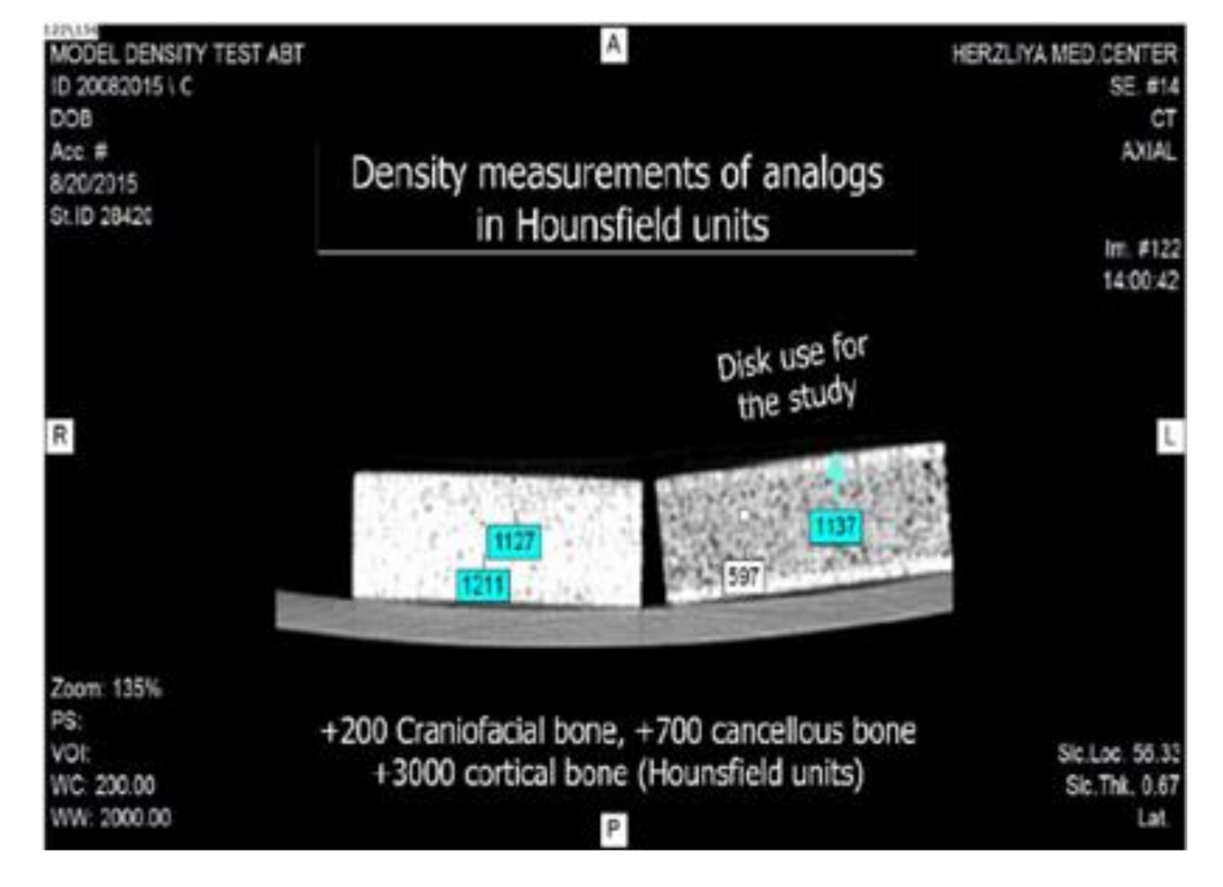
Between-group comparisons were performed with one-way ANOVA using statistical software (SPSS ver. 20.0). Statistical significance was defined by p < 0.05.

3. Results

The surface roughness analysis showed that the average surface roughness (Sa) in the control group was 2.24 ± 0.31 µm. The average Sa of the implants from the D2.8 osteotomy group was significantly lower, at 1.25 ± 0.35 µm (Table 2 and Figure 5 and Figure 6).
Similarly, the implants inserted in undersized osteotomies had significantly higher insertion torque and surface temperature values. The D2.8 group had the highest temperature and insertion torque values (44.82 ± 14.34 °C, 243.26 ± 36.77 Ncm), while the D3.65 group had the lowest values (25.7 ± 2.55 °C, 60.54 ± 12.59 Ncm) (p < 0.0001) (Figure 7 and Table 2). Among all the groups, the D2.8 group had the highest Temp-Amp value (20.83 ± 2.87 °C). Moreover, Temp-Amp in the D3.2 group (10.97 ± 1.48 °C) was significantly higher than that in the D3.65 group (Figure 8).

4. Discussion

This study examined the effects of inserting implants in vitro at high torques into undersized osteotomies on the cervical surface roughness and the implant surface temperature change during this process. It was demonstrated that the surface roughness values were significantly lower in the group of implants inserted in the smallest diameter preparation (2.8 mm) than in the other groups. The measured insertion torque values of this group were the highest. The high insertion torque associated with the implant placed in under-sized preparation stems in vivo from bone compression, which, in turn, leads to a higher friction coefficient between the bone walls and the implant [20]. Our findings on the changes in implant surface roughness following high-torque insertion are supported by those reported by Streckbein et al. [21], who observed smoothing of the surface in the region of the thread crests of the implants inserted at high torques. The flattening of the surface roughness might result clinically in a lower success rate. It has been reported that rough surfaced implants have significantly higher success rates compared to implants with more smooth surfaces [22]. Moreover, a finite element study reported that a coarse microthread profile is more favorable than a fine profile, since it reduces shear stress in the surrounding bone [23].
The high pressure associated with the high insertion torque can also affect crestal bone remodeling in the cervical area [24,25]. Aldahlawi reported significantly higher bone loss in implants placed with insertion torques higher than 55 Ncm than in implants placed with torques lower than 55 Ncm [24]. Additionally, Khayat et al. [25] reported that implants placed with insertion torques of 70.8–176 Ncm demonstrated greater bone loss than did implants placed with lower torques. A positive correlation was reported between bone density and insertion torque. Type 4 bone showed lowest insertion torque values, whereas type 1 bone showed the highest values. The peak insertion torque of implants inserted according to the manufacturer’s instruction in D1 bone reached 150 Ncm [26]. In the present study, the torque values observed in all study groups were higher than 55 Ncm. This result may be explained by a greater rigidity due to the elastic modulus of the synthetic bone being higher than that of natural bone [27]. Most likely, the rigidity of the synthetic bone contributed to the high insertion torque measured in the D3.65 group, even though the implants were inserted in sites prepared according to the manufacturer’s instructions.
Finally, the high friction associated with the insertion of implants at high torques results in excessive heat generation, which may lead to thermal injury. It has been reported that thermal injury occurs in osteoblast cell cultures that have been exposed to heat shock at 42 °C [28]. Heat-induced necrosis of the cortical bone of a rabbit occurs after 1 min of exposure to a temperature higher than 47 °C, demonstrating that damage can occur due to high insertion torques [29].
Since the average body temperature is 37 °C, 10 and 20 °C increases in temperature during the insertion of the implants in groups D3.2 and D2.8, respectively, exceed this threshold and may lead to bone necrosis [30]. Similar results were reported by Stocchero et al. [31], who demonstrated that implant insertion in undersized preparations leads to an increase in insertion torque concomitant with an increase in the surface temperature, which might surpass the threshold level above which bone necrosis occurs.
The main limitations of the current study are that nonvital synthetic bone with greater rigidity than that of vital bone was used, a single surface coating on the implants was used and the insertion of implants without a cooling solution. Additional studies with different implant coatings and roughnesses and vital bone models with the use of cooling irrigation should be conducted to confirm the findings reported above.

5. Conclusions

Within the limitations of the current study, we can conclude that the high levels of friction and resistance during implant placement in an undersized preparation increase the insertion torque, increase the implant surface temperature and flatten the implant surface roughness.
In a clinical setting, implant insertion at high torques can modify the implant surface topography and may lead to overheating of the surrounding bone and failed osseointegration. To avoid these detrimental effects, implants should be placed at the lowest torque values that ensure proper primary stability.

Author Contributions

Conceptualization, O.M., Z.O., O.C.; formal analysis, T.G., A.B., O.C.; investigation T.G., O.M.; Methodology, O.M., Z.O., Writing—original draft preparation, O.C.; writing—review and editing, O.M., Z.O., R.K. All authors have read and agreed to the published version of the manuscript.

Funding

This research received no external funding.

Acknowledgments

The authors thank Alpha Bio Tech company, Petach Tikva, Israel, for providing the materials and experimental system used in this study.

Conflicts of Interest

The authors declare no conflict of interest.

References

  1. Abrahamsson, I.; Linder, E.; Lang, N.P. Implant stability in relation to osseointegration: An experimental study in the Labrador dog. Clin. Oral Implants Res. 2009, 20, 313–318. [Google Scholar] [CrossRef]
  2. Martinez, H.; Davarpanah, M.; Missika, P.; Celletti, R.; Lazzara, R. Optimal implant stabilization in low density bone. Clin. Oral Implants Res. 2001, 12, 423–432. [Google Scholar] [CrossRef]
  3. Abuhussein, H.; Pagni, G.; Rebaudi, A.; Wang, H.L. The effect of thread pattern upon implant osseointegration. Clin. Oral Implants Res. 2010, 21, 129–136. [Google Scholar] [CrossRef] [PubMed] [Green Version]
  4. Orsini, E.; Giavaresi, G.; Trirè, A.; Ottani, V.; Salgarello, S. Dental implant thread pitch and its influence on the osseointegration process: An in vivo comparison study. Int. J. Oral Maxillofac. Implants 2012, 27, 383–392. [Google Scholar]
  5. Misch, C.E.; Strong, T.; Bidez, M.W. Scientific rationale for dental implant design. In Contemporary Implant Dentistry; Misch, C.E., Ed.; Mosby: St. Louis, MO, USA, 2008; pp. 200–229. [Google Scholar]
  6. Shalabi, M.M.; Wolke, J.G.; Jansen, J.A. The effects of implant surface roughness and surgical technique on implant fixation in an in vitro model. Clin. Oral Implants Res. 2006, 17, 172–178. [Google Scholar] [CrossRef]
  7. Campos, F.E.; Gomes, J.B.; Marin, C.; Teixeira, H.S.; Suzuki, M.; Witek, L.; Zanetta-Barbosa, D.; Coelho, P.G. Effect of drilling dimension on implant placement torque and early osseointegration stages: An experimental study in dogs. J. Oral Maxillofac. Surg. 2012, 70, e43–e50. [Google Scholar] [CrossRef]
  8. Shalabi, M.M.; Wolke, J.G.; de Ruijter, A.J.; Jansen, J.A. Histological evaluation of oral implants inserted with different surgical techniques into the trabecular bone of goats. Clin. Oral Implants Res. 2007, 18, 489–495. [Google Scholar] [CrossRef]
  9. Tabassum, A.; Meijer, G.J.; Wolke, J.G.; Jansen, J.A. Influence of the surgical technique and surface roughness on the primary stability of an implant in artificial bone with a density equivalent to maxillary bone: A laboratory study. Clin. Oral Implants Res. 2009, 20, 327–332. [Google Scholar] [CrossRef]
  10. Cohen, O.; Ormianer, Z.; Tal, H.; Rothamel, D.; Weinreb, M.; Moses, O. Differences in crestal bone-to-implant contact following an under-drilling compared to an over-drilling protocol. A study in the rabbit tibia. Clin. Oral Investig. 2016, 20, 2475–2480. [Google Scholar] [CrossRef]
  11. Scarano, A.; Carinci, F.; Lorusso, F.; Festa, F.; Bevilacqua, L.; Santos de Oliveira, P.; Maglione, M. Ultrasonic vs Drill Implant Site Preparation: Post-Operative Pain Measurement Through VAS, Swelling and Crestal Bone Remodeling: A Randomized Clinical Study. Materials 2018, 11, 2516. [Google Scholar] [CrossRef] [Green Version]
  12. Herrero-Climent, M.; Ferreira Lemos, B.; Herrero-Climent, F.; Falcao, C.; Oliveira, H.; Herrera, M.; Javier Gil, F.; Ríos-Carrasco, B.; Ríos-Santos, J.V. Influence of Implant Design and Under-Preparation of the Implant Site on Implant Primary Stability. An In Vitro Study. Int. J. Environ. Res. Public Health 2020, 17, 4436. [Google Scholar] [CrossRef]
  13. Skalak, R.; Zhao, Y. Interaction of force-fitting and surface roughness of implants. Clin. Implant Dent. Relat. Res. 2000, 2, 219–224. [Google Scholar] [CrossRef] [PubMed]
  14. Bratu, E.A.; Tandlich, M.; Shapira, L. A rough surface implant neck with microthreads reduces the amount of marginal bone loss: A prospective clinical study. Clin. Oral Implants Res. 2009, 20, 827–832. [Google Scholar] [CrossRef] [PubMed]
  15. Himmlová, L.; Dostálová, T.; Kácovský, A.; Konvicková, S. Influence of implant length and diameter on stress distribution: A finite element analysis. J. Prosthet. Dent. 2004, 91, 20–25. [Google Scholar] [CrossRef]
  16. Hansson, S. The implant neck: Smooth or provided with retention elements. A biomechanical approach. Clin. Oral Implants Res. 1999, 10, 394–405. [Google Scholar] [CrossRef]
  17. Abrahamsson, I.; Berglundh, T. Tissue characteristics at microthreaded implants: An experimental study in dogs. Clin. Implant Dent. Relat. Res. 2006, 8, 107–113. [Google Scholar] [CrossRef]
  18. Schrotenboer, J.; Tsao, Y.P.; Kinariwala, V.; Wang, H.L. Effect of microthreads and platform switching on crestal bone stress levels: A finite element analysis. J. Periodontol. 2008, 79, 2166–2172. [Google Scholar] [CrossRef] [Green Version]
  19. Hudieb, M.I.; Wakabayashi, N.; Kasugai, S. Magnitude and direction of mechanical stress at the osseointegrated interface of the microthread implant. J. Periodontol. 2011, 82, 1061–1070. [Google Scholar] [CrossRef]
  20. Trisi, P.; De Benedittis, S.; Perfetti, G.; Berardi, D. Primary stability, insertion torque and bone density of cylindric implant ad modum Branemark: Is there a relationship? An in vitro study. Clin. Oral Implants Res. 2011, 22, 567–570. [Google Scholar] [CrossRef]
  21. Streckbein, P.; Wilbrand, J.F.; Kähling, C.; Pons-Kühnemann, J.; Rehmann, P.; Wöstmann, B.; Howaldt, H.P.; Möhlhenrich, S.C. Evaluation of the surface damage of dental implants caused by different surgical protocols: An in vitro study. Int. J. Oral Maxillofac. Surg. 2019, 48, 971–981. [Google Scholar] [CrossRef]
  22. Cochran, D.L. A comparison of endosseous dental implant surfaces. J. Periodontol. 1999, 70, 1523–1539. [Google Scholar] [CrossRef]
  23. Golmohammadi, S.; Eskandari, A.; Movahhedy, M.R.; Shirmohammadi, A.; Amid, R. The effect of microthread design on magnitude and distribution of stresses in bone: A three-dimensional finite element analysis. Dent. Res. J. 2018, 15, 347–353. [Google Scholar] [CrossRef]
  24. Aldahlawi, S.; Demeter, A.; Irinakis, T. The effect of implant placement torque on crestal bone remodeling after 1 year of loading. Clin. Cosmet. Investig. Dent. 2018, 10, 203–209. [Google Scholar] [CrossRef] [Green Version]
  25. Khayat, P.G.; Arnal, H.M.; Tourbah, B.I.; Sennerby, L. Clinical outcome of dental implants placed with high insertion torques (up to 176Ncm). Clin. Implant Dent. Relat. Res. 2013, 15, 227–233. [Google Scholar] [CrossRef]
  26. Makary, C.; Rebaudi, A.; Mokbel, N.; Naaman, N. Peak insertion torque correlated to histologically and clinically evaluated bone density. Implant Dent. 2011, 20, 182–191. [Google Scholar] [CrossRef]
  27. Brown, A.D.; Walters, J.B.; Zhang, Y.X.; Saadatfar, M.; Escobedo-Diaz, J.P.; Hazell, P.J. The mechanical response of commercially available bone simulants for quasi-static and dynamic loading. J. Mech. Behav. Biomed. Mater. 2019, 90, 404–416. [Google Scholar] [CrossRef]
  28. Li, S.; Chien, S.; Brånemark, P.I. Heat shock-induced necrosis and apoptosis in osteoblasts. J. Orthop. Res. 1999, 17, 891–899. [Google Scholar] [CrossRef]
  29. Eriksson, A.R.; Albrektsson, T. Temperature threshold levels for heat-induced bone tissue injury: A vital-microscopic study in the rabbit. J. Prosthet. Dent. 1983, 50, 101–107. [Google Scholar] [CrossRef]
  30. Mishra, S.K.; Chowdhary, R. Heat generated by dental implant drills during osteotomy—A review: Heat generated by dental implant drills. J. Indian Prosthodont. Soc. 2014, 14, 131–143. [Google Scholar] [CrossRef]
  31. Stocchero, M.; Jinno, Y.; Toia, M.; Ahmad, M.; Papia, E.; Yamaguchi, S.; Becktor, J.P. Intraosseous temperature change during installation of dental implants with two different surfaces and different drilling protocols: An in vivo study in sheep. J. Clin. Med. 2019, 8, 1198. [Google Scholar] [CrossRef] [Green Version]
Figure 1. Illustration of MultiNeOTM implant.
Applsci 10 05231 g001
Figure 2. A photograph of the artificial bone.
Applsci 10 05231 g002
Figure 3. -Ray density measurement of the artificial bone expressed in Hounsfield units (HU).
Applsci 10 05231 g003
Figure 4. Testing measurement system.
Applsci 10 05231 g004
Figure 5. A topographic map of a 160 × 160 µm area at the highest point of the first cervical microthread that was obtained using Nanofocus µsurf explorer after photostimulation. The implant topography is presented on a unique color spectrum representing the range of surface height. (a) control group implant; (b) D3.65 group implant; (c) D3.2 group implant; (d) D2.8 group implant. The scan was performed with a 160 s lens using ×100 magnification.
Applsci 10 05231 g005
Figure 6. Surface roughness (Sa) values measured in the different study groups. The results are represented as averages and standard errors. * denotes p < 0.001.
Applsci 10 05231 g006
Figure 7. Insertion torque values in Ncm measured in the different study groups and expressed as averages and standard errors. * denotes p < 0.001.
Applsci 10 05231 g007
Figure 8. Implant surface temperature amplitude (Temp-Amp) for the different study groups. The amplitude was calculated by subtracting the room temperature from the measured implant surface temperature. The values are represented as averages and standard errors. * denotes p < 0.001.
Applsci 10 05231 g008
Table 1. Implant bed preparation protocols for the study groups. The values represent the drill diameters (in mm).
Drill 1 PilotDrill 2Drill 3Drill 4Drill 5
D3.652 mm2.5 mm2.8 mm3.2 mm3.65 mm final
D3.22 mm2.5 mm2.8 mm3.2 mm final
D2.82 mm2.5 mm2.8 mm final
Table 2. Average roughness, insertion torque and implant surface temperature values for the different implant groups. The values presented are averages and standard errors for each group of implants placed in implant beds of various diameters.
D2.80D3.20D3.65CONTROL
Surface roughness (Sa)1.25 ± 0.07 µm1.90 ± 0.13 µm2.08 ± 0.16 µm2.24 ± 0.06 µm
Insertion torque243.26 ± 7.36 Ncm158.40 ± 3.17 Ncm60.54 ± 2.25 Ncm
Temperature44.82 ± 2.87 °C34.97 ± 1.48 °C25.70 ± 0.51 °C

