Article

Multidisciplinary Approach to Characterize Archaeological Materials and Status of Conservation of the Roman Thermae of Reggio Calabria Site (Calabria, South Italy)

by
Michela Ricca
1,
Clara Enza Urzì
2,*,
Natalia Rovella
1,
Alessandro Sardella
3,
Alessandra Bonazza
3,
Silvestro Antonio Ruffolo
1,
Filomena De Leo
2,
Luciana Randazzo
1,
Anna Arcudi
4 and
Mauro Francesco La Russa
1
1
Department of Biology, Ecology and Earth Sciences (DiBEST), University of Calabria, 87036 Arcavacata, Rende CS, Italy
2
Department of Chemical, Biological, Pharmaceutical and Environmental Sciences, University of Messina, 98166 Messina, Italy
3
Institute of Atmospheric Sciences and Climate, National Research Council (ISAC-CNR), Via Gobetti 101, 40129 Bologna, Italy
4
Restauro e Conservazione Beni Culturali, 89131 Gallina RC, Italy
*
Author to whom correspondence should be addressed.
Appl. Sci. 2020, 10(15), 5106; https://doi.org/10.3390/app10155106
Submission received: 26 June 2020 / Revised: 12 July 2020 / Accepted: 21 July 2020 / Published: 24 July 2020

Abstract

:

Featured Application

This multidisciplinary diagnostic investigation should be considered as a fundamental and essential step that needs to be performed before any restoration and conservative intervention in the Cultural Heritage’s field.

Abstract

This multidisciplinary research focuses on diagnostic investigations to characterize the archaeological materials, as well as the alteration and degradation forms detected at the Roman Thermae of Reggio Calabria (Calabria, South Italy) site. The thermal complex, (dating I–II century B.C.), was built around three main rooms such as the caldarium (hot bath), the tepidarium (warm bath) and the frigidarium (cold bath), all connected to a central room through several entrances. The central hall still preserves a suggestive mosaic floor dating between the II and III century A.D., characterized by geometric motifs and black and white tesserae. Fragments of various archaeological stone materials, such as bricks, mortars, sedimentary, volcanic and metamorphic rocks have been studied with different and complementary techniques. Particularly, polarized optical microscopy (POM) and X-ray diffractometry (XRD) were performed to characterize the materials employed to construct the site and evaluate their state of preservation. Finally, laboratory microbiological culture analysis was conducted to identify the main microorganisms composing the biological patinas detected on the sampled materials. Results allowed us to evaluate the most suitable restoration procedures to conduct at the archaeological site, considering the different stone materials present in the studied area and their state of conservation.

1. Introduction

In the last few decades, the conservation of cultural heritage has become an increasing concern, because it includes historical construction and archaeological remains often affected by many agents and processes that endanger them [1,2,3,4,5,6,7,8,9,10,11,12,13].
Stone buildings, depending on the material they are made of and their location in a urban or rural context, especially in an outdoor environment, are particularly vulnerable to deterioration processes caused by environmental conditions (such as variability in temperature and humidity), pollution, biological colonization, lack of maintenance and neglecting.
Decay of building materials can be defined as the degradation over time of the materials’ properties (mineralogical, textural, structural, etc.), leading to their failure as construction components [14,15,16,17,18,19,20,21]. The damage process develops at the interface of materials with the environment [9,12,16,21] or at the interface of materials with other ones, also depending on other intrinsic and extrinsic factors. Intrinsic factors mainly refer to the type of stone and raw materials used, their properties, their origin processing technology, their “restoration history” (referring to prior conservation interventions) and, no less important, to their bioreceptivity [22]. This is defined as “the aptitude of a material (or any other inanimate object) to be colonised by one or several groups of living organisms without necessarily undergoing any biodeterioration” [22]. Extrinsic agents include climate factors and their variability (i.e., solar radiation, humidity, rain, wind, etc.), the prevailing microclimate, the type of the atmosphere (urban, marine, etc.), the anthropic action, the type and extension of biological factors (both macro- and micro-organisms) [23] and all their derivative and secondary products such as soluble salts, pollutants, deposits, crusts, etc. [5].
A description of degradation phenomena and related causes was conducted according to official glossaries such as the Italian UNI NORMAL [24] and the ICOMOS [25] in order to adopt a common language among the scientists and conservators dealing with the conservation of cultural heritage. Not secondary to the description of the type of alteration when biological factors are involved, a clear definition of the bioreceptivity [22] is very important to estimate the rate of colonization and what kind of intervention could be more successful. Nowadays, among conservators and scientists devoted to the preservation of cultural heritage, there is a general agreement that the diagnosis of the state of conservation of a monument can provide key information on the status of conservation of stone building materials to undertake sustainable preservation strategies [26,27,28]. Diagnosis includes a characterization of the materials as well as of their past and present decay and the main factors that could enhance a further deterioration. An overall interpretation of stone damage will lead to the most suitable procedures and protocols for a proper restoration intervention.
Starting from these considerations, the present research is addressed to the characterization of stone materials and degradation forms of the Roman Thermae of Reggio Calabria (Calabria, South Italy) site, selected as a case-study, to define the best practice for future adequate restoration work.
The site preserves the ancient remains, typical of the architecture of a Roman thermal bath (with structures such as the caldarium, frigidarium, tepidarium and a great mosaic floor) with a variability of materials ranging from sedimentary, metamorphic and volcanic rocks along with bricks and mortars [29,30,31,32]. For a complete study of the site, several analytical techniques and diagnosis investigation were employed to characterize both archaeological materials and decay forms mainly due to the biological colonization. Precisely, a polarizing optical microscope, an X-ray diffractometer, epifluorescence microscopy and biological analysis have been used.
The results achieved served as base for planning focused cleaning procedures and biocide treatments, taking into particular account the status of conservation and composition of constituent raw materials, as well as those used for previous restoration, and the consequent risk due to the periodical occurrence of biological growth. This overall approach should better respond to the demand of modern conservation strategies for archaeological sites. Particularly, conservation strategies through the use of eco-sustainable and biologically compatible products based on environment-friendly materials should be undertaken.

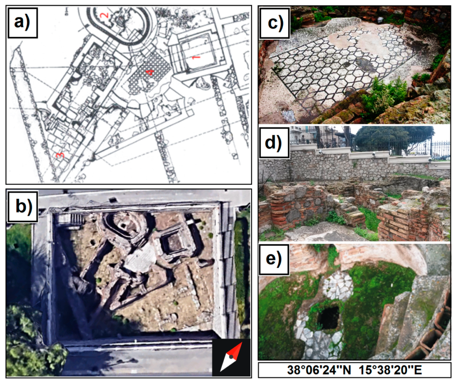
2. Archaeological Setting
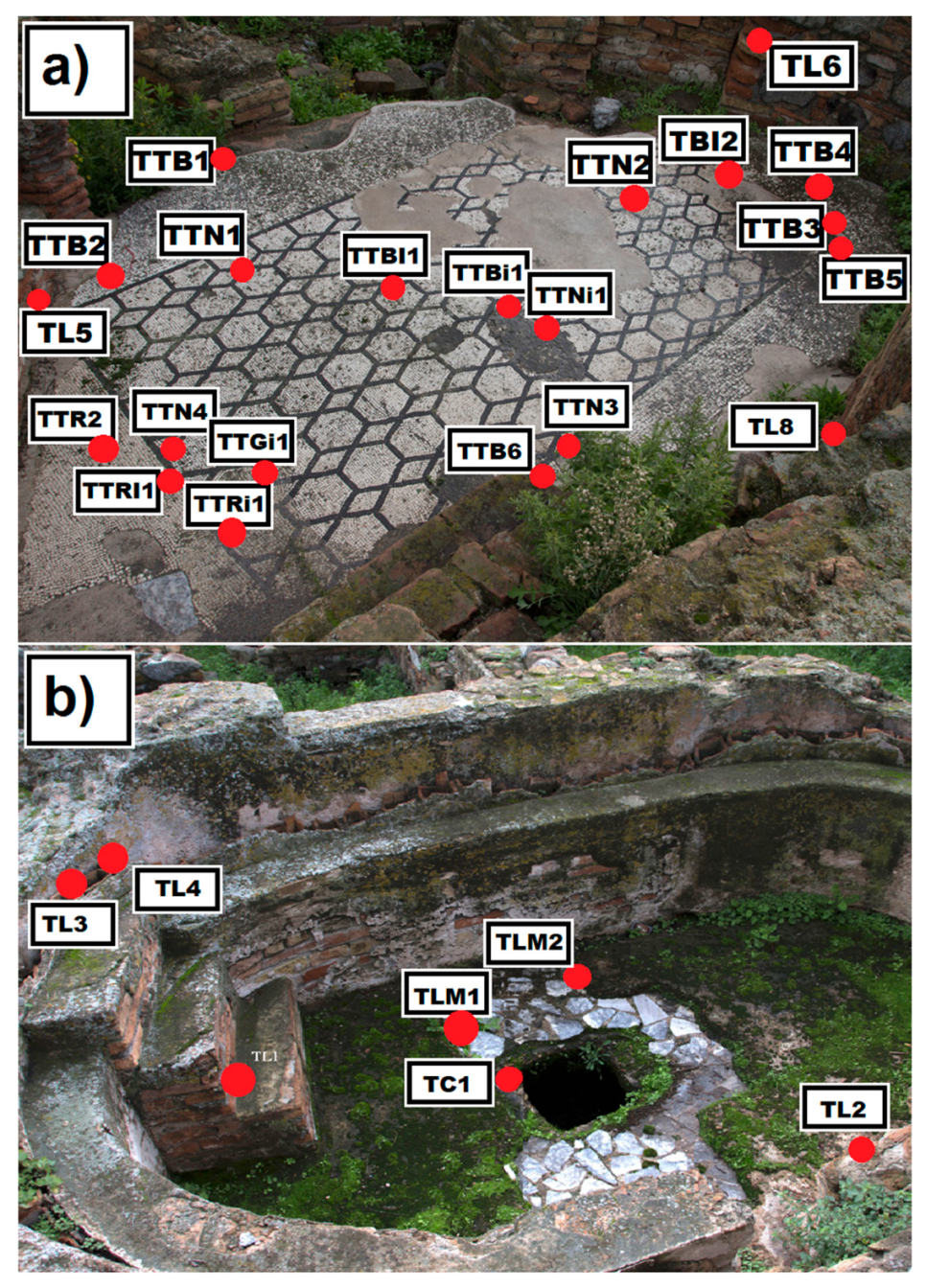
The Roman Baths of Reggio Calabria, located on the city’s seafront, came to light during the archaeological excavations carried out following the 1908 earthquake. Specifically, the first remains came to light starting from 1887, following the demolition of a medieval bastion, known as “Bastione di San Matteo” [29]. This demolition was necessary to expand the Via Marina, which became too narrow due to the construction of the railway headquarters [32].
Currently, it is possible to admire only the ruins of one of the eight thermal baths dating back to the first centuries AD (Figure 1a,b), while recent research assumes that part of the area is still below the road network, not yet excavated. The structures are arranged on a North-East/South-West axis, testifying the great example of the ancient Roman architecture to better take advantage of the sunlight exposure.
The visible part of the thermal building (Figure 1c,d) consists of a vast central area decorated with a mosaic floor with geometric motifs in black and white tesserae, which gives access to three different rooms, i.e., the caldarium (hot bath), the tepidarium (warm bath) and the frigidarium (cold bath). The central area, which also includes the mosaic, covers a space of approximately 540 cm in diameter and 340 cm at the point of maximum width, for a total area of 18.36 m2 [29,32].
As reported by the authors of [29], the frigidarium is an opus signinum, typically used by the Romans to cover structures for containing water; within the caldarium the brick tubules still show a good state of conservation; they consist of vertical elements leaning against the masonry structures used in ancient time for the circulation of hot air coming from the hypocaust (i.e., an empty space under the floor used as central heating system, conducting air heated by a furnace).
Finally, the brick pilae are still evident in the tepidarium, the warm bathroom designated for heating the human body from the walls and floor, by a hypocaust or underfloor heating system.
The construction technique of the masonry structures (i.e., walls) is in opus latericium; it defines both the semicircular rooms and the access stairs to the tanks, with evidence of bricks, mostly undamaged, showing different thicknesses. The brick walls leaning against each other, suggesting that they were built subsequently, at least twenty years later, compared to the realization of the mosaic. They were probably built during an ancient renovation of the thermal baths, also connected with the construction of two semi-circular rooms with the function of a dressing room (apodytera) and hallway, with niches obtained in the walls’ thickness, next to the bathing pools. The remaking of a large part of the edging of the central room (i.e., the one containing the mosaic) can also be dated at the same age as the masonry in the bricks. Such renovation intervention was probably carried out for compensating the damage caused to the mosaic during the construction of a second access staircase to the elliptical tanks, on the east side, and to the opening of a communication area between the caldarium and the central room.
In the past, the central area and the black and white mosaic underwent important restoration interventions due to a diffused biological patina causing swelling and detachments. Overall, the coloring of the mosaic tesserae is scarcely homogeneous; particularly, the light ones vary from milky white to pinkish beige; while, the dark ones, on a corner of the frame (south, south-west side) show a much lighter greyish color than the others.
Apart from coloring, generally, these tesserae respect the homogeneity in shapes and sizes with respect to the original installation. The reintegration areas are clearly visible also in the central portion of the mosaic and in the corner with west, north-west exposure. According to Andronico [29], these interventions were carried out in very ancient times (of unknown dating) as they faithfully reproduce the texture of the original floor. The only flaw is a clear final rolling operation, which induced in the smoothing of the floor surface, as well as the use of abundant bedding mortar with high lime content between the tesserae.
A known intervention covering the whole monumental complex is dated back to 1959 followed by another one, in 1995; the latter is more documented, thanks to archive data recovered at the Superintendence of Reggio Calabria [29]. The 1995 restoration interventions were addressed to: (a) the mechanical removal of earthy deposits and encrustations; (b) the removal of some cementitious layers, near some mosaic lacunae; (c) the application of biocidal products for the removal of biological weeds (i.e., microalgae and lichens); (d) the consolidation of decohese tesserae by mortar injections; (e) the reintegration of various lacunae by using mortar; (f) the creation of curbs for the containment of the tesserae along the external perimeter and in the areas in which major lacunae were evident (at undercut). Apart from the restoration intervention phases, there is no information and specifications about the biocides and mortars used.
From the latest surveys carried out in recent years, it has emerged that the structures of the thermal baths are affected by different degradation forms, mainly due to weeds and biological patinas (mosses, algae, lichens, etc.) and to superfetations in the mortars used in past restorations. In addition, patinas due to pollutants together with micro-cracks, swelling and abrasion phenomena are widely present on all the structures, and especially on the mosaic.
In this way, the present diagnostic approach, devoted to both archaeometric investigations and biological studies [23,28], accompanied by a biocidal treatment for cleaning purposes, can improve the management of future restoration intervention, especially in terms of choice of the best cleaning procedures and biocide treatments within the monumental complex.

3. Sampling

An extended sampling campaign of different stone materials was carried out on the remains of the archaeological site (Figure 2), covering bricks, mortars, volcanic, sedimentary and metamorphic rocks.
From a macroscopic point of view, almost all samples look rather compact and with no remarkable alteration and degradation forms, except for some of them, showing a superficial biological patina and microcracks. From a first observation with the naked eye, the biological colonization within the monumental complex is constituted mainly by a prevalence of ruderal plants between the bricks, lichens crusts and greenish-black patina. With particular reference to the mosaic, the biological colonization is especially accentuated along the external frame of the mosaic floor and where the tesserae are affected by microcracks. The heaviest colonization, however, was observed in correspondence to one cemented area (Figure 2 and Figure 3, sampled area MRC4) due to previous (but not well documented) restoration intervention.
A removal of exceeding biomass followed by a treatment with 2% of Rocima™ 103 (Rohm and Haas) (e.g., sampled areas MRC2 and MRC5) was performed one month before the biological sampling.

3.1. Archaeological Stone Materials

For the characterization of materials and the status of conservation, twenty-eight samples were selected for the study and chipped from different archaeological remains (i.e., walls, mosaics and floor coverings) using suitable stainless steel tools, such as lancets and small chisels. A list of the examined samples, of archaeological interest, along with their location within the site and a brief description, is summarized in Table 1.

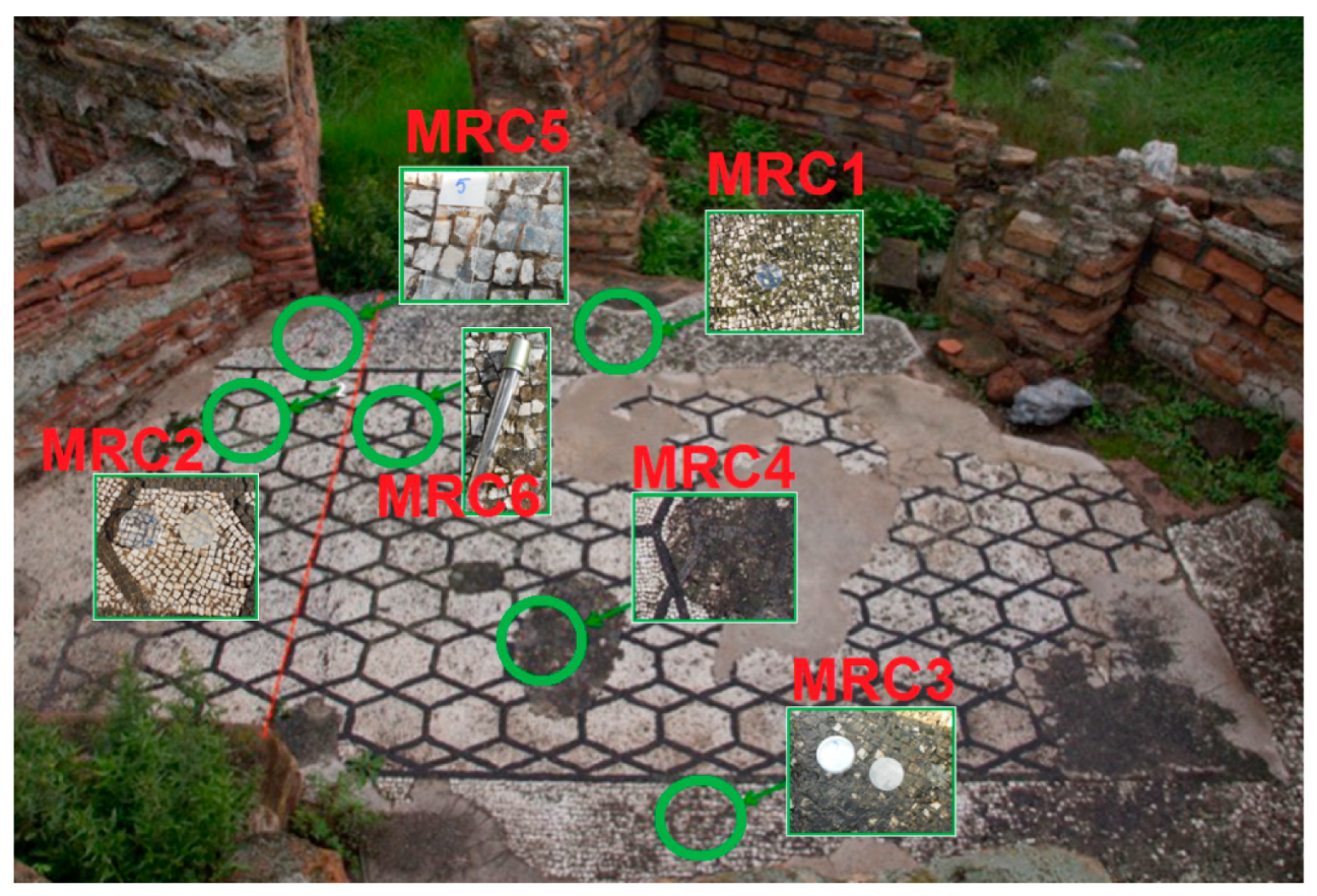
3.2. Microbiological Sampling

Microbiological analyses were focused only on the mosaic. Six samples (named MRC1 to MRC6) were taken from the mosaic area by using two non-invasive techniques aiming to obtain evidence through microscopic and qualitative cultural analysis of the microorganisms involved in the patina formation of mosaic surfaces. For this purpose, the adhesive strips technique as described by [33] and the technique of damp filter paper were applied. Samples were taken in correspondence to areas shown in Figure 3 and reported in Table 2.
MRC1-4 samples were taken by using a sterile filter paper, wet with about 800 µl of ultrapure H2O, to facilitate the algae detachment, and in parallel, adhesive tape strips were used.
For MRC5 and MRC6, sampling was performed only with adhesive tape (DID, Milan).
A list of the examined samples, of biological interest, along with their location within the site and a brief description, is summarized in Figure 3 and in Table 2.

4. Analytical Methods and Laboratory Procedures

4.1. Archaeometric and Diagnostic Analysis

Archaeometric and diagnostic investigations of the selected twenty-eight samples, including the mineralogical–petrographic characterization, were carried out on representative portions of the samples of ca. 0.5 × 1.0 × 1.0 cm.
Specifically, the analytical techniques applied for the material characterization include polarizing optical microscopy (POM) to define the mineralogical and textural features of the examined samples. Thin section observations were carried out through an Axioskope 40 (Axiolab Zeiss) microscope. Superficial patinas due to weathering forms were also detected.
X-ray Diffractometry (XRD) was used to identify the mineralogical phases constituting the samples. Measurements were performed using a Siemens D5000 diffractometer and spectra were taken in the range 5–65° 2Θ, using a step-size of 0.02° 2Θ and a step-time of 2 s/step.

4.2. Microbiological Analysis

Samples were processed by observing them under a microscope, followed by microbial cultivation. In particular, each adhesive tape was divided into small pieces (0.25 cm2) and used either for microscope observations or for cultural analysis.
A detailed observation under an epifluorescent microscope (EFM) Leica DMRE was performed by placing one piece on a glass slide after the deposition of a drop of diluted 1:1 in distilled water and Acridine Orange (AO) fluorescent staining (0.01 mg/mL). For cultural analysis, another piece was streaked (in double for each piece) on the surfaces of the agarized medium Dichlorane Rose Bengal Chloramphenicol, DRBC (Oxoid), for fungi. For the samples taken with a sterile filter paper (60 mm Ø), these were divided into two portions, half of which inoculated on Petri dishes containing agarized Blue Green Medium, BG11, for algae and cyanobacteria, the other half in flasks containing 30 mL of liquid BG11.
All Petri dishes containing DRBC were incubated in the dark at 25 °C for one month. Samples inoculated in BG11 (liquid and solid) were incubated in daylight and at room temperature (22 °C) for 2–3 months.
Phototrophic microorganisms were discriminated at group level (either eukaryotic algae or prokaryotic cyanobacteria) for their size and morphology, under light and epifluorescence microscopy Leica DMRE.
Fungi were identified either by using the diagnostic keys of Pitt [34], de Hoog [35], Samson et al. [36] and Domsch [37], or by molecular techniques for black fungi as described in detail by Ruffolo [7]. In brief, DNA was extracted and PCR PCR amplified with the universal primers ITS1/ITS4 which are specific for fungal interspacers ITS1, ITS2 and 5.8S rRNA genes. The reaction mixture contained 0.48 mM of each primer, 25 mL of MyTaq™ Mix 2x (Boline, London, UK) and 2 mL of template in the total reaction volume of 50 mL. PCR was performed with the following thermocycling program: 5 min denaturation at 95 °C, followed by 35 cycles of 1 min at 94 °C, 1 min at 55 °C and 1 min and 30 s at 72 °C, and a final extension was run at 72 °C for 10 min in the thermocycler TPersonal (Biometra, Germany). The PCR products were visualized on 1.5% agarose gel stained with ethidium bromide. Purification and sequencing of PCR products were performed by a commercial facility (Biofab, Rome, Italy). The closest relatives of isolates were determined by comparison with rDNA gene sequences in the NCBI GenBank by BLAST search (http://blast.ncbi.nlm.nih.gov/Blast.cgi).
Cultures allowed us to characterize the main groups of microorganisms involved in the formation of biological patina observed and then later were used to test the effectiveness of some biocides to be applied to slow down the biological colonization.

5. Results and Discussion

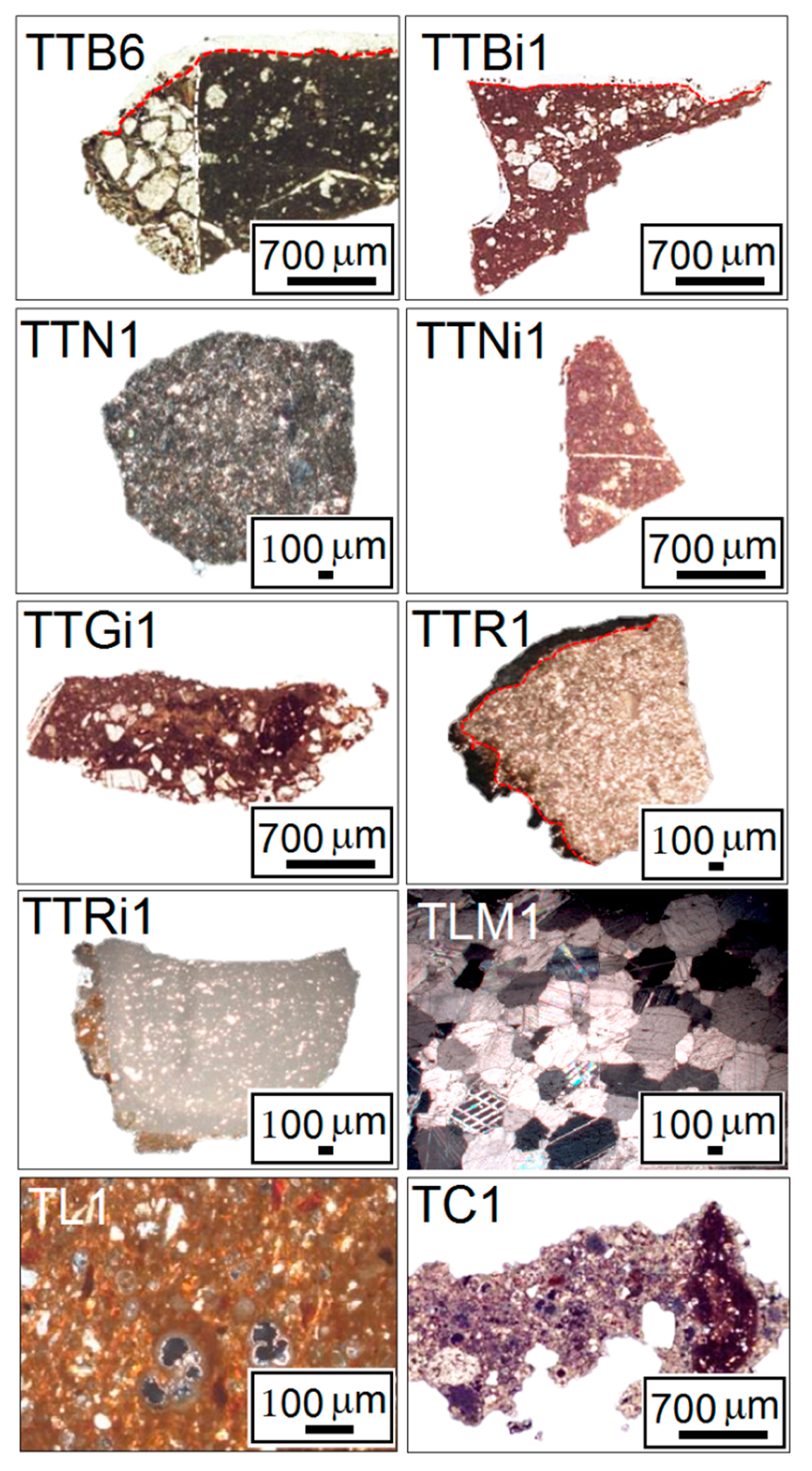
5.1. Archaeometric and Diagnostic Analysis

Petrographic analysis by POM was carried out to define the textural and compositional characteristics of all examined samples. Descriptions were made by grouping the samples (i.e., 10 groups) depending on the stone material type. Some representative photomicrographs are shown in Figure 4, while Table 3 synthesizes the main features of each sample. Regarding the mosaic’s tesserae, analyses permitted to characterize materials considering their originality within the mosaic, since some sampled tesserae are attributable to previous restoration intervention.
I group
On the basis of textural features, samples belonging to the first group, which includes original white mosaic’s tesserae (i.e., samples TTB1, TTB2, TTB3, TTB4, TTB5 and TTB6) can be classified such as biomicrites, according to Folk (1959) [38], or wackestones, according to Dunham (1962) [39]. They show a mud-supported texture (which contains more than 10% grains), where the allochemical component is mainly made up of bioclasts (25–30%) (mostly microforaminifera). Dissolution phenomena and recrystallization of secondary calcite occur in some fragments. Overall, tiles are compact, poorly degraded, with both primary and secondary porosity not exceeding 3%. Residues of the bedding mortar, mostly made up of quartz crystals with sub-angular to sub-rounded shapes, along with plagioclase and mica are also visible. Finally, regarding their state of conservation, samples show a thin and brownish superficial alteration layer with an average thickness of about 200 µm (Figure 4).
II group
The second group includes the samples TTBi1, TTBI1 and TTBI2, which are part of the replacement white mosaic’s tesserae. As for TTBi1, the fragment can be classified such as biomicrite, according to Folk (1959) [38], or wackestone, according to Dunham (1962) [39] where fossilized planktonic foraminifera (i.e., globigerinae and globotruncanae) and bioclasts represent the dominant allochemical components. Precipitation of secondary calcite within in bioclasts is also visible. The sample appears devoid of fractures and the porosity does not exceed 3%. Additionally, TTBi1 displays a thin and brownish superficial alteration layer with an average thickness of up to 100 µm. Regarding TTBI1 and TTBI2, both tesserae are carbonatic rocks with a grain-supported fabric, interstitial matrix and the presence of intraclasts, classifiable as packstones according to Dunham classification (1962) [39] and intrasparites according to the Folk one (1959) [38]. Samples show a relatively altered matrix characterized by highly altered plagioclase and quartz crystals. Recrystallization phenomena of secondary calcite grains in areas subject to dissolution are also common.
III group
The third group includes four original black mosaic’s tesserae (i.e., samples TTN1, TTN2, TTN3 and TTN4). These samples can be classified such as effusive igneous rocks with a porphyritic structure and a fine-grained groundmass ranging from crypto to microcrystalline. As phenocrysts, feldspars and plagioclase were mostly recognized, followed by pyroxenes and opaque mineral oxides. Overall, these tesserae are in a good state of preservation. No superficial alteration layers and patinas were detected.
IV group
The fourth group includes only the TTNi1 sample, as a replacement black mosaic’s tessera. It is a calcarenite and it can be classified such as wackestone, according to Dunham (1962) [39] and biomicrite, according to Folk (1959) [38]. The allochemical component consists mainly of planktonic microforaminifera (i.e., globigerinae and globotruncanae). Recrystallization phenomena of secondary calcite occur in bioclasts; also, both primary and secondary porosity (not exceeding 3%) is visible. Additionally, a thin superficial patina was detected (up to 100 µm).
V group
The fifth group includes a replacement grey mosaic’s tessera, initialled as TTGi1. From observation under the optical microscope, this fragment consists of two portions of material with distinct fabrics and compositions (Figure 4). The portion with greater thickness can be classified as wackestone, according to Dunham (1962) [39], where the main allochemical component is made up of planktonic microforaminifera and other bioclasts (biomicrite, according to Folk (1959) [38]. Dissolution phenomena and recrystallization of secondary calcite occur within some microfossils and microfractures. Overall, the sample shows a porosity not exceeding 3%. Additionally, samples show thin superficial layers with thicknesses of up to 100 µm. The less thick portion was a bedding mortar characterized by a slightly altered calcitic binder, from crypto to microcrystalline. As for the aggregate fraction, quartz, plagioclase and muscovite crystals, with sub-angular to sub-rounded shapes, were recognized. A fragment of subangular cocciopesto with a diameter of about 1 mm is also observed. Finally, both primary and secondary porosity mainly due to the binder alteration is visible.
VI group
The sixth group includes original pinkish mosaic’s tesserae, initialled as TTR1 and TTR2. Both tesserae show a superficial alteration layer with thickness up to 500 µm. In detail, TTR1 can be classified as Pelsparite, according to Folk (1959) [38], or packstone, according to Dunham (1962) [39]. It is rather compact, slightly altered, with primary and secondary porosity less than 3%. On the contrary, the TTR2 sample, which is slightly altered, can be classified as packstone, according to Dunham (1962) [39], or intrasparite according to Folk (1959) [38]. Quartz crystals were recognized along with dissolution phenomena and recrystallization of secondary calcite. Additionally, both tesserae show superficial layers with thicknesses of up to 500 µm.
VII group
The seventh group includes only a replacement pinkish mosaic’s tesserae, initialled as TTRi1. The specimen is a calcarenite, classifiable as biomicrite, according to Folk (1959) [38], or mudstone, according to Dunham (1962) [39]. The allochemical component is mainly made up of 15–20% of bioclasts (e.g., benthic foraminifers). The sample appears compact, with both primary and secondary porosity less than 3%. A superficial patina with a thickness of up to 350 µm is also present.
VIII group
The eighth group includes the TLM1 and TLM2 samples belonging to floor slabs. They are veined calcitic white marbles, characterized by a medium grain size (1–3 mm) and a heteroblastic texture. No shape preferred orientation (SPO) was observed, and crystals show curved and straight grain boundaries (GBS), occasionally dentate. A total of 120° triple junctions and traces of cleavage were also detected [40]. Accessory minerals such as quartz, in tiny crystals, were also recognized. No alteration forms were recognized.
IX group
The ninth group includes eight brick samples taken from masonry structures (i.e., TL1, TL2, TL3, TL4, TL5, TL6 and TL8). The observations were performed following the descriptive scheme proposed by Whitbread [41]. All fragments show a superficial alteration layer with thicknesses of up to 500 µm. The paste, in general, is rather homogeneous, with coloring, at crossed nicols, ranging from orange-reddish to dark brown, with the exception of the TL6 sample which is rather inhomogeneous with remarkable yellowish and dark red areas. As inclusions, single quartz, plagioclase, pyroxenes and mica crystals were recognized together with some rock fragments. The specimens are also rich in opaque minerals and microforaminifera fossils. The latter are subject to both dissolution and recrystallization phenomena. The optical activity is high in all samples, while isorientations are absent. The inclusions show subangular shapes, with low to medium sphericity, and a spatial distribution that varies from single-space to double-space. Additionally, they display a medium clustering and a bimodal particle size distribution. The coarse fraction is the most abundant and is characterized by fragments of metamorphic rocks, with dimensions between 500 and 2500 µm, followed by single crystals, which do not exceed 200 µm. As for the porosity, it varies from about 5% (in samples TL1, TL4, TL5 and TL6) up to 15% (in samples TL2, TL3 and TL8). According to Whitbread (1995), the voids can be classified as holes, irregular in shape and vesicles, with spherical shapes. Their dimensions also vary from meso- (0.05–0.5 mm) to macro- (0.5–2.0 mm), often showing secondary calcite recrystallization phenomena, along the edges. From a semi-quantitative visual estimation, the coarse/fine/voids ratio (c/f/v) is about 45/50/5 in TL1, TL4 and TL5; 40/45/15 in TL2 and TL3; 35/60/5 in TL6 and 30/55/15 in TL8. Amorphous concentration features (Acf), consisting of pure nodules with sizes between 0.5 and 2.5 mm, and impregnant portions, were also recognized.
X group
The tenth group includes only the TC1 sample, taken from a floor. The sample is a mortar characterized by a micritic binder with variable coloring from yellowish-brown to grey-brown (at parallel nicols). In general, the structure is heterogeneous with the presence of lime lumps with sub-rounded shapes, both grey-brown and whiteish (at crossed nicols), with a sparitic texture, consisting mainly of calcite. The structure of the calcite crystals is so well preserved as to suggest that the lumps may refer to a firing relict of a marble. The aggregate is poorly sorted and moderately clustered. The clasts show a poorly homogeneous distribution and are mainly constituted by quartz and plagioclase subangular in shapes; some of them appear dissolved with remarkable reactions occurring at the edges. Quartz is also present as a polycrystalline one, attributable to a metamorphic origin. Some plagioclase crystals are so heavily altered that the original mineral phase has been replaced by secondary minerals (sericitization phenomenon). Furthermore, millimetric fragments (cocciopesto) with a reddish-brown color (at crossed nicols) which incorporate quartz, mica, bioclasts and planktonic microforaminifera are present. As regards the porosity, it is less than 10% and includes both primary voids, with sizes between 80 and 150 µm, and secondary ones; the latter is probably due to the micro-fractures of the binder.
Based on observation by POM, final remarks are the following:
The six samples belonging to the first group (biomicrites/wackestones), which includes original white mosaic’s tesserae, display a good state of conservation, except for the presence of a very thin patina (up to 200 µm) present on the samples’ surfaces. There are no microcracks, this is probably due to both the compactness of the materials and the very low porosity, which never exceeds 3%. The low porosity and the absence of microfractures have probably limited the growth of the patina inside the materials.
The samples belonging to the second group includesthree replacement white mosaic’s tesserae. Particularly, TTBi1 (biomicrite/wackestone), of the same nature as the samples of the first group, appears in good condition, devoid of fractures and with a low porosity, which does not exceed 3%. Additionally, with respect to the samples from the first group, TTBi1 exhibits a thinner surface patina, up to 80 µm. The lower thickness of the patina could be due to the fact that the tessera, replaced in a past restoration intervention, was exposed for less time to the damage agents that favor its formation. As for the TTBI1 and TTBI2 (packstones/intrasparites), no surface layers and patina were observed, but only alteration within the matrix and intraclasts, along with recrystallization phenomena of secondary calcite grains in areas subject to dissolution.
The four original black mosaic’s tesserae belonging to the third group (effusive rocks) are well preserved, probably due to the high degree of compactness, which is typical in igneous rocks, therefore limiting the development of alteration forms. Additionally, no superficial patinas nor alterations in the material’ structure and groundmass were detected.
The fourth (TTNi1) and fifth (TTGi1) groups both include a replacement dark mosaic’s tesserae. They are both biomicrites/wackestones which differ mainly in color; TTNi1 is black, while TTGi1 is greyish. Additionally, TTGi1 present a higher percentage of allochemical components than TTNi1. Both show a porosity not exceeding 3%, with dissolution phenomena and recrystallization of secondary calcite within some bioclasts. As for the alteration forms, both tesserae display only the presence of a surface patina up to 100 µm thick, and not microfractures. Finally, in TTGi1, residuals of the bedding mortar having cocciopesto were recognized. The presence of cocciopesto suggests that during one of the ancient restorations, hydraulic mortars were applied to faithfully reproduce the ancient Roman recipes, as also testified in Andronico et al. [29].
The sixth (TTR1 and TTR2) and seventh group (TTRi1) include tesserae with a light pinkish colour. However, TTR1 and TTR2 are from the original installation while TTRi1 is a replacement tessera. TTR1 is a rather compact pelsparite/packstone with primary and secondary porosity less than 3%, while TTR2 is an intrasparite/packstone showing dissolution phenomena and recrystallization of secondary calcite. As for alteration phenomena, both tesserae display superficial layers with thicknesses of up to 500 µm. On the contrary, TTRi1 is a biomicrite/mudstone with both primary and secondary porosity (less than 3%) and a superficial patina up to 300 µm in thickness. The lower thickness of the superficial patina in TTRi1 with respect to TTR1 and TTR2, may depend on the fact that such is a replacement tessera, exposed for less time to the weathering forms causing alteration phenomena.
The two fragments of the eighth group are veined white marbles, from floor slabs of the caldarium, that are in a good state of preservation, devoid of surface alteration and microfractures. The high compactness of the material but probably also the less exposed location (caldarium) with respect to the mosaic (central area), has prevented or at least limited the development of damage forms. In detail, the marbles are very resistant metamorphic rocks, with a very low porosity (maximum 2%) which barely undergo remarkable degradation forms (i.e., fractures, erosion, etc.), except in certain and extreme environmental conditions which are not the case with the site being studied.
The ninth group includes eight brick fragments taken from masonry structures within the caldarium. They show very similar properties, both in the pastes and in the microstructures, with porosity ranging from 5% to 15%. Additionally, all fragments show a superficial patina with a thickness up to 500 µm. Their thicknesses are very thin compared to those found in specimens sampled in the central compartment with the mosaic. The good state of conservation of brick masonry structures could depend on several factors: (a) the type of raw materials used, that conferred high resistance, limiting the alteration phenomena; (b) the different exposure to the weathering forms compared to the central area with the mosaic; (c) their subsequent construction (as testified by the literature) compared to the realization of the mosaic, which probably occurred during an ancient renovation of the thermal baths.
The ninth group includes eight brick fragments taken from masonry structures within the caldarium. They show very similar properties, both in the pastes and in the microstructures, with porosity ranging from 5% and 15%. Additionally, all fragments show a superficial patina with thickness up to 500 µm.
The tenth group includes a mortar sample (TC1) from a floor with a sparitic texture, consisting mainly of calcite and with evidence of lumps. Calcite grains are so well preserved as to suggest that the lumps may refer to a firing relict of a marble. Fragments of cocciopesto suggest that it is an ancient mortar [3] and not a remake due to previous restoration work.

5.2. Characterization of Archaeological Stone Materials by XRD

X-ray diffraction (XRD) measurements carried out on all the samples, revealed the presence of several mineralogical phases according to the different materials sampled, as summarized in Table 3. As regards mosaic tesserae, the data show that (a) the white and pinkish tesserae, both original (TTB1; TTB2; TTB3; TTB4; TTB5; TTB6; TTR1 and TTR2) and used for restorations (TTRi1; TBI1 and TBI2), are sedimentary rocks with a predominant calcitic component, sometimes with accessory phases such as quartz and albite; (b) the black tesserae, belonging to the original mosaic (TTN1; TTN2; TTN3 and TTN4), have the typical mineralogical composition of volcanic rocks, with a predominance of feldspars (Sanidine; Orthoclase); amphibole (Hornblende); pyroxene (Clinopiroxene); plagioclase (Albite); followed by Calcite and Forsterite; (c) the black and dark greyish tiles used during the restoration (TTGi1 and TTNi1) are also sedimentary rocks, thus attesting to the use of a different material from the original one.
Regarding the bricks, sampled both from the caldarium and in adjacent masonry structures, mineralogical analyses reveal a similar mineralogical composition in all the samples, characterized by the predominant presence of calcite, quartz and feldspars. Additionally, the detection of neoformation mineralogical phases such as Diopside and Gehlenite, only in samples (i.e., TL1; TL3 and TL8), suggest their higher firing temperatures (generally > 850 °C) [3,41,42,43].
As regards the two veined white marbles taken from the caldarium floor, respectively, TLM1 and TLM2, they display a calcitic composition with traces of quartz.
Finally, the analysis carried out on the only cementitious sample (i.e., TC1) revealed that it was mainly characterized by dominant calcite and quartz, followed by lower intensity diffraction peaks of feldspars.

5.3. Microbiological Analysis

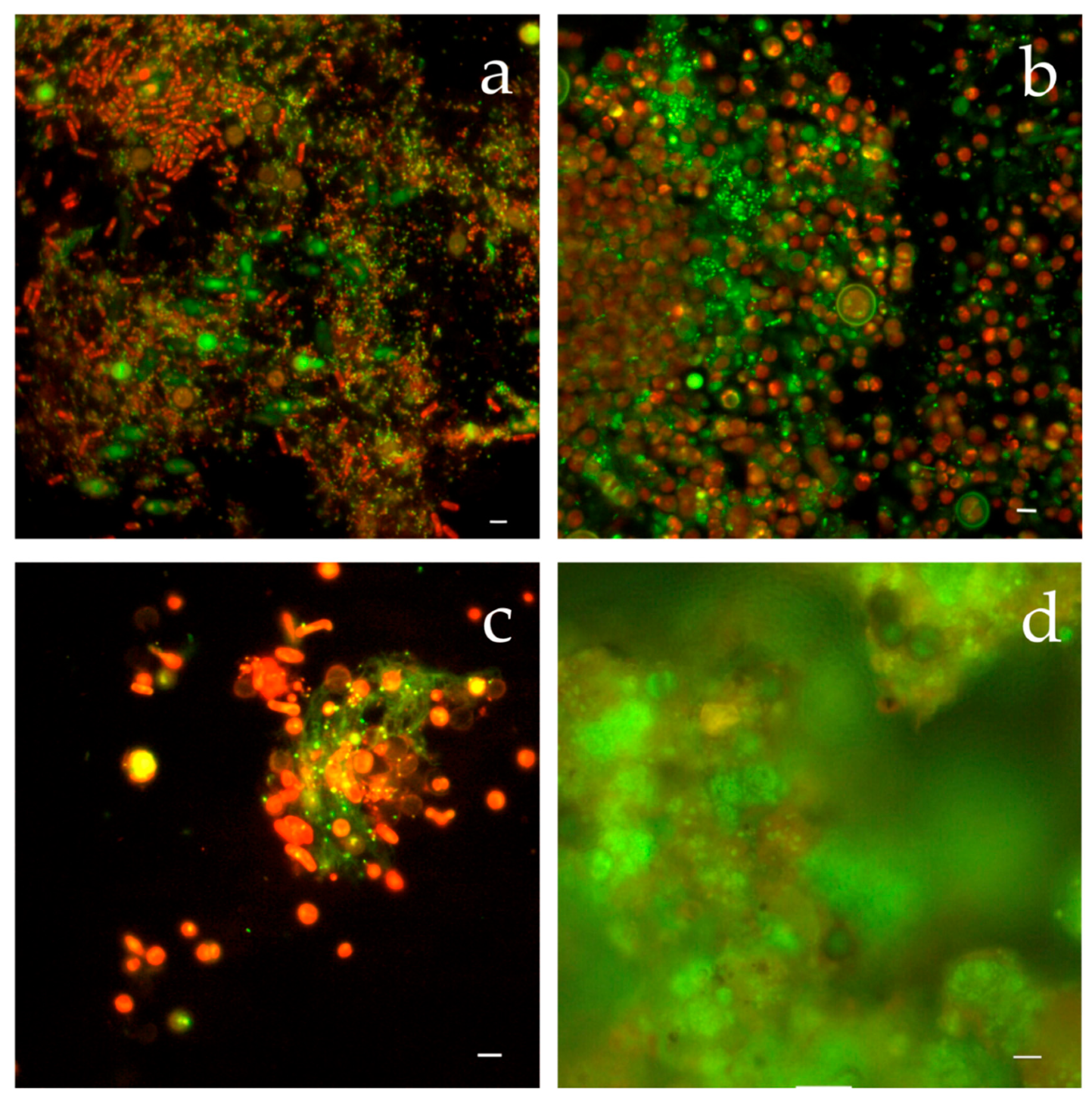
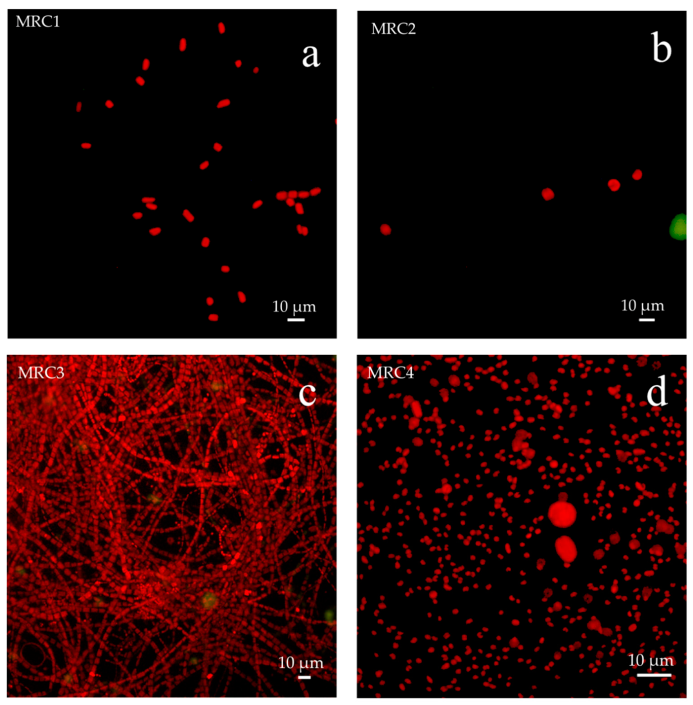
5.3.1. Epifluorescence Microscope Observations

Observations of untreated samples (MRC1, MRC3, MRC4 and MRC6) under epifluorescence microscope revealed the presence of various microorganisms as shown in Figure 5; while samples pre-treated with 2% of biocide Rocima™ 103 (MRC2, and MRC5) showed a scarce or none presence of microorganisms.

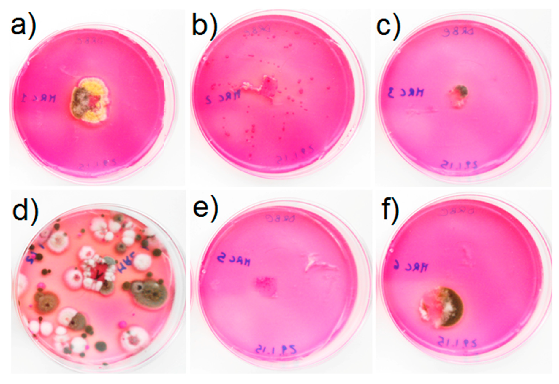
5.3.2. Cultural Analysis

Culture techniques confirmed what was seen under microscopy. In fact, fungi and photosynthetic microorganisms were the main microorganisms isolated and involved in the biological patinas observed on the tesserae surfaces. Results are summarized in Figure 6, Figure 7 and Figure 8 and Table 4 and Table 5. Fungi were isolated from all samples both from the adhesive tape streaked on DRBC (with the exception of the treated samples MRC2 and MRC5) (Figure 7) and from the paper disc inoculated in BG11 liquid medium (except sample MRC2) (Figure 6 and Figure 8).
Fungal growth was observed in the DRBC plates mainly in strict contact with the adhesive tape; only sample MRC4 showed abundant and composite fungal growth (Figure 7). Black fungi were isolated with the technique of a wet paper disc placed in BG11 liquid medium (Figure 6 and Table 5).
Algae and/or cyanobacteria were isolated from samples MRC1, MRC3 and MRC4 as shown in Figure 6. In addition, a slight growth was visible after three months for the disc sample MRC2 inoculated in BG11. No growth was observed for MRC5 and MRC6. Considering the abundant evidence of photosynthetic species in the MRC1-4 samples, an aliquot of culture grown in the flasks was taken and re-isolated in BG11 agar plates, and the type of photosynthetic microorganisms were observed under an EFM microscope as shown in Figure 8.
In general, the mosaic was affected by different alteration forms, caused by a massive colonization of several microbial populations varying in relation to the sampling areas and to the bioreceptivity of the material, also considering portions that underwent previous restoration interventions.
In particular, the MRC4 sample showed a greater and massive colonization than the other 5 specimens (MRC1–3, 5–6), displaying that mortar, as also clearly visible to the naked eye (Figure 3), had the higher bioreceptivity, allowing the colonization of both fungi and photosynthetic organisms (Table 4 and Table 5). According to literature [29], the area in which MRC4 was sampled, underwent previous restoration works. The thesis was further confirmed by POM investigations, showing that in the same area the mosaic tesserae were made of different stone materials (i.e., sedimentary and volcanic rocks), some of which were replaced during the restoration intervention.
Additionally, some of the tesserae sampled in the same area of sample MRC4 (Figure 2, such as the limestone ones) showed numerous micro-cracks, becoming suitable for the colonization of endolithic microorganisms.
In the portions of the mosaic where the Rocima™ 103 biocide was used (one month before the microbiological sampling in correspondence to samples MRC2 and MRC5) (Figure 3), a good reduction in the microbial biomass was observed, although it has not been completely removed. For this reason, cultural analysis showed a recolonization even of scarce entity. In fact, single-celled algae and numerous bacteria have been isolated (Table 4 and Table 5). The results achieved, thus, demonstrate that on one hand, the treatment with 2% of Rocima™ 103 was quite effective to eliminate the actual colonization, but on the other hand does not have long-term efficacy, allowing the recolonization of the mosaic tesserae.

6. Conclusions

The results achieved in this work improved the knowledge of the monumental complex, in terms of materials used in the various eras and of alteration and degradation forms, mainly due to the biological colonization.
The study carried out on the materials of the Roman Thermae of Reggio Calabria detected the variability in the alteration and degradation forms, mainly in relation to the types of material, their bioreceptivity and of the restoration interventions carried out in past times within the monumental complex.
The archaeometric and diagnostic study allowed to define the following:
  • Overall, all the sampled stone materials show a good state of conservation from a textural point of view, except for the presence of a superficial patina.
  • According to the different types of materials identified, the sedimentary samples (i.e., white, pinkish and greyish tesserae), coming from the central area of the thermal bath having a mosaic floor, show more marked alteration forms compared to the tesserae of volcanic origin (i.e., black). Unlike sedimentary samples, volcanic ones do not have superficial alteration layers, such as patinas.
  • By comparing the light colored and greyish tesserae and all sedimentary rocks, it is possible to detect some differences in terms of alteration forms, mainly attributable to biological colonization. Many of these are replacement tesserae and show superficial patinas with lower thicknesses than the original ones.
  • In some replacement tesserae, the bedding mortar is much more degraded than that found in some original tesserae.
  • As for the samples taken in the caldarium room, including both brick, marble and mortar, they show an overall good state of conservation with slighter superficial patinas compared to those found in specimens sampled in the central compartment with mosaic. Additionally, the mortar is intact, well preserved and rich in fragments of cocciopesto that surely increased its resistance and limited the degradation.
  • Biological colonization resulted to be one of the main problems causing esthetical (coverage of surfaces), mechanical (endolithic colonization) and chemical deterioration patina formation and alteration layers ranging from 200 to 500 µm depending on the nature of the colonized material. In fact, volcanic rock (dark tesserae) is less affected by biological attack, followed by compact sedimentary calcareous stone, while those stones with cracks and fissurations are affected not only by a biological patina on the surface but also by a colonization of endolithic microorganisms, mainly black fungi.
The research represents a further milestone to better manage future restoration intervention, especially regarding the choice of the best cleaning procedures to carry out within the archaeological site. Suitable conservative procedures will allow protecting the stone materials against the degradation phenomena mainly related to the biological colonization, type of stones and exposure in the site.
From this study, the need for periodical intervention (at least one before the starting of the vegetative period) with the use of classical biocide (e.g., Rocima™ 103) for the removal of already grown biomass, followed by the application of products that can slow down the regrowth, is clear.

Author Contributions

Conceptualization, M.F.L.R.; methodology, M.F.L.R., M.R., C.E.U., F.D.L.; formal analysis, M.R., N.R., S.A.R.; investigation, M.R., N.R., S.A.R., A.S., A.B., C.E.U., F.D.L., A.A., L.R.; resources, M.F.L.R.; data curation, M.F.L.R., M.R.; writing—original draft preparation, M.F.L.R., M.R., N.R.; writing—review and editing, M.F.L.R., M.R., R.N., C.E.U., A.B.; supervision, M.F.L.R.; project administration, M.F.L.R.; funding acquisition, M.F.L.R. All authors have read and agreed to the published version of the manuscript.

Funding

This research is part of the POR Calabria FESR project “NANOPROTECH” (NANO PROtection Technology for Cultural Heritage) (J24E07000380005).

Conflicts of Interest

The authors declare no conflict of interest.

References

  1. Alberghina, M.F.; Casanova Municchia, A.; Germinario, G.; Macchia, A.; Matteini, M.; Milazzo, G.; Ruffolo, S.A.; Sabbatini, L.; Schiavone, S.; Sodo, A.; et al. A multi-analitycal approach to adress a sustanaible conservation of the main marble portal of the Monreale Cathedral. Int. J. Conserv. Sci. 2020, 11, 353–362. [Google Scholar]
  2. Randazzo, L.; Collina, M.; Ricca, M.; Barbieri, L.; Bruno, F.; Arcudi, A.; La Russa, M.F. Damage Indices and Photogrammetry for Decay Assessment of Stone-Built Cultural Heritage: The Case Study of The San Domenico Church Main Entrance Portal (South Calabria, Italy). Sustainability 2020, 12, 5198. [Google Scholar] [CrossRef]
  3. Ricca, M.; Galli, G.; Ruffolo, S.A.; Sacco, A.; Aquino, M.; La Russa, M.F. An archaeometric approach of historical mortars taken from Foligno City (Umbria, Italy): News insight of Roman Empire in Italy. Archaeol. Anthrop. Sci. 2019, 11, 2649–2657. [Google Scholar] [CrossRef]
  4. Ricca, M.; Le Pera, E.; Licchelli, M.; Macchia, A.; Malagodi, M.; Randazzo, L.; Rovella, N.; Ruffolo, S.A.; Weththimuni, M.L.; La Russa, M.F. The CRATI project: New insights on the consolidation of salt weathered stone and the case study of San Domenico Church in Cosenza (South Calabria, Italy). Coatings 2019, 9, 330. [Google Scholar] [CrossRef] [Green Version]
  5. La Russa, M.F.; Ricca, M.; Cerioni, A.M.; Chilosi, M.G.; Comite, V.; De Santis, M.; Rovella, N.; Ruffolo, S.A. The colors of the Fontana di Trevi: An analytical approach. Int. J. Archit. Herit. 2018, 12, 1. [Google Scholar] [CrossRef]
  6. Rovella, N.; Arcudi, A.; Crupi, V.; La Russa, M.F.; Majolino, D.; Osanna, M.; Pace, R.; Ruffolo, S.A.; Ricca, M.; Ruggieri, N.; et al. Tituli Picti in the archaeological site of Pompeii: Diagnostic analysis and conservation strategies. Eur. Phys. J. Plus 2018, 133, 539. [Google Scholar] [CrossRef]
  7. Ruffolo, S.A.; La Russa, M.F.; Ricca, M.; Belfiore, C.M.; Macchia, A.; Comite, V.; Pezzino, A.; Crisci, G.M. New insights on the consolidation of salt weathered limestone: The case study of Modica stone. Bull. Eng. Geol. Environ. 2017, 76, 11–20. [Google Scholar] [CrossRef]
  8. La Russa, M.F.; Ruffolo, S.A.; Álvarez de Buergo, M.; Ricca, M.; Belfiore, C.M.; Pezzino, A.; Crisci, G.M. The behaviour of consolidated Neapolitan yellow Tuff against salt weathering. Bull. Eng. Geol. Environ. 2017, 76, 115–124. [Google Scholar] [CrossRef]
  9. Di Turo, F.; Proietti, C.; Screpanti, A.; Fornasier, M.F.; Cionni, I.; Favero, G.; De Marco, A. Impacts of air pollution on cultural heritage corrosion at European level: What has been achieved and what are the future scenarios. Environ. Pollut. 2016, 218, 586–594. [Google Scholar] [CrossRef]
  10. Moropoulou, A.; Labropoulos, K.C.; Delegou, E.T.; Karoglou, M.; Bakolas, A. Non-destructive techniques as a tool for the protection of built cultural heritage. Constr. Build. Mater. 2013, 48, 1222–1239. [Google Scholar] [CrossRef]
  11. La Russa, M.F.; Ruffolo, S.A.; Belfiore, C.M.; Aloise, P.; Rovella, N.; Randazzo, L.; Pezzino, A.; Montana, G. Study of the effects of salt crystallisation on degradation of limestone rocks. Period. Miner. 2013, 82, 113–127. [Google Scholar]
  12. La Russa, M.F.; Belfiore, C.M.; Comite, V.; Barca, D.; Bonazza, A.; Ruffolo, S.A.; Crisci, G.M.; Pezzino, A. Geochemical study of black crusts as a diagnostic tool in cultural heritage. App. Phys. A 2013, 113, 1151–1162. [Google Scholar] [CrossRef]
  13. Belfiore, C.M.; Fichera, G.V.; La Russa, M.F.; Pezzino, A. The Baroque architecture of Scicli (south-eastern Sicily): Characterization of degradation materials and testing of protective products. Period. Miner. 2012, 81. [Google Scholar] [CrossRef]
  14. Doehne, E.; Price, C.A. Stone Conservation: An Overview of Current Research, 2nd ed.; The Getty Conservation Institute: Los Angeles, CA, USA, 2010; p. 176. [Google Scholar]
  15. Zehnde, K.; Schoch, O. Efflorescence of mirabilite, epsomite and gypsum traced by automated monitoring on-site. J. Cult. Herit. 2009, 10, 319–330. [Google Scholar] [CrossRef]
  16. Grossi, C.M.; Brimblecombe, P. Effect of long-term changes in air pollution and climate on the decay and blackening of European Stone buildings. In Building Stone Decay: From Diagnosis to Conservation, Geological Society, Special Publication, N. 271; Prikryl, R., Smith, B.J., Eds.; The Geological Society of London: London, UK, 2007; pp. 117–130. [Google Scholar]
  17. Cardell, C.; Delalieux, F.; Roumpopoulos, K.; Moropoulou, A.; Auger, F.; VanGrieken, R. Salt-induced decay in calcareous stone monuments and buildings in a marine environment in SW France. Constr. Build. Mater. 2003, 17, 165–179. [Google Scholar] [CrossRef]
  18. Primerano, P.; Marino, G.; Di Pasquale, S.; Mavilia, L.; Corigliano, F. Possible alteration of monuments caused by Primerano particles emitted into the atmosphere carrying strong primary acidity. Atmos. Environ. 2000, 34, 3889–3896. [Google Scholar] [CrossRef]
  19. Diakumaku, E.; Gorbushina, A.A.; Krumbein, W.E.; Panina, L.; Soukharjevski, S. Black fungi in marble and limestones an aesthetical, chemical and physical problem for the conservation of monuments. Sci. Total Environ. 1995, 167, 295–304. [Google Scholar] [CrossRef]
  20. Amoroso, G.G.; Fassina, V. Stone Decay and Conservation: Atmospheric Pollution, Cleaning Consolidation and Protection; Elsevier: Amsterdam, The Netherlands, 1983; p. 474. [Google Scholar]
  21. Del Monte, M.; Sabbioni, C.; Vittori, O. Airborne carbon particles and marble deterioration. Atmos. Environ. 1981, 15, 645–665. [Google Scholar] [CrossRef]
  22. Guillitte, O. Bioreceptivity: A new concept for building ecology studies. Sci. Total Environ. 1995, 167, 215–220. [Google Scholar] [CrossRef]
  23. Urzì, C. Microbial deterioration of rocks and marble monuments of the Mediterranean basin: A review. Corros. Rev. 2004, 22, 441–457. [Google Scholar] [CrossRef]
  24. UNI 11182:2006 Beni Culturali—Materiali Lapidei Naturali Ed Artificiali—Descrizione Della Forma Di Alterazione—Termini E Definizioni ICS: [01.020] [91.100.15] Commissioni Tecniche: [Beni Culturali—NORMAL] Aree Tematiche: [PRODOTTI FINITI]. Available online: http://store.uni.com/catalogo/uni-11182-2006/ (accessed on 26 June 2020).
  25. ICOMOS-ISCS. Illustrated Glossary on Stone Deterioration Patterns. Available online: http://iscs.icomos.org/glossary.html (accessed on 26 June 2020).
  26. Pinna, D. Coping with Biological Growth on Stone Heritage Objects. Methods, Products, Applications, and Perspectives; Apple Academic Press: Palm Bay, FL, USA, 2017; ISBN 9781771885324. [Google Scholar]
  27. Pinna, D.; Salvadori, B.; Galeotti, M. Monitoring the performance of innovative and traditional biocides mixed with consolidants and water-repellents for the prevention of biological growth on stone. Sci. Total Environ. 2012, 423, 132–141. [Google Scholar] [CrossRef] [PubMed]
  28. Ruffolo, S.A.; De Leo, F.; Ricca, M.; Arcudi, A.; Silvestri, C.; Bruno, L.; Urzì, C.; La Russa, M.F. Medium-term in situ experiment by using organic biocides and titanium dioxide for the mitigation of microbial colonization on stone surfaces. Int. Biodeter. Biodegr. 2017, 123, 17–26. [Google Scholar] [CrossRef]
  29. Andronico, E.; Micalizzi, C.; Palmeri, S.; Repaci, G.; Triscari, M. Reggio Calabria: Il pavimento musivo delle terme romane. Studio per un intervento di restauro. In Scienza e Beni Culturali XVIII. Atti del Convegno di Studi Bressanone, a Cura di Guido Biscontin e Guido Driussi; Arcadia: Venezia, Italy, 2002. [Google Scholar]
  30. Amici, C.M. Le Terme romane di Reggio Calabria, rilievo ed analisi tecnica. Quad. Dip. PAU Univ. Reggio Calabria Fac. Archit. 1992, 1, 9–14. [Google Scholar]
  31. Fiorelli, G. Reggio Calabria, Avanzi di edificio termale scoperti in Piazza delle Caserme. Rapporto del can. In Notizie degli Scavi di Antichità; 1886; pp. 459–460. [Google Scholar]
  32. Balmelle, C.; Prudhomme, R. Le Décor Geometrique de la Mosaique Romaine: Répertoire Graphique et Descriptif des Compositions Linéaires et Isotropes, 1st ed.; Picard: Paris, France, 1985. [Google Scholar]
  33. Urzì, C.; De Leo, F. Sampling with adhesive tape strips: An easy and rapid method to monitor microbial colonization on monument surfaces. J. Microbiol. Methods 2001, 44, 1–11. [Google Scholar] [CrossRef]
  34. Pitt, J.I. A Laboratory Guide to Common Penicillium Species. Commonwealth Scientific and Industrial Research Organization; Division of Food Processing: North Ryde, Australia, 1991. [Google Scholar]
  35. De Hoog, G.S.; Guarro, J.; Gené, J.; Figueras, M.J. Atlas of Clinical Fungi, 2nd ed.; Centraalbureau voor Schimmelcultures: Delft, The Netherlands, 2000. [Google Scholar]
  36. Samson, R.A.; Hoekstra, E.S.; Frisvad, J.C.; Filtenborg, O. Introduction to Food-Borne Fungi; Centraalbureau voor Schimmelcultures: Delft, The Netherlands, 1996. [Google Scholar]
  37. Domsch, K.H.; Gams, W.; Anderson, T.H. Compendium of Soil Fungi; Academic Press: London, UK, 1980. [Google Scholar]
  38. Folk, R.L. Practical Petrographic Classification of Limestones. AAPG Bull. 1959, 43, 1–38. [Google Scholar]
  39. Dunham, R.J. Classification of carbonate rocks according to depositional texture. Classification of Carbonate Rocks. Am. Assoc. Pet. Geol. Mem. 1962, 1, 108–121. [Google Scholar]
  40. Belfiore, C.M.; Ricca, M.; La Russa, M.F.; Ruffolo, S.A.; Galli, G.; Barca, D.; Malagodi, M.; Vallefuoco, M.; Sprovieri, M.; Pezzino, A. Provenance study of building and statuary marbles from the Roman archaeological site of “Villa dei Quintili” (Rome, Italy). Ital. J. Geosci. 2016, 135, 236–249. [Google Scholar] [CrossRef]
  41. Whitbread, I.K. Greek Transport Amphorae. A Petrological and Archaeological Study; The British School at Athens: Athens, Greece, 1995. [Google Scholar]
  42. Rovella, N.; Comite, V.; Ricca, M. The methodology of investigation on red-and black-figured pottery of unknown provenance. Int. J. Conserv. Sci. 2016, 72, 954–964. [Google Scholar]
  43. Kreimeyer, R. Some notes on the Firing Colour of Clay Bricks. Appl. Clay Sci. 1987, 2, 175–183. [Google Scholar] [CrossRef]
  44. Whitney, D.L.; Evans, B.W. Abbreviations for names of rock-forming minerals. Am. Mineral. 2010, 95, 185–187. [Google Scholar] [CrossRef]
Figure 1. The Roman Thermae of Reggio Calabria site (Calabria, South Italy). (a) Graphic relief by R. Amodeo; (b) Aerial view of the archaeological complexes by Google Earth; (c) Floor mosaic; (d,e) Brick wall structures and pavement in cementitious and marbles.
Applsci 10 05106 g001
Figure 2. Sampling points of archaeological materials collected at the Roman Thermae of Reggio Calabria site. (a) Central area decorated with a mosaic floor with geometric motifs in black and white tesserae; (b) Caldarium.
Applsci 10 05106 g002
Figure 3. Sampling points of biological specimens collected at the Roman Thermae of Reggio Calabria site) Central area decorated with a mosaic floor with geometric motifs in black and white tesserae.
Applsci 10 05106 g003
Figure 4. Microphotograph (by polarized optical microscopy (POM), in parallel (PPL) and crossed (CPL) polarized light) showing textural features of some selected brick, mortar, metamorphic, sedimentary and volcanic rock fragments, according to the 10 groups, with some evidence of the degradation layer on the surface. TTB6 (I group); TTBi1 (II group); TTN1 (III group); TTNi1 (IV group); TTGi1 (V group); TTR1 (VI group); TTRi1 (VII group); TLM1 (VIII group); TL1 (IX group) and TC1 (X group). The red dotted line in TTB6, TTBi1, TTR1 demarcates the layer of the alteration patina from the substrate. The white dotted line in TTB6 demarcates the layer of the residues of bedding mortar from the substrate.
Applsci 10 05106 g004
Figure 5. Adhesive tape samples stained with Acridine Orange (AO), seen under an epifluorescent microscope (EFM). Photosynthetic microorganisms are colored in orange/red being autofluorescent; bacteria and fungi are colored in green by the effect of fluorochrome AO. (a) Sample MRC1: mixed microbial community with cyanobacteria, algae, bacteria and fungi; (b) sample MRC3: mixed population mainly eukaryotic Chlorella like type algae and bacteria; (c) sample MRC4: mixed population of eukaryotic algae (Chlorella-like and Desmodesus-like and bacteria embedded in a polysaccharide matrix, EPS; (d) sample MRC6: massive presence of fungi, mainly black fungi as was seen by the thick dark cell wall. Bars are 10 µm.
Applsci 10 05106 g005
Figure 6. Main microorganisms growing in BG11 liquid. MRC1, MRC3 and MRC4 both fungi and algae were isolated. Scarce growth of algae in MRC2. Only fungi were isolated from MRC5 and MRC6.
Applsci 10 05106 g006
Figure 7. Growth of fungi in Dichlorane Rose Bengal Chloramphenicol (DRBC) medium. (a) Sample MRC1 with evident fungal colonies around and below the adhesive tape; (b) sample MRC2 with only yeasts; (c) sample MRC3 with fungi around the adhesive tape; (d) sample MRC4 with composite fungal growth; (e) MRC5 no fungal growth; (f) sample MRC6 with fungal growth around the adhesive tape.
Applsci 10 05106 g007
Figure 8. Isolated algae from samples MRC1, MRC2, MRC3 and MRC4. The isolation procedure, in general, confirms what was already observed under microscopy, except in MRC3 where filamentous cyanobacteria were isolated, but were not observed under microscopy. (a) Single-celled eukaryotic algae isolated from sample MRC1; (b) Scarce growth of single-celled Chlorella-like eukaryotic algae in sample MRC2; (c) Filamentous Leptolyngbya and Nostoc-like cyanobacteria isolated from sample MRC3; (d) Single-celled eukaryotic Chlorella-like algae and prokaryotic Synechococcus-like cyanobacteria in sample MRC4.
Applsci 10 05106 g008
Table 1. List of the examined archaeological samples taken from different areas within the monumental complex of the Roman Thermae of Reggio Calabria.
Archaeological Stone Materials
Sample IDSampling AreaType
TTB1Floor mosaic—sampling in an area with East exposureWhite mosaic’s tessera
TTB2Floor mosaic—sampling in an area with East exposureWhite mosaic’s tessera
TTB3Floor mosaic—sampling in an area with South exposureWhite mosaic’s tessera
TTB4Floor mosaic—sampling in an area with South exposure White mosaic’s tessera
TTB5Floor mosaic—sampling in an area with West exposureWhite mosaic’s tessera
TTB6Floor mosaic—sampling in an area with West exposureWhite mosaic’s tessera
TTR1Floor mosaic—sampling in an area with North exposurePinkish mosaic’s tessera
TTR2Floor mosaic—sampling in an area with North exposurePinkish mosaic’s tessera
TTRi1Floor mosaic—sampling in an area with North exposurePinkish mosaic’s tessera *
TTGi1Floor mosaic—sampling in an area with North exposureGreyish mosaic tessera *
TTBi1Floor mosaic—sampling in the central areaWhite mosaic’s tessera *
TTNi1Floor mosaic—sampling in the central areaBlack mosaic’s tessera *
TTN1Floor mosaic—sampling in an area with East exposureBlack mosaic’s tessera
TTN2Floor mosaic—sampling in an area with South exposureBlack mosaic’s tessera
TTN3Floor mosaic—sampling in an area with West exposureBlack mosaic’s tessera
TTN4Floor mosaic—sampling in an area with North exposureBlack mosaic’s tessera
TBI1Floor mosaic—sampling in an area with East exposureWhite mosaic’s tessera *
TBI2Floor mosaic—sampling in an area with West exposureWhite mosaic’s tessera *
TLM1Calidarium—sampling in an area with South exposure Floor slab in veined white marble
TLM2Calidarium—sampling in an area with South exposureFloor slab in veined white marble
TL1Calidarium—sampling in an area with South exposure near the entrance stairsBrick fragment
TL2Calidarium—sampling in an area with South exposure on an inner wall Brick fragment
TL3Calidarium—sampling on fictile tubulesBrick fragment
TL4Calidarium—sampling on fictile tubulesBrick fragment
TC1Calidarium—sampling on a concrete baseConcrete base
TL5Wall structure adjacent to the mosaic, Westside exposureBrick fragment
TL6Wall structure adjacent to the mosaic, Westside exposure Brick fragment
TL8Wall structure adjacent to the mosaic, Westside exposureBrick fragment
Note: * area previously restored.
Table 2. List of the examined samples of microbial colonization taken from different areas of the mosaic of the Roman Thermae of Reggio Calabria.
Biological Samples
Sample IDSampling AreaType of Colonization
MRC1Floor mosaic—Untreated area (North exposure), in correspondence of original white tesseraeGreenish to dark green patina
MRC2Floor mosaic—Area treated with 2% Rocima™ 103 (North exposure), in correspondence of original white tesseraeNo apparent colonization on the surface, but visible between the tesserae
MRC3Floor mosaic—Untreated area, seafront, in correspondence of non-original white tesseraeBiological patina
MRC4Floor mosaic—Restored area, seafront, in correspondence of cemented areaBiological patina
MRC5Floor mosaic—External area treated with 2% Rocima™ 103 (North exposure), in correspondence of original white tesseraeLight greyish patina
MRC6Floor mosaic—Untreated internal area (North exposure), in correspondence of original white tesseraeHeavy black patina
Table 3. Main features of the examined samples (by POM) along with the main mineralogical phase and accessory minerals (by (XRD) occurring in the analyzed samples.
Tesserae
GroupSample IDTypeClassificationMineralogical phases by XRD
ITTB1SedimentaryCalcarenite (biomicrites/wackestones)Cal; Qz
TTB2Cal; Qz
TTB3Cal
TTB4Cal
TTB5Cal
TTB6Cal; Qz; Ab
IITTBi1SedimentaryCalcarenite (intrasparite/packstone)Cal; Qz
TBI1Calcarenite (biomicrites/wackestones)Cal
TBI2Calcarenite (biomicrites/wackestones)Cal
IIITTN1VolcanicEffusive igneous rockCal; Sa; Hbl; Cpx; Fo
TTN2Cal; Or; Sa; Cpx
TTN3Cal; Ab; Cpx
TTN4Cal; Cpx; Fo; Sa; Hbl
IVTTNi1SedimentaryCalcarenite (biomicrites/wackestones)Cal; Qz
VTTGi1SedimentaryCalcarenite (biomicrites/wackestones)Cal; Qz; Ab
VITTR1SedimentaryCalcarenite (pelsparite/packstone)Cal
TTR2Calcarenite (intrasparite/packstone)Cal; Qz
VIITTRi1SedimentaryCalcarenite (biomicrite/mudstone)Cal; Qz
Veined white marble
GroupSample IDTypeClassificationMineralogical phases by XRD
VIIITLM1MetamorphicWhite marbleCal; Qz
TLM2Cal
Brick fragment
GroupSample IDTypeMineralogical phases by XRD
IXTL1BrickCal; Qz; Ab; Di
TL2Cal; Qz; Ab; Ilt
TL3Cal; Qz; Di; Gh
TL4Cal; Qz; Ab; Ilt
TL5Cal; Qz; Ab; Ilt
TL6Cal; Qz; Ab; Ilt
TL8Cal; Qz; Ab; Ilt; Di; Gh
Concrete base
GroupSample IDTypeMineralogical phases by XRD
XTC1MortarCal; Qz; Ab
Notes: Ab: Albite; Cal: calcite; Cpx: Clinopyroxene; Di: Diopside; Fo: Forsterite; Gh: Gehlenite; Hbl: Hornblende; Ilt: Illite series; Qz: quartz; Or: Orthoclase; Sa: Sanidine. Mineral abbreviations were made according to Whitney [44].
Table 4. Microorganism’s growth and frequency in the MRC samples.
Sample IDFilamentous FungiYeastsAlgae and/or CyanobacteriaBacteria
MRC1+++++-
MRC2--+++++
MRC3++++++++
MRC4+++++++++
MRC5+---
MRC6+++-++-
Notes: ++++ = very abundant, +++ = abundant, ++ = medium, + = scarce, - = not present.
Table 5. Taxonomic groups and strains isolated from the MRC samples.
GroupsSample ID
Preliminary IdentificationMRC1MRC2MRC3MRC4MRC5MRC6
FungiAlternaria sp.+--+-+
Cladosporium sp.---+++
Aspergillus sp.+-----
Trichosporon sp.--++--
Phoma sp.-----+
Acremonium sp.+-+---
Coniosporium apollinis+--+--
ni yeasts+-----
Algae and/or Cyanobacteriani unicellular+++++++++++-
ni filamentous--+++--
Notes: +++ = abundant, ++ = medium, + = scarce, - = not present.

