Article

Effect of Diagonal Pattern Training on Trunk Function, Balance, and Gait in Stroke Patients

1
Department of Physical Therapy, Gangdong University, 278, Daehak-gil, Gamgok-myeon, Eumseong-gun, Chungcheongbuk-do 27600, Korea
2
Department of Physical Therapy, Graduate School, Dankook University, 119, Dandae-ro, Dongnam-gu, Cheonan-si, Chungnam 330-714, Korea
*
Author to whom correspondence should be addressed.
Appl. Sci. 2020, 10(13), 4635; https://doi.org/10.3390/app10134635
Submission received: 3 June 2020 / Revised: 29 June 2020 / Accepted: 1 July 2020 / Published: 4 July 2020
(This article belongs to the Special Issue Physical Therapy and Health)

Abstract

:
Introduction: Trunk control disability commonly occurs after stroke. This study investigated the effect of diagonal pattern training in the sitting position to improve trunk control ability and gait performance. Method: 46 stroke patients were enrolled in this study. We used single plane training and diagonal pattern training. Additionally, the subjects were randomly assigned to the experiment group (diagonal pattern training) and the control group (single plane training). Diagonal pattern training was modified with proprioceptive neuromuscular facilitation technique’s chopping and lifting pattern to create 10 movements. Results: the trunk impairment scale score, Berg balance scale score, 10 m walking test result, and gait significantly increased in the experiment group compared to the control group. Conclusions: diagonal pattern training can be regarded as a promising method to improve postural control and increase balance and gait in stroke patients

1. Introduction

The sitting balance and gait performance with trunk control is one of the important prognostic indicators for the functional outcome of stroke patients. Thus, restoring the sitting balance and gait performance is a major goal of rehabilitation after stroke [1,2,3].
It is observed that trunk control disability frequently occurs after a stroke [4,5,6,7]. Additionally, trunk control disability after stroke causes reduced trunk muscle activity [7], decreased contractility [4], reduced trunk control performance [6], changes in the position sense [5], and decreased pelvic movements [8]. Moreover, the trunk control ability of stroke patients significantly alters their sitting balance and gait ability [9,10]. Specifically, as the thoracic and pelvic movements occur together due to the lack of the trunk motion that separates the thorax and pelvis during walking, stroke patients walk with a pathologically increased trunk movement [11]. Trunk control impairment in stroke patients not only shows spinal problems which can be seen in the sagittal plane, but also shows asymmetric changes in the pelvis [12]. Therefore, these impairments alter the balance and gait performance causing pathological movement.
Diagonal pattern training has been conducted for trunk control rehabilitation in several studies for the selective movement of the spine and pelvis in stroke patients [13,14,15]. Diagonal pattern training is one of the trunk rehabilitation training methods that improve the movement, trunk asymmetry, flexibility, and strength provided by various planes [13]. It produces greater adaptive plasticity as the neural recruitment increases and dorsolateral prefrontal cortex and the primary motor cortex changes when compared to single plane movements [16]. Diagonal pattern movement can also rotate the trunk diagonally, separate the thorax and pelvis, and include repetitive weight movements to promote the facilitation of trunk muscles [13]. Although diagonal pattern training provides well-matched intervention in patients with trunk disability by stroke, the effect of trunk control ability for balance and gait relationship has not been verified yet.
Therefore, the purpose of this study was to investigate the effect of diagonal pattern training in the sitting position to improve trunk control ability for balance and gait in stroke-affected patients.

2. Materials and Methods

2.1. Participants

This study is an assessor-blinded and randomized controlled clinical trial. This study was designed for 46 stroke patients admitted to a rehabilitation hospital. A total of 50 subjects were enrolled and 46 patients who met the criteria were selected. All participants were given the full instructions of the study and agreed to participate in the experiment.

2.2. Inclusion Criteria

The inclusion criteria were as follows: (1) had been diagnosed with a stroke on magnetic resonance imaging (MRI) for more than 6 months, (2) were aged 45–70 years, (3) had a Korea mini-mental state examination (KMMSE) score of 24 or higher, (4) Brunstrom scale score of 4 or higher, (5) had a modified Ashworth scale (MAS) score for elbow flexion and shock of 1+ or less, (6) and consented to participate in the study after receiving a clear explanation of the purpose and characteristics of this clinical trial.

2.3. Exclusion Criteria

The inclusion criteria were as follows: (1) sensory ataxia or cerebellar ataxia, (2) neglect, (3) coronary heart disease (CHD) or peripheral arterial disease (PAD), (4) cardiorespiratory problems, and (5) spine surgery.

2.4. Sample Size Calculation

The sample size of this study was determined using G-power software (G* Power 3.1.9.2, Heinrich-Heine-Universität, Düsseldorf, Germany) [17]. First, a pilot study was conducted in 12 stroke patients to determine the effect size. Using the independent sample t-test, a total of 42 study subjects were calculated as 21 experiment subjects and 21 control participants, where the effect size was 0.90, significance level was 0.05, and the power was 0.80. The subjects considered to be suitable for this study were randomly assigned (randomization website: http://www.randomization.com) to the experiment group (diagonal pattern training) and the control group (single plane training).

2.5. Study Design

The experimental group performed diagonal pattern training 20 times for 4 weeks; the control group performed single plane training 20 times for 4 weeks. With the exception of this training, general treatment was allowed in both groups. Additionally, evaluation was conducted before and 4 weeks after the intervention. In this study, 2 patients were discharged from the experimental group, 1 patient was discharged from the control group, and 1 patient was removed during the intervention. The experiment group was assigned using random numbers obtained through a computer program for random selection.
Four evaluators were assigned for conducting the evaluation. The evaluator blinding was conducted by four pediatric physiotherapists from the Department of Pediatric Physiotherapy to ensure that they were not aware of the therapeutic effects and data processing background of this study prior to initiating the intervention. The blinding was not released until the end of the intervention and the evaluator was unaware of whether the subject of evaluation was in the experimental or control group.

2.6. Protocol

The diagonal pattern training was modified with chopping and lifting pattern of the proprioceptive neuromuscular facilitation (PNF) technology to create 10 movements [13]. All the movements were performed in a sitting position on a height-adjustable mat and ten fingers were crossed with each other for the movement of the paralyzed hand. The diagonal pattern movements group ten movements into five items (Figure 1, Table 1). In the single plane motion, ten movements were grouped into five items (Table 2).
The training was carried out for 1 min per item. Additionally, a 30 s break was provided. All the five items of diagonal pattern training were performed as 1 set. The rest between each set was 150 s and the total training time was 30 min when 3 sets were performed. The participants were asked to continuously look at the ends of the two clasped hands moving diagonally during the training procedure.

2.7. Evaluation

2.7.1. Trunk Impairment Scale (TIS)

To check the effect of the diagonal pattern training on the trunk performance, the trunk impairment scale was used. The trunk impairment scale measures the static balance, dynamic balance, and the coordination of body adjustment in a sitting position. The trunk impairment scale consists of a total of 23 points with 7 points, 10 points, and 6 points, respectively. The higher the score, the better the trunk control ability. Notably, the trunk impairment scale tool reported a high reliability of 0.96 for stroke patients [18].

2.7.2. Berg Balance Scale (BBS)

A Berg balance scale was used to check the effect of the diagonal pattern movement on this balance ability. The Bergug balance scale consists of sitting posture balance (1 question), standing posture balance (8 questions), and dynamic balance (5 questions). The total score is calculated as 0–56 points [19]. The higher the score, the better the balancing ability. This evaluation tool reported a high reliability of 0.98 for stroke patients [20].

2.7.3. Meters Walk Test (10 MWT)

A 10 m gait test was used to check the effect of the diagonal pattern motion on the gait speed. This test measures the time by marking a straight walking path (14 m) on the floor and having the patient walk the walking path with the fastest trajectory. During the measurement, the first acceleration section (2 m) and the last deceleration section (2 m) minus 10 m walking time were measured and analyzed in 1/100 s increments. The intra-tester reliability and inter-tester reliability are both 0.95–0.99 and 0.87–0.88, which are reported to be highly reliable [21].

2.7.4. Walking Measurement Equipment (G-Walk)

The gait measuring equipment (G-walk, BTS Inc., Milan, Italy) was used to check the effect of the diagonal pattern motion on spatiotemporal gait variables. In order to measure the gait parameters, this measuring device was fixed with a portable device (G-sensor) on the patient’s fifth lumbar bone (L5) with a belt and walked at a comfortable distance of 8 m [22]. Among the spatiotemporal gait variables measured by this equipment, the measured variables for cadence, speed, and stride length were analyzed.

2.7.5. Statistical Analysis

By using SPSS software (SPSS Inc., Chicago, IL, USA, Ver. 21.0), all statistical analyses were conducted. The chi-squared distribution and the independent t-test was used to determine the significances of general characteristics of the subjects. The Shapiro–Wilk test was performed to confirm the normal distribution of the pre-test for base line test. The two-way repeated-measure analysis was applied to determine the training effect. The time (within-factors) were the pre- and post-training results. The group-by-time (between- factors) were the experimental and control group results. When significant differences were observed in group-by-time (interactions or main effects) analyses, the t-test was performed. The significance level was set to α = 0.05.

3. Result

The general characteristics of the participants are shown in Table 3. In the control group, both the experimental and the control groups showed a significant increase in the TIS score, except for the coordination (Table 4). In addition, the experimental group displayed a significant increase in the static, dynamic, coordination, and total score of TIS compared to the control group (Table 4).
Both the experimental and the control groups displayed a significant increase in the BBS and 10 MWT (Table 5). In addition, the experimental group showed a significant increase in the BBS and 10 MWT when compared to the control group (Table 5).
There was a significant increase in the cadence, speed, and stride length in both the experimental and control groups (Table 6). In addition, the experimental group had significantly increased the cadence, speed, and stride length than the control group (Table 6).

4. Discussion

This study was conducted to investigate the effect of diagonal pattern training on the trunk control, balance, and gait ability of stroke patients. As a result of the study, the experimental group that underwent the four-week diagonal pattern training showed significantly improved TIS scores, BBS scores, and gait parameters than the control group that received single-plane training.
The TIS score showed increases in the experimental group when compared to the control group. The trunk performance including movements such as symmetrical trunk rotation, flexion, extension, lateral bending, and rotation movement is required to improve the TIS score [18]. Previous studies have demonstrated the single-plane training to show the effect of exercise on a single plane such as trunk flexion, extension, and rotation movements [3,23]. However, diagonal pattern movements simultaneously performed the flexion, lateral bending, and rotation movements, resulting in a greater range of motion and participation of various muscles. It is notable that the diagonal pattern movement conducted in our study is a movement made based on the PNF technique and consists of repeated chopping and lift [13]. In a previous study, the chopping and lifting technique of the PNF significantly increased the TIS of stroke patients [24]. These results are consistent with our findings. Thus, the diagonal pattern training can be regarded as more efficient than single-plane training.
The BBS score showed increases in the experimental group compared to the control group. BBS is widely used to evaluate the ability to maintain balance during functional activities [19,25]. In 2015, Kim et al., reported that the total TIS score correlated with BBS positively and 10 m walking negatively [26]. In 2019, Sag et al. reported that the TIS showed a strong correlation with BBS and the selective trunk muscles exercise in the sitting posture can be regarded as improving the BBS score due to its use of the leg muscles [25]. According to these reports, our increased BBS score correlated with an increased TIS score which increased by diagonal pattern training to improve the trunk ability.
The gait parameters such as speed and stride length showed increases in the experimental group when compared to the control group. The diagonal pattern movement conducted in this study induced separate movement of the thorax and pelvis through a diagonal downward trunk rotation and a diagonal upward trunk rotation using the upper trunk and lower trunk [27]. Stroke patients experience difficulty in separating the thorax and pelvis during gait; additionally, the anteroposterior and mediolateral trunk movements increase [11]. As the trunk coordination and trunk motion depend on the walking speed [11], trunk control through diagonal movement was also connected in walking. In addition, the diagonal pattern training composed of repetitive weight movements [27] can be regarded as having increased the walking speed by contributing the most to maintaining the balance of the trunk located on the pelvis during walking. Consequently, diagonal pattern training improved the gait speed by separated movements of the thorax and pelvis. The patients who participated in this study had mild and moderate physical impairment and thus it is not representative of all stroke patients. Additionally, daily activity level of hospitalized stroke patients was not controlled.

5. Conclusion

We observed changes in the trunk control, balance, and gait in stroke patients with diagonal pattern movements in a sitting position. Our results indicate that diagonal pattern training can be regarded as a potential method to improve the postural control and increase the balance and gait in stroke patients.

Author Contributions

Conceptualization, S.J.P.; validation, S.O.; investigation, S.J.P. and S.O.; data curation, S.J.P. and S.O; writing—original draft preparation, S.J.P. and S.O.; writing—review and editing, S.J.P. and S.O.; supervision, S.O.; All authors have read and agreed to the published version of the manuscript.

Funding

This research received no external funding.

Acknowledgments

The authors would like to thank all subjects for the study. Special thanks should be given to Kyung-Ok Min and Soon-Hee Kim who provided useful recommendations for this study.

Conflicts of Interest

The authors declare no conflict of interest.

References

  1. Dean, C.M.; Channon, E.F.; Hall, J.M. Sitting training early after stroke improves sitting ability and quality and carries over to standing up but not to walking: A randomised controlled trial. Aust. J. Physiother. 2007, 53, 97–102. [Google Scholar] [CrossRef]
  2. Morgan, P. The relationship between sitting balance and mobility outcome in stroke. Aust. J. Physiother. 1994, 40, 91–96. [Google Scholar] [CrossRef] [Green Version]
  3. Karthikbabu, S.; John, M.S.; Manikandan, N.; Bhamini, K.R.; Chakrapani, M.; Akshatha, N. Role of trunk rehabilitation on trunk control, balance and gait in patients with chronic stroke: A pre-post design. Neurosci. Med. 2011, 2, 61–67. [Google Scholar] [CrossRef] [Green Version]
  4. Kim, H.D.; You, J.M.; Han, N.; Eom, M.J.; Kim, J.G. Ultrasonographic measurement of transverse abdominis in stroke patients. Ann. Rehabil. Med. 2014, 38, 317–326. [Google Scholar] [CrossRef] [PubMed]
  5. Ryerson, S.; Byl, N.N.; Brown, D.A.; Wong, R.A.; Hidler, J.M. Altered trunk position sense and its relation to balance functions in people post-stroke. J. Neurol. Phys. Ther. JNPT 2008, 32, 14–20. [Google Scholar] [CrossRef] [Green Version]
  6. Verheyden, G.; Nieuwboer, A.; Feys, H.; Thijs, V.; Vaes, K.; De Weerdt, W. Discriminant ability of the Trunk Impairment Scale: A comparison between stroke patients and healthy individuals. Disabil. Rehabil. 2005, 27, 1023–1028. [Google Scholar] [CrossRef]
  7. Dickstein, R.; Shefi, S.; Marcovitz, E.; Villa, Y. Electromyographic activity of voluntarily activated trunk flexor and extensor muscles in post-stroke hemiparetic subjects. Clin. Neurophysiol. Off. J. Int. Fed. Clin. Neurophysiol. 2004, 115, 790–796. [Google Scholar] [CrossRef]
  8. Verheyden, G.; Ruesen, C.; Gorissen, M.; Brumby, V.; Moran, R.; Burnett, M.; Ashburn, A. Postural alignment is altered in people with chronic stroke and related to motor and functional performance. J. Neurol. Phys. Ther. JNPT 2014, 38, 239–245. [Google Scholar] [CrossRef]
  9. Verheyden, G.; Vereeck, L.; Truijen, S.; Troch, M.; Herregodts, I.; Lafosse, C.; Nieuwboer, A.; De Weerdt, W. Trunk performance after stroke and the relationship with balance, gait and functional ability. Clin. Rehabil. 2006, 20, 451–458. [Google Scholar] [CrossRef]
  10. Cho, Y.H.; Cho, K.H.; Park, S.J. Effects of trunk rehabilitation with kinesio and placebo taping on static and dynamic sitting postural control in individuals with chronic stroke: A randomized controlled trial. Top. Stroke Rehabil. 2020, 6, 1–10. [Google Scholar] [CrossRef]
  11. Van Criekinge, T.; Saeys, W.; Hallemans, A.; Velghe, S.; Viskens, P.J.; Vereeck, L.; De Hertogh, W.; Truijen, S. Trunk biomechanics during hemiplegic gait after stroke: A systematic review. Gait Posture 2017, 54, 133–143. [Google Scholar] [CrossRef] [PubMed]
  12. Iyengar, Y.R.; Vijayakumar, K.; Abraham, J.M.; Misri, Z.K.; Suresh, B.V.; Unnikrishnan, B. Relationship between postural alignment in sitting by photogrammetry and seated postural control in post-stroke subjects. NeuroRehabilitation 2014, 35, 181–190. [Google Scholar] [CrossRef] [PubMed]
  13. Dubey, L.; Karthikbabu, S. Trunk proprioceptive neuromuscular facilitation influences pulmonary function and respiratory muscle strength in a patient with pontine bleed. Neurol. India 2017, 65, 183. [Google Scholar] [PubMed]
  14. Khanal, D.; Singaravelan, R.; Khatri, S.M. Effectiveness of pelvic proprioceptive neuromuscular facilitation technique on facilitation of trunk movement in hemiparetic stroke patients. Dent. Med. Sci. 2013, 3, 29–37. [Google Scholar] [CrossRef]
  15. Sharma, V.; Kaur, J. Effect of core strengthening with pelvic proprioceptive neuromuscular facilitation on trunk, balance, gait, and function in chronic stroke. J. Exerc. Rehabil. 2017, 13, 200–205. [Google Scholar] [CrossRef] [PubMed] [Green Version]
  16. Moreira, R.; Lial, L.; Teles Monteiro, M.G.; Aragao, A.; Santos David, L.; Coertjens, M.; Silva-Junior, F.L.; Dias, G.; Velasques, B.; Ribeiro, P.; et al. Diagonal movement of the upper limb produces greater adaptive plasticity than sagittal plane flexion in the shoulder. Neurosci. Lett. 2017, 643, 8–15. [Google Scholar] [CrossRef]
  17. Faul, F.; Erdfelder, E.; Buchner, A.; Lang, A.G. Statistical power analyses using G*Power 3.1: Tests for correlation and regression analyses. Behav. Res. Methods 2009, 41, 1149–1160. [Google Scholar] [CrossRef] [Green Version]
  18. Verheyden, G.; Nieuwboer, A.; Mertin, J.; Preger, R.; Kiekens, C.; De Weerdt, W. The Trunk Impairment Scale: A new tool to measure motor impairment of the trunk after stroke. Clin. Rehabil. 2004, 18, 326–334. [Google Scholar] [CrossRef]
  19. Berg, K.; Wood-Dauphine, S.; Williams, J.; Gayton, D. Measuring balance in the elderly: Preliminary development of an instrument. Physiother. Can. 1989, 41, 304–311. [Google Scholar] [CrossRef]
  20. Liston, R.A.; Brouwer, B.J. Reliability and validity of measures obtained from stroke patients using the Balance Master. Arch. Phys. Med. Rehabil. 1996, 77, 425–430. [Google Scholar] [CrossRef]
  21. Green, J.; Forster, A.; Young, J. Reliability of gait speed measured by a timed walking test in patients one year after stroke. Clin. Rehabil. 2002, 16, 306–314. [Google Scholar] [CrossRef] [PubMed]
  22. Pau, M.; Leban, B.; Collu, G.; Migliaccio, G.M. Effect of light and vigorous physical activity on balance and gait of older adults. Arch. Gerontol. Geriatr. 2014, 59, 568–573. [Google Scholar] [CrossRef] [PubMed]
  23. Chan, B.K.; Ng, S.S.; Ng, G.Y. A home-based program of transcutaneous electrical nerve stimulation and task-related trunk training improves trunk control in patients with stroke: A randomized controlled clinical trial. Neurorehabil. Neural Repair 2015, 29, 70–79. [Google Scholar] [CrossRef] [PubMed]
  24. Chitra, J.; Joshi, D.D. The effect of proprioceptive neuromuscular facilitation techniques on trunk control in hemiplegic subjects: A pre post design. Physiother. J. Indian Assoc. Physiother. 2017, 11, 40. [Google Scholar]
  25. Sag, S.; Buyukavci, R.; Sahin, F.; Sag, M.S.; Dogu, B.; Kuran, B. Assessing the validity and reliability of the Turkish version of the Trunk Impairment Scale in stroke patients. North. Clin. Istanb. 2019, 6, 156–165. [Google Scholar] [CrossRef] [PubMed]
  26. Kim, J.H.; Lee, S.M.; Jeon, S.H. Correlations among trunk impairment, functional performance, and muscle activity during forward reaching tasks in patients with chronic stroke. J. Phys. Ther. Sci. 2015, 27, 2955–2958. [Google Scholar] [CrossRef] [Green Version]
  27. Voight, M.L.; Hoogenboom, B.J.; Cook, G. The chop and lift reconsidered: Integrating neuromuscular principles into orthopedic and sports rehabilitation. N. Am. J. Sports Phys. Ther. NAJSPT 2008, 3, 151–159. [Google Scholar]
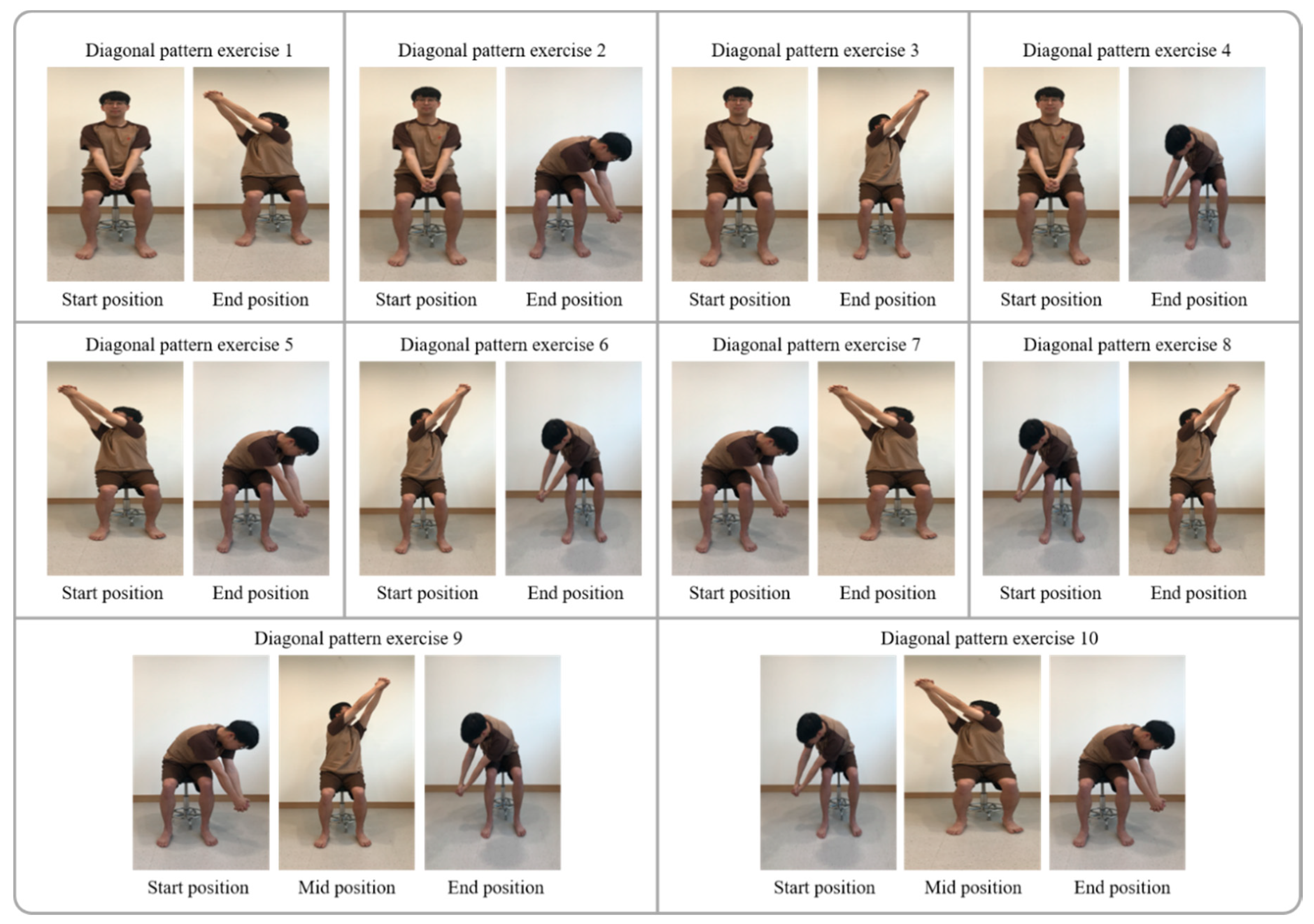
Figure 1. Diagonal pattern training.
Applsci 10 04635 g001
Table 1. Diagonal pattern training.
ClassificationTimeExercise TimeRest Time
Diagonal pattern exercise 1 1st stage51 min30 s
Diagonal pattern exercise 25
Diagonal pattern exercise 3 2nd stage51 min30 s
Diagonal pattern exercise 4 5
Diagonal pattern exercise 5 3rd stage51 min30 s
Diagonal pattern exercise 6 5
Diagonal pattern exercise 7 4th stage51 min30 s
Diagonal pattern exercise 8 5
Diagonal pattern exercise 9 5th stage51 min30 s
Diagonal pattern exercise 105
Table 2. Single plane training.
ClassificationTimeExercise TimeRest Time
Trunk flexion1st stage51 min30 s
Trunk extension5
Trunk flexion to extension2nd stage51 min30 s
Trunk Extension to flexion5
Paretic side lateral flexion3rd stage51 min30 s
Non-paretic side lateral flexion 5
Mid position to paretic side rotation4th stage51 min30 s
Mid position to non-paretic side rotation5
Paretic side to non-paretic side rotation5th stage51 min30 s
Non paretic side to paretic side rotation5
Table 3. General characteristics.
Experimental Group (n = 21)Control Group (n = 21)p
Gender (male/female)16/514/70.495
Affected side (left/right)8/1310/110.533
stroke type (infarction/hemorrhage)12/911/100.757
Onset (month)13.00 ± 2.6813.48 ± 2.820.578
K-MMSE (point)26.14 ± 1.6226.33 ± 1.710.713
Age (years)67.43 ± 4.7467.57 ± 3.280.910
Height (cm)165.05 ± 6.10163.76 ± 5.440.475
Weight (kg)69.86 ± 6.6070.43 ± 6.220.774
K-MMSE, Korean version of Mini Mental State Examination.
Table 4. Comparison of TIS in before and after.
Classification Experimental Group (n = 21)Control Group (n = 21)Fp
TIS (score)BeforeAfterBeforeAfter
Static (7)6.19 ± 0.686.76 ± 0.44 1,26.29 ± 0.646.52 ± 0.60 125.1300.01 *,a
Dynamic (10)3.24 ± 0.543.81 ± 0.68 1,23.62 ± 0.743.81 ± 0.75 129.0910.01 *,a
Coordination (6)1.52 ± 0.512.05 ± 0.81 1,21.76 0 ± 0.441.90 ± 0.5415.8060.01 *,a
Total (23)10.95 ± 1.4012.62 ± 1.28 1,211.67 ± 1.3912.24 ± 1.22 170.5750.01 *,a
* p < 0.05, 1 There was a significant difference between before test and after test (p < 0.05), 2 The experimental group improved more than the control group (interaction), a: Analyzed by two-way repeated measures ANOVA, TIS: trunk impairment scale, Experimental group: diagonal pattern training group, Control group: single plane training group.
Table 5. Comparison of BBS, 10 MWT in before and after.
Classification Experimental Group (n = 21)Control Group (n = 21)Fp
BeforeAfterBeforeAfter
BBS (score)38.48 ± 4.1740.43 ± 4.50 1,238.43 ± 3.9039.05 ± 4.32 116.3450.01 *,a
10 MWT (s)25.00 ± 5.2422.48 ± 5.50 1,224.48 ± 5.0323.48 ± 4.32 122.7500.01 *,a
* p < 0.05, 1 There was a significant difference between before test and after (p < 0.05), 2 The experimental group improved more than the control group (interaction), a: Analyzed by two-way repeated measures ANOVA, BBS: Berg balance scale, 10 MWT: 10 meters walk test, Experimental group: diagonal pattern training group, Control group: single plane training group.
Table 6. Comparison of cadence, speed, stride length in before and after.
Classification Experimental Group (n = 21)Control Group (n = 21)Fp
BeforeAfterBeforeAfter
Cadence (step/min)69.28 ± 10.4079.18 ± 15.28 1,270.00 ± 11.4072.63 ± 12.40 113.1670.01 *,a
Speed (m/s)0.61 ± 0.100.74 ± 0.12 1,20.61 ± 0.110.67 ± 0.13 122.2680.01 *,a
Stride length (m)0.77 ± 0.220.85 ± 0.26 1,20.78 ± 0.210.81 ± 0.19 124.1610.01 *,a
* p < 0.05, 1 There was a significant difference between before test and after (p < 0.05), 2 The experimental group improved more than the control group (interaction), a Analysed by two-way repeated measures ANOVA, Experimental group: diagonal pattern training group, Control group: single plane training group.