Article

Pesticide Pollution Provokes Histopathological Alterations in Apis mellifera (Linnaeus, 1758) Drone Gonads

1
Department of Developmental Biology, Faculty of Biology, Paisii Hilendarski University of Plovdiv, 4000 Plovdiv, Bulgaria
2
Department of Animal Sciences, Faculty of Agronomy, Agricultural University of Plovdiv, 4000 Plovdiv, Bulgaria
3
Department of Ecology and Environmental Conservation, Faculty of Biology, Paisii Hilendarski University of Plovdiv, 4000 Plovdiv, Bulgaria
4
Department of Hydrobiology, Faculty of Science and Technology, Institute of Biology and Ecology, University of Debrecen, H-4032 Debrecen, Hungary
5
National Laboratory for Water Science and Water Security, University of Debrecen, H-4032 Debrecen, Hungary
*
Author to whom correspondence should be addressed.
Environments 2025, 12(6), 173; https://doi.org/10.3390/environments12060173
Submission received: 27 March 2025 / Revised: 16 May 2025 / Accepted: 20 May 2025 / Published: 22 May 2025

Abstract

:
Honey bees are one of the most significant pollinators and contribute to the pollination of various crops. The honey bee, Apis mellifera (Linnaeus, 1758), has unique characteristics that could be successfully used to improve biomonitoring approaches in assessing environmental interactions. Three apiaries with different rates of honey bee colony losses were included in the study—Dimovtsi, Plovdiv, and Krasnovo, Bulgaria. Male individuals (immature and mature) were collected from five colonies for each of the three apiaries and studied for histopathological changes in the gonads. The results concerning the rate of honey bee colony losses in the studied apiaries from 2022 and 2023 showed honey bee losses in the tested colonies due to queen problems, which were reported for Plovdiv, as well as the death of honey bees or a reduction in their number to a few hundred bees in the colony. The chemical analysis showed the presence of different organic substances, such as Coumaphos, DEET (N, N-diethyl-M-toluamide), Fluvalinate, and Piperonyl-butoxide, in the alive and dead honey bee samples and those of food stocks (wax, pollen, and honey) within the hives. Among the sample types, those of the dead honey bees contained the greatest variety of pesticide residues, particularly in Plovdiv and Dimovtsi, reinforcing the link between pesticide exposure and honey bee mortality. The histopathological alterations were mainly associated with the thinning of the covering epithelium of the seminiferous tubules and the detachment of the basement membrane of the seminiferous tubules. The more severe histopathological lesion, necrosis, was observed in a higher degree of expression in the drones from Plovdiv, indicating a higher pollution level in this region.

1. Introduction

Modern agricultural practices are considered one of the primary causes of insect biodiversity loss [1,2]. The control of pesticide usage is regulated in the European Union by Regulation (EC) No. 1107/2009 and Directive 2009/128/EC. The most crucial steps that should be taken to protect honey bees are the following three: pesticide companies provide studies on the effects of active substances on honey bees and, if needed, on residues in nectar and pollen, one reporter Member State assesses the delivered data, and the European Food Safety Authority together with Member States peer reviews the active substance assessment [3]. Moreover, Regulation (EC) No. 396/2005 indicates the maximum pesticide residue levels in or on food and feed of plant and animal origin. According to Martinello et al. [4], biomonitoring programs could be used as a suitable method to assess the uptake and potential health risks caused by direct and indirect exposure to these environmental pollutants. As it is well known, honey bees are among the most significant pollinators and contribute to the pollination of various crops [5], making them excellent bioindicators for environmental pollution. In addition, as explained by Papa et al. [6], the honey bee has unique characteristics that could be successfully used to improve biomonitoring approaches in assessing environmental interactions. According to Jiang et al. [7], unintentional pesticide exposure includes contact with contaminated nectar, pollen, and water sources because honey bees can cover a wide area during a flight for foraging [8]. Eissa and Taha [9] pointed out that beekeepers utilise different pesticides to control the infestation with the Varroa destructor, Acarapis woodi, and Paenibacillus spp. larvae [10,11,12] in honey bee colonies. Therefore, honey bees’ products could be considered indicators for environmental biomonitoring purposes [13,14,15]. There has been a worrying trend in recent decades because of the increasing mortality of honey bees worldwide. This harms the genetic richness of honey bee populations in Europe. It turns out that they are threatened by genetic contamination of local populations adapted to specific environmental conditions and stress caused by global climate change and agrochemical pollution. In addition, intensive agriculture with long-term use of pesticides, the shortage of suitable food, habitat loss, and the emergence of new pathogens and pests are significant factors negatively affecting the vitality of honey bees. The harmful effects of pesticides are based on their accumulation potential as residues in the ecological food chain and their diverse toxic effects on living organisms. In recent years, agricultural practice has seen a trend toward widespread usage of neonicotinoids as a new class of pesticides applied in modern agrarian production to protect crops from pests. Although this class of insecticides is currently used in many countries worldwide, scientific data on their toxic effects on honey bees and other pollinators in nature are building up. This is also the reason for limiting or altogether banning their use in some European countries. In addition to their direct toxicity, which can lead to lethal effects, pesticides also affect the normal functioning of bee colonies. Several studies [16,17,18] aimed to clarify the relationship between bee colony mortality, declining bee populations, and the increased use of pesticides, including neonicotinoids. Moreover, several other studies presented data showing that neonicotinoid pollution negatively affects queen bee production, individual growth, and development, increases worker mortality, and leads to a decrease in colony size [19,20,21]. In beekeeping practice, various agrochemicals are widely used to combat Varroa. Although they are adequate to varying degrees against Varroa, they also pose a potential toxicological risk to honey bees, which has not been sufficiently studied. The synergistic interactions between plant protection chemicals and acaricides against Varroa have not been studied in detail.
Appropriate methodological tools and biomarkers are currently being sought to investigate the relationship between pesticide pollution and increasing honey bee colony losses in the populations of Apis mellifera. In this regard, histopathological biomarkers in bees could be successfully applied to monitor environmental effects and those of treatment with different classes of pesticides. According to Gusso-Choueri et al. [22] and Tanabe et al. [23], applying histopathological biomarkers to determine the effects of environmental pollution is a critical approach, as they precisely reflect the health status of the organism. Histopathological methods allow the differentiation of morphological changes caused by environmental factors or pollution at the tissue level. Furthermore, the histopathological changes represent an actual response of the organism to the influence of various environmental stressors, including pesticides. The growing number of studies based on the usage of histopathological biomarkers is related to the fact that they more accurately reflect the health status of the affected organisms compared to other biological methods. However, there is not much information on histopathological lesions in bees caused by environmental pollution with pesticides.
Therefore, the present study aims to determine how pesticide stress affects the reproductive system of honey bee drones from three populations with different rates of colony losses in Bulgaria. To our knowledge, this is the first study on histopathological alterations in drone gonads in the context of their relationship with pesticide contamination in situ.

2. Material and Methods

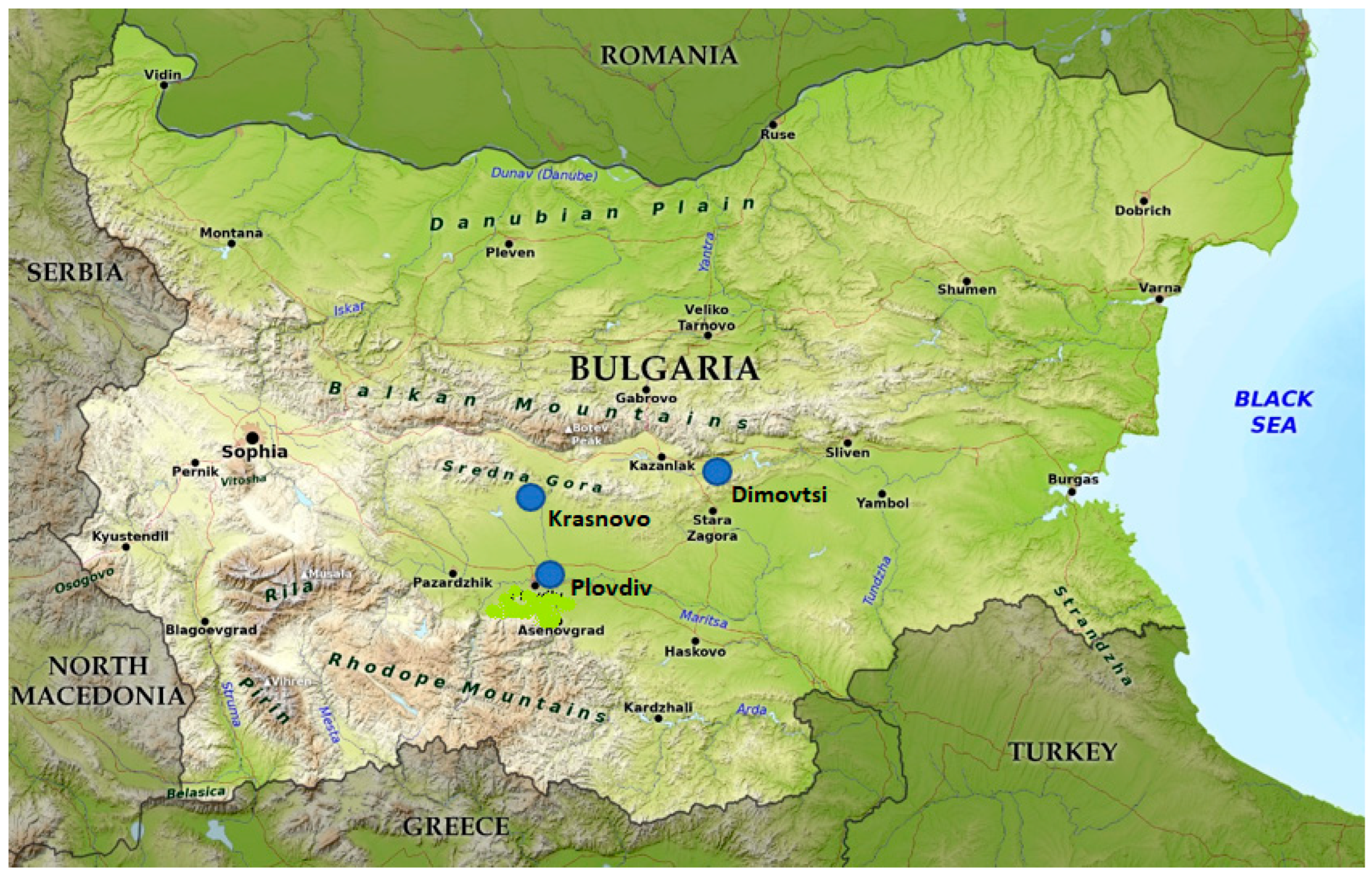
2.1. Sample Collection

Three apiaries with different rates of honey bee colony losses were included in the study—Dimovtsi (42.672887, 25.734201), Plovdiv (42.136097, 24.742168), and Krasnovo (42.4667, 24.4833)—Figure 1. The Dimovtsi apiary is situated in the Pre-Balkan region, where orchards and oil-bearing roses have been identified as potentially risky vegetation for honey bees due to the pesticide treatment. The Plovdiv apiary is a scientific beekeeping base of the Agricultural University in Bulgaria. It is located in the Thracian Valley, close to intensively pesticide-treated agricultural zones. The Krasnovo apiary is in the Sredna Gora Mountain—a region free of pesticide-treated crop fields. Drones (immature and mature) were collected from five colonies for each of the three apiaries for the period from May to June 2022 and transported alive in plastic bee cages with escape-proof lids, which allow airflow to ensure their health, safety, and legal compliance with the histopathology laboratory. The data on honey bee colony losses, the presence of risky honey vegetation, which is actively and permanently treated with pesticides in the region of the apiary, and the agrochemicals used by beekeepers against Varroa were obtained by applying the international COLOSS questionnaire during the period from April 2022 to March 2023 [24]. Given the reported low levels of honey bee colony losses (≤1%), the apiary in Krasnovo was used as a reference site.

2.2. Chemical Analysis

Pooled samples of bees, both for the alive and dead honey bees, and pooled samples of food stocks containing wax, honey, and pollen, collected from the studied colonies and pooled for each of the apiaries included in the study, were submitted for chemical analysis. The sample collection period for chemical analysis (May–June 2022) coincided with the collection of samples for histopathological analysis.
Pesticide residue concentrations (mg/kg) in the bee samples and food stocks in the hive collected from the same five colonies of the three studied apiaries were analysed by GMS-GC-MSMS and LMS-LC-MSMS chromatographic analysis, performed in the licensed international chemical laboratory Primoris in Gent, Belgium (the accredited methods were reported under the accreditation of Primoris Holding BELAC 057-TEST/ISO17025) [25]. Additionally, samples of oil-bearing rose foliage of Rosa damascena, commonly treated with plant protection products, were tested for the Dimovtsi apiary area. A calibration curve with a linear range from 0.01 mg/kg to 0.100 mg/kg for the bees and from 0.01 mg/kg to 0.150 mg/kg for the food stocks (wax, pollen, and honey) within the hives, respectively, was created with values lower than 0.01 mg/kg.

2.3. Histopathological Analysis

The dissection was performed in laboratory conditions. Immature and mature drone gonads (5 drones per colony, 5 colonies per apiary, 3 apiaries, and total number of gonads = 150 total) from each apiary were fixed in 10% neutral buffered formaldehyde solution (pH = 7.0) and used for the histopathological analyses. After fixation in formaldehyde, the gonads were rinsed in tap water, dehydrated in a graded series of ethanol concentrations, cleared in xylene, embedded in paraffin wax with a melting point of 54–56 °C, sectioned to a thickness of 5–7 µm using a semi-automated rotary microtome (Leica RM 2125 RTS, Leica Microsystems, Wetzlar, Germany), and mounted on sterilised glass slides. The sections were deparaffinised, stained with haematoxylin and eosin (H&E), and prepared for light microscopy analysis [26], which was performed by using a microscope with an attached high-quality camera (Leica DM 2000 LED, Leica Microsystems, Wetzlar, Germany). Ten paraffin sections were produced from each specimen. Each section was taken from a different location on the paraffin block. The histopathological alterations were analysed by observing the whole organ surface according to Power et al. [27]. The degree of expression was studied, including the corresponding changes in the organ surface about the standard histological structure of the organ. The histopathological lesions were determined semi-quantitatively using the grading systems of Gibson-Corley et al. [28] and Saraiva et al. [29], which we combined and modified. Each grade represented specific histopathological characteristics and was categorised as follows: (0)—no lesions, which represented standard histological structure (<10%), (1)—mild lesions (10–30%), (2)—moderate lesions (30–50%), (3)—severe lesions (50–80%), and (4)—very severe lesions (>80%). The histopathological slides from each individual were examined, and each alteration’s percentage value was determined from ten visual fields. The indicated value of histopathological changes is an average of all histopathological slides we observed.

2.4. Statistical Analysis

All statistical analyses were conducted using Python 3.11 with SciPy (v1.11.3) and Statsmodels (v0.14.0) [30,31]. The results are presented as mean ± standard deviation (SD), and statistical significance was set at p < 0.05. For the chemical residue analysis, one-way analysis of variance (ANOVA) was used to assess differences in pesticide concentrations among sampling sites and sample types. When significant differences were detected, Tukey’s HSD post hoc test was applied to determine which groups differed significantly. In cases where data were non-normally distributed, the Kruskal–Wallis test was performed instead of ANOVA, followed by the Mann–Whitney U test for pairwise comparisons. The Shapiro–Wilk test was used to assess the normality of data distributions, while Levene’s test was applied to check for homogeneity of variances. For the histopathological lesion scores, which were measured on a semi-quantitative scale, one-way ANOVA was performed to compare lesion severity among groups. Tukey’s HSD test was used for post hoc comparisons where significant differences were detected. For categorical data where numerical variability was insufficient for parametric analysis, a Chi-square test was used to compare the frequency of lesions across groups.
All statistical procedures were carried out using custom Python scripts, which are available from the corresponding author upon reasonable request.

3. Results and Discussion

3.1. International COLOSS Questionnaire

The results concerning the rate of honey bee colony losses in the studied apiaries from 2022 and 2023 are presented in Table 1. As could be seen, there were no reported honey bee colony losses for Krasnovo, but the total losses for Dimovtsi and Plovdiv were 19.74% and 40.96%, respectively. Colony losses in connection with queen problems were reported for Plovdiv, and those due to the death of honey bees or a reduction in their number to a few hundred bees in the colony were reported for Dimovtsi and Plovdiv. Treatment against Varroa has been previously performed in Krasnovo (by oxalic acid) and Dimovtsi (by oxalic acid and amitraz), in contrast to Plovdiv, where acaricide treatment has not been carried out in the last ten years (Table 1). Furthermore, the Krasnovo apiary is located in a semi-mountainous area without agrarian fields, and the risk of applying plant protection chemicals is lower. However, during the study period, the interviewed beekeepers reported spraying with mosquito repellents. The Dimovtsi apiary is near orchards and oil-bearing roses, which are regularly treated with pesticides, and the one in Plovdiv is located on the territory of an agricultural educational complex, where the experimental gardens (orchards, rapeseed, sunflower, and maize) are periodically treated with various pesticides.

3.2. Chemical Levels

The chemical analysis showed the presence mainly of Coumaphos in the alive honey bee samples and in those of food stocks (wax, pollen, and honey) within the hives. DEET (N,N-diethyl-M-toluamide), Fluvalinate, and Piperonyl-butoxide were also detected in the samples collected from some of the apiaries included in this study (Table 2). Additionally, data from the chemical analysis of the samples from the Dimovtsi region showed the presence of eight pesticide residues (Cypermethrin—0.68 mg/kg; Lambda-Cyhalothrin—0.32 mg/kg; Clothianidin—0.014 mg/kg; Difenoconazole—0.013 mg/kg; Flutriafol—0.023 mg/kg; Fluxapyroxad—6.4 mg/kg; Pyraclostrobin—6.6 mg/kg; Thiamethoxam—0.23 mg/kg) in the studied leaf mass of oil-bearing rose, five of which were also found in the dead bee samples. In addition, residues of fourteen pesticides were also detected in the dead bee samples from the apiary area in Plovdiv (Table 2). Significant differences in the pesticide residues were found among sampling sites and sample types. Clothianidin concentrations in dead honey bee samples differed significantly among sites (ANOVA F = 379.8, p < 0.001), with Dimovtsi showing higher levels than Plovdiv. Similarly, the Coumaphos levels varied significantly among alive honey bee samples and food stocks (ANOVA F = 455.1, p < 0.001), with Dimovtsi food stocks containing significantly higher concentrations than other sample types. The Fluvalinate concentrations in food stocks also differed among locations (ANOVA F = 208.3, p < 0.001), with higher levels detected in Plovdiv compared to Krasnovo (Table 2). For several pesticides, statistical comparisons were impossible, as they were detected in only one sample type or location, with concentrations below the detection limit (BDL) in all other cases. These include Cypermethrin, DEET, Dimethoate, Fenamidone, Fenbutatin oxide, Fenoxycarb, Flonicamid, Fluxapyroxad, Hexythiazox, Linuron, Piperonyl-butoxide, Prosulfocarb, Pyraclostrobin, Tebuconazole, Thiamethoxam, and Thiophanate-methyl (Table 2). Regarding the contamination levels across sites, Plovdiv (dead honey bee samples) exhibited the highest number of detected pesticides, suggesting higher environmental exposure. Conversely, Krasnovo (food stocks) had the lowest contamination levels, with only three detected pesticides in the samples, indicating lower pesticide transfer into stored honey and nectar sources. Moreover, among the sample types, the dead honey bee samples contained the highest variety of pesticide residues, particularly in Plovdiv and Dimovtsi, reinforcing the link between pesticide exposure and honey bee mortality (Table 2).
According to the European Food Safety Authority (EFSA), the application of pesticides should be restricted to reduce their negative effect on non-target organisms, such as honey bees [32]. Kaila et al. [3] explained that continual monitoring of pesticide exposure and its negative effects on bees and other wild pollinators should be established regarding pesticide legislation and its implementation across the EU to protect pollinators and their products in agricultural environments. Moreover, many honey bee colonies are exposed to multiple pesticides, which beekeepers apply to control pests and pathogens on bees. As a result of the widespread use of these miticides, contamination of the wax in different colonies could be established [33]. We agree with Traynor et al. [34], who stated that some of these pesticides can be widespread, especially in colonies owned by commercial beekeepers, and can reach high concentrations in wax, honey, pollen, and bees. In this regard, Traynor et al. [34] and Mullins et al. [35] found the product Amitraz and its derivatives in bee wax. Moreover, for the first time, Rinderer et al. [36] evaluated the effects of Fluvalinate used against Varroa mites on drone reproductive health. Similarly to Benito-Murcia et al. [37], we consider that acaricide residues, such as Fluvalinate and Coumaphos, are widespread in honey bee colonies. The authors found that chronic exposure to these acaricides resulted in significant changes in key biomarkers, such as neurotoxicity, xenobiotic biotransformation, and oxidative stress. In addition to the statement of Benito-Murcia et al. [37], the chronic toxicity of the mentioned acaricides presents the first LD50 values for Fluvalinate and Coumaphos in honey bees, providing insights into the risks for the contaminated bee colonies. Furthermore, short-term application and moderate concentrations are necessary to prevent Varroa resistance. We support the opinion of Ilyasov et al. [38], who recommended reducing the use of miticides and producing organic honey for less harmful effects on honey bees.

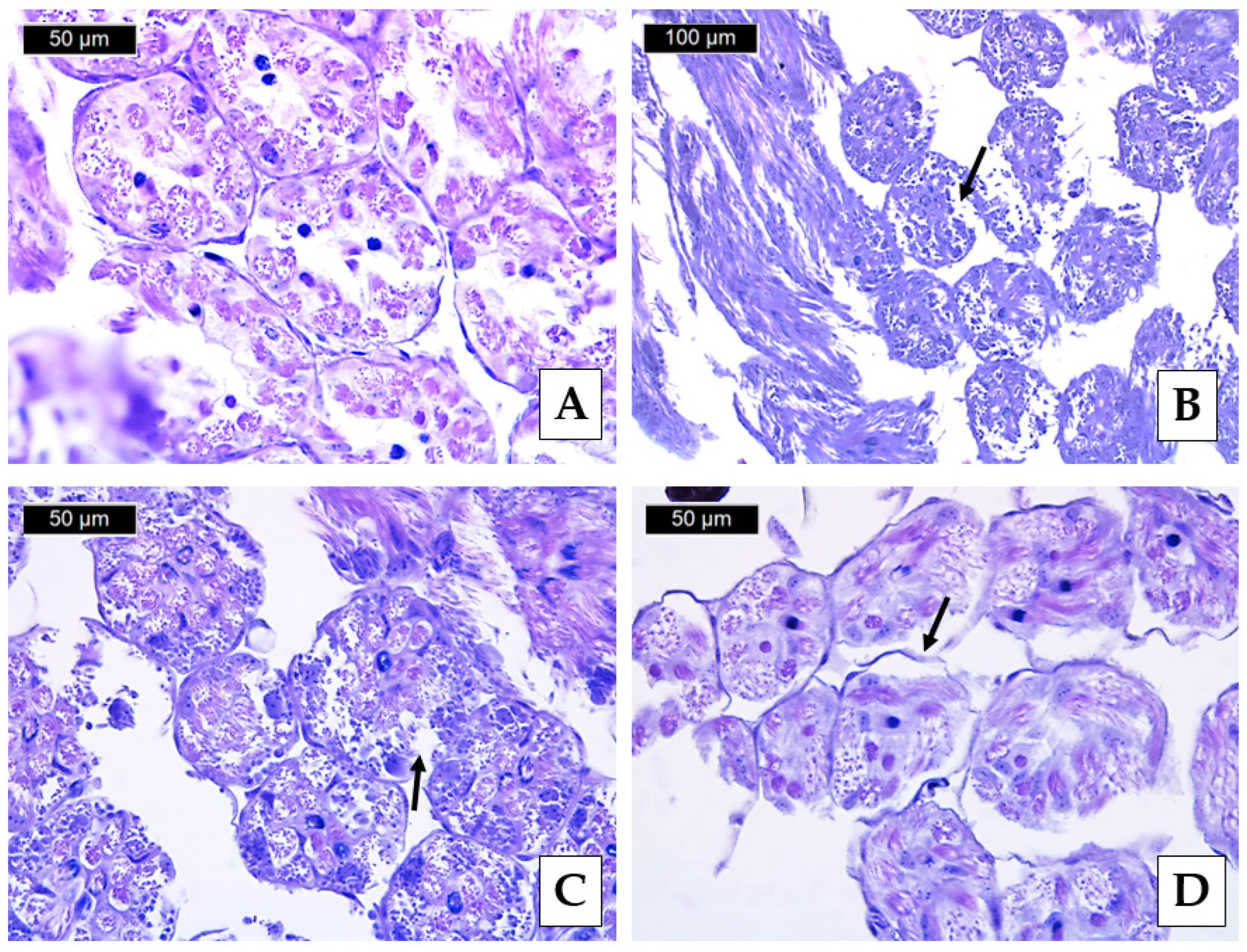
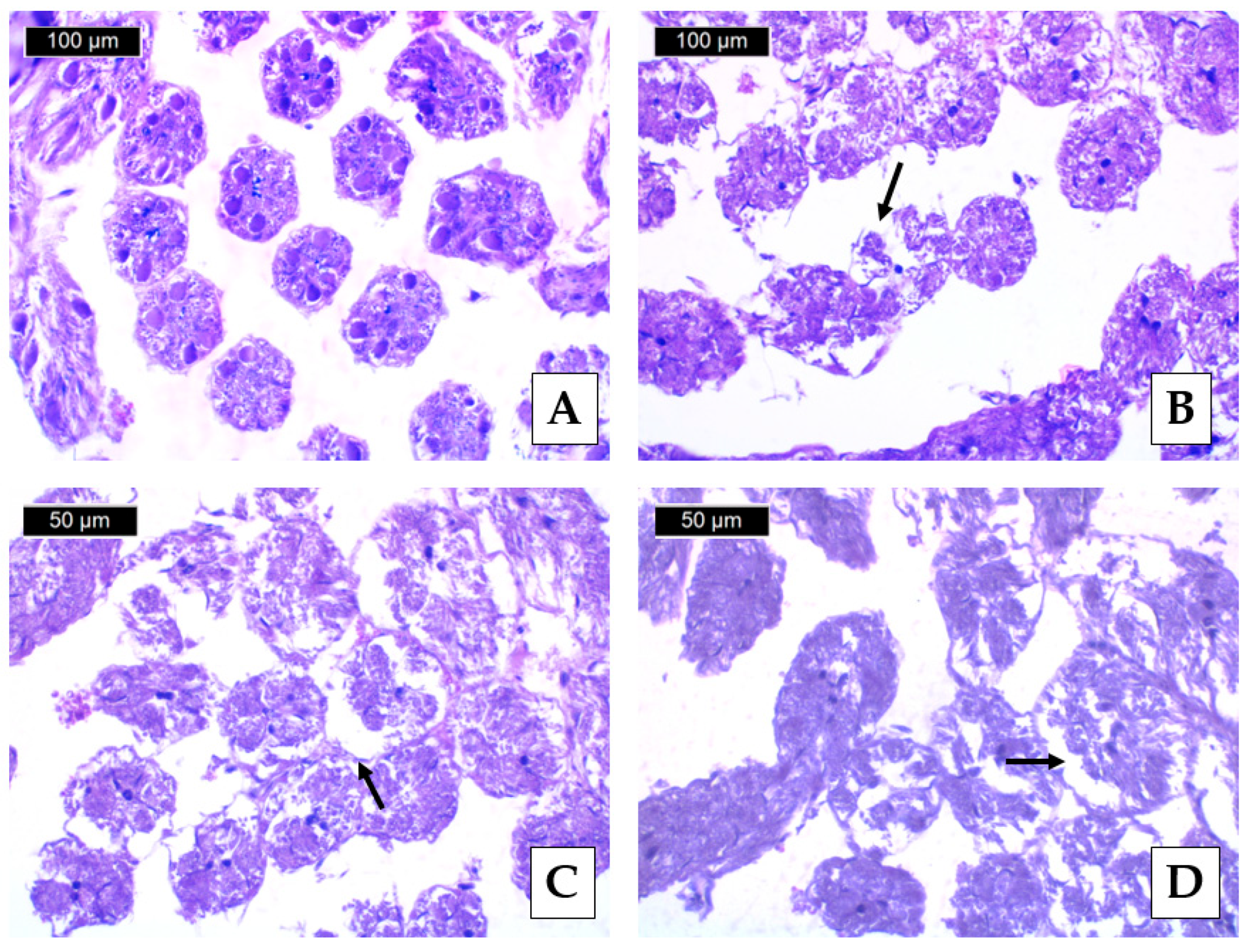
3.3. Histopathological Lesions

In the reference samples (Figure 2A and Figure 3A), we found a typical morphology of the examined organs, which follows the previously described one by Bishop [39] and Klein et al. [5]. On the other hand, we found histopathological changes regarding the sperm cells in the gonads of immature and mature drones. They were expressed mainly in thinning the epithelium covering the tubules, separating the seminiferous tubules’ basal membrane (Figure 3C,D and Figure 4A,B), and establishing separate areas with necrotic changes (Figure 2B,C and Figure 3B). The listed lesions affected drones more than immature males from the two sampling sites—Dimovtsi and Plovdiv. Compared with the Krasnovo apiary used as a reference (Figure 2A), we observed a higher degree of histopathological alterations in the drones from the Plovdiv apiary (Figure 1). In the drones from Dimovtsi, a higher degree of separation of the basal membrane from the seminiferous tubules was also found (Figure 4C,D). In the seminiferous tubules of all mature drones, mature spermatozoa have passed the spermiogenesis process. However, spermatids were less frequently detected. In the immature individuals, we observed seminiferous tubules without a lumen due to the earlier stage of development. Spermatogenic cells and spermatocytes were also found in the spermatogonia stage. Similar to the mature drones, separation of the basement membrane from the seminiferous tubules was present, as well as thinning of the covering epithelium, again to a higher degree in the individuals from the city of Plovdiv (Figure 3C,D). Significant differences were observed among the sampling sites and maturity groups in several histopathological parameters. Thinning of the covering epithelium of the seminiferous tubules showed significant variation among the groups (ANOVA F = 17.7662, p < 0.001; Figure 4D). The Tukey’s HSD test indicated that the Dimovtsi mature drones (Table 3, B)—exhibited significantly higher thinning than the Dimovtsi immature male individuals and the Plovdiv immature and mature drones (Table 3, C), which showed intermediate values. In contrast, the Krasnovo immature and mature drones (Table 3, A) exhibited no signs of this alteration, reinforcing the assumption that this change is related to pollution exposure in Dimovtsi and Plovdiv (Table 3). A similar trend was found in the detachment of the basement membrane of the seminiferous tubules, where a significant difference was detected (ANOVA F = 23.9962, p < 0.001). The Tukey’s test revealed that the Dimovtsi mature drones (Table 3, B) had the highest detachment scores. In contrast, the Dimovtsi immature male individuals and Plovdiv immature and mature male individuals (Table 3, C) had lower but notable levels of detachment. The Krasnovo immature and mature drones (Table 3, A) exhibited no detachment, indicating that this lesion is likely linked to external stressors. In the case of necrotic areas, a significant difference was observed (ANOVA F = 24.4948, p < 0.001). The Plovdiv immature and mature drones (Table 3, B) exhibited significantly more necrotic tissue than the Dimovtsi immature and mature drones (Table 3, A), showing only minor necrosis signs. No necrotic areas were detected in Krasnovo (Table 3, A), highlighting a marked difference between the control site and the impacted locations. No significant differences were observed among the groups for the lack of spermatozoa in mature drones (Chi-square = 4.2857, p = 0.1171). While a few cases of spermatozoa absence were recorded in the Dimovtsi mature drones (0.20 ± 0.42 A), no instances were found in Krasnovo or Plovdiv (Table 3, A). The rarity of this alteration suggests that it may not be directly linked to site-specific pollution effects. A significant difference was found in the absence of lumen in mature drones (Chi-square = 6.4286, p = 0.0403). The Dimovtsi mature drones (Table 3, B) had significantly more lumen absence cases than the Krasnovo and Plovdiv mature drones, which showed no instances (Table 3, A). The absence of this alteration in Plovdiv suggests that the factors contributing to lumen loss may be site-specific and more prominent in Dimovtsi. The Plovdiv immature and mature drones exhibited the most severe histopathological alterations, particularly in necrotic areas, indicating increased tissue damage at this site. The Dimovtsi mature drones showed notable alterations in detachment of the basement membrane and the absence of a lumen, suggesting a high degree of tissue damage in this location. In contrast, Krasnovo (the reference site) had the lowest alteration scores across all examined parameters, reinforcing the assumption that environmental stressors contribute to the observed histopathological changes in Dimovtsi and Plovdiv (Table 3).
Data are presented as mean ± SD for histopathological semi-quantitative scores, which are categorised as follows:
(0)—no lesions, representing standard histological structure (<10% affected),
(1)—mild lesions (10–30% affected),
(2)—moderate lesions (30–50% affected),
(3)—severe lesions (50–80% affected),
(4)—very severe lesions (>80% affected).
According to Elhamalawy et al. [40], pesticides cause adverse effects on non-target invertebrates, their vital roles in agriculture, and their economic value. Moreover, as the authors explained, pesticides also affect individual survival, physiological state, growth, behaviour, development, and reproduction. In addition to this opinion, Mitkovska et al. [41,42] provided evidence that there are statistically significant relationships between pesticide contamination in apiary areas, the rate of honey bee colony losses in populations, the presence of various morphological abnormalities, and DNA integrity disorders in spermatozoa of drones from similar locations to the described ones. Our results for histopathological alterations in gonads in male individuals in different stages of development align with those of Power et al. [27], who observed degenerative changes in the seminiferous tubules of the Italian bee (Apis mellifera ligustica Spinola, 1806), expressed in changes of the covering epithelium, as well as the more severe degree of degeneration—necrosis. According to Ebrahimi et al. [43] and Babazadeh et al. [44], such degenerative changes in the seminiferous tubules may be due to the presence of high levels of pesticides, such as organophosphorus chemicals and neonicotinoids. In addition, the effects of different toxicants could lead to the induction of oxidative stress in various tissues and the presence of degenerative changes. As Finkel and Holbrook [45] explained, oxidative stress causes the accumulation of reactive oxygen species (ROS), leading to alterations at the cellular and tissue levels. According to Sandroc et al. [46] and Baines et al. [47], persistent organic pollutants, such as neonicotinoids, induce hormonal and morphological changes in the male reproductive system of honey bees. What is more, the negative changes in the development of the reproductive system of male individuals are observed even at lower concentrations but after prolonged exposure to pesticides. However, we agree with Power et al. [27] that the degenerative changes found may also be due to parasites. Therefore, we suggest further detailed research in this field. Basak et al. [48] stated that Piperonyl-butoxide could cause environmental hazards and is toxic to non-targeted organisms, resulting in haematological, biochemical, and histopathological alterations. In addition, the external treatment of honey bee colonies by miticides Amitraz and Fluvalinate, along with a positive effect of pest control, harms reproductivity and honey productivity and probably can reduce learning and memory, gustation, and olfaction of honey bees [38]. Our results also align with those of Abderkader et al. [49], who established a significant decrease in semen volume and sperm concentration and an increase in sperm mortality rate in honey bee drones, which indicates the negative effect of insecticides, such as Clothianidin. Moreover, Benito-Murcia et al. [37] found that chronic exposure to Fluvalinate and Coumaphos resulted in significant changes in key biomarkers, indicating potential neurotoxicity, xenobiotic biotransformation, and oxidative stress. Ko et al. [50] studied the impact of different concentrations of Fluvalinate as a widely used insecticide against Varroa mites on the larvae of honey bee experimental colonies in Taiwan. The authors found that the applied concentrations harmed the adult honey bee workers, affecting their subsequent associative ability. In addition, Arslan et al. [51] stated that increasing doses of Coumaphos resulted in increased locomotor activity with differences in sensitivity across honey bee populations in Turkey. Swiatly-Blaszkiewicz et al. [52] quantified pesticide residues in different honey bee products collected from Poland and, similarly to us, found a presence of different pesticide residues, such as Fluvalinate, Cypermethrin, and Dimethoate. The negative impact of the studied pesticides was confirmed by Gregorc et al. [53], who conducted a comparative immunohistological TUNEL analysis concerning the negative effects of two insecticides (Coumaphos and Imidacloprid) on the honey bee Apis mellifera worker’s lifespan, food consumption, mortality, and expression of antioxidant genes. We agree with the authors who recommend applying the integrated biomarker response approach with histopathological analysis to assess the health status of bee colonies under the influence of different environmental pollutants.
It should be noted that the impaired reproductive potential of drones negatively affects the fertility of queen bees, which leads to a weakening of the honey bee colony and the population as a whole. This circumstance, together with factors such as pests and parasites, pathogens, nutrition, habitat loss and fragmentation, and poor beekeeping practices, is the basis for knowledge and opportunities to improve the health status and ecological well-being of honey bees.

4. Conclusions

According to the obtained results concerning the histopathological changes in the reproductive system of immature and mature drones of bee colonies from the studied areas, a tendency was established towards a more severe degree of expression of the established degenerative changes in the experimental individuals from the apiary of Plovdiv compared to the other studied apiaries—reference and experimental. The identified histopathological changes can be successfully applied as biomarkers of contamination, assessing the reproductive potential of apiaries. In this way, the potential negative effects of chronic pesticide exposure on bee colonies can be predicted. Moreover, regulatory authorities should revise pesticide regulations, implementing the scientific data and potential risks to reduce pollution and protect honey bee colonies.

Author Contributions

Investigation, S.S.; methodology, S.S.; conceptualisation, E.G.; data curation, E.N.I.; investigation, E.G.; methodology, E.G.; resources, P.P.; software, D.S. and K.N.; supervision, E.N.I.; writing—review and editing, P.P., V.Y., L.A., D.S., K.N. and E.N.I. All authors have read and agreed to the published version of the manuscript.

Funding

The National Research Fund of Bulgaria supported this study under the contract No. KP-06-H5112/2021, “Complex assessment of genetic and environmental factors related to the losses of honey bees (Apis mellifera L.) in Bulgaria”. This study is also financed by the European Union, NextGenerationEU, through the National Recovery and Resilience Plan of the Republic of Bulgaria, Project No. BG-RRP-2.004-0001-C01.

Data Availability Statement

The original contributions presented in this study are included in the article. Further inquiries can be directed to the corresponding author(s).

Conflicts of Interest

The authors declare no conflicts of interest.

References

  1. Raven, P.H.; Wagner, D.L. Agricultural intensification and climate change are rapidly decreasing insect biodiversity. Proc. Natl. Acad. Sci. USA 2021, 118, e2002548117. [Google Scholar] [CrossRef]
  2. Seibold, S.; Gossner, M.M.; Simons, N.K.; Blüthgen, N.; Müller, J.; Ambarlı, D.; Ammer, C.; Bauhus, J.; Fischer, M.; Habel, J.C.; et al. Arthropod decline in grasslands and forests is associated with landscape-level drivers. Nature 2019, 574, 671–674. [Google Scholar] [CrossRef]
  3. Kaila, L.; Ketola, J.; Toivonen, M.; Loukola, O.; Hakala, K.; Raiskio, S.; Hurme, T.; Jalli, M. Pesticide residues in honey bee-collected pollen: Does the EU regulation protect honey bees from pesticides? Environ. Sci. Pollut. Res. 2022, 29, 18225–18244. [Google Scholar] [CrossRef]
  4. Martinello, M.; Manzinello, C.; Dainese, N.; Giuliato, I.; Gallina, A.; Mutinelli, F. The Honey Bee: An Active Biosampler of Environmental Pollution and a Possible Warning Biomarker for Human Health. Appl. Sci. 2021, 11, 6481. [Google Scholar] [CrossRef]
  5. Klein, C.D.; Kozii, I.V.; Wood, S.C.; Koziy, R.V.; Zabrodski, M.W.; Dvylyuk, I.; Medici de Mattos, I.; Moshynskyy, I.; Honaramooz, A.; Simko, E. Testicular changes of honey bee drones, Apis mellifera (Hymenoptera: Apidae), during sexual maturation. J. Insect Sci. 2021, 21, 3. [Google Scholar] [CrossRef]
  6. Papa, G.; Maier, R.; Durazzo, A.; Lucarini, M.; Karabagias, I.K.; Plutino, M.; Bianchetto, E.; Aromolo, R.; Pignatti, G.; Ambrogio, A.; et al. The Honey bee Apis mellifera: An insect at the interface between human and ecosystem health. Biology 2022, 11, 233. [Google Scholar] [CrossRef]
  7. Jiang, J.; Ma, D.; Zou, N.; Yu, X.; Zhang, Z.; Liu, F.; Mu, W. Concentrations of imidacloprid and thiamethoxam in pollen, nectar and leaves from seed-dressed cotton crops and their potential risk to honey bees (Apis mellifera L.). Chemosphere 2018, 201, 159–167. [Google Scholar] [CrossRef]
  8. Chiesa, L.M.; Labella, G.F.; Giorgi, A.; Panseri, S.; Pavlovic, R.; Bonacci, S.; Arioli, F. The occurrence of pesticides and persistent organic pollutants in Italian organic honeys from different productive areas in relation to potential environmental pollution. Chemosphere 2016, 154, 482–490. [Google Scholar] [CrossRef]
  9. Eissa, F.; Taha, E.K.A. Contaminants in honey: An analysis of EU RASFF notifications from 2002 to 2022. J. Consum. Prot. Food Saf. 2023, 18, 393–402. [Google Scholar] [CrossRef]
  10. Johnson, R.M.; Dahlgren, L.; Siegfried, B.D.; Ellis, M.D. Acaricide, Fungicide and Drug Interactions in Honey Bees (Apis mellifera). PLoS ONE 2013, 8, e54092. [Google Scholar] [CrossRef]
  11. Margaoan, R.; Papa, G.; Nicolescu, A.; Cornea-Cipcigan, M.; Kösoğlu, M.; Topal, E.; Negri, I. Environmental pollution effect on honey bees and their derived products: A comprehensive analysis. Environ. Sci. Pollut. Res. 2024, 32, 10370–10391. [Google Scholar] [CrossRef]
  12. Kunat-Budzyńska, M.; Łabuć, E.; Ptaszyńska, A.A. Seasonal detection of pathogens in honey bees kept in natural and laboratory conditions. Parasitol. Int. 2025, 104, 102978. [Google Scholar] [CrossRef]
  13. Kasiotis, K.M.; Anagnostopoulos, C.; Anastasiadou, P.; Machera, K. Pesticide residues in honey bees, honey and bee pollen by LC–MS/MS screening: Reported death incidents in honey bees. Sci. Total Environ. 2014, 485, 633–642. [Google Scholar] [CrossRef]
  14. Malhat, F.M.; Haggag, M.N.; Loutfy, N.M.; Osman, M.A.M. Residues of organochlorine and synthetic pyrethroid pesticides in honey, an indicator of ambient environment, a pilot study. Chemophere 2015, 120, 457–461. [Google Scholar] [CrossRef]
  15. Catalano, P.; Della Sala, F.; Cavaliere, M.; Caputo, C.; Pecoraro, D.; Crispino, G.; Lettera, S.; Caioni, G.; Esposito, M.; Verre, A.; et al. Use of honey bees and hive products as bioindicators to assess environmental contamination in targeted areas of the Campania region (Italy). Animals 2024, 14, 1446. [Google Scholar] [CrossRef]
  16. Blacquière, T.; Smagghe, G.; van Gestel, C.A.; Mommaerts, V. Neonicotinoids in bees: A review on concentrations, side-effects and risk assessment. Ecotoxicology 2012, 21, 973–992. [Google Scholar] [CrossRef]
  17. van der Sluijs, J.P.; Simon-Delso, N.; Goulson, D.; Maxim, L.; Bonmatin, J.-M.; Belzunces, L.P. Neonicotinoids, bee disorders and the sustainability of pollinator services. Curr. Opin. Environ. Sustain. 2013, 5, 293–305. [Google Scholar] [CrossRef]
  18. De Jong, D.; Lester, P.J. The global challenge of improving bee protection and health. Front. Bee Sci. 2023, 1, 1118292. [Google Scholar] [CrossRef]
  19. Cresswell, J.E.; Desneux, N.; van Engelsdorp, D. Dietary traces of neonicotinoid pesticides as a cause of population declines in honey bees: An evaluation by Hill’s epidemiological criteria. Pest. Manag. Sci. 2012, 68, 819–827. [Google Scholar] [CrossRef]
  20. Gill, R.J.; Ramos-Rodriguez, O.; Raine, N.E. Combined pesticide exposure severely affects individual- and colony-level traits in bees. Nature 2012, 491, 105–108. [Google Scholar] [CrossRef]
  21. Whitehorn, P.R.; O’Connor, S.; Wackers, F.L.; Goulson, D. Neonicotinoid pesticide reduces bumble bee colony growth and queen production. Science 2012, 336, 351–352. [Google Scholar] [CrossRef]
  22. Gusso-Choueri, P.K.; Choueri, R.B.; de Araújo, G.S.; Cruz, A.C.F.; de Oliveira Stremel, T.R.; de Campos, S.X.; de Souza Abessa, D.M.; de Oliveira Ribeiro, C.A. Univariate or multivariate approaches for histopathological biomarkers in the context of environmental quality assessments? Mar. Pollut. Bull. 2022, 181, 113828. [Google Scholar] [CrossRef]
  23. Tanabe, P.; Schlenk, D.; Forsgren, K.L.; Pampanin, D.M. Using digital pathology to standardize and automate histological evaluations of environmental samples. Environ. Toxicol. Chem. 2025, 44, 306–317. [Google Scholar] [CrossRef]
  24. Gray, A.; Adjlane, N.; Arab, A.; Ballis, A.; Brusbardis, V.; Bugeja Douglas, A.; Cadahía, L.; Charrière, J.-D.; Chlebo, R.; Coffey, M.F.; et al. Honey bee colony loss rates in 37 countries using the COLOSS survey for winter 2019–2020: The combined effects of operation size, migration and queen replacement. J. Apic. Res. 2022, 62, 204–210. [Google Scholar] [CrossRef]
  25. ISO/IEC 17025; General Requirements for the Competence of Testing and Calibration Laboratories. ISO: Geneva, Switzerland, 2017.
  26. Alturkistani, H.A.; Tashkandi, F.M.; Mohammedsaleh, Z.M. Histological stains: A literature review and case study. Glob. J. Health Sci. 2015, 8, 72–79. [Google Scholar] [CrossRef] [PubMed]
  27. Power, K.; Martano, M.; Altamura, G.; Maiolino, P. Histopathological findings in testes from apparently healthy drones of Apis mellifera ligustica. Vet. Sci. 2020, 7, 124. [Google Scholar] [CrossRef]
  28. Gibson-Corley, K.N.; Olivier, A.K.; Meyerholz, D.K. Principles for valid histopathologic scoring in research. Vet. Pathol. 2013, 50, 1007–1015. [Google Scholar] [CrossRef]
  29. Saraiva, A.; Costa, J.; Serrão, J.; Cruz, C.; Eiras, J.C. A histology-based fish health assessment of farmed seabass (Dicentrarchus labrax L.). Aquaculture 2015, 448, 375–381. [Google Scholar] [CrossRef]
  30. Virtanen, P.; Gommers, R.; Oliphant, T.E.; Haberland, M.; Reddy, T.; Cournapeau, D.; Burovski, E.; Peterson, P.; Weckesser, W.; Bright, J.; et al. SciPy 1.0: Fundamental algorithms for scientific computing in Python. Nat. Methods 2020, 17, 261–272. [Google Scholar] [CrossRef]
  31. Seabold, S.; Perktold, J. Statsmodels: Econometric and statistical modeling with Python. SciPy 2010, 7, 92–96. [Google Scholar]
  32. European Food Safety Authority (EFSA). Scientific Services to Support EFSA Systematic Reviews: Lot 5 Systematic Literature Review on the Neonicotinoids (Namely Active Substances Clothianidin, Thiamethoxam and Imidacloprid) and the Risks to Bees. EFSA Supporting Publication 2015: EN-756, 2013. Available online: http://www.efsa.europa.eu/en/supporting/doc/756e.pdf (accessed on 15 February 2025).
  33. Boncristiani, H.; Underwood, R.; Schwarz, R.; Evans, J.D.; Pettis, J.; van Engelsdorp, D. Direct effect of acaricides on pathogen loads and gene expression levels in honey bees Apis mellifera. J. Insect Physiol. 2012, 58, 613–620. [Google Scholar] [CrossRef] [PubMed]
  34. Traynor, K.S.; Pettis, J.S.; Tarpy, D.R.; Mullin, C.A.; Frazier, J.L.; Frazier, M. In-hive pesticide exposome: Assessing risks to migratory honey bees from in-hive pesticide contamination in the eastern United States. Sci. Rep. 2016, 6, 33207. [Google Scholar] [CrossRef] [PubMed]
  35. Mullins, C.A.; Frazier, M.; Frazier, J.L.; Ashcroft, S.; Simonds, R.; van Engelsdorp, D.; Pettis, J.S. High levels of miticides and agrochemicals in North American apiaries: Implications for honey bee health. PLoS ONE 2010, 5, e9754. [Google Scholar] [CrossRef]
  36. Rinderer, T.E.; De Guzman, L.I.; Lancaster, V.A.; Delatte, G.T.; Seltzer, J.A. Varroa in the mating yard: I. The effects of Varroa jacobsoni and Apistan® on drone honey bees. Am. Bee J. 1999, 139, 134–139. [Google Scholar]
  37. Benito-Murcia, M.; Botías, C.; Martín-Hernández, R.; Higes, M.; Soler, F.; Pérez-López, M.; Míguez-Santiyán, M.P.; Martínez-Morcillo, S. 2024. Biomarker responses and lethal dietary doses of tau-fluvalinate and coumaphos in honey bees: Implications for chronic acaricide toxicity. Environ. Toxicol. Pharmacol. 2024, 105, 104330. [Google Scholar] [CrossRef]
  38. Ilyasov, R.A.; Lim, S.; Lee, L.M.L.; Kwon, H.W.; Nikolenko, A.G. Effect of miticides amitraz and fluvalinate on reproduction and productivity of honey bee Apis mellifera. Uludağ Arıcılık Derg. 2021, 21, 21–30. [Google Scholar] [CrossRef]
  39. Bishop, G.H. Fertilization in the honey-bee. I. The male sexual organs: Their histological structure and physiological functioning. J. Exp. Zool. 1920, 31, 224–265. [Google Scholar] [CrossRef]
  40. Elhamalawy, O.; Bakr, A.; Eissa, F. Impact of pesticides on non-target invertebrates in agricultural ecosystems. Pest. Biochem. Physiol. 2024, 202, 105974. [Google Scholar] [CrossRef] [PubMed]
  41. Mitkovska, V.; Chassovnikarova, T.; Vasileva, P.; Stoyanov, I.; Petrov, P.; Petkov, N.; Ivanova, E.N. Sperm comet assay as a novel tool in assessing genotoxicity in high-mortality honey bee (Apis mellifera) populations. Apidologie 2025, 56, 13. [Google Scholar] [CrossRef]
  42. Mitkovska, V.; Chassovnikarova, T.; Vasileva, P.; Stoyanov, I.; Petrov, P.; Ivanova, E.N. Pesticide stress induces spermatozoa DNA damage and morphological abnormalities in Apis mellifera populations. Environ. Toxicol. Pharmacol. 2025, 116, 104710. [Google Scholar] [CrossRef]
  43. Ebrahimi, M.; Taherianfard, M. The effects of heavy metals exposure on reproductive system of cyprinid fish from Kor River. Iran. J. Fish. Sci. 2011, 10, 13–26. [Google Scholar]
  44. Babazadeh, M.; Najafi, G. Effect of chlorpyrifos on sperm characteristics and testicular tissue changes in adult male rats. Vet. Res. Forum. 2017, 8, 319–326. [Google Scholar]
  45. Finkel, T.; Holbrook, N.J. Oxidants, oxidative stress and the biology of ageing. Nature 2000, 408, 239–247. [Google Scholar] [CrossRef]
  46. Sandroc, C.; Tanadini, L.G.; Pettis, J.S.; Biesmeijer, J.C.; Potts, S.G.; Neumann, P. Sublethal neonicotinoid insecticide exposure reduces solitary bee reproductive success. Agric. For. Entomol. 2014, 16, 119–128. [Google Scholar] [CrossRef]
  47. Baines, D.; Wilton, E.; Pawluk, A.; de Gorter, M.; Chomistek, N. Neonicotinoids act like endocrine disrupting chemicals in newly-emerged bees and winter bees. Sci. Rep. 2017, 7, 10979. [Google Scholar] [CrossRef]
  48. Basak, M.; Choudhury, R.A.; Goswami, P.; Dey, B.K.; Laskar, M.A. A review on non-target toxicity of deltamethrin and piperonyl butoxide. J. Pharm. Res. Int. 2021, 33, 85–89. [Google Scholar] [CrossRef]
  49. Abderkader, F.B.; Kairo, G.; Tchamitchian, S.; Bonnet, M.; Cousin, M.; Barbouche, N.; Belzunces, L.P.; Brunet, J.L. Effects of Clothianidin exposure on semen parameters of honey bee drones. J. New Sci. 2018, 59, 3791–3798. [Google Scholar]
  50. Ko, C.-Y.; Nai, Y.-S.; Lo, W.; Chen, C.-T.; Chen, Y.-W. Low-level fluvalinate treatment in the larval stage induces impaired olfactory associative behavior of honey bee workers in the field. Insects 2022, 13, 273. [Google Scholar] [CrossRef]
  51. Arslan, O.C.; Erdem, B.; Somel, M.; Giray, T.; Kence, M. Effects of coumaphos on locomotor activities of different honey bee (Apis mellifera L.) subspecies and ecotypes. Apidologie 2023, 54, 39. [Google Scholar] [CrossRef]
  52. Swiatly-Blaszkiewicz, A.; Klupczynska-Gabryszak, A.; Matuszewska-Mach, E.; Matysiak, J.; Attard, E.; Kowalczyk, D.; Adamkiewicz, A.; Kupcewicz, B.; Matysiak, J. Pesticides in honey bee products—Determination of pesticides in bee pollen, propolis, and royal jelly from Polish apiary. Molecules 2025, 30, 275. [Google Scholar] [CrossRef]
  53. Gregorc, A.; Alburaki, M.; Rinderer, N.; Sampson, B.; Knight, P.R.; Karim, S.; Adamczyk, J. Effects of coumaphos and imidacloprid on honey bee (Hymenoptera: Apidae) lifespan and antioxidant gene regulations in laboratory experiments. Sci. Rep. 2018, 8, 15003. [Google Scholar] [CrossRef] [PubMed]
Figure 1. Locations of the apiaries included in the study and sample collection sites.
Environments 12 00173 g001
Figure 2. Histopathological lesions in drone gonads from Plovdiv (H&E staining, 5–7 µm sections): (A) reference group, (B,C) necrotic areas in seminiferous tubules (arrow), and (D) detachment of the basement membrane of the seminiferous tubule (arrow).
Environments 12 00173 g002
Figure 3. Degenerative lesions in gonads of honey bee immature drones (H&E staining, 5–7 µm sections): (A) control group with seminiferous tubules without the presence of a lumen, (B) a necrotic area in a seminal duct, affecting some of the follicles in drones from Dimovtsi (arrow), and (C,D) separation of the basement membrane of the seminiferous tubules in drones from Plovdiv (arrow).
Environments 12 00173 g003
Figure 4. Histopathological lesions in drone gonads from Dimovtsi (H&E staining, 5–7 µm sections): (A,B) detachment of the basement membrane of the seminiferous tubule (arrow), (C) seminiferous tubule with the presence of spermatozoa without the presence of a lumen (arrow), and (D) thinning of the covering epithelium of the seminiferous tubules (arrow).
Environments 12 00173 g004
Table 1. Rate of honey bee colony losses (%), risky honey vegetation, and agrochemicals used by beekeepers against Varroa during 2022–2023.
ParameterKrasnovoDimovtsiPlovdiv
Queen problem losses %008.43
Losses due to bee mortality %019.7432.53
Total loss %019.7440.96
Reported treatmentOxalic acidOxalic acid
Amitraz
No treatment
Risky vegetationNoOrchardsOrchards, rapeseed,
sunflower, maize
Table 2. Pesticide residue levels (mean ± SD) in honey bee samples from different locations and sample types (mg/kg).
ChemicalDimovtsiKrasnovoPlovdiv
Dead Honey BeesAlive Honey BeesFood StocksDead Honey BeesAlive Honey BeesFood StocksDead Honey BeesAlive Honey BeesFood Stocks
AmitrazBDLBDLBDLBDLBDLBDL0.0039 ± 0.0003BDLBDL
CarbendazimBDLBDLBDLBDLBDLBDL0.0011 ± 0.0001BDLBDL
Clothianidin0.0203 ± 0.0015 ABDLBDLBDLBDLBDL0.0030 ± 0.0002 BBDLBDL
CoumaphosBDL0.0433 ± 0.0012 A0.3133 ± 0.0208 BBDLBDLBDLBDL0.0617 ± 0.0021 A0.0603 ± 0.0015 A
Cypermethrin0.0103 ± 0.0006BDLBDLBDLBDLBDLBDLBDLBDL
DEET
(N,N-diethyl-M-toluamide)
BDLBDLBDLBDLBDLBDLBDL0.0387 ± 0.0015BDL
DimethoateBDLBDLBDLBDLBDLBDL0.0011 ± 0.0002BDLBDL
FenamidoneBDLBDLBDLBDLBDLBDL0.0013 ± 0.0002BDLBDL
Fenbutatin oxideBDLBDLBDLBDLBDLBDL0.0032 ± 0.0002BDLBDL
FenoxycarbBDLBDLBDLBDLBDLBDL0.0018 ± 0.0001BDLBDL
FlonicamidBDLBDLBDLBDLBDLBDL0.0052 ± 0.0002BDLBDL
FluvalinateBDLBDLBDLBDLBDL0.0057 ± 0.0015 ABDLBDL0.0237 ± 0.0015 B
Fluxapyroxad0.0620 ± 0.0020BDLBDLBDLBDLBDLBDLBDLBDL
HexythiazoxBDLBDLBDLBDLBDLBDL0.0015 ± 0.0002BDLBDL
LinuronBDLBDLBDLBDLBDLBDL0.0023 ± 0.0002BDLBDL
Piperonyl-butoxideBDLBDLBDLBDLBDL0.0073 ± 0.0021BDLBDLBDL
ProsulfocarbBDLBDLBDLBDLBDLBDL0.0035 ± 0.0002BDLBDL
Pyraclostrobin0.1433 ± 0.0208BDLBDLBDLBDLBDLBDLBDLBDL
TebuconazoleBDLBDLBDLBDLBDLBDL0.0026 ± 0.0004BDLBDL
Thiamethoxam0.0071 ± 0.0052BDLBDLBDLBDLBDLBDLBDLBDL
Thiophanate-methyl BDLBDLBDLBDLBDLBDL0.1367 ± 0.0153BDLBDL
A,B Values with different letters in the same row are significantly different (Tukey’s test, p < 0.05). BDL—below detection limit.
Table 3. Histopathological lesion assessment in the male reproductive system of honey bees (N = 10 per group).
Histopathological LesionKrasnovo (Control)DimovtsiPlovdiv
Mature DronesImmature DronesMature DronesImmature DronesMature DronesImmature Drones
Thinning of the covering epithelium of the seminiferous tubules0 A0 A1.90 ± 0.74 B1.00 ± 0.67 C0.80 ± 0.63 C0.90 ± 0.57 C
Detachment of the basement membrane of the seminiferous tubule0 A0 A2.20 ± 0.79 B1.30 ± 0.48 C1.00 ± 0.67 C1.00 ± 0.67 C
Necrotic areas0 A0 A0.70 ± 0.67 A0.60 ± 0.52 A2.10 ± 0.74 B2.00 ± 0.94 B
Lack of spermatozoa
(only in mature drones)
0 A0 A0.20 ± 0.42 A-0 A-
Absence of lumen (only in mature drones)0 A0 A1.00 ± 0.67 B-0 A-
A,B,C Values with different letters in the same row are significantly different (Tukey’s test, p < 0.05).
Disclaimer/Publisher’s Note: The statements, opinions and data contained in all publications are solely those of the individual author(s) and contributor(s) and not of MDPI and/or the editor(s). MDPI and/or the editor(s) disclaim responsibility for any injury to people or property resulting from any ideas, methods, instructions or products referred to in the content.

