Article

Chronic Heat Exposure Modulates Innate and Adaptive Immune Responses in Firefighters

by
Brijesh Yadav
1,
Afzaal Nadeem Mohammed
1,
Brittney Graham
1,
Amit Bhattacharya
2 and
Jagjit Singh Yadav
1,*
1
Pulmonary Pathogenesis and Immunotoxicology Laboratory, Division of Environmental Genetics and Molecular Toxicology, Department of Environmental and Public Health Sciences, University of Cincinnati College of Medicine, Cincinnati, OH 45267, USA
2
Division of Environmental and Industrial Hygiene, Department of Environmental and Public Health Sciences, University of Cincinnati College of Medicine, Cincinnati, OH 45267, USA
*
Author to whom correspondence should be addressed.
Environments 2024, 11(6), 131; https://doi.org/10.3390/environments11060131
Submission received: 16 April 2024 / Revised: 8 June 2024 / Accepted: 16 June 2024 / Published: 19 June 2024
(This article belongs to the Special Issue Environments: 10 Years of Science Together)

Abstract

:
Global fire activities, which are getting worse due to climate change, cause both environmental and human health hazards. Firefighters, being the first responders, are frequently exposed to heat which may impact their immune system and overall health. However, the nature of the impact of chronic heat exposure on immune function has not been studied in-depth in firefighters. In this study, 22 firefighters exposed to “heavy-smoke fires (structural fires)”, categorized as the “high-exposure group” (>0.15 structural fires/week) and “low-exposure group” (<0.15 structural fires/week), were sampled. Peripheral blood was examined for immune cell profile based on total and differential cell counts, immune function based on the transcriptional expression of drivers of innate and adaptive immunity and key inflammation mediators, and heat stress marker HSP70. The white blood cell (WBC) count, mean corpuscular volume, mean corpuscular hemoglobin, and absolute and segmented neutrophil counts decreased below the normal range in both exposure groups. The gene transcript levels for toll-like receptors (TLR2, TLR4, but not TLR7) and their adaptor protein MYD88 were lower whereas those for T-cell transcription factors (RORC/RORγ, FoxP3) and inflammatory mediators (TNF-α, Granzyme-B) were higher in the “high-exposure group”, indicating mixed response; however, the ratios between pro-inflammatory and anti-inflammatory transcription factors of adaptive immunity, namely T-bet/FoxP3 (Th1/Treg) and RORC/FoxP3 (Th17/Treg), were lower. Collectively, decreased immune cell landscape, downregulated key innate immunity receptors, and Tregs’ dominance suggested that chronic heat exposure in firefighters dysregulated innate and adaptive immunity, skewed towards an overall immunosuppressive condition with inflammation.

1. Introduction

Fire activities are showing an increasing global trend, coinciding with escalating climate change patterns, causing hazardous environmental conditions for the public and first responders. Firefighting is an extremely strenuous occupation due to the repeated exposure of firefighters to harmful environmental conditions such as heat, toxic chemicals, biological agents, and a variety of mechanical hazards [1,2]. Heat exposure and stresses in the workplace environment have been linked to physiological impairments in firefighters [3,4,5] and various acute and chronic illnesses such as sudden cardiac arrest, diabetes, cancer, hypertension, and neuromotor dysfunction, leading to high mortality [1,6,7]. In particular, occupational exposure to extreme heat in firefighters may possibly exacerbate the underlying immunological factors [8,9] contributing to their propensity for immune-mediated diseases including cancer and infections [10,11,12].
Deleterious effects of heat stress on immunity have been explored in animals [13,14,15,16]. However, there are limited human studies on the effects of heat stress on immunity [8,9], especially on the role of occupational chronic heat exposure in immune dysfunction [17]. For instance, in experimental mouse models, chronic heat exposure was shown to reduce the local respiratory tract immunity via epithelial cell necrosis and reduced tracheal and mucosal epithelial cell number, red blood cell infiltration in tracheal tube, reduced alveolar macrophage count, reduced expression of dendritic cell maturation markers major histocompatibility complex class II (MHC-II), cluster of differentiation 40 (CD40), CD80 and CD86, and regulation of cytokines including downregulation of interleukin-6 (IL-6) and interferon-β (IFN-β) and upregulation of IL-10, leading to higher H5N1 influenza virus infection [16]. Studies on dairy cattle showed that heat stress reduces the level of anti-inflammatory cytokines such as IL-4 and IL-10 [13]. Calves exposed to chronic heat during gestation and early post-natal period had either dysregulated immune systems or significantly delayed development of their immune system [13,18]. Heat stress in calves impacted peripheral blood mononuclear cells (PBMCs) by reducing neutrophils, eosinophils, and lymphocytes, and their stimulation with lipopolysaccharide (LPS) increased serotonin secretion [18] which is known to modulate adaptive immune response [19]. The impact of heat stress on immune system dysregulation is also supported by recent epidemiological studies. Heat stress during physical activities affects human immunity via activation of the innate immune response [20]. A decline in the percentage of T-helper cells and an increased percentage of natural killer (NK) cells were reported in heat-stroke patients as compared to healthy controls [21]. Male bakers exposed to high heat showed reduced count and cytotoxic activity of NK cells [17]. Immune system dysregulation may increase the risk of developing chronic allergic and/or inflammatory diseases as well as susceptibility to infections. In this context, studies have reported that exposure to extreme heat is associated with higher hospitalization rates related to asthma and chronic kidney disease and increased plasma osmolality [21,22,23].
Dysregulation of immune function may occur at the level of innate and/or adaptive immunity. Among the innate immunity mediators, toll-like receptors (TLRs) expressed by both the immune and non-immune cells sense the conserved pathogen-associated molecular patterns (PAMPs) and damage-associated molecular patterns (DAMPs). They bind to conserved motifs and non-specifically trigger the innate immune response for eventual activation of the adaptive immune cells. While cell surface TLRs (TLR1, TLR2, TLR4, TLR5, TLR6, TLR10) bind to extracellular floating ligands, intracellular TLRs (TLR3, TLR7, TLR8, TLR9) bind to cytoplasmic DAMPs such as heat shock protein (HSP), altered self-peptides, mitochondrial DNA (hypomethylated CpG DNA motifs), among other ligands. Among the cell surface TLRs, TLR2 binds to microbial products and ligands released from stressed tissue including heat shock proteins (HSPs). TLR2 signaling is known to induce the expression of cytochrome P450 1A1 (CYP1A1) gene that mediates detoxification of carcinogenic polycyclic aromatic hydrocarbons (PAHs) such as benzo(a)pyrene [24] that are commonly associated with fire emissions. This is significant considering that downregulation of TLR2 has been associated with neoplasia [25]. TLR4 binds to microbial endotoxins, and DAMPs like high mobility group box protein 1 (HMGB1), HSP70, hyaluronans, and histone proteins [26,27]. DMAPs-driven TLR4 signaling leading to the secretion of inflammatory cytokines was required for tissue repair in the lungs due to hyaluronan exposure [27]. Among the intracellular TLRs, TLR7 recognizes the single-stranded RNA genome of viruses and its upregulation is associated with the formation of higher intensity of cytokines and auto-antibodies leading to auto-immune diseases such as rheumatoid arthritis [28]. TLR9 recognizes the hypomethylated DNA of bacteria as well as mitochondria and triggers pro-inflammatory responses. Heat stress in mitochondria reduces its biogenesis and enhances reactive oxygen species formation and spills of CpG-rich mitochondrial DNA in the extracellular matrix, leading to the secretion of cytokines [29].
Innate immune mediators activate adaptive immune responses including antibody response (by B-cells) or cell-mediated response (by T-cells). In the presence of a predominant local cytokines milieu, activated T-cell subsets arising from the naïve CD4+ T-cell lineage differentiate either in pro-inflammatory T-helper (Th) cell subsets such as Th1, Th17 or in anti-inflammatory T-regulatory (Treg) cell subset [30,31,32]. Specific T-cell lineage commitments driven by cytokine signaling result from the induction of cell-specific transcription factor expressions. The presence of IL-12 leads to T-bet expression and Th1 cell development, IL-6 plus transforming growth factor-β (TGF-β) leads to Th17 cell development, and TGF-β leads to Treg cell development [31]. The T-helper cell subsets Th1 and Th17 secrete cytokines, leading to an inflammatory response whereas the Treg subset inhibits the inflammatory cells to maintain immune homeostasis [30]. Cytotoxic T-lymphocytes (CTLc) arising from activation of CD8+ T cell lineage release granzyme-B which cleaves the extracellular matrix, cytoskeleton proteins such as nuclear lamins, and mitochondrial membrane and activates the release of protein-bound pro IL-1 and pro TGF-β into their active forms resulting in inflammation and fibrosis [27,33], causing immune dysfunction.
There is a paucity of information on immune dysfunction in firefighters. As firefighters are occupationally exposed to extreme heat conditions, our working hypothesis is that firefighters experience compromised innate and adaptive immunity due to heat stress. Thus, our goal was to investigate the direct impacts of heat stress on innate and adaptive immune cells and their responses in firefighters differentially exposed to occupational heat stress conditions.

2. Materials and Methods

2.1. Study Design

In this pilot study, duly approved by the Institutional Review Board (IRB 2016-2342), 22 medically fit employer-recommended professional firefighters with matching age and BMI were recruited between 1 January 2019 to 30 April 2020. Firefighters with current smoking history or any underlying medical complications were excluded. Their demographic and service-associated details were collected as listed in Table 1. Firefighters were categorized into “low-exposure group” (n = 14) and “high-exposure group” (n = 8). Low exposure and high exposure were defined as exposure to <0.15 and >0.15 structural firefighting/week, respectively. Structural firefighting/week was determined by the total number of fires fought in a year divided by 52 weeks and a cut-off value of 0.15 was selected for grouping to compare the high versus low exposure as per the previous part of this pilot study on postural stability [34]. Each firefighter was informed about the study in advance and signed a written consent form before donating blood samples for the study. Venous blood samples were collected by a trained phlebotomist and subjected to initial immune cell profiling by complete blood cell count (CBC) with differential analysis. Innate and adaptive immune responses were evaluated by measuring gene expression levels of inflammatory mediators, toll-like receptors (TLRs) and their adaptive protein myeloid differentiation primary response protein-88 (MyD88), and T-cell subset-specific transcription factors. Heat exposure marker HSP70 protein level was measured in blood plasma.

2.2. Blood Sample Collection and Processing

For plasma separation and whole blood immune cell profiling, venous blood samples were collected in 10 mL- and 2-mL lavender-capped collection tubes containing dipotassium ethylene diamine tetra acetic acid (K2EDTA), respectively; the samples were mixed properly by repeated inversions. For total RNA isolation, another venous blood sample was collected in a 2 mL-PAXgene tube (Pre-Analytix, GmbH, Hombrechtikon, Germany) and mixed properly by shaking followed by incubation for 2–3 h at room temperature.
Blood from the 10 mL-K2EDTA vials was used for the plasma separation. Plasma was separated after centrifugation of blood at 1500 RPM for 5 min and stored at −80 °C until the analysis was completed. The 2 mL-lavender capped vial of blood with K2EDTA was used for the complete blood cell count (CBC) analysis at the Cincinnati Children’s Hospital Medical Center hematology lab using the Cell-Dyn Sapphire (Abbott, Diagnostics, IL, USA) hematology analyzer. The 2 mL-PAXgene tube was processed for RNA extraction.

2.3. RNA Isolation

Total RNA was isolated from the PAXgene blood sample using the PAXgene blood RNA isolation kit according to the manufacturer’s instructions (Qiagen, GmbH Germany). Briefly, the tube was centrifuged at 3000× g for 10 min to pellet cells. The resulting pellet was washed (2×) with RNAase-free water (4 mL) by vertexing and centrifugation (3000× g for 10 min). The washed pellet was dissolved in 350 µL Buffer BR1 by vertexing. The solution was transferred to a new 1.5-mL microfuge tube and 300 µL of Buffer BR2 and 40 µL of proteinase K were added and mixed by vertexing for 5 s. The mixture was incubated at 55 °C for 10 min for cell lysis. The lysate was passed through a shredder spin column placed in a 2-mL microfuge tube by centrifugation at 15,000× g for 3 min. The eluate was transferred to a new 1.5-mL microfuge tube and 350 µL of absolute ethanol was added and mixed by vertexing. The mixture was passed through a PAXgene RNA spin column by centrifugation at 15,000× g for 1 min. The spin column was placed in a new 2-mL tube and washed with 350 µL Buffer BR3 by centrifugation (15,000× g for 1 min). The washed column was transferred to a new 1.5-mL microfuge tube and 80 µL of DNase-I diluted in RDD buffer was poured into the column followed by incubation at room temperature for 15 min. The column was further washed with 350 µL Buffer BR3 by centrifugation (15,000× g for 1 min), placed in a new 2-mL microfuge tube, and washed with 500 µL Buffer BR4 by centrifugation (15,000× g for 1 min). An additional 500 µL Buffer BR4 was added, and the column was re-washed by centrifugation (15,000× g for 3 min). The PAXgene column was then placed in a new 1.5-mL microfuge tube and RNA was eluted (2×) with 40 µL Buffer BR5 by centrifugation (15,000× g for 1 min). The eluted RNA was incubated at 65 °C for 5 min and chilled on ice. The quantity and quality of RNA were analyzed using Nanodrop (Thermofisher Scientific, Bend, OR, USA).

2.4. Gene Expression Analysis

A 10 ng aliquot of total RNA was used for quantitative reverse transcriptase-polymerase chain reaction (qRT-PCR) based gene expression analysis using one-step Brilliant-III Ultra-Fast SYBER Green qRT-PCR Master Mix (Agilent technology, Santa Clara, CA, USA). Gene-specific primers synthesized by Integrated DNA technology (IDT, San Diego, CA, USA) were used for the analysis of the following targets: housekeeping gene glyceraldehyde-3-phosphate dehydrogenase (GAPDH) and the genes encoding transcription factors T-box transcription factor (TBX21 or T-bet in Th1 cells), Retinoic acid receptor-related orphan receptor-C (RORC encoding RORγt protein in Th17 cells), and Forehead box P3 (FoxP3 in Treg cells), toll-like receptors TLR 2, TLR4, TLR7, and TLR9, Granzyme-B (for NK cells and Cytotoxic T cells), the adaptor protein MyD88, and the typical proinflammatory cytokine TNF-α. For each target gene, one step qRT-PCR reaction (10 µL reaction volume) was performed in triplicate on the real-time thermocycler ABI 7500 (Applied Biosystems, Foster, CA, USA). The cycling conditions were as follows: reverse transcription step at 50 °C for 10 min followed by denaturation step at 94 °C for 10 min, and 35 cycles of amplification (each cycle involving denaturation at 94 °C for 5 s followed by annealing and amplification at optimized target-specific annealing temperature (Ta) for 60 s). Primer sequences and annealing temperatures used were those described elsewhere [35,36]. Melting curve analysis was performed using a ramp rate of 0.5 °C/10 s between 65 to 95 °C. Delta Ct was calculated as Cttarget gene − Ctgapdh. Fold-change compared to the endogenous housekeeping control gene GAPDH was calculated using the 2−Δct method [37]. The ratio between the fold-change values for the high-exposure group versus the low-exposure group (2−Δct of higher exposure/2−Δct of lower exposure) implied the following status of differential gene regulation in the high-exposure group: >1, upregulation; <1, downregulation; 1, no difference in gene expression. The downregulation was then expressed as a negative reciprocal of the fold-change ratio.

2.5. Plasma HSP70 Protein Analysis (ELISA)

Plasma derived from the K2EDTA-containing blood was used for measuring the heat stress marker HSP70 using a Duo set ELISA kit (R&D system, Minneapolis, MN, USA; Catalog Number DYC1663-2), following the manufacturer’s instructions. Briefly, the plate was coated with capture antibody (100 µL) overnight in the dark at room temperature. After aspiration, the wells were washed (3×) with 400 µL of wash buffer and non-specific binding sites were blocked with block buffer (1% BSA, 0.05% NaN3 in PBS) for 1 h at room temperature. The wells were then loaded with 100 µL of either the standard or sample plasma solution (diluted 1:100 in diluent containing 1 mM EDTA, 0.5% Triton™ X-100 in PBS) and incubated for 2 h at room temperature. The wells were washed (3×) with wash buffer and loaded with 100 µL of detection antibody followed by incubation for 1 h. The wells were further washed and loaded with Streptavidin-conjugated horseradish-peroxidase (HRP-streptavidin) solution and incubated for 20 min. After further washing (5×) with wash buffer, the wells were loaded with 100 µL of 3,3,5,5 tetra-methyl benzidine substrate solution (TMB) and incubated for 20 min. The reaction was stopped with 50 µL of 2N H2SO4 solution. The optical density of the color product developed was measured at 450 nm and 540 nm wavelengths on a Bio-Tek Epoch microplate reader (Agilent Technology, Santa Clara, CA, USA). The analyte concentration was calculated by computer-generated plotting of a 4-parameter logistic curve between standard concentrations and their respective optical density values. The minimum detection limit for the HSP70 kit was 156 pg/mL.

2.6. Statistical Analysis

Data were compared between the two groups, i.e., low-exposure versus high-exposure groups, using independent t-tests assuming non-equal standard deviation (Welch correction) on GraphPad Prism version 8.0 for Windows (GraphPad Software, La Jolla, CA, USA). Results were expressed as mean ± standard error of the mean (SEM). p ≤ 0.05 was considered statistically significant.

3. Results

3.1. Demographic Profiles and Exposure Grouping of Study Subjects

The demographic profiles of firefighter participants in terms of age, body mass index (BMI), and total years of firefighting service were similar between the low- and high-exposure groups (Table 1). While the high-exposure group had more experience working with structural heavy smoke fires, other parameters namely, average hours worked over the last month, average shift hours per year, and total working shift hours throughout the career were significantly higher in the low-exposure group (Table 1).

3.2. Blood Immune Cell Landscape

Complete blood cell count (CBC) profile was deranged in all firefighters when compared with the normal range recommended for adult healthy individuals but was similar between the low- and high-exposure groups (Table 2). Likewise, white blood cell (WBC) count, mean corpuscular volume (MCV)/hemoglobin (MCH), and absolute neutrophil count and segmented neutrophil percentage were lower in firefighters in comparison with the normal recommended range for healthy adults, suggesting suppression of hematopoiesis in firefighters (Table 2).

3.3. Dysregulation of Immune Functions

Transcriptional expression of genes associated with innate and adaptive immune responses was used to assess the status of immune function (Table 3). Among the innate immunity drivers/mediators, mRNA levels of toll-like receptor TLR2 and adapter protein MyD88 for this and other receptors in the NF-kB pathway showed a downward trend whereas TLR4 showed significant downregulation (p = 0.041) in the high-exposure group (2.21-fold). However, the genes for intracellular receptor TLR7 (1.85-fold) and the proinflammatory cytokine TNF-α were upregulated in the high-exposure group as compared to the low-exposure group (Table 3 and Figure 1A).
Among the adaptive immunity mediators, expression of the transcription factor T-bet (for Th1 cells) was similar between the two exposure groups. The transcription factors RORC (for Th17 cells) and FoxP3 (for Treg cells) showed higher expression levels in the high-exposure group (1.69-fold and 3.52-fold, respectively). However, the quantitative ratios between the pro-inflammatory and anti-inflammatory transcription factors, namely T-bet/FoxP3 (Th1/Treg) and RORC/FoxP3 (Th17/Treg), were lower in the high-exposure group. Expression of the cytolytic protease Granzyme-B (secreted from the NK and Cytotoxic T-cells) was upregulated in the high-exposure group (Figure 1B).
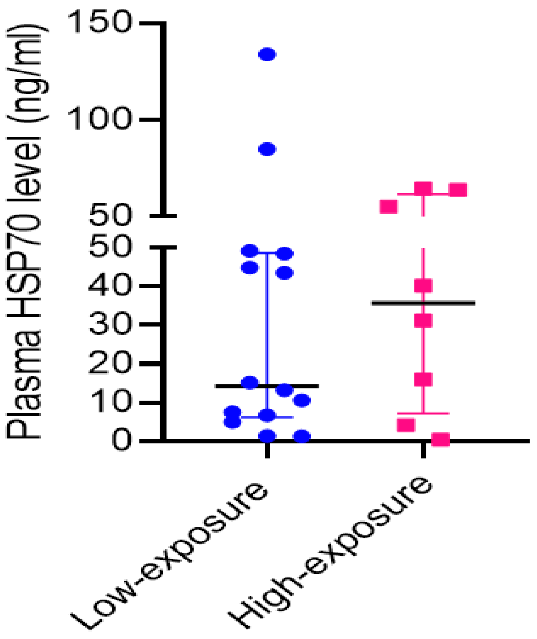
Plasma levels of HSP protein were above the normal reference range (5–15 ng/mL) for both the exposure groups. The median HSP protein level in plasma was higher in the high-exposure group (35.7 ng/mL) as compared to the low-exposure group (14.2 ng/mL), though there was a large inter-individual variability (Figure 2).

4. Discussion

The results showed a deranged blood immune cell profile, dysregulated innate and adaptive immune response mediators, and altered ratios between T-helper cells and regulatory T-cells along with elevated plasma heat shock protein in firefighters, as elaborated in the following discussion. This implied that role of chronic heat exposure in skewing immune homeostasis may be the underlying cause of immune-mediated diseases such as cancer, cardiometabolic syndrome, neurological conditions, and higher infections commonly experienced by firefighters [16].
Elevated blood HSP70 protein levels as compared to the normal reference range (5–15 ng/mL) in both exposure groups of firefighters indicated heat stress response. Higher median plasma HSP70 levels in the high-exposure group in our study coincided with greater exposure to structural/high smoke fires by this group. However, there was a high inter-individual variability in both groups which may be due to exhaustion or tolerance of body cells to respond further to heat stress and diminished compensating capacity and/or temporal instability of HSP70 protein in prolonged exposures. In fact, an animal study reported that acute heat exposure can lead to an increased level of HSP70 in the early days of exposure with a return to normal level after 21 days of chronic exposure [16]. HSP70 is a molecular chaperone protein that mitigates heat-induced stress by renaturing the denatured proteins [38]. HSP has also been reported to reduce cellular apoptosis in cardiomyocytes and neuronal cells [39], implying its significance in cardiac function; however, prolonged heat exposure may skew physiological and immune homeostasis leading to adverse cardiac and other consequences usually experienced by firefighters.
The most striking impact of chronic heat stress on firefighters was a significant reduction in the absolute count of circulating immune cells in peripheral blood. An overall lower CBC count in all firefighters as compared to the recommended normal healthy range suggested a suppressive effect of heat exposure on the proliferation and differentiation of different immune cell lineages. This is in agreement with the findings of other studies on dairy cattle and bakery workers that reported curtailed mesenchymal stem cell proliferation and differentiation due to heat stress, leading to reduced numbers and altered immunophenotype of blood cells [40,41].
The differential cell count analysis showed a reduction in the WBC and RBC counts, mean corpuscular volume (MCV), mean hematocrit value (MHV), and mean corpuscular hemoglobin (MCH). WBCs being critical immune cells required for eliminating pathogens, reduced WBC count implies susceptibility to infections. Decreased RBC count and hemoglobin content may trigger the development of anemia and chronic fatigue, the health conditions often experienced by firefighters [42,43]. We also observed that segmented and absolute counts of neutrophils in firefighters were lower than the normal reference range. This observation is in agreement with the previous study that reported lower neutrophil count in blood samples of bakery workers frequently exposed to chronic heat stress [16,22]. Neutrophils are the most abundant leukocytes patrolling the tissues and are the first responders to pathogen attacks. A diminished neutrophil count (neutropenia) can cause delayed viral clearance, arthritis, inflammatory bowel disease, and non-Hodgkin’s lymphoma. Recent studies documented a higher prevalence of these conditions in firefighters [44,45,46]. A higher count of immature granulocytes and a lower absolute count of neutrophils in the high-exposure group (Table 2), suggested an impairment of granulocyte differentiation into mature neutrophils due to heat stress [41,42].
Myeloid lineage monocytes differentiated into macrophages and dendritic cells (DCs) are required for phagocytosis and antigen presentation to lymphoid cells (T and B lymphocytes) for triggering the adaptive immune response. These antigen-presenting cells (APCs) express different types of toll-like receptors (TLRs) that are involved in triggering the immune response against extracellular stimuli including heat stress and coordinate with the adaptive immune response. Heat stress leads to tissue injury and the release of DAMPs such as HSP70, which are sensed by TLR4 expressed on DCs and macrophages [47]. Higher expression levels of TLR4 in the low-exposure group of firefighters suggest a responsiveness of immune cells to other stresses such as endotoxins that might have been experienced more by this group (Table 3). TLR binding of DAMP ligands triggers the NF-kB pathway eventually leading to the release of inflammatory cytokines such as TNF-α [48]. In our study, the TNF-α gene was 2.52-fold upregulated in the high-exposure group. In contrast, mRNA levels of TLR2, TLR4, and MyD88 were decreased in this group. Downregulation of TLRs and MyD88 may be an indication of compromised ability to trigger innate immune response due to prolonged exposure in firefighters, which may be the cause for delayed pathogen clearance as observed in animal studies [15].
We also observed decreased lymphocyte count in the high-exposure group as compared to the normal reference range (Table 2), consistent with a previous study on heat stroke patients that reported a low abundance of CD3+ and CD4+ T-helper cells [49]. A dysregulation of lymphoid cell lineages is associated with various metabolic disorders frequently reported in human and animal studies [6,14,17,50]. Dysregulation of lymphocyte subpopulations of T, B and NK cells may adversely affect the pathogen-specific adaptive immunity. Based on transcriptional expression levels of T-cell subset-specific transcription factors, we observed a dysregulation of T-helper (Th) cell subsets in firefighters. The high-exposure group showed greater activation of Th17 (RORC) and Treg (FoxP3) subsets and lower activation of the Th1 (T-bet) subset, measured in terms of the respective transcription factor genes shown in parenthesis; this suggested a skewed T-cell balance towards Th17 and Treg cells. Th17 cells secrete cytokines (IL-17, IL-21, IL-22) that protect from extracellular pathogens such as Candida and Klebsiella. IL-21 is required for B cell maturation and antibody formation. IL-17 has been associated with lung pathologies including fibrosis. IL-17A also induces expression of CXCL8 and granulocyte colony-stimulating factors (G-CSF) from epithelial cells and helps in neutrophil infiltration at the inflammation site [51]. Furthermore, the active ingredients of fire smoke such as polycyclic aromatic hydrocarbons (PAHs), dioxins, monocyclic hydrocarbons (benzene, toluene, styrene), and metals, may serve as receptor ligands and trigger the aryl hydrocarbon receptor (AHR) signaling that promotes Th17 cell differentiation and inflammatory IL-17 secretion but inhibits the Th1-driven IFN-ϒ pathway [52]. This is consistent with the decreased expression of T-bet, the transcription factor for IFN-ϒ-producing Th1 cells, in the current study. The inflammatory diseases in firefighters may be driven by Th17 cells and their cytokines. In this regard, a higher incidence of inflammatory interstitial lung diseases has been reported in firefighters [11]. Additionally, the survivors exposed to the 9/11 attack involving heat exposure experienced a higher incidence of auto-immune diseases like arthritis, scleroderma, myositis, and connective tissue diseases [10].
In terms of the Treg subset, we observed upregulation of its transcription factor FoxP3 in the high-exposure group. FoxP3-positive Treg cells normally secrete the anti-inflammatory cytokine IL-10 which attenuates the overactivation of inflammatory cells. In this context, previous animal studies have reported that chronic heat exposure induces Treg differentiation, increases IL-10 cytokine levels in circulation [38,53] and suppresses the antigen-specific Th1 and Th2-mediated immune responses. This eventually leads to an immunosuppressive profile, which may partially explain the higher susceptibility of firefighters to chronic infections and malignancy [7]. However, it is also reported that, under inflammatory cytokines milieu, Treg cells may become functionally impaired and convert into inflammatory Th17 cell phenotypes and become less suppressive to the inflammatory cells [54]. A higher frequency and functional activity of Treg cells was associated with acute myeloid leukemia [55]. Under certain cellular microenvironments, Tregs can also secrete granzyme B, a cytotoxic mediator normally produced by CD8+ T cells and NK cells, which regulates proinflammatory responses. Notably, we observed a 4.5-fold higher expression of granzyme-B in the high-exposure group. Granzyme-B activates pro-IL-1α into mature IL-1α, and degrades extracellular matrix proteins, cleaves mitochondrial membrane, facilitates reactive oxygen species (ROS) formation, thereby leading to systemic inflammation, apoptosis, microvascular injury and various inflammatory diseases, as commonly observed in firefighters [56,57].
Notably, the ratio of T-helper cells (Th1 or Th17) to Tregs was lower in the high-exposure group. This suggests the immunosuppressive nature of adaptive immune response in firefighters [50]. Such immunocompromised status may favor various immune-mediated conditions such as the poor defense against pathogens leading to infections or the unhindered proliferation of malignant cells leading to cancer.
Limitation of the study: The study data largely suggest that chronic heat stress in firefighters disrupts normal immune function and causes immunosuppressive conditions in the backdrop of the inflammatory response, which may be the underlying cause for the propensity for higher infection and cancer risks in firefighters. However, this being a pilot study is limited in terms of sample size and inability to control the other fire-associated chemicals, gases, biological hazards generated, and other environmental exposures experienced by firefighters.

5. Conclusions

Taken together, our results suggest that chronic heat stress in firefighters induces a non-linear impact on immune cell differentiation and function and a skewed immune response. Specifically, prolonged exposure to heat led to a decreased immune cell landscape in terms of WBC, MCV, MCH, absolute and segmented neutrophils and lymphocytes in firefighters when compared to the recommended normal ranges of these measurements in healthy individuals. While the immune response included increase in certain proinflammatory T-cell subsets (as indicated by higher RORC of Th17 cells and Granzyme-B of cytotoxic T-cells) and secreted cytokines (TNF-α), the overall milieu remained immunosuppressive, as indicated by downregulated innate immunity receptors (TLRs) and a lower quantitative ratio of T-helper cells (Th1 or Th17) to Tregs in the high-exposure group

Author Contributions

Designing of the experiments was performed by J.S.Y., A.B. and B.Y.; Experimentation and data analyses were conducted by B.Y., B.G., A.N.M. and J.S.Y.; Data presentation and drafting of the manuscript were performed by B.Y., A.N.M., B.G. and J.S.Y. All authors have read and agreed to the published version of the manuscript.

Funding

This research study was supported by the National Institute for Occupational Safety and Health (NIOSH) in part through the NIOSH Contract # 200-2015-M-87462 (to A.B. and J.S.Y.) and the Pilot Research Project Training Program of the University of Cincinnati’s Education and Research Center (UC-ERC) Grant #T42OH008432 (to B.Y. and J.S.Y.).

Institutional Review Board Statement

The study was conducted in accordance with the approval by the Institutional Review Board of The University of Cincinnati (protocol code 2016-2342; date of approval: 23 November 2016).

Informed Consent Statement

Informed consent was obtained from all subjects involved in the study.

Data Availability Statement

The data presented in this study are available on request from the corresponding author.

Conflicts of Interest

The authors declare no conflicts of interest.

References

  1. Kales, S.N.; Soteriades, E.S.; Christophi, C.A.; Christiani, D.C. Emergency Duties and Deaths from Heart Disease among Firefighters in the United States. N. Engl. J. Med. 2007, 356, 1207–1215. [Google Scholar] [CrossRef]
  2. Fent, K.W.; Evans, D.E.; Babik, K.; Striley, C.; Bertke, S.; Kerber, S.; Smith, D.; Horn, G.P. Airborne Contaminants during Controlled Residential Fires. J. Occup. Environ. Hyg. 2018, 15, 399–412. [Google Scholar] [CrossRef]
  3. Abrard, S.; Bertrand, M.; De Valence, T.; Schaupp, T. Physiological, Cognitive and Neuromuscular Effects of Heat Exposure on Firefighters after a Live Training Scenario. Int. J. Occup. Saf. Ergon. 2018, 27, 185–193. [Google Scholar] [CrossRef]
  4. Hemmatjo, R.; Motamedzade, M.; Aliabadi, M.; Kalatpour, O.; Farhadian, M. The Effect of Various Hot Environments on Physiological Responses and Information Processing Performance Following Firefighting Activities in a Smoke-Diving Room. Saf. Health Work. 2017, 8, 386–392. [Google Scholar] [CrossRef]
  5. Aljaroudi, A.M.; Bhattacharya, A.; Strauch, A.; Quinn, T.D.; Jon Williams, W. Effect of Cooling on Static Postural Balance While Wearing Firefighter’s Protective Clothing in a Hot Environment. Int. J. Occup. Saf. Ergon. 2023, 29, 1460–1466. [Google Scholar] [CrossRef]
  6. Daniels, R.D.; Bertke, S.; Dahm, M.M.; Yiin, J.H.; Kubale, T.L.; Hales, T.R.; Baris, D.; Zahm, S.H.; Beaumont, J.J.; Waters, K.M.; et al. Exposure-Response Relationships for Select Cancer and Non-Cancer Health Outcomes in a Cohort of U.S. Firefighters from San Francisco, Chicago and Philadelphia (1950–2009). Occup. Environ. Med. 2015, 72, 699–706. [Google Scholar] [CrossRef]
  7. Daniels, R.D.; Kubale, T.L.; Yiin, J.H.; Dahm, M.M.; Hales, T.R.; Baris, D.; Zahm, S.H.; Beaumont, J.J.; Waters, K.M.; Pinkerton, L.E. Mortality and Cancer Incidence in a Pooled Cohort of US Firefighters from San Francisco, Chicago and Philadelphia (1950–2009). Occup. Environ. Med. 2014, 71, 388–397. [Google Scholar] [CrossRef]
  8. Bouchama, A.; al-Sedairy, S.; Siddiqui, S.; Shail, E.; Rezeig, M. Elevated Pyrogenic Cytokines in Heatstroke. Chest 1993, 104, 1498–1502. [Google Scholar] [CrossRef]
  9. Lefferts, W.K.; Heffernan, K.S.; Hultquist, E.M.; Fehling, P.C.; Smith, D.L. Vascular and Central Hemodynamic Changes Following Exercise-Induced Heat Stress. Vasc. Med. 2015, 20, 222–229. [Google Scholar] [CrossRef]
  10. Miller-Archie, S.A.; Izmirly, P.M.; Berman, J.R.; Brite, J.; Walker, D.J.; Dasilva, R.C.; Petrsoric, L.J.; Cone, J.E. Systemic Autoimmune Disease Among Adults Exposed to the September 11, 2001 Terrorist Attack. Arthritis Rheumatol. 2020, 72, 849–859. [Google Scholar] [CrossRef]
  11. Lee, C.T.; Ventura, I.B.; Phillips, E.K.; Leahy, A.; Jablonski, R.; Montner, S.; Chung, J.H.; Vij, R.; Adegunsoye, A.; Strek, M.E. Interstitial Lung Disease in Firefighters: An Emerging Occupational Hazard. Front. Med. 2022, 9, 864658. [Google Scholar] [CrossRef]
  12. International Association of Firefighters (IAFF), Disease and Illness, Infectious Disease. Available online: https://www.iaff.org/Infectious-Disease/ (accessed on 2 April 2024).
  13. Bagath, M.; Krishnan, G.; Devaraj, C.; Rashamol, V.P.; Pragna, P.; Lees, A.M.; Sejian, V. The Impact of Heat Stress on the Immune System in Dairy Cattle: A Review. Res. Vet. Sci. 2019, 126, 94–102. [Google Scholar] [CrossRef]
  14. Cartwright, S.L.; McKechnie, M.; Schmied, J.; Livernois, A.M.; Mallard, B.A. Effect of In-Vitro Heat Stress Challenge on the Function of Blood Mononuclear Cells from Dairy Cattle Ranked as High, Average and Low Immune Responders. BMC Vet. Res. 2021, 17, 233. [Google Scholar] [CrossRef]
  15. Yamamoto, S.; Katagiri, K.; Ando, M. The Effect of High Temperature on Pulmonary Antibacterial Defense in Mice. Jpn. J. Biometeorol. 1999, 36, 145–151. [Google Scholar]
  16. Jin, Y.; Hu, Y.; Han, D.; Wang, M. Chronic Heat Stress Weakened the Innate Immunity and Increased the Virulence of Highly Pathogenic Avian Influenza Virus H5N1 in Mice. J. Biomed. Biotechnol. 2011, 2011, 367846. [Google Scholar] [CrossRef]
  17. Al-Otaibi, S.T. Hematological Parameters in a Population of Male Bakers Exposed to High Heat Work Environment. PLoS ONE 2022, 17, e0274782. [Google Scholar] [CrossRef]
  18. Marrero, M.G.; Dado-Senn, B.; Field, S.L.; Yang, G.; Driver, J.P.; Laporta, J. Chronic Heat Stress Delays Immune System Development and Alters Serotonin Signaling in Pre-Weaned Dairy Calves. PLoS ONE 2021, 16, e0252474. [Google Scholar] [CrossRef]
  19. O’Connell, P.J.; Wang, X.; Leon-Ponte, M.; Griffiths, C.; Pingle, S.C.; Ahern, G.P. A Novel Form of Immune Signaling Revealed by Transmission of the Inflammatory Mediator Serotonin between Dendritic Cells and T Cells. Blood 2006, 107, 1010–1017. [Google Scholar] [CrossRef]
  20. Presbitero, A.; Melnikov, V.R.; Krzhizhanovskaya, V.V.; Sloot, P.M.A. A Unifying Model to Estimate the Effect of Heat Stress in the Human Innate Immunity during Physical Activities. Sci. Rep. 2021, 11, 16688. [Google Scholar] [CrossRef]
  21. Glaser, J.; Lemery, J.; Rajagopalan, B.; Diaz, H.F.; García-Trabanino, R.; Taduri, G.; Madero, M.; Amarasinghe, M.; Abraham, G.; Anutrakulchai, S.; et al. Climate Change and the Emergent Epidemic of CKD from Heat Stress in Rural Communities: The Case for Heat Stress Nephropathy. Clin. J. Am. Soc. Nephrol. 2016, 11, 1472–1483. [Google Scholar] [CrossRef]
  22. Soneja, S.; Jiang, C.; Fisher, J.; Upperman, C.R.; Mitchell, C.; Sapkota, A. Exposure to Extreme Heat and Precipitation Events Associated with Increased Risk of Hospitalization for Asthma in Maryland, U.S.A. Environ. Health 2016, 15, 57. [Google Scholar] [CrossRef]
  23. Norloei, S.; Jafari, M.J.; Omidi, L.; Khodakarim, S.; Bashash, D.; Abdollahi, M.B.; Jafari, M. The Effects of Heat Stress on a Number of Hematological Parameters and Levels of Thyroid Hormones in Foundry Workers. Int. J. Occup. Saf. Ergon. 2017, 23, 481–490. [Google Scholar] [CrossRef]
  24. Uno, S.; Dalton, T.P.; Dragin, N.; Curran, C.P.; Derkenne, S.; Miller, M.L.; Shertzer, H.G.; Gonzalez, F.J.; Nebert, D.W. Oral Benzo[a]Pyrene in Cyp1 Knockout Mouse Lines: CYP1A1 Important in Detoxication, CYP1B1 Metabolism Required for Immune Damage Independent of Total-Body Burden and Clearance Rate. Mol. Pharmacol. 2006, 69, 1103–1114. [Google Scholar] [CrossRef]
  25. Rotondo, J.C.; Bosi, S.; Bassi, C.; Ferracin, M.; Lanza, G.; Gafà, R.; Magri, E.; Selvatici, R.; Torresani, S.; Marci, R.; et al. Gene Expression Changes in Progression of Cervical Neoplasia Revealed by Microarray Analysis of Cervical Neoplastic Keratinocytes. J. Cell Physiol. 2015, 230, 806–812. [Google Scholar] [CrossRef]
  26. Yang, H.; Wang, H.; Ju, Z.; Ragab, A.A.; Lundbäck, P.; Long, W.; Valdes-Ferrer, S.I.; He, M.; Pribis, J.P.; Li, J.; et al. MD-2 Is Required for Disulfide HMGB1-Dependent TLR4 Signaling. J. Exp. Med. 2015, 212, 5–14. [Google Scholar] [CrossRef]
  27. Jiang, D.; Liang, J.; Fan, J.; Yu, S.; Chen, S.; Luo, Y.; Prestwich, G.D.; Mascarenhas, M.M.; Garg, H.G.; Quinn, D.A.; et al. Regulation of Lung Injury and Repair by Toll-like Receptors and Hyaluronan. Nat. Med. 2005, 11, 1173–1179. [Google Scholar] [CrossRef]
  28. Ramirez-Ortiz, Z.G.; Prasad, A.; Griffith, J.W.; Pendergraft, W.F.; Cowley, G.S.; Root, D.E.; Tai, M.; Luster, A.D.; El Khoury, J.; Hacohen, N.; et al. The Receptor TREML4 Amplifies TLR7-Mediated Signaling during Antiviral Responses and Autoimmunity. Nat. Immunol. 2015, 16, 495–504. [Google Scholar] [CrossRef]
  29. Lu, J.; Li, H.; Yu, D.; Zhao, P.; Liu, Y. Heat Stress Inhibits the Proliferation and Differentiation of Myoblasts and Is Associated with Damage to Mitochondria. Front. Cell Dev. Biol. 2023, 11, 1171506. [Google Scholar] [CrossRef]
  30. Martinez-Sanchez, M.E.; Huerta, L.; Alvarez-Buylla, E.R.; Villarreal Luján, C. Role of Cytokine Combinations on CD4+ T Cell Differentiation, Partial Polarization, and Plasticity: Continuous Network Modeling Approach. Front. Physiol. 2018, 9, 877. [Google Scholar] [CrossRef]
  31. Leung, S.; Liu, X.; Fang, L.; Chen, X.; Guo, T.; Zhang, J. The Cytokine Milieu in the Interplay of Pathogenic Th1/Th17 Cells and Regulatory T Cells in Autoimmune Disease. Cell Mol. Immunol. 2010, 7, 182–189. [Google Scholar] [CrossRef]
  32. Yadav, B.; Prasad, N.; Agrawal, V.; Jain, M.; Agarwal, V. Role of Pathogenic T-helper Cells-17 in Chronic Antibody-mediated Rejection in Renal Allograft Recipients. Indian J. Transplant. 2022, 16, 88–95. [Google Scholar] [CrossRef]
  33. Boivin, W.A.; Shackleford, M.; Vanden Hoek, A.; Zhao, H.; Hackett, T.L.; Knight, D.A.; Granville, D.J. Granzyme B Cleaves Decorin, Biglycan and Soluble Betaglycan, Releasing Active Transforming Growth Factor-Β1. PLoS ONE 2012, 7, e33163. [Google Scholar] [CrossRef]
  34. Zeiler, R.J.; Bhattacharya, A.B. Effects of Chronic Heat Stress and Shift Work on Postural Stability in Firefighters: A Pilot Study; AIHce EXP 2023: Phoenix, AZ, USA, 2023; Available online: https://www.coeh.berkeley.edu/21ew1117 (accessed on 13 May 2024).
  35. Yao, C.; Oh, J.-H.; Lee, D.H.; Bae, J.-S.; Jin, C.L.; Park, C.-H.; Chung, J.H. Toll-like Receptor Family Members in Skin Fibroblasts Are Functional and Have a Higher Expression Compared to Skin Keratinocytes. Int. J. Mol. Med. 2015, 35, 1443–1450. [Google Scholar] [CrossRef]
  36. Available online: https://www.origene.com/Catalog/Gene-Expression/Qpcr-Primer-Pairs/Hp212370/Tlr-Human-Qpcr-Primer-Pair-Nm (accessed on 1 July 2018).
  37. Schmittgen, T.D.; Livak, K.J. Analyzing Real-Time PCR Data by the Comparative C(T) Method. Nat. Protoc. 2008, 3, 1101–1108. [Google Scholar] [CrossRef]
  38. Żwirowski, S.; Kłosowska, A.; Obuchowski, I.; Nillegoda, N.B.; Piróg, A.; Ziętkiewicz, S.; Bukau, B.; Mogk, A.; Liberek, K. Hsp70 Displaces Small Heat Shock Proteins from Aggregates to Initiate Protein Refolding. EMBO J. 2017, 36, 783–796. [Google Scholar] [CrossRef]
  39. Xu, J.; Tang, S.; Song, E.; Yin, B.; Bao, E. Inhibition of Heat Shock Protein 70 Intensifies Heat-Stressed Damage and Apoptosis of Chicken Primary Myocardial Cells in Vitro. Mol. Med. Rep. 2017, 15, 2881–2889. [Google Scholar] [CrossRef]
  40. Shimoni, C.; Goldstein, M.; Ribarski-Chorev, I.; Schauten, I.; Nir, D.; Strauss, C.; Schlesinger, S. Heat Shock Alters Mesenchymal Stem Cell Identity and Induces Premature Senescence. Front. Cell Dev. Biol. 2020, 8, 565970. [Google Scholar] [CrossRef]
  41. Gharibi, V.; Khanjani, N.; Heidari, H.; Ebrahimi, M.H.; Hosseinabadi, M.B. The Effect of Heat Stress on Hematological Parameters and Oxidative Stress among Bakery Workers. Toxicol. Ind. Health 2020, 36, 1–10. [Google Scholar] [CrossRef]
  42. Paley, M.J.; Tepas, D.I. Fatigue and the Shiftworker: Firefighters Working on a Rotating Shift Schedule. Hum. Factors 1994, 36, 269–284. [Google Scholar] [CrossRef]
  43. Justiz Vaillant, A.A.; Zito, P.M. Neutropenia. In StatPearls; StatPearls Publishing: Treasure Island, FL, USA, 2022. [Google Scholar]
  44. Starkebaum, G. Chronic Neutropenia Associated with Autoimmune Disease. Semin. Hematol. 2002, 39, 121–127. [Google Scholar] [CrossRef]
  45. Visser, G.; Rake, J.P.; Fernandes, J.; Labrune, P.; Leonard, J.V.; Moses, S.; Ullrich, K.; Smit, G.P. Neutropenia, Neutrophil Dysfunction, and Inflammatory Bowel Disease in Glycogen Storage Disease Type Ib: Results of the European Study on Glycogen Storage Disease Type I. J. Pediatr. 2000, 137, 187–191. [Google Scholar] [CrossRef]
  46. Kroemer, G.; Galassi, C.; Zitvogel, L.; Galluzzi, L. Immunogenic Cell Stress and Death. Nat. Immunol. 2022, 23, 487–500. [Google Scholar] [CrossRef]
  47. Arumugam, T.V.; Okun, E.; Tang, S.-C.; Thundyil, J.; Taylor, S.M.; Woodruff, T.M. Toll-like Receptors in Ischemia-Reperfusion Injury. Shock 2009, 32, 4–16. [Google Scholar] [CrossRef]
  48. Hammami, M.M.; Bouchama, A.; Shail, E.; Aboul-Enein, H.Y.; Al-Sedairy, S. Lymphocyte Subsets and Adhesion Molecules Expression in Heatstroke and Heat Stress. J. Appl. Physiol. 1998, 84, 1615–1621. [Google Scholar] [CrossRef]
  49. Glick, A.B.; Wodzinski, A.; Fu, P.; Levine, A.D.; Wald, D.N. Impairment of Regulatory T-Cell Function in Autoimmune Thyroid Disease. Thyroid 2013, 23, 871–878. [Google Scholar] [CrossRef]
  50. Xu, S.; Cao, X. Interleukin-17 and Its Expanding Biological Functions. Cell Mol. Immunol. 2010, 7, 164–174. [Google Scholar] [CrossRef]
  51. Kimura, A.; Naka, T.; Nohara, K.; Fujii-Kuriyama, Y.; Kishimoto, T. Aryl Hydrocarbon Receptor Regulates Stat1 Activation and Participates in the Development of Th17 Cells. Proc. Natl. Acad. Sci. USA 2008, 105, 9721–9726. [Google Scholar] [CrossRef]
  52. Hu, Y.; Jin, H.; Du, X.; Xiao, C.; Luo, D.; Wang, B.; She, R. Effects of Chronic Heat Stress on Immune Responses of the Foot-and-Mouth Disease DNA Vaccination. DNA Cell Biol. 2007, 26, 619–626. [Google Scholar] [CrossRef]
  53. Kleinewietfeld, M.; Hafler, D.A. The Plasticity of Human Treg and Th17 Cells and Its Role in Autoimmunity. Semin. Immunol. 2013, 25, 305–312. [Google Scholar] [CrossRef]
  54. Wan, Y.; Zhang, C.; Xu, Y.; Wang, M.; Rao, Q.; Xing, H.; Tian, Z.; Tang, K.; Mi, Y.; Wang, Y.; et al. Hyperfunction of CD4 CD25 Regulatory T Cells in de Novo Acute Myeloid Leukemia. BMC Cancer 2020, 20, 472. [Google Scholar] [CrossRef]
  55. Wensink, A.C.; Hack, C.E.; Bovenschen, N. Granzymes Regulate Proinflammatory Cytokine Responses. J. Immunol. 2015, 194, 491–497. [Google Scholar] [CrossRef]
  56. Velotti, F.; Barchetta, I.; Cimini, F.A.; Cavallo, M.G. Granzyme B in Inflammatory Diseases: Apoptosis, Inflammation, Extracellular Matrix Remodeling, Epithelial-to-Mesenchymal Transition and Fibrosis. Front. Immunol. 2020, 11, 587581. [Google Scholar] [CrossRef]
  57. Yadav, B.; Prasad, N.; Agrawal, V.; Agarwal, V.; Jain, M. Lower Circulating Cytotoxic T-Cell Frequency and Higher Intragraft Granzyme-B Expression Are Associated with Inflammatory Interstitial Fibrosis and Tubular Atrophy in Renal Allograft. Medicina 2023, 59, 1175. [Google Scholar] [CrossRef]
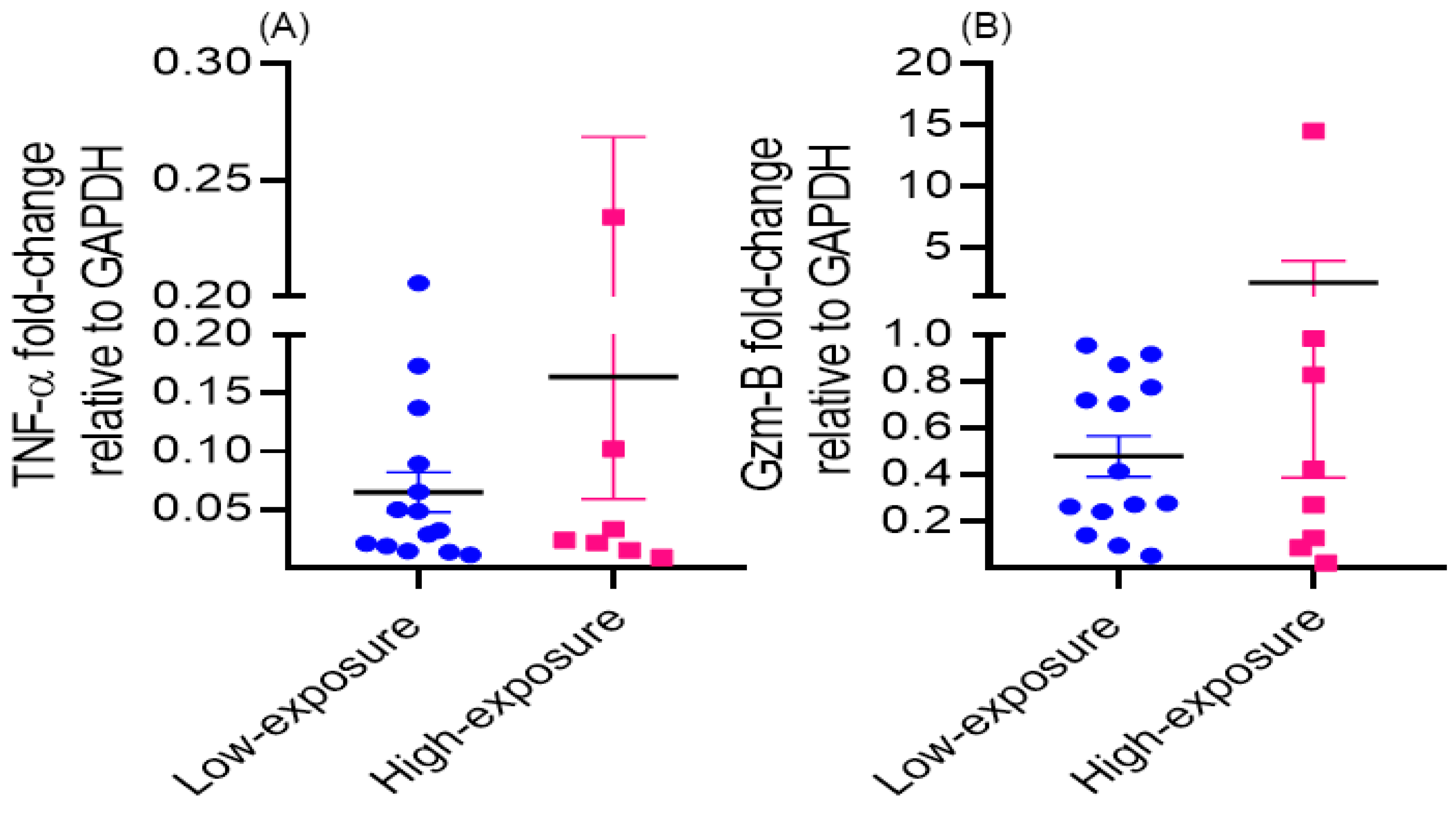
Figure 1. Gene expression levels of the inflammatory mediators TNF-α (A) and Granzyme-B (B) in the low-exposure group versus the high-exposure group of firefighters, measured in peripheral blood.
Environments 11 00131 g001
Figure 2. Plasma levels of heat shock protein-70 (HSP-70) in low exposure group versus high exposure group of firefighters.
Environments 11 00131 g002
Table 1. Demographic and exposure profiles of firefighter subjects exposed to low versus high “heavy smoke fires (Structural fires)” per week (Low-exposure group: <0.15/week or high-exposure group: >0.15/week).
Firefighter CharacteristicsLow-Exposure Group
N = 14
(Mean ± SE)
High-Exposure Group
N = 8
(Mean ± SE)
p-Value
Age (Years)36.42 ± 1.1334.25 ± 1.560.27
BMI (kg/m2)28.96 ± 0.9229.04 ± 0.800.95
Structure fires/week (number of heavy smoke conditions or structure fires in previous year ÷ number of weeks in the year)0.10 ± 0.0170.26 ± 0.0940.051
Number of times total in the last year worked in a structure fire or heavy smoke conditions5.50 ± 0.9213.50 ± 4.900.051
Years of full-time firefighter work10.10 ± 1.808.1 ± 2.40.52
Additional years worked as a part-time firefighter5.64 ± 0.944.75 ± 1.470.60
Total Years as firefighter (full-time + part-time)15.75 ± 1.3312.87 ± 2.530.28
Average hours per week worked as a firefighter over the past 6 months60.43 ± 2.4341.62 ± 4.470.001
Average shiftwork hours/year (per week × 52 weeks)3141.98 ± 122.552164.50 ± 232.950.001
Average shiftwork hours/month (per week × 52 weeks)/12261.83 ± 10.21180.37 ± 19.410.001
Career shift work (hours/year × years as firefighter)49,539.65 ± 4429.4828,236.0 ± 6876.900.013
On average, number of EMS runs per week over the past 6 months14.78 ± 3.1015.87 ± 5.370.852
BMI—Body mass index, EMS—Emergency medical service.
Table 2. Complete blood count (CBC) with differential cellular profiles of firefighter subjects exposed to low versus high “heavy smoke fires (Structural fires)” per week (Low-exposure group: <0.15/week or High-exposure group: >0.15/week).
CBC Parameters *Low-Exposure Group
N = 14
(Mean ± SE)
High-Exposure Group
N = 8
(Mean ± SE)
p-ValueReference Range ϒ
(Normal Value)
WBC (k/mcl)6.8 ± 0.476.07 ± 0.890.299.00–30.00
RBC(m/mcl)5.18 ± 0.085.07 ± 0.140.483.90–5.50
HGB (gm/dcl)15.40 ± 0.2314.98 ± 0.230.2513.5–19.5
HCT (%)46.19 ± 0.6244.61 ± 0600.1142.0–60.0
MCV (fL)89.18 ± 0.7486.96 ± 0.940.0898.0–118
MCH (pg)29.73 ± 0.2329.18 ± 0.280.1631.0–37.0
MCHC (gm/dL)33.34 ± 0.2533.56 ± 0.220.5630.0–36.0
RDWCV (%)14.18 ± 1.9112.43 ± 0.140.50NA
Platelets(k/mcl)264.78 ± 11.56253.25 ± 12.350.52135–466
MPV (fL)10.22 ± 0.2210.43 ± 0.290.5610.2–11.9
SEGS (%)58.73 ± 1.6661.11 ± 3.690.5067.0–87.0
Lymphocyte (%)30.55 ± 1.6028.18 ± 3.040.4528.0–34.0
Monocyte (%)7.8 ± 0.368.13 ± 0.660.630.0–12.0
Eosinophils (%)1.88 ± 0.331.51 ± 0.310.460.0–4.0
Basophils (%)0.43 ± 0.090.76 ± 0.080.0250.0–1.0
Immature granulocyte (%)0.25 ± 0.0340.52 ± 0.280.21NA
Neutrophils absolute (K/mcl)4.05 ± 0.353.76 ± 0.400.606.00–28.00
Lymphocyte absolute (K/mcl)3.70 ± 1.671.67 ± 0.150.382.00–11.00
Monocyte absolute (K/mcl)0.86 ± 0.320.52 ± 0.050.430.00–2.20
Eosinophils absolute counts (K/mcl)0.47 ± 0.250.08 ± 0.010.270.00–1.20
Basophils absolute counts (K/mcl)0.06 ± 0.0260.046 ± 0.0030.680.00–0.10
Immature Granulocyte absolute counts0.099 ± 0.0330.033 ± 0.0230.170.0–0.7
* Abbreviations: WBC (White blood cells); RBC (Red blood cells); HGB (Hemoglobin); HCT (Hemocrit); MCV (Mean corpuscular volume); MCH (Mean corpuscular hemoglobin): MCHC (Mean corpuscular hemoglobin concentration); RDWCV (Red cell distribution width); MPV (Mean platelets volume); SEGS (Segmented neutrophils). ϒ Normal reference range accepted for clinical diagnosis in the US healthy population. NA means the normal range value is ‘Not Available’.
Table 3. Gene expression levels for Innate and adaptive immune targets in firefighter subjects exposed to low versus high heavy smoke fires/Structural fires (Low exposure group: <0.15/week versus High exposure group: >0.15/week).
Target Gene/ProteinFold-Change (FC) Relative to House-Keeping GeneFold-Difference
Low-Exposure Group
N = 14
(Mean ± SE)
High-Exposure Group
N = 8
(Mean ± SE)
(high-Exposure FC/Low-Exposure FC)
TLR20.48 ± 0.090.31 ± 0.12−1.51
TLR40.44 ± 0.090.20 ± 0.07−2.21
TLR70.088 ± 0.0360.16 ± 0.121.58
TLR90.15 ± 0.050.15 ± 0.10−1.03
MYD880.40 ± 0.100.27 ± 0.11−1.48
T-bet (Th1)5.46 ± 2.125.58 ± 2.65−1.19
RORC (Th17)54.86 ± 17.2192.78 ± 68.471.69
FoxP3 (Treg)(0.03 ± 0.006) × 10−2(0.12 ± 0.07) × 10−23.52
T-bet/FoxP3 (Th1/Treg)(15.64 ± 4.02) × 103(11.9 ± 5.93) × 103−1.31
RORC/FoxP3 (Th17/Treg) (180.6 ± 53.3) × 103(102.6 ± 3.5) × 103−1.76
Disclaimer/Publisher’s Note: The statements, opinions and data contained in all publications are solely those of the individual author(s) and contributor(s) and not of MDPI and/or the editor(s). MDPI and/or the editor(s) disclaim responsibility for any injury to people or property resulting from any ideas, methods, instructions or products referred to in the content.