Endotype-Guided Imaging in Chronic Rhinosinusitis: HRCT/CBCT and MRI Metrics, Structured Reporting, and Radiomics-A Systematic Review



Abstract
1. Introduction
2. Materials and Methods
Eligibility Criteria (PICO)
3. Results
3.1. Study Selection and Included Evidence
3.2. HRCT and CBCT Acquisition Protocols and Quantitative Indices
3.3. MRI Indications and Clinically Relevant Performance Domains
3.4. Imaging Patterns Associated with CRS Phenotypes and Inflammatory Endotypes
3.5. Complications, Skull Base Defects, and Unilateral Disease
3.6. Radiomics and Artificial Intelligence
3.7. Environmental Exposure Studies (Hypothesis-Generating)
4. Discussion
5. Conclusions
Supplementary Materials
Author Contributions
Funding
Institutional Review Board Statement
Informed Consent Statement
Data Availability Statement
Conflicts of Interest
Abbreviations
| AFRS | Allergic Fungal Rhinosinusitis |
| AI | Artificial Intelligence |
| AUC | Area Under the Curve |
| CBCT | Cone-Beam Computed Tomography |
| CRS | Chronic Rhinosinusitis |
| CRSsNP | Chronic Rhinosinusitis without Nasal Polyps |
| CRSwNP | Chronic Rhinosinusitis with Nasal Polyps |
| CTA | Computed Tomography Angiography |
| CT | Computed Tomography |
| DLP | Dose Length Product |
| FESS | Functional Endoscopic Sinus Surgery |
| FLAIR | Fluid-Attenuated Inversion Recovery |
| FOV | Field of View |
| GOSS | Global Osteitis Scoring Scale |
| HRCT | High-Resolution Computed Tomography |
| IBSI | Image Biomarker Standardisation Initiative |
| IFRS | Invasive Fungal Rhinosinusitis |
| LM | Lund–Mackay Score |
| LOEM | Lamella Ostium Extent Mucosa |
| MRI | Magnetic Resonance Imaging |
| PM | Particulate Matter |
| PRISMA | Preferred Reporting Items for Systematic Reviews |
| ROI | Region of Interest |
| RARS | Recurrent Acute Rhinosinusitis |
| SR | Structured Reporting |
| T2-high | Type 2 Inflammation-High Endotype |
Appendix A

| Database | Search Strategy (Verbatim) | Limits/Notes |
|---|---|---|
| MEDLINE (PubMed) | (“Rhinosinusitis”[Mesh] OR “Rhinosinusitis, Chronic”[Mesh] OR “chronic rhinosinusitis”[tiab] OR CRS[tiab] OR CRSwNP[tiab] OR CRSsNP[tiab] OR “nasal polyp*”[tiab]) AND (“Tomography, X-Ray Computed”[Mesh] OR CT[tiab] OR “computed tomography”[tiab] OR HRCT[tiab] OR “cone-beam computed tomography”[tiab] OR CBCT[tiab] OR “Magnetic Resonance Imaging”[Mesh] OR MRI[tiab]) AND (endotype*[tiab] OR phenotype*[tiab] OR eosinophil*[tiab] OR “type 2”[tiab] OR “type II”[tiab] OR T2[tiab] OR IL-4[tiab] OR IL-5[tiab] OR IL-13[tiab] OR IgE[tiab] OR biologic*[tiab] OR dupilumab[tiab] OR omalizumab[tiab] OR mepolizumab[tiab] OR benralizumab[tiab] OR radiomic*[tiab] OR “artificial intelligence”[tiab] OR “machine learning”[tiab] OR “deep learning”[tiab] OR “structured reporting”[tiab] OR “Lund-Mackay”[tiab] OR GOSS[tiab] OR osteitis[tiab] OR “olfactory cleft”[tiab]) AND (“1 January 2000”[Date-Publication]: “30 September 2025”[Date-Publication]) | Date limits applied within query; no language filter at search stage. |
| Embase | (‘chronic rhinosinusitis’/exp OR ‘rhinosinusitis’/exp OR ‘chronic rhinosinusitis’:ti,ab OR rhinosinusitis:ti,ab OR CRS:ti,ab OR CRSwNP:ti,ab OR CRSsNP:ti,ab OR ‘nasal polyp’/exp OR ‘nasal polyp*’:ti,ab) AND (‘computed tomography’/exp OR ‘computed tomography’:ti,ab OR CT:ti,ab OR HRCT:ti,ab OR ‘cone beam computed tomography’/exp OR CBCT:ti,ab OR ‘magnetic resonance imaging’/exp OR MRI:ti,ab) AND (endotype*:ti,ab OR phenotype*:ti,ab OR eosinophil*:ti,ab OR ‘type 2’:ti,ab OR ‘type II’:ti,ab OR T2:ti,ab OR IL-4:ti,ab OR IL-5:ti,ab OR IL-13:ti,ab OR IgE:ti,ab OR biologic*:ti,ab OR dupilumab:ti,ab OR omalizumab:ti,ab OR mepolizumab:ti,ab OR benralizumab:ti,ab OR radiomic*:ti,ab OR ‘artificial intelligence’:ti,ab OR ‘machine learning’:ti,ab OR ‘deep learning’:ti,ab OR ‘structured reporting’:ti,ab OR ‘Lund-Mackay’:ti,ab OR GOSS:ti,ab OR osteitis:ti,ab OR ‘olfactory cleft’:ti,ab) AND [2000–2025]/py | Year filter [2000–2025]/py; no language filter at search stage. |
| Scopus | TITLE-ABS-KEY ((“chronic rhinosinusitis” OR rhinosinusitis OR CRS OR CRSwNP OR CRSsNP OR “nasal polyp*”) AND (HRCT OR “computed tomography” OR CT OR CBCT OR “cone beam” OR MRI OR “magnetic resonance”) AND (endotype* OR phenotype* OR eosinophil* OR “type 2” OR “type II” OR IL-4 OR IL-5 OR IL-13 OR IgE OR biologic* OR dupilumab OR omalizumab OR mepolizumab OR benralizumab OR radiomic* OR “artificial intelligence” OR “machine learning” OR “deep learning” OR “structured reporting” OR “Lund-Mackay” OR GOSS OR osteitis OR “olfactory cleft”)) AND PUBYEAR > 1999 AND PUBYEAR < 2026 | Publication year 2000–2025; TITLE-ABS-KEY fields. |
| Cochrane Library | (chronic next rhinosinusitis OR rhinosinusitis OR CRS OR CRSwNP OR CRSsNP OR “nasal polyp*”):ti,ab,kw AND (CT OR “computed tomography” OR HRCT OR CBCT OR “cone beam” OR MRI OR “magnetic resonance”):ti,ab,kw AND (endotype* OR phenotype* OR eosinophil* OR “type 2” OR “type II” OR IL-4 OR IL-5 OR IL-13 OR IgE OR biologic* OR dupilumab OR omalizumab OR mepolizumab OR benralizumab OR radiomic* OR “artificial intelligence” OR “machine learning” OR “deep learning” OR “structured reporting” OR “Lund-Mackay” OR GOSS OR osteitis OR “olfactory cleft”):ti,ab,kw | Trials and reviews searched; year limits set in interface (2000–2025). |
| Evidence Type | Tool/Framework | Most Frequent Concerns | Interpretation Notes |
|---|---|---|---|
| Diagnostic accuracy imaging studies (CT/MRI tasks) | QUADAS-2 | Patient selection (retrospective/non-consecutive sampling); heterogeneous reference standards (biomarkers/histology thresholds); incomplete blinding or unclear thresholds for index tests. | Applicability is generally acceptable for tertiary CRS settings; caution when extrapolating performance estimates to community practice. |
| Non-randomized comparative studies (e.g., surgical pathway or protocol comparisons) | ROBINS-I | Confounding (disease severity, prior surgery, comorbid asthma); selection bias; outcome measurement heterogeneity. | Findings should be interpreted as associative rather than causal; prospective designs are needed. |
| Radiomics/AI prediction or classification models | CLAIM/TRIPOD principles (plus IBSI where applicable) | Small datasets and single-center designs; variable segmentation and feature extraction settings; limited external validation; inconsistent calibration/clinical utility reporting. | Reported AUCs are task-specific and may overestimate generalizability without external validation. |
| Environmental exposure observational analyses | Observational quality appraisal (confounding/temporality) | Potential confounding (seasonality, treatment changes, comorbid allergy/asthma); exposure misclassification; outcomes often symptom-based rather than standardized imaging endpoints. | Hypothesis-generating; not sufficient to define imaging indications or schedules. |
| Category | Parameter/Indicator | Modality/Group | Key Finding (Range/% Where Reported) | Notes/Caveats |
|---|---|---|---|---|
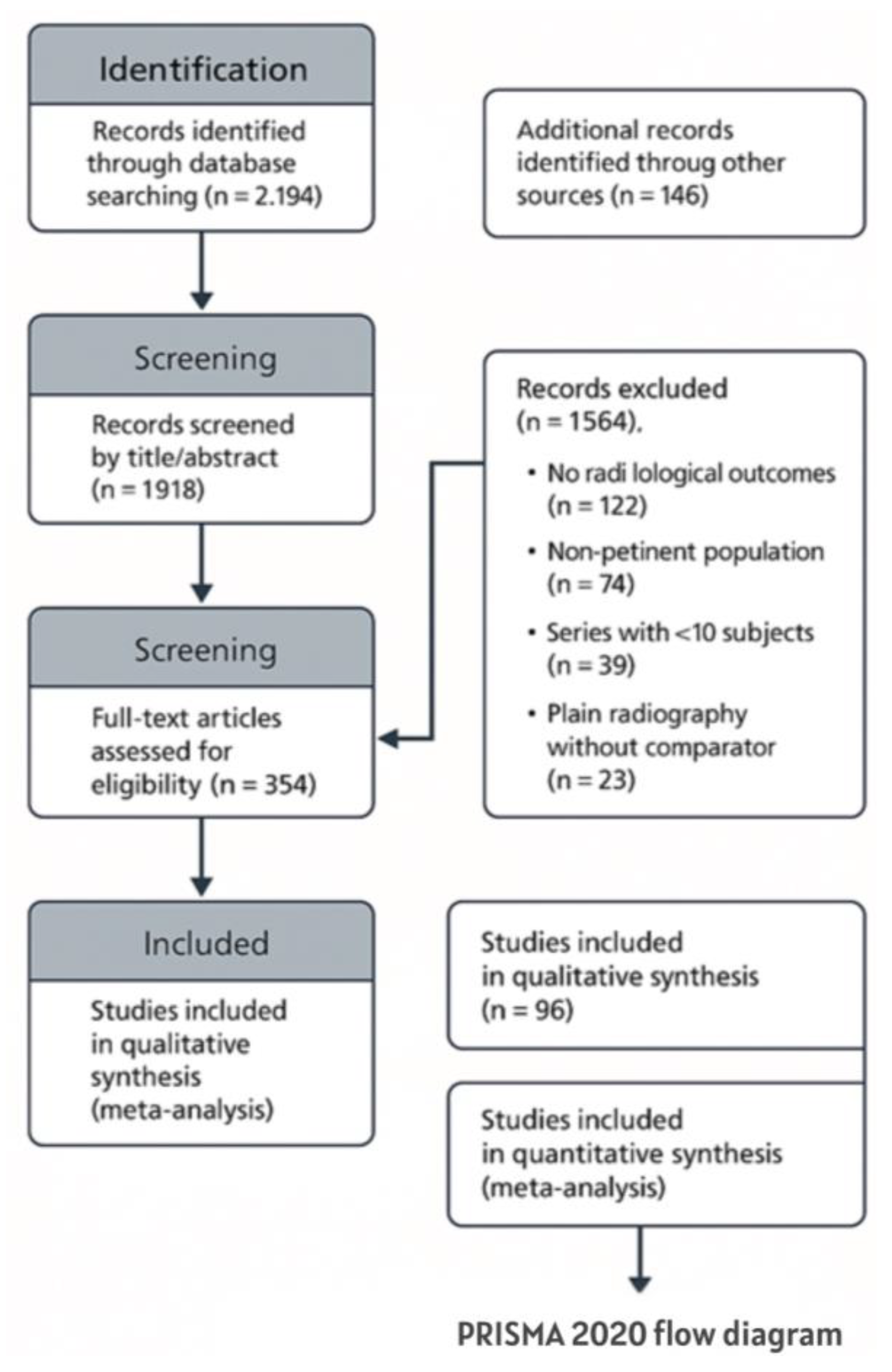
| Study identification | Total articles (2000–2025) | Databases + other sources | 2330 records | 96 included in qualitative synthesis; 38 studies reported quantitative outcomes suitable for descriptive summary. |
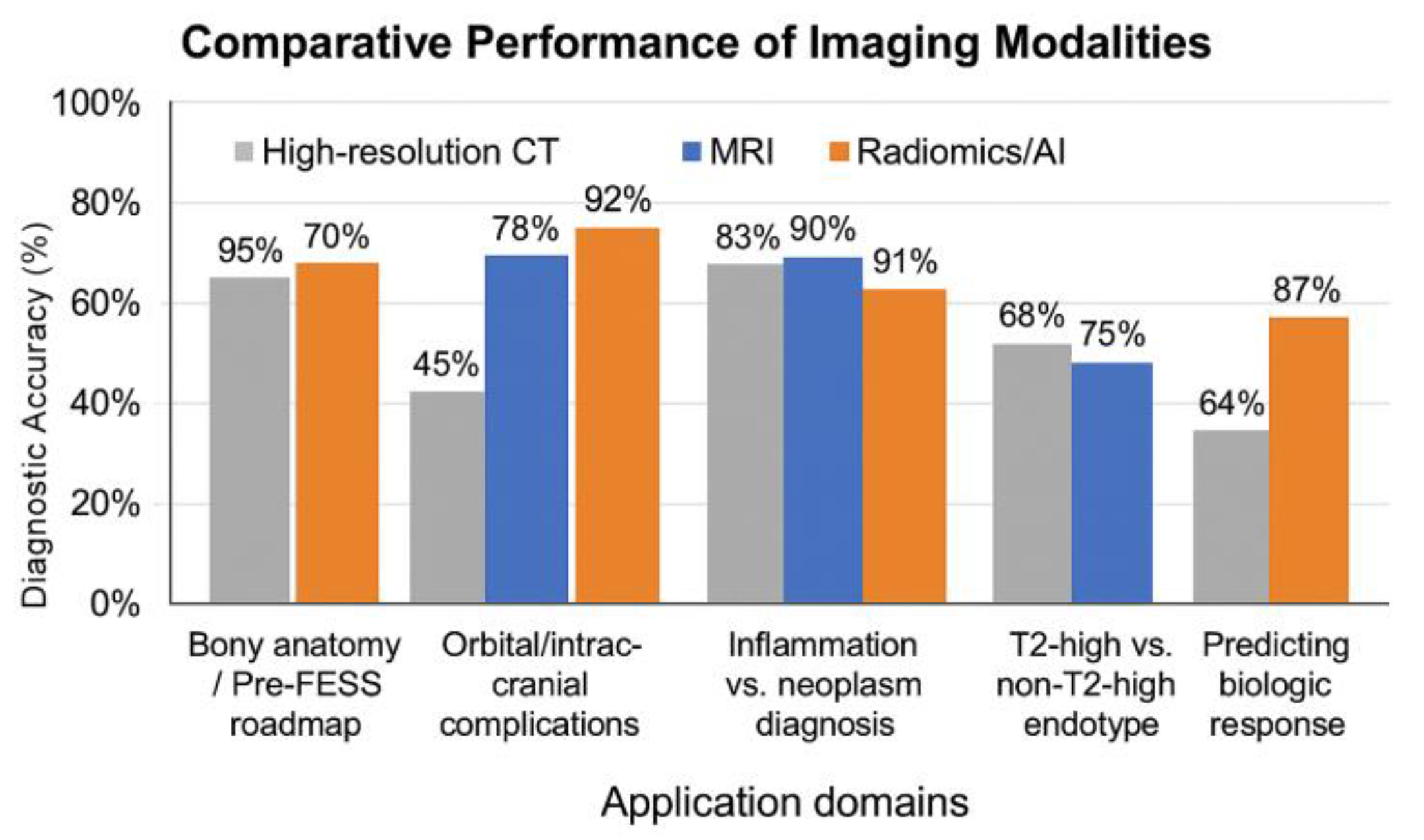
| CT HR | Utilization across studies | - | 92% | Reference method for pre-FESS planning and bony anatomy assessment. |
| CT HR | Mean slice thickness | CT HR | Typically submillimetric | Reconstructed thickness commonly 0.6–1.0 mm; reporting varied across years and scanners. |
| CT HR | Lund–Mackay score | CT (HRCT/CBCT where applicable) | Frequently used | Standard 0–24 staging metric; useful for standardization but not a standalone predictor of endotype or outcomes. |
| CT HR | Osteitis/bony remodeling grading (e.g., GOSS) | CT (HRCT) | Reported in selected cohorts | Supports characterization of bony remodeling; definitions and thresholds varied across studies. |
| CT HR | Inter-reader agreement | CT HR | κ = 0.84 | High reproducibility |
| MRI | Utilization across studies | - | 58% | Mainly used for soft-tissue characterization, unilateral disease, and suspected complications. |
| MRI | ADC (inflammatory lesions) | MRI | 1.45 ± 0.3 × 10⁻³ mm2/s | Free diffusion |
| MRI | ADC (neoplastic lesions) | MRI | 0.85 ± 0.2 × 10⁻³ mm2/s | Marked restriction |
| MRI | Diagnostic performance (retained secretions vs. enhancing soft tissue) | MRI | Up to ~92% (selected studies) | Task-specific estimates; depends on sequences and reference standard. |
| CRS phenotypes/endotypes | Type 2-high CRSwNP (biomarker-defined when reported) | CT/MRI | Ethmoid-dominant pattern; olfactory cleft involvement | Supportive imaging pattern; endotype remains biomarker-based. Osteitis was more frequently reported in several cohorts. |
| CRS phenotypes/endotypes | CRSsNP/odontogenic CRS | CT (HRCT/CBCT) | Maxillary predominance; dental root relationship | Imaging supports etiologic suspicion (e.g., odontogenic source) when correlated with dental findings. |
| Complications | Orbital/intracranial | CT + MRI | 14% | Represents proportion in selected surgical series; definitions varied. |
| Complications | CSF leak/meningoencephalocele | CT HR + MRI | 4% | High accuracy pertains to selected skull base defects (e.g., CSF leak/meningoencephalocele). |
| Bone erosion | Inflammatory | CT HR/MRI | 41% | Smooth margins, reactive cortical sclerosis |
| Bone erosion | Neoplastic | CT HR/MRI | 18% | Irregular margins, cortical disruption |
| Radiomics/AI | Studies with a radiomics-ready pipeline | - | 12% of included studies | Radiomics-ready pipelines; most used HRCT. IBSI alignment was variably reported. |
| Radiomics/AI | AUC (type 2-high vs. non-type 2-high discrimination) | CT/MRI | 0.79–0.93 | Reported across heterogeneous models; external validation in 38% of studies. Not pooled. |
| Radiomics/AI | AUC (inflammation vs. neoplasia discrimination in unilateral disease workflows) | CT/MRI | Around 0.91 (selected studies) | Task-specific; depends on case mix and reference standard. Not pooled. |
| Radiomics/AI | ΔRad-score in biologic therapy (16–24 weeks) | MRI | r = 0.71 | Reported correlation in selected studies; interpretation limited by heterogeneous definitions and short follow-up. |
| Environmental factors | PM/NO2/O3 (seasonal burden) | - | Heterogeneous associations | Hypothesis-generating observational data; insufficient to define imaging indications or schedules. |
| Endotype/Phenotype | CT/MRI Hallmarks | Suggested Reporting Statement (Interpretation/Next Steps) |
|---|---|---|
| CRSwNP T2-high (eosinophilic) | Ethmoid-dominant pattern, olfactory cleft involvement, osteitis (elevated GOSS) | Report Lund–Mackay score per sinus/side, olfactory cleft status, and osteitis/remodeling (e.g., GOSS). Comment: ‘pattern suggestive of type 2-high inflammation; correlate with endoscopic findings and available biomarkers to support endotype assignment and treatment planning.’ |
| CRSsNP/odontogenic | Maxillary predominance, asymmetric mucosal thickening, and dental root involvement | Indicate imaging features suggestive of an odontogenic source (e.g., periapical disease, oroantral communication) when present; recommend correlation with dental/endoscopic evaluation. |
| AFRS | CT: central hyperattenuation; MRI: T2-void signal | Suggest AFRS in the appropriate clinical context; describe features relevant to surgical planning and postoperative medical therapy (e.g., corticosteroids). |
| IFRS | Periantral or pterygopalatine infiltration; MRI black turbinate sign | If clinical suspicion of invasive fungal rhinosinusitis exists, explicitly report extrasinus spread and vascular/orbital/intracranial red flags; recommend urgent ENT evaluation. Contrast-enhanced MRI can be used to assess the extent of disease. |
| Cerebrospinal fluid leak/meningoencephalocele | Bony defect on HRCT; CSF signal hyperintense on T2, suppressed on FLAIR | Report the site and size of the bony defect and associated herniation/CSF signal; recommend referral to a skull base team for endoscopic repair planning. |
Appendix B

References
- Fokkens, W.J.; Lund, V.J.; Hopkins, C.; Hellings, P.W.; Kern, R.; Reitsma, S.; Toppila-Salmi, S.; Bernal-Sprekelsen, M.; Mullol, J.; Alobid, I.; et al. European Position Paper on Rhinosinusitis and Nasal Polyps 2020. Rhinology 2020, 29, 1–464. [Google Scholar] [CrossRef]
- Nyaiteera, V.; Nakku, D.; Nakasagga, E.; Llovet, E.; Kakande, E.; Nakalema, G.; Byaruhanga, R.; Bajunirwe, F. The burden of chronic rhinosinusitis and its effect on quality of life among patients reattending an otolaryngology clinic in Southwestern Uganda. BMC Ear Nose Throat Disord. 2018, 18, 10. [Google Scholar] [CrossRef] [PubMed]
- Expert Panel on Neurological Imaging; Hagiwara, M.; Policeni, B.; Juliano, A.F.; Agarwal, M.; Burns, J.; Dubey, P.; Friedman, E.R.; Gule-Monroe, M.K.; Jain, V.; et al. ACR Appropriateness Criteria® Sinonasal Disease: 2021 Update. J. Am. Coll. Radiol. JACR 2022, 19, S175–S193. [Google Scholar] [CrossRef] [PubMed]
- Georgalas, C.; Videler, W.; Freling, N.; Fokkens, W. Global Osteitis Scoring Scale and chronic rhinosinusitis: A marker of revision surgery. Clin. Otolaryngol. 2010, 35, 455–461. [Google Scholar] [CrossRef]
- Huang, Z.Q.; Liu, J.; Sun, L.Y.; Ong, H.H.; Ye, J.; Xu, Y.; Wang, D.Y. Updated epithelial barrier dysfunction in chronic rhinosinusitis: Targeting pathophysiology and treatment response of tight junctions. Allergy 2024, 79, 1146–1165. [Google Scholar] [CrossRef]
- Toppila-Salmi, S.; Reitsma, S.; Hox, V.; Gane, S.; Eguiluz-Gracia, I.; Shamji, M.; Maza-Solano, J.; Jääskeläinen, B.; Väärä, R.; Escribese, M.M.; et al. Endotyping in Chronic Rhinosinusitis-An EAACI Task Force Report. Allergy 2025, 80, 132–147. [Google Scholar] [CrossRef] [PubMed]
- Darbari Kaul, R.; Sacks, P.L.; Thiel, C.; Rimmer, J.; Kalish, L.; Campbell, R.G.; Sacks, R.; Di Ieva, A.; Harvey, R.J. Radiomics of the Paranasal Sinuses: A Systematic Review of Computer-Assisted Techniques to Assess Computed Tomography Radiological Data. Am. J. Rhinol. Allergy 2025, 39, 147–158. [Google Scholar] [CrossRef]
- Zwanenburg, A.; Vallières, M.; Abdalah, M.A.; Aerts, H.J.W.L.; Andrearczyk, V.; Apte, A.; Ashrafinia, S.; Bakas, S.; Beukinga, R.J.; Boellaard, R.; et al. The Image Biomarker Standardization Initiative: Standardized Quantitative Radiomics for High-Throughput Image-Based Phenotyping. Radiology 2020, 295, 328–338. [Google Scholar] [CrossRef]
- Mongan, J.; Moy, L.; Kahn, C.E. Checklist for Artificial Intelligence in Medical Imaging (CLAIM): A Guide for Authors and Reviewers. Radiol. Artif. Intell. 2020, 2, e200029. [Google Scholar] [CrossRef]
- Bachert, C.; Hicks, A.; Gane, S.; Peters, A.T.; Gevaert, P.; Nash, S.; Horowitz, J.E.; Sacks, H.; Jacob-Nara, J.A. The interleukin-4/interleukin-13 pathway in type 2 inflammation in chronic rhinosinusitis with nasal polyps. Front. Immunol. 2024, 15, 1356298. [Google Scholar] [CrossRef]
- Castelnuovo, P.; Battaglia, P.; Simoncello, F. Attualità nella rinosinusite: Diagnosi, imaging, terapia e chirurgia. Acta Otorhinolaryngol. Ital. 2008, 2, 13–56. [Google Scholar]
- Page, M.J.; McKenzie, J.E.; Bossuyt, P.M.; Boutron, I.; Hoffmann, T.C.; Mulrow, C.D.; Shamseer, L.; Tetzlaff, J.M.; Akl, E.A.; Brennan, S.E.; et al. The PRISMA 2020 Statement: An Updated Guideline for Reporting Systematic Reviews. J. Clin. Epidemiol. 2021, 134, 178–189. [Google Scholar] [CrossRef]
- Sanchez-Gomez, S.; Moreno-Luna, R.; Martin-Jimenez, D.; Maza-Solano, J.; del Cuvillo, A.; Villacampa-Auba, J.-M.; Gonzalez-Garcia, J.; Fernandez-Liesa, R.; Alobid, I.; Bernal-Sprekelsen, M. The Lamella Ostium Extent Mucosa (LOEM) system: A new classification for endoscopic sinus surgery. Rhinology 2025, 63, 125–128. [Google Scholar] [CrossRef] [PubMed]
- Collins, G.S.; Reitsma, J.B.; Altman, D.G.; Moons, K.G.M. Transparent Reporting of a Multivariable Prediction Model for Individual Prognosis or Diagnosis (TRIPOD): The TRIPOD Statement. Ann. Intern. Med. 2015, 162, 55–63. [Google Scholar] [CrossRef]
- Castelnuovo, P.; Battaglia, P.; Simoncello, F. Quando è necessaria la chirurgia. Acta Otorhinolaryngol. Ital. 2008, 2, 50–56. [Google Scholar]
- Dykewicz, M.S.; Wallace, D.V.; Amrol, D.J.; Baroody, F.M.; Bernstein, J.A.; Craig, T.J.; Dinakar, C.; Ellis, A.K.; Finegold, I.; Golden, D.B.; et al. Rhinitis 2020: Practice Parameter Update. J. Allergy Clin. Immunol. 2020, 146, 721–767. [Google Scholar] [CrossRef]
- Kim, D.W.; Kim, J.Y.; Jeon, S.Y. The status of the olfactory cleft may predict postoperative olfactory function in chronic rhinosinusitis with nasal polyposis. Am. J. Rhinol. Allergy 2011, 25, e90–e94. [Google Scholar] [CrossRef] [PubMed]
- Castelnuovo, R.; Battaglia, P.; Simoncello, F. Le forme monolaterali: Diagnosi differenziale. Acta Otorhinolaryngol. Ital. 2008, 2, 46–54. [Google Scholar]
- Whiting, P.F.; Rutjes, A.W.S.; Westwood, M.E.; Mallett, S.; Deeks, J.J.; Reitsma, J.B.; Leeflang, M.M.G.; Sterne, J.A.C.; Bossuyt, P.M.M.; QUADAS-2 Group. QUADAS-2: A Revised Tool for the Quality Assessment of Diagnostic Accuracy Studies. Ann. Intern. Med. 2011, 155, 529–536. [Google Scholar] [CrossRef]
- Sterne, J.A.C.; Hernán, M.A.; Reeves, B.C.; Savović, J.; Berkman, N.D.; Viswanathan, M.; Henry, D.; Altman, D.G.; Ansari, M.T.; Boutron, I.; et al. ROBINS-I: A Tool for Assessing Risk of Bias in Non-Randomised Studies of Interventions. BMJ 2016, 355, i4919. [Google Scholar] [CrossRef] [PubMed]
- Higgins, J.P.T.; Savović, J.; Page, M.J.; Elbers, R.G.; Sterne, J.A.C. RoB 2: A Revised Tool for Assessing Risk of Bias in Randomized Trials. BMJ 2019, 366, l4898. [Google Scholar] [CrossRef]
- Mullol, J.; Azar, A.; Buchheit, K.M.; Hopkins, C.; Bernstein, J.A. Chronic Rhinosinusitis with Nasal Polyps: Quality of Life in the Biologics Era. J. Allergy Clin. Immunol. Pract. 2022, 10, 1434–1453.e9. [Google Scholar] [CrossRef]
- Becker, S.; Huppertz, T.; Möller, W.; Havel, M.; Schuster, M.; Becker, A.M.; Sailer, M.; Schuschnig, U.; Johnson, T.R. Xenon-Enhanced Dynamic Dual-Energy CT Is Able to Quantify Sinus Ventilation Using Laminar and Pulsating Air-/Gas Flow Before and After Surgery: A Pilot Study in a Cadaver Model. Front. Allergy 2022, 3, 829898. [Google Scholar] [CrossRef] [PubMed]
- Poto, R.; Pelaia, C.; di Salvatore, A.; Saleh, H.; Scadding, G.W.; Varricchi, G. Imaging of chronic rhinosinusitis with nasal polyps in the era of biological therapies. Curr. Opin. Allergy Clin. Immunol. 2024, 24, 243–250. [Google Scholar] [CrossRef] [PubMed]


| Section | Essential Elements (Minimum Reporting Set) |
|---|---|
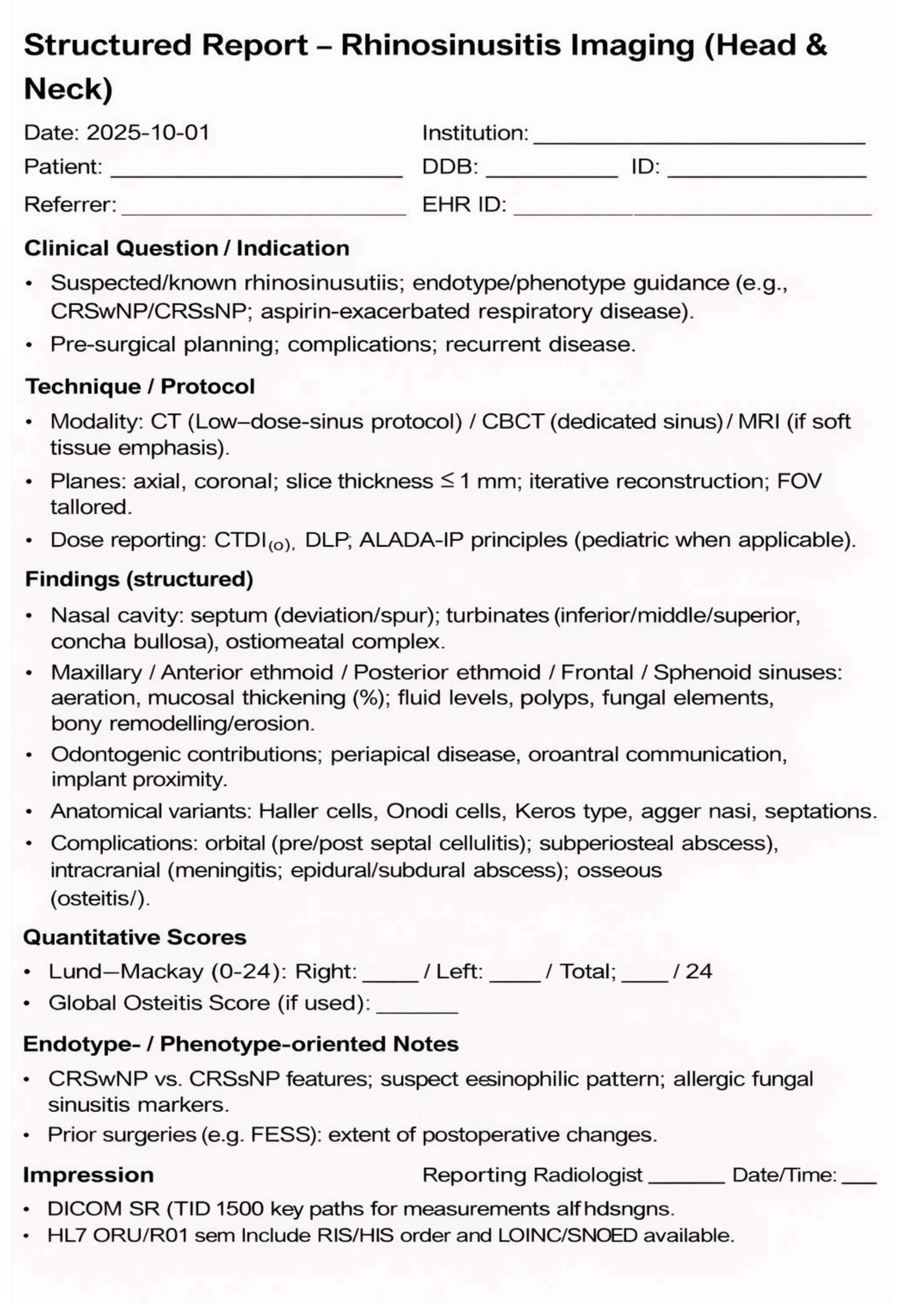
| Technique/dose (Essential) | CT: kVp/mAs, kernel, slice thickness ≤ 1 mm, iterative reconstruction, CTDIvol/DLP; CBCT: bone assessment only (no soft-tissue characterization); MRI: T2-weighted and T1-weighted ± gadolinium sequences; isotropic voxels when available. |
| Critical variants (Essential) | Onodi cell; Haller cell; Keros classification; uncinate process; lamina papyracea; cribriform plate. |
| Staging | Lund–Mackay score per sinus and side; ethmoid vs. maxillary-dominant distribution. |
| Osteitis (Optional /Advanced) | Global Osteitis Scoring Scale (GOSS) or a standardized description of cortical thickening and bony remodeling. |
| Olfactory cleft (Optional /Advanced) | Olfactory cleft opacification/edema; correlation with olfactory symptoms and T2-high pattern (when clinically available). |
| Complications (Essential) | Orbital complications (e.g., subperiosteal abscess, cavernous sinus thrombosis) and intracranial complications (e.g., empyema/abscess); flags suggestive of IFRS when present. |
| Radiomics (if applicable) (Optional /Advanced) | IBSI compliance statement; ROI definition (ethmoid, olfactory cleft, maxillary sinuses, bony walls); feature extraction settings (resampling/discretization); declared radiomic score (rad-score), calibration, and external validation if available. |
| Operational conclusion (Essential) | “Findings-to-management” statement: pre-FESS roadmap; indicate when MRI is recommended based on the clinical scenario; highlight red flags and propose follow-up imaging only when clinically indicated (i.e., when results are expected to change management). |
Disclaimer/Publisher’s Note: The statements, opinions and data contained in all publications are solely those of the individual author(s) and contributor(s) and not of MDPI and/or the editor(s). MDPI and/or the editor(s) disclaim responsibility for any injury to people or property resulting from any ideas, methods, instructions or products referred to in the content. |
© 2026 by the authors. Licensee MDPI, Basel, Switzerland. This article is an open access article distributed under the terms and conditions of the Creative Commons Attribution (CC BY) license.
Share and Cite
Messineo, D.; Frisina, P.; Begvarfaj, E.; Panebianco, V. Endotype-Guided Imaging in Chronic Rhinosinusitis: HRCT/CBCT and MRI Metrics, Structured Reporting, and Radiomics-A Systematic Review. Med. Sci. 2026, 14, 274. https://doi.org/10.3390/medsci14020274
Messineo D, Frisina P, Begvarfaj E, Panebianco V. Endotype-Guided Imaging in Chronic Rhinosinusitis: HRCT/CBCT and MRI Metrics, Structured Reporting, and Radiomics-A Systematic Review. Medical Sciences. 2026; 14(2):274. https://doi.org/10.3390/medsci14020274
Chicago/Turabian StyleMessineo, Daniela, Pasquale Frisina, Elona Begvarfaj, and Valeria Panebianco. 2026. "Endotype-Guided Imaging in Chronic Rhinosinusitis: HRCT/CBCT and MRI Metrics, Structured Reporting, and Radiomics-A Systematic Review" Medical Sciences 14, no. 2: 274. https://doi.org/10.3390/medsci14020274
APA StyleMessineo, D., Frisina, P., Begvarfaj, E., & Panebianco, V. (2026). Endotype-Guided Imaging in Chronic Rhinosinusitis: HRCT/CBCT and MRI Metrics, Structured Reporting, and Radiomics-A Systematic Review. Medical Sciences, 14(2), 274. https://doi.org/10.3390/medsci14020274