Invasive Alien Species of European Union Concern: A Systematic Review of High-Priority Pathogens in 22 Species in a One Health Framework




Simple Summary
Abstract
1. Introduction
2. Materials and Methods
2.1. Study Design and Species Selection
2.2. Systematic Review of the Literature
2.2.1. Inclusion and Exclusion Criteria
2.2.2. Quality Assessment and Risk of Bias
2.3. Data Integration and Prioritization of Pathogens of Public and Animal Health Significance in the European Context
2.4. Epidemiological Risk Analysis
2.4.1. Assessment of Invasion Pressure and Spatial Expansion
2.4.2. Epidemiologically Relevant IAS
- Host breadth (specialist, restricted multi-host, generalist),
- Transmission route complexity (complex/rare, moderate, direct/environmental),
- Life-cycle structure (obligate indirect, flexible indirect, direct),
- One Health impact level (low, high).
2.4.3. Final Composite Index
- represents dynamic invasion pressure,
- represents the proportion of epidemiologically relevant IAS,
- represents pathogen infectivity potential,
- represents observed pathogen evidence.
2.4.4. Post Hoc Validation and Statistical Robustness Analysis
3. Results
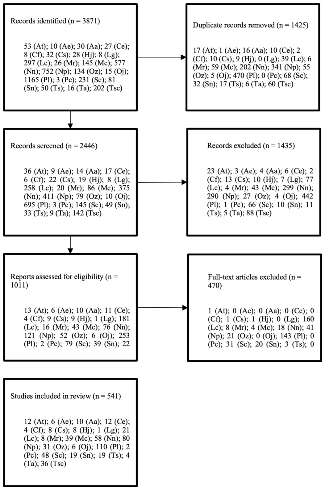
3.1. Systematic Review of the Literature
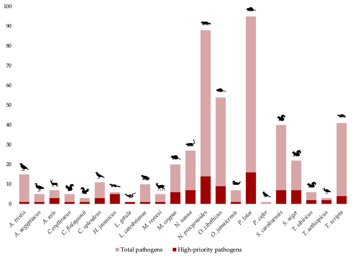
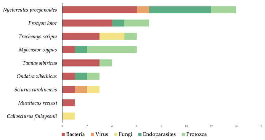
3.2. Pathogen Species in Invasive Alien Species (IAS)
3.2.1. High-Priority Bacteria Species in IAS

| Bacteria | Document | IAS Involved | Pathogen Species | Location EU | Location EXTRA EU |
|---|---|---|---|---|---|
| Borrelia spp. | Directive Zoonoses 2003/99/EC | Mammals: Procyon lotor, Nyctereutes procyonoides, Sciurus carolinensis, Sciurus niger, Tamias sibiricus | B. spp., B. afzelli, B. burgdorferi, B. garinii, B. lonestarii, B. lusitaniae, B. miyatomotoi, B. theileri, B. turicatae, B. valaisiana | France (Vourc’h et al. (2007) [62], Marsot et al. (2011) [64], Marsot et al. (2013) [104], Marchant et al. (2017) [63]), Denmark (Kjær et al. (2021) [105]), Italy (Cruciani et al. (2022) [106]), Poland (Wodecka et al. (2016) [60], Myśliwy et al. (2022) [61]). | Republic of Korea (Kang et al. (2018) [107], Myśliwy et al. (2022) [61]), UK (Millins et al. (2015) [108]), USA (Salkeld et al. (2008) [109], Roy et al. (2017) [110]). |
| Brucella abortus | Animal Health Law Reg. UE 2016/429 | Mammals: Nasua nasua | B. abortus | Brazil (Oliveira-Filho et al. (2012) [111]). | |
| Brucella spp. | Directive Zoonoses 2003/99/EC | Mammals: Nasua nasua | B. abortus, B. canis | Brazil (Oliveira-Filho et al. (2012) [111]). | |
| Campylobacter spp. | Directive Zoonoses 2003/99/EC | Mammals: Herpestes javanicus, Nyctereutes procyonoides, Procyon lotor, Ondatra zibethicus. Birds: Corvus splendes | C. spp., C. jejuni, C. coli | Barbados (Rhynd et al. 2014) [77]), Canada (Mutschall et al. (2020) [66]), Japan (Lee et al. (2011) [112], Takako et al. (2021) [65]), Malaysia (Ganapathy et al. 2007) [102]), USA (Pacha et al. (1985) [67], Rainwater et al. (2017) [90]). | |
| Chlamydia psittaci | Directive Zoonoses 2003/99/EC | Mammals: Myocastor coypus | C. psittaci | Argentina (Martino et al. (2014) [113]). | |
| Clostridium botulinum, C. butyricum, C. baratii | Directive Zoonoses 2003/99/EC | Reptiles: Trachemys scripta | C. butyricum type E, C. butyricum BoNT/E | Ireland (Shelley et al. (2015) [114]). | |
| Francisella spp. | Directive Zoonoses 2003/99/EC | Mammals: Nyctereutes procyonoides, Procyon lotor, Ondatra zibethicus, Sciurus carolinensis, Sciurus niger | F. tularensis | Germany (Kuehn et al. (2013) [76]), Sweden (Hestvik et al. (2019) [71]). | Canada (Ganoe et al. (2020) [68]), USA (Bischof and Rogers (2005) [75], Duncan et al. (2012) [74], Nelson et al. (2014) [69], Vincent et al. (2020) [115], Ganoe et al. (2020) [68], Jahan et al. (2021) [70]). |
| Klebsiella pneumoniae | WHO Pathogens Prioritization | Mammals: Ondatra zibethicus. Birds: Corvus splendens | K. pneumoniae | Bangladesh (Hasan et al. (2015) [116]), USA (Grear et al. (2019) [117]). | |
| Leptospira spp. | Directive Zoonoses 2003/99/EC | Mammals: Herpestes javanicus, Nasua nasua, Nyctereutes procyonoides, Procyon lotor, Myocastor coypus, Ondatra zibethicus, Sciurus carolinensis, Sciurus niger.Reptiles: Trachemys scripta | L. interrogans | Belgium (Mori et al. (2015) [118]), France (Michel et al. (2001) [81], Vein et al. (2014) [83]), Germany (Hurd et al. (2017) [119], Reinhardt et al. (2023) [80]), Italy (Bollo et al. (2003) [120], Bonacina et al. (2021) [121]), The Netherlands (Kik et al. (2006) [82]), Slovenia (Žele-Vengušt et al. (2021) [122]). | Brazil (Lilenbaum et al. (2002) [123], Minervino et al. (2018) [124], Fornazari et al. (2018) [78], Cutolo et al. (2020) [125]) Canada (Jardine et al. (2011) [126]), Costa Rica (Baldi et al. (2019) [127]), Japan (Yamashita et al. (2021) [79]), Saint Kitts and Nevis (Shiokawa et al. (2019) [128]), USA (Mitchell et al. (1999) [87], Hamir et al. (2001) [129], Richardson and Gauthier (2003) [130]; Bischof and Rogers (2005) [75], Raizman et al. (2009) [86], Duncan et al. (2012) [74], Dirsmith et al. (2013) [88], Tan et al. (2014) [85], Pedersen et al. (2018) [84], Straub et al. (2020) [131], Anderson et al. (2022) [89], Helman et al. (2023) [132]). |
| Listeria monocytogenes | Directive Zoonoses 2003/99/EC | Mammals: Nyctereutes procyonoides, Procyon lotor. | L. monocytogenes | Japan (Aoyagi et al. (2000) [133]), USA (Hamir et al. (2000) [134]). | |
| Mycobacterium bovis complex | Directive Zoonoses 2003/99/EC Animal Health Law Reg. UE 2016/429 | Mammals: Muntiacus reevesi, Axis axis, nasua nasua, Procyon lotor. Amphibians: Lithobates catesbeianus | M. bovis, M. orygis | Poland (Bruczyńska et al. (2022) [135]) | Brazil (Murakami et al. (2012) [136], Albertti et al. (2015) [137], India (Refaya et al. (2022) [138]), UK (Ward et al. (2009) [139]), USA (Bruning-Fann et al. (2001) [140], Palmer et al. (2002) [141], Witmer et al. (2010) [142]). |
| Salmonella spp. | Directive Zoonoses 2003/99/EC | Mammals: Procyon lotor, Herpestes javanicus, Procyon lotor, Sciurus carolinensis. Birds: Acridotheres tristis. Reptiles: Trachemys scripta | S. spp., S. enterica | Spain (Marin et al. (2013) [98]). | Australia (Chang et al. (2021) [101]), Barbados (Rhynd et al. (2014) [77]), Canada (Bondo et al. (2016) [91], Bondo et al. (2019) [143], Vogt et al. (2021) [144]), China (Gong et al. (2014) [94]), Japan (Nagano et al. (2006) [97], Lee et al. (2011) [112]), USA (Jijón et al. (2007) [145], Gaertner et al. (2008) [93], Very et al. (2016) [92], Rainwater et al. (2017) [90], Jahan et al. (2021) [70], Vogt et al. (2022) [146]). |
| Salmonella enterica subsp. enterica serotype Enteritidis, S. Typhimurium, S. Newport, S. Heidelberg, S. Javiana | WHO Pathogens Prioritization | Mammals: Herpestes javanicus | S. enterica subsp. Enterica serotype Javiana | Barbados (Rhynd et al. (2014) [77]). | |
| Yersinia pestis | WHO Pathogens Prioritization | Mammals: Sciurus niger | Y. pestis | USA (Malberg et al. (2012) [147]). | |
| Yersinia spp. | Directive Zoonoses 2003/99/EC | Mammals: Axis axis, Nyctereutes procyonoides, Procyon lotor. | Y. pseudotubercolosis | Australia (Jerrett et al. (1990) [148]), Japan (Fukushima and Gomyoda (1991) [149]). |
3.2.2. High-Priority Virus Species in IAS
| Virus | Document | IAS Involved | Pathogen Species | Location EU | Location EXTRA EU |
|---|---|---|---|---|---|
| Alphainfluenzavirus influenzae | Directive Zoonoses 2003/99/EC Animal Health Law Reg. UE 2016/429 WHO Pathogens Prioritization | Mammals: Nyctereutes procyonoides, Procyon lotor Birds: Alopochen aegyptiacus, Corvus splendens, Oxyura jamaicensis, Threskiornis aethiopicus | Avian influenza virus | Bangladesh (Hassan et al. (2017) [150]), China (Qi et al. (2009) [154]), India (Verma et al. (2023) [151]), Japan (Yamaguchi et al. (2018) [165]), Oman (Body et al. (2015) [166]), South Africa (Thompson et al. (2008) [167], Burger et al. (2012) [168], Abolnik et al. (2016) [153]), USA (Hall et al. (2008) [169], Spackman et al. (2017) [152]). | |
| Apthovirus vesciculae | Animal Health Law Reg. UE 2016/429 | Mammals: Axis axis | Foot and mouth virus | India (Rout et al. (2017) [170]). | |
| Betacoronavirus pandemicum | WHO Pathogens Prioritization | Mammals: Nasua nasua, Nyctereutes procyonoides | SARS-CoV-2 | Brazil (Stoffella-Dutra et al. (2023) [171]), India (Sharun et al. (2021) [172]). | |
| Caliciviridae | Directive Zoonoses 2003/99/EC | Birds: Acridotheres tristis | Calicivirus | Australia (Chang et al. (2021) [101]). | |
| Lyssavirus rabies | Directive Zoonoses 2003/99/EC | Mammals: Herpestes javanicus, Nasua nasua, Nyctereutes procyonoides, Ondatra zibethicus, Procyon lotor | Rabies virus | Lithuania (Zienius et al. (2011) [157]), Estonia (Kulonen and Boldina (1993) [162]), Finland (Holmala and Kauhana (2009) [161]). | Brazil (Araujo et al. (2014) [173]), Canada (Ganoe et al. (2020) [68]), China (Wang et al. (2014) [156]), USA (Puskas et al. (2010) [158], Ganoe et al. (2020) [68]). |
| Orthoflavivirus japonicum | Animal Health Law Reg. UE 2016/429 | Mammals: Nyctereutes procyonoides, Procyon lotor | Japanese encephalitis virus | Japan (Ohno et al. (2009) [174]) | |
| Orthoflavivirus nilense | Animal Health Law Reg. UE 2016/429 | Mammals: Procyon lotor, Sciurus carolinensis, Sciurus niger | West Nile virus | Italy (Romeo et al. (2018) [164]). | USA (Kiupel et al. (2003) [175], Dietrich et al. (2005) [176], Heinz-Taheny et al. (2004) [177], Padgett et al. (2007) [178]). |
| Orthohantavirus hantanense | WHO Pathogens Prioritization | Mammals: Myocastor coypsu | Hantavirus | Brazil (Lemos et al. (2004) [179]). | |
| Varicellovirus suidalpha 1 | Animal Health Law Reg. UE 2016/429 | Mammals: Procyon lotor | Aujesky virus | USA (Kirkpatrick et al. (1980) [180]). |
3.2.3. High-Priority Endoparasites in IAS
| Parasites | Document | IAS Involved | Pathogen Species | Location EU | Location EXTRA EU |
|---|---|---|---|---|---|
| Echinococcus multilocularis | Directive Zoonoses 2003/99/EC Animal Health Law Reg. UE 2016/429 Directive | Mammals: Nyctereutes procyonoides, Myocastor coypus, Ondatra zibethicus | E. multilocularis | Belgium (Mathy et al. (2009) [186]), Estonia (Süld et al. (2014) [196], Laurimaa et al. (2015) [197]), France (Boussinesq et al. (1986) [198], Umhang et al. (2013) [181], Umhang et al. (2013) [182], Umhang et al. (2016) [183]), Latvia (Bagrade et al. (2016) [189]), Lithuania (Mažeika et al. (2009) [187], Bružinskaitė-Schmidhalter et al. (2012) [190]), The Netherlands (Maas et al. (2016) [199]), Poland (Pilarczyk et al. (2022) [188]) | Russia (Masur and Fomina (2012) [185]), Switzerland (Thiele et al. (2023) [184]) |
| Trichinella spp. | Directive Zoonoses 2003/99/EC | Mammals: Nyctereutes procyonoides, Procyon lotor, | T. spp., T. britovi, T. murrelli, T. nativa, T. pseudospiralis, T. spiralis | Estonia (Kärssin et al. (2017) [193]), Finland (Airas et al. (2010) [191]), Germany (Langner et al. (2022) [200]), Lithuania (Bružinskaitė-Schmidhalter et al. (2012) [190]), Netherlands (Maas et al. (2016) [199]), Poland (Cybulska et al. (2019) [192]) | Japan (Kobayashi et al. (2007) [201]), Russia (Seryodkin et al. (2020) [194]), USA (Hill et al. (2008) [195]) |
3.2.4. High-Priority Protozoa Species in IAS
| Protozoa | Document | IAS Involved | Pathogen Species | Location EU | Location EXTRA EU |
|---|---|---|---|---|---|
| Cryptosporidium spp. | Directive Zoonoses 2003/99/EC | Mammals: Callosciurus erythraeus, Myocastor coypus, Nyctereutes procyonoides, Ondatra zibethicus, Procyon lotor, Sciurus carolinensis, Sciuris niger, Tamias sibiricus.Birds: Acridotheres tristis Reptiles: Lampropeltis getula, Trachemys scripta | C. spp., C. canis, C. myocastoris, C. muris, C. parvum, C. serpentis, C. ubiquitum, C. wrairi | Czechia (Hůrková et al. (2003) [218], Ježková et al. (2021) [219]), Italy (Lombardo et al. (2023) [220]), Germany (Rentería-Solís et al. (2020) [221]), Poland (Pavlásek and Kozakiewicz (1991) [222], Rzeżutka et al. (2020) [205]), Slovakia (Ježková et al. (2021) [219]) | Canada (Ganoe et al. (2020) [68]), China (Qi et al. (2011) [223], Chai et al. (2019) [224], Wang et al. (2022) [202]), Iran (Mohammad Rahimi et al. (2022) [225]), Japan (Masuda et al. (2021) [226]), Thailand (Yimming et al. (2016) [204]), USA (Perz and Le Blancq (2001) [227], Stenger et al. (2015) [203], Stenger et al. (2018) [228], Ganoe et al. (2020) [68], Davis et al. (2022) [229]) |
| Sarcocystis spp. | Directive Zoonoses 2003/99/EC | Mammals: Ondatra zibethicus, Procyon lotor | S. jaypeedubey, S. kirkpatricki, S. neurona, S. sebeki | Germany (Stolte et al. (1996) [230]) | Canada (Ganoe et al. (2020) [68]), USA (Hamir et al. (1999) [231], Dubey et al. (2001) [232], Hamir and Dubey (2001) [233], Stanek et al. (2002) [234], Dryburgh et al. (2015) [235], Ganoe et al. (2020) [68], Gupta et al. (2023) [236]) |
| Toxoplasma spp. | Directive Zoonoses 2003/99/EC | Mammals: Myocastor coypus, Nyctereutes procyonoides, Ondatra zibethicus, Sciurus carolinensis, Sciurus niger | T. gondii | Czechia (Hejlíček et al. (1997) [206]), Denmark (Kjær et al. (2021) [105]), Germany (Beltrán-Beck et al. (2012) [73]), Italy (Bollo et al. (2003) [120], Nardoni et al. (2011) [207]) | Canada (Ganoe et al. (2020) [68]), China (Zheng et al. (2017) [237], Qin et al. (2020) [238]), Israel (Salant et al. (2013) [239]), USA (Mitchell et al. (1999) [87], Dubey et al. (2006) [240], Dubey et al. (2007) [241], Dubey et al. (2008) [242], Rainwater et al. (2017) [90], Kumar et al. (2019) [243], Ganoe et al. (2020) [68]) |
| Trypanosoma envasi | Animal Health Law Reg. UE 2016/429 | Mammals: Nasua nasua | T. envasii | Brazil (Alves et al. (2011) [213], Alves et al. (2016) [212], Martins Santos et al. (2018) [211], de Oliveira Porfirio et al. (2018) [210]) |
3.2.5. High-Priority Fungal Species in IAS
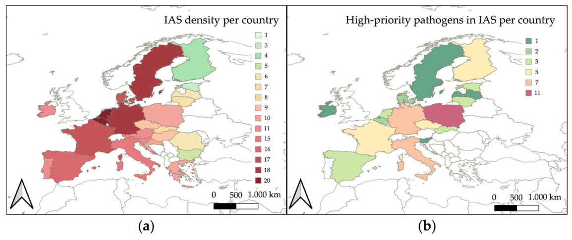
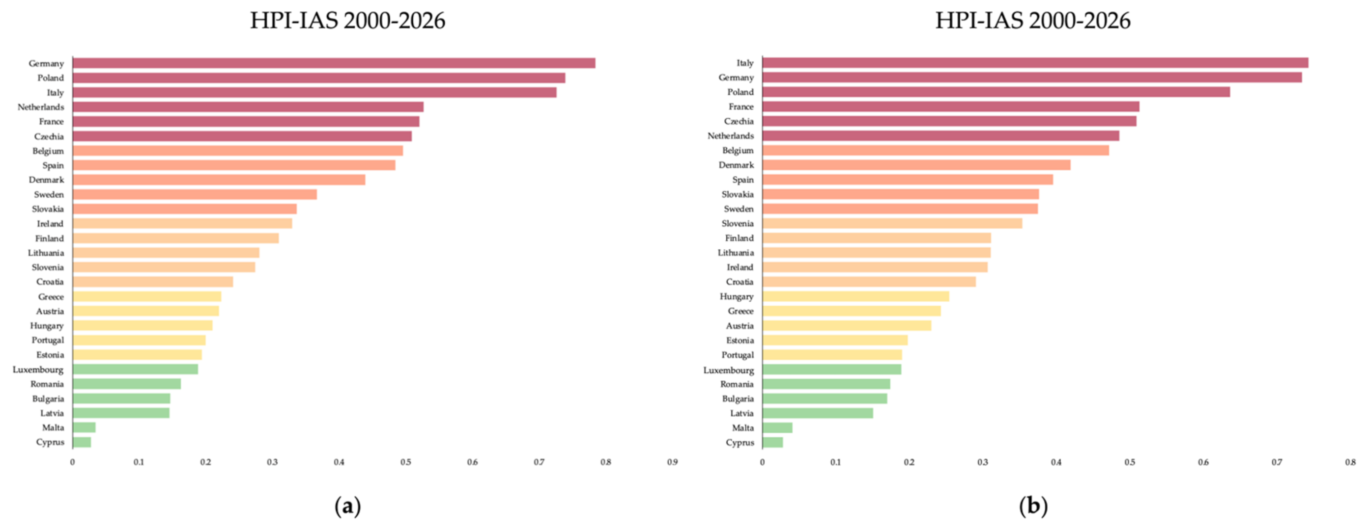
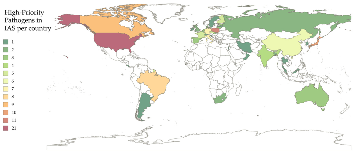
3.3. Spatial Distribution and Epidemiological Risk of High-Priority Pathogens in Europe and in the World
4. Discussion
5. Conclusions
Supplementary Materials
Author Contributions
Funding
Institutional Review Board Statement
Informed Consent Statement
Data Availability Statement
Conflicts of Interest
References
- Pyšek, P.; Hulme, P.E.; Simberloff, D.; Bacher, S.; Blackburn, T.M.; Carlton, J.T.; Dawson, W.; Essl, F.; Foxcroft, L.C.; Genovesi, P.; et al. Scientists’ Warning on Invasive Alien Species. Biol. Rev. Camb. Philos. Soc. 2020, 95, 1511–1534. [Google Scholar] [CrossRef]
- Thomas, S.M. Invasive Alien Species: A Rising Global Threat That Needs Control. Lancet Planet Health 2023, 7, e875–e876. [Google Scholar] [CrossRef]
- Dalla Pozza, G.; Majori, G. First Record of Aedes Albopictus Establishment in Italy. J. Am. Mosq. Control Assoc. 1992, 8, 318–320. [Google Scholar] [PubMed]
- Lencioni, V.; Bertola, F.; Franceschini, A.; Ferrarese, U.; Zandonai, F.; Stancher, G.; Spitale, D. Multi-Year Dynamics of the Aedes Albopictus Occurrence in Two Neighbouring Cities in the Alps. Eur. Zool. J. 2023, 90, 101–112. [Google Scholar] [CrossRef]
- Menegale, F.; Manica, M.; Del Manso, M.; Bella, A.; Zardini, A.; Gobbi, A.; Mignuoli, A.D.; Mattei, G.; Vairo, F.; Vezzosi, L.; et al. Risk Assessment and Perspectives of Local Transmission of Chikungunya and Dengue in Italy, a European Forerunner. Nat. Commun. 2025, 16, 6237. [Google Scholar] [CrossRef]
- Finch, D.M.; Butler, J.L.; Runyon, J.B.; Fettig, C.J.; Kilkenny, F.F.; Jose, S.; Frankel, S.J.; Cushman, S.A.; Cobb, R.C.; Dukes, J.S.; et al. Effects of Climate Change on Invasive Species. In Invasive Species in Forests and Rangelands of the United States; Poland, T.M., Patel-Weynand, T., Finch, D.M., Miniat, C.F., Hayes, D.C., Lopez, V.M., Eds.; Springer International Publishing: Cham, Switzerland, 2021; pp. 57–83. [Google Scholar] [CrossRef]
- Flickinger, H.D.; Dukes, J.S. A Review of Theory: Comparing Invasion Ecology and Climate Change-Induced Range Shifting. Glob. Change Biol. 2024, 30, e17612. [Google Scholar] [CrossRef]
- Seebens, H.; Blackburn, T.M.; Dyer, E.E.; Genovesi, P.; Hulme, P.E.; Jeschke, J.M.; Pagad, S.; Pyšek, P.; Winter, M.; Arianoutsou, M.; et al. No Saturation in the Accumulation of Alien Species Worldwide. Nat. Commun. 2017, 8, 14435. [Google Scholar] [CrossRef]
- Jia, J.; Dai, Z.; Li, F.; Liu, Y. How Will Global Environmental Changes Affect the Growth of Alien Plants? Front. Plant Sci. 2016, 7, 1623. [Google Scholar] [CrossRef]
- Stephens, K.L.; Dantzler-Kyer, M.E.; Patten, M.A.; Souza, L. Differential Responses to Global Change of Aquatic and Terrestrial Invasive Species: Evidences from a Meta-analysis. Ecosphere 2019, 10, e02680. [Google Scholar] [CrossRef]
- Roy, H.E.; Pauchard, A.; Stoett, P.; Renard Truong, T.; Bacher, S.; Galil, B.S.; Hulme, P.E.; Ikeda, T.; Sankaran, K.; McGeoch, M.A.; et al. IPBES Invasive Alien Species Assessment: Summary for Policymakers; Zenodo: Geneva, Switzerland, 2024. [Google Scholar] [CrossRef]
- Blackburn, T.M.; Pyšek, P.; Bacher, S.; Carlton, J.T.; Duncan, R.P.; Jarošík, V.; Wilson, J.R.U.; Richardson, D.M. A Proposed Unified Framework for Biological Invasions. Trends Ecol. Evol. 2011, 26, 333–339. [Google Scholar] [CrossRef] [PubMed]
- Sandvik, H. Expansion Speed as a Generic Measure of Spread for Alien Species. Acta Biotheor. 2020, 68, 227–252. [Google Scholar] [CrossRef] [PubMed]
- Goldstein, J.; Park, J.; Haran, M.; Liebhold, A.; Bjørnstad, O.N. Quantifying Spatio-Temporal Variation of Invasion Spread. Proc. R. Soc. B Biol. Sci. 2019, 286, 20182294. [Google Scholar] [CrossRef]
- United Nations. Convention on Biological Diversity. 1992. Available online: https://www.cbd.int/convention/ (accessed on 17 March 2024).
- European Commission. Directorate General for Environment. In EU Biodiversity Strategy for 2030: Bringing Nature Back into Our Lives; Publications Office: Luxembourg, 2021. [Google Scholar]
- European Commission. Commission Implementing Regulation. European Commission, Environment, Invasive Alien Species, Commission Implementing Regulation (EU) 2022/1203 of 12 July 2022. 2022. Available online: https://eur-lex.europa.eu/legal-content/EN/TXT/?uri=CELEX%3A32022R1203 (accessed on 18 July 2024).
- Rato, J.; Brandão, P.; Gama, M.; Banha, F.; Anastácio, P. Effectiveness of legislative tools to stop biological invasions: Freshwater turtles’ invasion in Europe as a study case. NeoBiota 2025, 102, 313–329. [Google Scholar] [CrossRef]
- Katsanevakis, S.; Bogucarskis, K.; Gatto, F. Building the European Alien Species Information Network (EASIN): A novel approach for the exploration of distributed alien species data. BioInvasions Rec. 2012, 1, 235–245. [Google Scholar] [CrossRef]
- Booy, O.; Robertson, P.A.; Moore, N.; Ward, J.; Roy, H.E.; Adriaens, T.; Mill, A.C. Using structured eradication feasibility assessment to prioritize the management of new and emerging invasive alien species in Europe. Glob. Change Biol. 2020, 26, 6235–6250. [Google Scholar] [CrossRef]
- Kumschick, S.; Gaertner, M.; Vilà, M.; Essl, F.; Jeschke, J.M.; Pyšek, P.; Ricciardi, A.; Bacher, S.; Blackburn, T.M.; Dick, J.T.A.; et al. Ecological Impacts of Alien Species: Quantification, Scope, Caveats, and Recommendations. BioScience 2015, 65, 55–63. [Google Scholar] [CrossRef]
- Blackburn, T.M.; Bellard, C.; Ricciardi, A. Alien versus Native Species as Drivers of Recent Extinctions. Front. Ecol. Environ. 2019, 17, 203–207. [Google Scholar] [CrossRef]
- Doherty, T.S.; Glen, A.S.; Nimmo, D.G.; Ritchie, E.G.; Dickman, C.R. Invasive Predators and Global Biodiversity Loss. Proc. Natl. Acad. Sci. USA 2016, 113, 11261–11265. [Google Scholar] [CrossRef]
- Tobin, P.C. Managing Invasive Species. F1000Research 2018, 7, 1686. [Google Scholar] [CrossRef]
- O’Connor, L.M.J.; Cosentino, F.; Harfoot, M.B.J.; Maiorano, L.; Mancino, C.; Pollock, L.J.; Thuiller, W. Vulnerability of Terrestrial Vertebrate Food Webs to Anthropogenic Threats in Europe. Glob. Change Biol. 2024, 30, e17253. [Google Scholar] [CrossRef]
- Crowl, T.A.; Crist, T.O.; Parmenter, R.R.; Belovsky, G.; Lugo, A.E. The Spread of Invasive Species and Infectious Disease as Drivers of Ecosystem Change. Front. Ecol. Environ. 2008, 6, 238–246. [Google Scholar] [CrossRef]
- Rulli, M.C.; D’Odorico, P.; Galli, N.; John, R.S.; Muylaert, R.L.; Santini, M.; Hayman, D.T.S. Land Use Change and Infectious Disease Emergence. Rev. Geophys. 2025, 63, e2022RG000785. [Google Scholar] [CrossRef]
- Shackleton, R.T.; Shackleton, C.M.; Kull, C.A. The Role of Invasive Alien Species in Shaping Local Livelihoods and Human Well-Being: A Review. J. Environ. Manag. 2019, 229, 145–157. [Google Scholar] [CrossRef] [PubMed]
- Patz, J.A.; Daszak, P.; Tabor, G.M.; Aguirre, A.A.; Pearl, M.; Epstein, J.; Wolfe, N.D.; Kilpatrick, A.M.; Foufopoulos, J.; Molyneux, D.; et al. Unhealthy Landscapes: Policy Recommendations on Land Use Change and Infectious Disease Emergence. Environ. Health Perspect. 2004, 112, 1092–1098. [Google Scholar] [CrossRef]
- Chinchio, E.; Crotta, M.; Romeo, C.; Drewe, J.A.; Guitian, J.; Ferrari, N. Invasive Alien Species and Disease Risk: An Open Challenge in Public and Animal Health. PLoS Pathog. 2020, 16, e1008922. [Google Scholar] [CrossRef]
- VanderWaal, K.L.; Ezenwa, V.O. Heterogeneity in Pathogen Transmission: Mechanisms and Methodology. Funct. Ecol. 2016, 30, 1606–1622. [Google Scholar] [CrossRef]
- Aguirre, A.A.; Longcore, T.; Barbieri, M.; Dabritz, H.; Hill, D.; Klein, P.N.; Lepczyk, C.; Lilly, E.L.; McLeod, R.; Milcarsky, J.; et al. The One Health Approach to Toxoplasmosis: Epidemiology, Control, and Prevention Strategies. EcoHealth 2019, 16, 378–390. [Google Scholar] [CrossRef]
- Vredenburg, V.T.; Felt, S.A.; Morgan, E.C.; McNally, S.V.; Wilson, S.; Green, S.L. Prevalence of Batrachochytrium dendrobatidis in Xenopus Collected in Africa (1871–2000) and in California (2001–2010). PLoS ONE 2013, 8, e63791. [Google Scholar] [CrossRef]
- Cabe, P.R. European Starlings (Sturnus vulgaris) as Vectors and Reservoirs of Pathogens Affecting Humans and Domestic Livestock. Animals 2021, 11, 466. [Google Scholar] [CrossRef] [PubMed]
- Martelli, L.; Fornasiero, D.; Scarton, F.; Spada, A.; Scolamacchia, F.; Manca, G.; Mulatti, P. Study of the Interface between Wild Bird Populations and Poultry and Their Potential Role in the Spread of Avian Influenza. Microorganisms 2023, 11, 2601. [Google Scholar] [CrossRef] [PubMed]
- Ellis, J.W.; Root, J.J.; McCurdy, L.M.; Bentler, K.T.; Barrett, N.L.; VanDalen, K.K.; Shriner, S.A. Avian influenza A virus susceptibility, infection, transmission, and antibody kinetics in European starlings. PLoS Pathog. 2021, 17, e1009879. [Google Scholar] [CrossRef]
- Burdon, J.; Thrall, P.; Ericson, L. Genes, Communities & Invasive Species: Understanding the Ecological and Evolutionary Dynamics of Host―Pathogen Interactions. Curr. Opin. Plant Biol. 2013, 16, 400–405. [Google Scholar] [CrossRef]
- de Toro Salas, A.; Dueñas Díez, J.; de Jaime Revuelta, E. Concentrations of calcium and bone remodeling biomarkers in umbilical cord blood and urine of newborn infants during delivery. An. Esp. Pediatr. 2001, 54, 290–296. [Google Scholar] [CrossRef]
- Jia, B.; Colling, A.; Stallknecht, D.E.; Blehert, D.; Bingham, J.; Crossley, B.; Eagles, D.; Gardner, I.A. Validation of Laboratory Tests for Infectious Diseases in Wild Mammals: Review and Recommendations. J. Vet. Diagn. Investig. 2020, 32, 776–792. [Google Scholar] [CrossRef] [PubMed]
- Stallknecht, D.E. Impediments to Wildlife Disease Surveillance, Research, and Diagnostics. Curr. Top. Microbiol. Immunol. 2007, 315, 445–461. [Google Scholar] [CrossRef] [PubMed]
- Michel, A.L.; Van Heerden, H.; Crossley, B.M.; Al Dahouk, S.; Prasse, D.; Rutten, V. Pathogen Detection and Disease Diagnosis in Wildlife: Challenges and Opportunities. Rev. Sci. Tech. OIE 2021, 40, 105–118. [Google Scholar] [CrossRef]
- European Commission. Commission Implementing Regulation. Regulation (EU) 2016/429 of the European Parliament and of the Council of 9 March 2016 on Transmissible Animal Diseases and Amending and Repealing Certain Acts in the Area of Animal Health (‘Animal Health Law’). 2016. Available online: https://eur-lex.europa.eu/legal-content/EN/ALL/?uri=CELEX%3A32016R0429 (accessed on 24 July 2024).
- World Health Organization. R&D Blue Print (RDB). Pathogens Prioritization: A Scientific Framework for Epidemic and Pandemic Research Preparedness. 2024. Available online: https://www.who.int/publications/m/item/pathogens-prioritization-a-scientific-framework-for-epidemic-and-pandemic-research-preparedness (accessed on 6 April 2026).
- WHO. Fungal Priority Pathogens List to Guide Research, Development and Public Health Action, 1st ed.; World Health Organization: Geneva, Switzerland, 2022. [Google Scholar]
- WHO. Bacterial Priority Pathogens List 2024: Bacterial Pathogens of Public Health Importance, to Guide Research, Development, and Strategies to Prevent and Control Antimicrobial Resistance, 1st ed.; World Health Organization: Geneva, Switzerland, 2024. [Google Scholar]
- European Parliament. Directive 2003/99/EC of the European Parliament and of the Council of 17 November 2003 on the Monitoring of Zoonoses and Zoonotic Agents, Amending Council Decision 90/424/EEC and Repealing Council Directive 92/117/EEC. 2003. Available online: https://eur-lex.europa.eu/eli/dir/2003/99/oj (accessed on 6 April 2026).
- Jones, K.E.; Patel, N.G.; Levy, M.A.; Storeygard, A.; Balk, D.; Gittleman, J.L.; Daszak, P. Global trends in emerging infectious diseases. Nature 2008, 451, 990–993. [Google Scholar] [CrossRef]
- Woolhouse, M.E.J.; Gowtage-Sequeria, S. Host Range and Emerging and Reemerging Pathogens. Emerg. Infect. Dis. 2005, 11, 1842–1847. [Google Scholar] [CrossRef]
- Olival, K.J.; Hosseini, P.R.; Zambrana-Torrelio, C.; Ross, N.; Bogich, T.L.; Daszak, P. Host and viral traits predict zoonotic spillover from mammals. Nature 2017, 546, 646–650. [Google Scholar] [CrossRef] [PubMed]
- European Commission, Directorate-General for Health and Food Safety. Commission Implementing Regulation (EU) 2020/2002 of 7 December 2020 Laying down Rules for the Application of Regulation (EU) 2016/429 of the European Parliament and of the Council with Regard to Union Notification and Union Reporting of Listed Diseases, to Formats and Procedures for Submission and Reporting of Union Surveillance Programmes and of Eradication Programmes and for Application for Recognition of Disease-Free Status, and to the Computerised Information System. 2022. Available online: https://eur-lex.europa.eu/legal-content/EN/TXT/?uri=CELEX%3A32020R2002 (accessed on 6 April 2026).
- Galanidi, M.; Zenetos, A. Data-Driven Recommendations for Establishing Threshold Values for the NIS Trend Indicator in the Mediterranean Sea. Diversity 2022, 14, 57. [Google Scholar] [CrossRef]
- European Commission, Organisation for Economic Co-operation and Development. Handbook on Constructing Composite Indicators: Methodology and User Guide; OECD: Paris, France, 2008. [Google Scholar]
- Van Rossum, G.; Drake, F.L. The Python Language Reference Manual: For Python Version 3.2; Network Theory Ltd.: Bristol, UK, 2011. [Google Scholar]
- Mckinney, W. Pandas: A Foundational Python Library for Data Analysis and Statistics. Python High Perform. Sci. Comput. 2011, 14, 1–9. [Google Scholar]
- Harris, C.R.; Millman, K.J.; Van Der Walt, S.J.; Gommers, R.; Virtanen, P.; Cournapeau, D.; Wieser, E.; Taylor, J.; Berg, S.; Smith, N.J.; et al. Array Programming with NumPy. Nature 2020, 585, 357–362. [Google Scholar] [CrossRef]
- Greco, S.; Ishizaka, A.; Tasiou, M.; Torrisi, G. On the Methodological Framework of Composite Indices: A Review of the Issues of Weighting, Aggregation, and Robustness. Soc. Indic. Res. 2019, 141, 61–94. [Google Scholar] [CrossRef]
- Saltelli, A.; Tarantola, S.; Campolongo, F.; Ratto, M. Sensitivity Analysis in Practice: A Guide to Assessing Scientific Models; Saltelli, A., Ed.; Wiley: Hoboken, NJ, USA, 2004. [Google Scholar]
- Becker, W.; Saisana, M.; Paruolo, P.; Vandecasteele, I. Weights and Importance in Composite Indicators: Closing the Gap. Ecol. Indic. 2017, 80, 12–22. [Google Scholar] [CrossRef]
- Conover, W.J. Practical Nonparametric Statistics, 3rd ed.; Wiley Series in Probability and Statistics; Applied Probability and Statistics Section; Wiley: Hoboken, NJ, USA, 1999. [Google Scholar]
- Wodecka, B.; Michalik, J.; Lane, R.S.; Nowak-Chmura, M.; Wierzbicka, A. Differential Associations of Borrelia Species with European Badgers (Meles Meles) and Raccoon Dogs (Nyctereutes Procyonoides) in Western Poland. Ticks Tick-Borne Dis. 2016, 7, 1010–1016. [Google Scholar] [CrossRef]
- Myśliwy, I.; Perec-Matysiak, A.; Hildebrand, J. Invasive Raccoon (Procyon Lotor) and Raccoon Dog (Nyctereutes Procyonoides) as Potential Reservoirs of Tick-Borne Pathogens: Data Review from Native and Introduced Areas. Parasit. Vectors 2022, 15, 126. [Google Scholar] [CrossRef]
- Vourc’h, G.; Marmet, J.; Chassagne, M.; Bord, S.; Chapuis, J.-L. Borrelia Burgdorferi Sensu Lato in Siberian Chipmunks (Tamias Sibiricus) Introduced in Suburban Forests in France. Vector-Borne Zoonotic Dis. 2007, 7, 637–642. [Google Scholar] [CrossRef] [PubMed]
- Marchant, A.; Le Coupanec, A.; Joly, C.; Perthame, E.; Sertour, N.; Garnier, M.; Godard, V.; Ferquel, E.; Choumet, V. Infection of Ixodes Ricinus by Borrelia Burgdorferi Sensu Lato in Peri-Urban Forests of France. PLoS ONE 2017, 12, e0183543. [Google Scholar] [CrossRef] [PubMed]
- Marsot, M.; Sigaud, M.; Chapuis, J.L.; Ferquel, E.; Cornet, M.; Vourc’h, G. Introduced Siberian Chipmunks (Tamias Sibiricus Barberi) Harbor More-Diverse Borrelia Burgdorferi Sensu Lato Genospecies than Native Bank Voles (Myodes Glareolus). Appl. Environ. Microbiol. 2011, 77, 5716–5721. [Google Scholar] [CrossRef]
- Takako, T.; Elpita, T.; Hiroyuki, S.; Chiho, K.; Naoaki, M. Prevalence of Campylobacter Spp. in Raccoon Dogs and Badgers in Miyazaki Prefecture, Japan. EcoHealth 2021, 18, 241–249. [Google Scholar] [CrossRef] [PubMed]
- Mutschall, S.K.; Hetman, B.M.; Bondo, K.J.; Gannon, V.P.J.; Jardine, C.M.; Taboada, E.N. Campylobacter Jejuni Strain Dynamics in a Raccoon (Procyon Lotor) Population in Southern Ontario, Canada: High Prevalence and Rapid Subtype Turnover. Front. Vet. Sci. 2020, 7, 27. [Google Scholar] [CrossRef]
- Pacha, R.E.; Clark, G.W.; Williams, E.A. Occurrence of Campylobacter Jejuni and Giardia Species in Muskrat (Ondatra Zibethica). Appl. Environ. Microbiol. 1985, 50, 177–178. [Google Scholar] [CrossRef]
- Ganoe, L.S.; Brown, J.D.; Yabsley, M.J.; Lovallo, M.J.; Walter, W.D. A Review of Pathogens, Diseases, and Contaminants of Muskrats (Ondatra Zibethicus) in North America. Front. Vet. Sci. 2020, 7, 233. [Google Scholar] [CrossRef]
- Nelson, D.D.; Haldorson, G.J.; Stanton, J.B.; Noh, S.M.; Bradway, D.S.; Mansfield, K.G.; Baszler, T.V. Francisella Tularensis Infection without Lesions in Gray Tree Squirrels (Sciurus Griseus): A Diagnostic Challenge. J. Vet. Diagn. Investig. Off. Publ. Am. Assoc. Vet. Lab. Diagn. Inc. 2014, 26, 312–315. [Google Scholar] [CrossRef]
- Jahan, N.A.; Lindsey, L.L.; Larsen, P.A. The Role of Peridomestic Rodents as Reservoirs for Zoonotic Foodborne Pathogens. Vector-Borne Zoonotic Dis. 2021, 21, 133–148. [Google Scholar] [CrossRef] [PubMed]
- Hestvik, G.; Uhlhorn, H.; Koene, M.; Åkerström, S.; Malmsten, A.; Dahl, F.; Åhlén, P.-A.; Dalin, A.-M.; Gavier-Widén, D. Francisella Tularensis in Swedish Predators and Scavengers. Epidemiol. Infect. 2019, 147, e293. [Google Scholar] [CrossRef] [PubMed]
- Sutor, A.; Schwarz, S.; Conraths, F.J. The Biological Potential of the Raccoon Dog (Nyctereutes Procyonoides, Gray 1834) as an Invasive Species in Europe―New Risks for Disease Spread? Acta Theriol. 2014, 59, 49–59. [Google Scholar] [CrossRef]
- Beltrán-Beck, B.; García, F.J.; Gortázar, C. Raccoons in Europe: Disease Hazards Due to the Establishment of an Invasive Species. Eur. J. Wildl. Res. 2012, 58, 5–15. [Google Scholar] [CrossRef]
- Duncan, C.; Krafsur, G.; Podell, B.; Baeten, L.A.; LeVan, I.; Charles, B.; Ehrhart, E.J. Leptospirosis and Tularaemia in Raccoons (Procyon Lotor) of Larimer Country, Colorado. Zoonoses Public Health 2012, 59, 29–34. [Google Scholar] [CrossRef]
- Bischof, R.; Rogers, D.G. Serologic Survey of Select Infectious Diseases in Coyotes and Raccoons in Nebraska. J. Wildl. Dis. 2005, 41, 787–791. [Google Scholar] [CrossRef]
- Kuehn, A.; Schulze, C.; Kutzer, P.; Probst, C.; Hlinak, A.; Ochs, A.; Grunow, R. Tularaemia Seroprevalence of Captured and Wild Animals in Germany: The Fox (Vulpes Vulpes) as a Biological Indicator. Epidemiol. Infect. 2013, 141, 833–840. [Google Scholar] [CrossRef] [PubMed]
- Rhynd, K.J.R.; Leighton, P.A.; Elcock, D.A.; Whitehall, P.J.; Rycroft, A.; Macgregor, S.K. PREVALENCE OF SALMONELLA SPP. AND THERMOPHILIC CAMPYLOBACTER SPP. IN THE SMALL ASIAN MONGOOSE (HERPESTES JAVANICUS) IN BARBADOS, WEST INDIES. J. Zoo Wildl. Med. 2014, 45, 911–914. [Google Scholar] [CrossRef] [PubMed]
- Fornazari, F.; Langoni, H.; Marson, P.M.; Nóbrega, D.B.; Teixeira, C.R. Leptospira Reservoirs among Wildlife in Brazil: Beyond Rodents. Acta Trop. 2018, 178, 205–212. [Google Scholar] [CrossRef]
- Yamashita, R.; Yoshida, T.; Kobayashi, M.; Uomoto, S.; Shimizu, S.; Takesue, K.; Maeda, N.; Hara, E.; Ohshima, K.; Zeng, W.; et al. Leptospiral Meningoencephalitis in a Raccoon Dog. J. Vet. Diagn. Investig. 2021, 33, 1137–1141. [Google Scholar] [CrossRef]
- Reinhardt, N.P.; Köster, J.; Thomas, A.; Arnold, J.; Fux, R.; Straubinger, R.K. Bacterial and Viral Pathogens with One Health Relevance in Invasive Raccoons (Procyon Lotor, Linné 1758) in Southwest Germany. Pathogens 2023, 12, 389. [Google Scholar] [CrossRef] [PubMed]
- Michel, V.; Ruvoen-Clouet, N.; Menard, A.; Sonrier, C.; Fillonneau, C.; Rakotovao, F.; Ganière, J.P.; André-Fontaine, G. Role of the Coypu (Myocastor Coypus) in the Epidemiology of Leptospirosis in Domestic Animals and Humans in France. Eur. J. Epidemiol. 2001, 17, 111–121. [Google Scholar] [CrossRef]
- Kik, M.J.L.; Goris, M.G.; Bos, J.H.; Hartskeerl, R.A.; Dorrestein, G.M. An Outbreak of Leptospirosis in Seals (Phoca Vitulina) in Captivity. Vet. Q. 2006, 28, 33–39. [Google Scholar] [CrossRef]
- Vein, J.; Leblond, A.; Belli, P.; Kodjo, A.; Berny, P.J. The Role of the Coypu (Myocastor Coypus), an Invasive Aquatic Rodent Species, in the Epidemiological Cycle of Leptospirosis: A Study in Two Wetlands in the East of France. Eur. J. Wildl. Res. 2014, 60, 125–133. [Google Scholar] [CrossRef]
- Pedersen, K.; Anderson, T.D.; Maison, R.M.; Wiscomb, G.W.; Pipas, M.J.; Sinnett, D.R.; Baroch, J.A.; Gidlewski, T. LEPTOSPIRA ANTIBODIES DETECTED IN WILDLIFE IN THE USA AND THE US VIRGIN ISLANDS. J. Wildl. Dis. 2018, 54, 450–459. [Google Scholar] [CrossRef]
- Tan, C.G.; Dharmarajan, G.; Beasley, J.; Rhodes, O.; Moore, G.; Wu, C.C.; Lin, T.L. Neglected Leptospirosis in Raccoons (Procyon Lotor) in Indiana, USA. Vet. Q. 2014, 34, 1–10. [Google Scholar] [CrossRef]
- Raizman, E.A.; Dharmarajan, G.; Beasley, J.C.; Wu, C.C.; Pogranichniy, R.M.; Rhodes, O.E. Serologic Survey for Selected Infectious Diseases in Raccoons (Procyon Lotor) in Indiana, USA. J. Wildl. Dis. 2009, 45, 531–536. [Google Scholar] [CrossRef] [PubMed]
- Mitchell, M.A.; Hungerford, L.L.; Nixon, C.; Esker, T.; Sullivan, J.; Koerkenmeier, R.; Dubey, J.P. SEROLOGIC SURVEY FOR SELECTED INFECTIOUS DISEASE AGENTS IN RACCOONS FROM ILLINOIS. J. Wildl. Dis. 1999, 35, 347–355. [Google Scholar] [CrossRef]
- Dirsmith, K.; VanDalen, K.; Fry, T.; Charles, B.; VerCauteren, K.; Duncan, C. Leptospirosis in Fox Squirrels (Sciurus Niger) of Larimer County, Colorado, USA. J. Wildl. Dis. 2013, 49, 641–645. [Google Scholar] [CrossRef]
- Anderson, A.N.; Long, A.M.; LaCour, J.M.; Bresnan, A.M.; Bushaw, J.D.; Gerrits, A.P.; Hunt, J.D.; Moscicki, D.J.; Raginski, N.M.; Stafford, B.; et al. Seroprevalence of Leptospira Among Selected Mammals on a Wildlife Management Area in Louisiana, USA. J. Wildl. Dis. 2022, 58, 183–187. [Google Scholar] [CrossRef]
- Rainwater, K.L.; Marchese, K.; Slavinski, S.; Humberg, L.A.; Dubovi, E.J.; Jarvis, J.A.; McAloose, D.; Calle, P.P. HEALTH SURVEY OF FREE-RANGING RACCOONS (PROCYON LOTOR) IN CENTRAL PARK, NEW YORK, NEW YORK, USA: IMPLICATIONS FOR HUMAN AND DOMESTIC ANIMAL HEALTH. J. Wildl. Dis. 2017, 53, 272–284. [Google Scholar] [CrossRef]
- Bondo, K.J.; Pearl, D.L.; Janecko, N.; Boerlin, P.; Reid-Smith, R.J.; Parmley, J.; Jardine, C.M. Epidemiology of Salmonella on the Paws and in the Faeces of Free-Ranging Raccoons (Procyon Lotor) in Southern Ontario, Canada. Zoonoses Public Health 2016, 63, 303–310. [Google Scholar] [CrossRef] [PubMed]
- Very, K.J.; Kirchner, M.K.; Shariat, N.; Cottrell, W.; Sandt, C.H.; Dudley, E.G.; Kariyawasam, S.; Jayarao, B.M. Prevalence and Spatial Distribution of Salmonella Infections in the Pennsylvania Raccoon (P Rocyon Lotor). Zoonoses Public Health 2016, 63, 223–233. [Google Scholar] [CrossRef] [PubMed]
- Gaertner, J.P.; Hahn, D.; Rose, F.L.; Forstner, M.R.J. Detection of Salmonellae in Different Turtle Species within a Headwater Spring Ecosystem. J. Wildl. Dis. 2008, 44, 519–526. [Google Scholar] [CrossRef]
- Gong, S.; Wang, F.; Shi, H.; Zhou, P.; Ge, Y.; Hua, L.; Liu, W. Highly Pathogenic Salmonella Pomona Was First Isolated from the Exotic Red-Eared Slider (Trachemys Scripta Elegans) in the Wild in China: Implications for Public Health. Sci. Total Environ. 2014, 468–469, 28–30. [Google Scholar] [CrossRef]
- Mermin, J.; Hutwagner, L.; Vugia, D.; Shallow, S.; Daily, P.; Bender, J.; Koehler, J.; Marcus, R.; Angulo, F.J.; the Emerging Infections Program FoodNet Working Group. Reptiles, Amphibians, and Human Salmonella Infection: A Population-Based, Case-Control Study. Clin. Infect. Dis. 2004, 38, S253–S261. [Google Scholar] [CrossRef]
- Garamszegi, L.Z.; Møller, A.P. Prevalence of Avian Influenza and Host Ecology. Proc. R. Soc. B Biol. Sci. 2007, 274, 2003–2012. [Google Scholar] [CrossRef]
- Nagano, N.; Oana, S.; Nagano, Y.; Arakawa, Y. A Severe Salmonella Enterica Serotype Paratyphi B Infection in a Child Related to a Pet Turtle, Trachemys Scripta Elegans. Jpn. J. Infect. Dis. 2006, 59, 132–134. [Google Scholar] [CrossRef]
- Marin, C.; Ingresa-Capaccioni, S.; González-Bodi, S.; Marco-Jiménez, F.; Vega, S. Free-Living Turtles Are a Reservoir for Salmonella but Not for Campylobacter. PLoS ONE 2013, 8, e72350. [Google Scholar] [CrossRef] [PubMed]
- Pasmans, F.; De Herdt, P.; Dewulf, J.; Haesebrouck, F. Pathogenesis of Infections with Salmonella enterica Subsp. Enterica Serovar Muenchen in the Turtle Trachemys Scripta Scripta. Vet. Microbiol. 2002, 87, 315–325. [Google Scholar] [CrossRef]
- Smyth, C.W.; Sarmiento-Ramírez, J.M.; Short, D.P.G.; Diéguez-Uribeondo, J.; O’Donnell, K.; Geiser, D.M. Unraveling the Ecology and Epidemiology of an Emerging Fungal Disease, Sea Turtle Egg Fusariosis (STEF). PLoS Pathog. 2019, 15, e1007682. [Google Scholar] [CrossRef] [PubMed]
- Chang, W.-S.; Rose, K.; Holmes, E.C. Meta-Transcriptomic Analysis of the Virome and Microbiome of the Invasive Indian Myna (Acridotheres tristis) in Australia. One Health 2021, 13, 100360. [Google Scholar] [CrossRef]
- Ganapathy, K.; Saleha, A.A.; Jaganathan, M.; Tan, C.G.; Chong, C.T.; Tang, S.C.; Ideris, A.; Dare, C.M.; Bradbury, J.M. Survey of Campylobacter, Salmonella and Mycoplasmas in House Crows (Corvus splendens) in Malaysia. Vet. Rec. 2007, 160, 622–624. [Google Scholar] [CrossRef]
- Vogler, B.R.; Zurfluh, K.; Mattmann, P.; Schmitt, K.; Albini, S. Low Occurrence of Salmonella Spp. in Wild Birds from a Swiss Rehabilitation Centre. Vet. Rec. Open 2021, 8, e17. [Google Scholar] [CrossRef]
- Marsot, M.; Chapuis, J.-L.; Gasqui, P.; Dozières, A.; Masséglia, S.; Pisanu, B.; Ferquel, E.; Vourc’h, G. Introduced Siberian Chipmunks (Tamias sibiricus barberi) Contribute More to Lyme Borreliosis Risk than Native Reservoir Rodents. PLoS ONE 2013, 8, e55377. [Google Scholar] [CrossRef]
- Kjær, L.J.; Jensen, L.M.; Chriél, M.; Bødker, R.; Petersen, H.H. The Raccoon Dog (Nyctereutes procyonoides) as a Reservoir of Zoonotic Diseases in Denmark. Int. J. Parasitol. Parasites Wildl. 2021, 16, 175–182. [Google Scholar] [CrossRef] [PubMed]
- Cruciani, D.; Crotti, S.; Paoloni, D.; La Morgia, V.; Felici, A.; Papa, P.; Cosseddu, G.M.; Moscati, L.; Gobbi, P. Health Status of the Eastern Grey Squirrel (Sciurus carolinensis) Population in Umbria: Results of the LIFE Project ‘U-SAVEREDS’. Animals 2022, 12, 2741. [Google Scholar] [CrossRef]
- Kang, J.-G.; Chae, J.-B.; Cho, Y.-K.; Jo, Y.-S.; Shin, N.-S.; Lee, H.; Choi, K.-S.; Yu, D.-H.; Park, J.; Park, B.-K.; et al. Molecular Detection of Anaplasma, Bartonella, and Borrelia Theileri in Raccoon Dogs (Nyctereutes procyonoides) in Korea. Am. J. Trop. Med. Hyg. 2018, 98, 1061–1068. [Google Scholar] [CrossRef]
- Millins, C.; Magierecka, A.; Gilbert, L.; Edoff, A.; Brereton, A.; Kilbride, E.; Denwood, M.; Birtles, R.; Biek, R. An Invasive Mammal (the Gray Squirrel, Sciurus carolinensis) Commonly Hosts Diverse and Atypical Genotypes of the Zoonotic Pathogen Borrelia burgdorferi Sensu Lato. Appl. Environ. Microbiol. 2015, 81, 4236–4245. [Google Scholar] [CrossRef]
- Salkeld, D.J.; Leonhard, S.; Girard, Y.A.; Hahn, N.; Mun, J.; Padgett, K.A.; Lane, R.S. Identifying the Reservoir Hosts of the Lyme Disease Spirochete Borrelia burgdorferi in California: The Role of the Western Gray Squirrel (Sciurus griseus). Am. J. Trop. Med. Hyg. 2008, 79, 535–540. [Google Scholar] [CrossRef]
- Roy, A.N.; Straub, M.H.; Stephenson, N.; Sholty, K.E.; Foley, J. Distribution and Diversity of Borrelia burgdorferi Sensu Lato Group Bacteria in Sciurids of California. Vector-Borne Zoonotic Dis. 2017, 17, 735–742. [Google Scholar] [CrossRef] [PubMed]
- Oliveira-Filho, E.F.; Júnior, J.W.P.; Souza, M.M.A.; Santana, V.L.A.; Silva, J.C.R.; Mota, R.A.; Sá, F.B. SEROLOGIC SURVEY OF BRUCELLOSIS IN CAPTIVE NEOTROPICAL WILD CARNIVORES IN NORTHEAST BRAZIL. J. Zoo Wildl. Med. 2012, 43, 384–387. [Google Scholar] [CrossRef] [PubMed]
- Lee, K.; Iwata, T.; Nakadai, A.; Kato, T.; Hayama, S.; Taniguchi, T.; Hayashidani, H. Prevalence of Salmonella, Yersinia and Campylobacter Spp. in Feral Raccoons (Procyon lotor) and Masked Palm Civets (Paguma larvata) in Japan. Zoonoses Public Health 2011, 58, 424–431. [Google Scholar] [CrossRef] [PubMed]
- Martino, P.E.; Stanchi, N.O.; Silvestrini, M.; Brihuega, B.; Samartino, L.; Parrado, E. Seroprevalence for Selected Pathogens of Zoonotic Importance in Wild Nutria (Myocastor coypus). Eur. J. Wildl. Res. 2014, 60, 551–554. [Google Scholar] [CrossRef]
- Shelley, E.B.; O’Rourke, D.; Grant, K.; McArdle, E.; Capra, L.; Clarke, A.; McNamara, E.; Cunney, R.; McKeown, P.; Amar, C.F.L.; et al. Infant Botulism Due to C. butyricum Type E Toxin: A Novel Environmental Association with Pet Terrapins. Epidemiol. Infect. 2015, 143, 461–469. [Google Scholar] [CrossRef]
- Vincent, E.C.; Ruder, M.G.; Yabsley, M.J.; Hesting, V.S.; Keel, M.K.; Brown, J.D.; Nemeth, N.M. A Baylisascaris Outbreak in Fox Squirrels (Sciurus niger) and Subsequent Detection of Francisella Tularensis in Kansas, USA. J. Wildl. Dis. 2020, 56, 457. [Google Scholar] [CrossRef]
- Hasan, B.; Olsen, B.; Alam, A.; Akter, L.; Melhus, Å. Dissemination of the Multidrug-Resistant Extended-Spectrum β-Lactamase-Producing Escherichia Coli O25b-ST131 Clone and the Role of House Crow (Corvus splendens) Foraging on Hospital Waste in Bangladesh. Clin. Microbiol. Infect. 2015, 21, 1000.E1–1000.E4. [Google Scholar] [CrossRef]
- Grear, D.A.; Lankton, J.S.; Zaleski, S.; Witt, M.; Lorch, J.M. Mortality Due to Tyzzer’s Disease of Muskrats (Ondatra zibethicus) in Northern Ohio, USA. J. Wildl. Dis. 2019, 55, 982. [Google Scholar] [CrossRef]
- Mori, M.; Van Esbroeck, M.; Depoorter, S.; Decaluwe, W.; Vandecasteele, S.J.; Fretin, D.; Reynders, M. Outbreak of Leptospirosis during a Scout Camp in the Luxembourg Belgian Province, Belgium, Summer 2012. Epidemiol. Infect. 2015, 143, 1761–1766. [Google Scholar] [CrossRef]
- Hurd, J.; Berke, O.; Poljak, Z.; Runge, M. Spatial Analysis of Leptospira Infection in Muskrats in Lower Saxony, Germany, and the Association with Human Leptospirosis. Res. Vet. Sci. 2017, 114, 351–354. [Google Scholar] [CrossRef]
- Bollo, E.; Pregel, P.; Gennero, S.; Pizzoni, E.; Rosati, S.; Nebbia, P.; Biolatti, B. Health Status of a Population of Nutria (Myocastor coypus) Living in a Protected Area in Italy. Res. Vet. Sci. 2003, 75, 21–25. [Google Scholar] [CrossRef] [PubMed]
- Bonacina, E.; Oltolina, M.; Robbiati, R.; Pinzauti, P.; Ebani, V.V. Serological Survey on the Occurrence of Anti-Leptospira Spp. Antibodies in Red-Eared Terrapins (Trachemys scripta elegans) Living in a Natural Park of Northern Italy. Animals 2021, 11, 602. [Google Scholar] [CrossRef]
- Žele-Vengušt, D.; Lindtner-Knific, R.; Mlakar-Hrženjak, N.; Jerina, K.; Vengušt, G. Exposure of Free-Ranging Wild Animals to Zoonotic Leptospira Interrogans Sensu Stricto in Slovenia. Animals 2021, 11, 2722. [Google Scholar] [CrossRef] [PubMed]
- Lilenbaum, W.; Monteiro, R.V.; Ristow, P.; Fraguas, S.; Cardoso, V.S.; Fedullo, L.P.L. Leptospirosis Antibodies in Mammals from Rio de Janeiro Zoo, Brazil. Res. Vet. Sci. 2002, 73, 319–321. [Google Scholar] [CrossRef]
- Minervino, A.H.H.; Soares, H.S.; Barrêto-Júnior, R.A.; Neves, K.A.L.; Morini, A.C.; Ortolani, E.L.; Vasconcellos, S.A.; Soares, R.M.; Gennari, S.M. ANTIBODIES AGAINST BRUCELLA ABORTUS AND LEPTOSPIRA SPP. IN CAPTIVE MAMMALS IN THE STATES OF PARá AND RIO GRANDE DO NORTE, BRAZIL. J. Zoo Wildl. Med. 2018, 49, 355–360. [Google Scholar] [CrossRef]
- Cutolo, A.A.; Wanderley, G.G.; Langoni, H.; Teixeira, C.R. Anti-Leptospira Spp. Antibodies in Wild Mammals in the Municipality of Monte Mor, São Paulo State, Brazil. Semin. Ciênc. Agrár. 2020, 41, 3465–3472. [Google Scholar] [CrossRef]
- Jardine, C.; Lindsay, L.R.; Nicholson, V.M.; Ojkic, D.; Prescott, J.F. Longitudinal Study on the Seroprevalence of Avian Influenza, Leptospirosis, and Tularemia in an Urban Population of Raccoons (Procyon lotor) in Ontario, Canada. Vector-Borne Zoonotic Dis. 2011, 11, 37–42. [Google Scholar] [CrossRef] [PubMed]
- Baldi, M.; Hernández-Mora, G.; Jimenez, C.; Hutter, S.E.; Alfaro, A.; Walzer, C. Leptospira Seroprevalence Detection and Rabies Virus Absence in an Urban Raccoon (Procyon lotor) Population in a Highly Populated Area, Costa Rica. Vector-Borne Zoonotic Dis. 2019, 19, 889–895. [Google Scholar] [CrossRef] [PubMed]
- Shiokawa, K.; Llanes, A.; Hindoyan, A.; Cruz-Martinez, L.; Welcome, S.; Rajeev, S. Peridomestic Small Indian Mongoose: An Invasive Species Posing as Potential Zoonotic Risk for Leptospirosis in the Caribbean. Acta Trop. 2019, 190, 166–170. [Google Scholar] [CrossRef] [PubMed]
- Hamir, A.N.; Hanlon, C.A.; Niezgoda, M.; Rupprecht, C.E. The Prevalence of Interstitial Nephritis and Leptospirosis in 283 Raccoons (Procyon lotor) from 5 Different Sites in the United States. Can. Vet. J. Rev. Vet. Can. 2001, 42, 869–871. [Google Scholar]
- Richardson, D.J.; Gauthier, J.L. A Serosurvey of Leptospirosis in Connecticut Peridomestic Wildlife. Vector-Borne Zoonotic Dis. 2003, 3, 187–193. [Google Scholar] [CrossRef]
- Straub, M.H.; Church, M.; Glueckert, E.; Foley, J.E. Raccoons (Procyon lotor) and Striped Skunks (Mephitis mephitis) as Potential Reservoirs of Leptospira Spp. in California. Vector-Borne Zoonotic Dis. 2020, 20, 418–426. [Google Scholar] [CrossRef]
- Helman, S.K.; Tokuyama, A.F.N.; Mummah, R.O.; Stone, N.E.; Gamble, M.W.; Snedden, C.E.; Borremans, B.; Gomez, A.C.R.; Cox, C.; Nussbaum, J.; et al. Pathogenic Leptospira Are Widespread in the Urban Wildlife of Southern California. Sci. Rep. 2023, 13, 14368. [Google Scholar] [CrossRef]
- Aoyagi, T.; Sato, Y.; Matsuura, S.; Wada, H. Listeriosis in a Raccoon Dog (Nyctereutes procyonoides) Associated with Canine Distemper. J. Vet. Med. Sci. 2000, 62, 639–641. [Google Scholar] [CrossRef]
- Hamir, A.; Mattson, D.; Sonn, R.; Stasko, J.; Habecker, P. Concurrent Candidiasis, Listeriosis and Adenovirus Infections in a Raccoon (Procyon lotor). Vet. Res. Commun. 2000, 146, 320–322. [Google Scholar] [CrossRef]
- Bruczyńska, M.; Didkowska, A.; Michalski, M.; Brzezińska, S.; Augustynowicz-Kopeć, E.; Anusz, K. Bovine Tuberculosis in a Reeves’s Muntjac (Muntiacus reevesi) in a Private Animal Collection in Poland―Management and Legal Implications. Ann. Agric. Environ. Med. 2022, 29, 365–369. [Google Scholar] [CrossRef]
- Murakami, P.S.; Monego, F.; Ho, J.L.; Gibson, A.; De Castro Vilani, R.G.D.; Soresini, G.C.G.; Brockelt, S.R.; Biesdorf, S.M.; Fuverki, R.B.N.; Nakatani, S.M.; et al. AN OUTBREAK OF TUBERCULOSIS BY MYCOBACTERIUM BOVIS IN COATIS (NASUA NASUA). J. Zoo Wildl. Med. 2012, 43, 338–341. [Google Scholar] [CrossRef]
- Albertti, L.A.G.; Souza-Filho, A.F.; Fonseca-Júnior, A.A.; Freitas, M.E.; De Oliveira-Pellegrin, A.; Zimmermann, N.P.; Tomás, W.M.; Péres, I.A.H.F.S.; Fontana, I.; Osório, A.L.A.R. Mycobacteria Species in Wild Mammals of the Pantanal of Central South America. Eur. J. Wildl. Res. 2015, 61, 163–166. [Google Scholar] [CrossRef]
- Refaya, A.K.; Ramanujam, H.; Ramalingam, M.; Rao, G.V.S.; Ravikumar, D.; Sangamithrai, D.; Shanmugam, S.; Palaniyandi, K. Tuberculosis Caused by Mycobacterium orygis in Wild Ungulates in Chennai, South India. Transbound. Emerg. Dis. 2022, 69, e3327–e3333. [Google Scholar] [CrossRef]
- Ward, A.I.; Smith, G.C.; Etherington, T.R.; Delahay, R.J. ESTIMATING THE RISK OF CATTLE EXPOSURE TO TUBERCULOSIS POSED BY WILD DEER RELATIVE TO BADGERS IN ENGLAND AND WALES. J. Wildl. Dis. 2009, 45, 1104–1120. [Google Scholar] [CrossRef]
- Bruning-Fann, C.S.; Schmitt, S.M.; Fitzgerald, S.D.; Fierke, J.S.; Friedrich, P.D.; Kaneene, J.B.; Clarke, K.A.; Butler, K.L.; Payeur, J.B.; Whipple, D.L.; et al. BOVINE TUBERCULOSIS IN FREE-RANGING CARNIVORES FROM MICHIGAN. J. Wildl. Dis. 2001, 37, 58–64. [Google Scholar] [CrossRef]
- Palmer, M.V.; Waters, W.R.; Whipple, D.L. SUSCEPTIBILITY OF RACCOONS (PROCYON LOTOR) TO INFECTION WITH MYCOBACTERIUM BOVIS. J. Wildl. Dis. 2002, 38, 266–274. [Google Scholar] [CrossRef] [PubMed]
- Witmer, G.; Fine, A.E.; Gionfriddo, J.; Pipas, M.; Shively, K.; Piccolo, K.; Burke, P. EPIZOOTIOLOGIC SURVEY OF MYCOBACTERIUM BOVIS IN WILDLIFE AND FARM ENVIRONMENTS IN NORTHERN MICHIGAN. J. Wildl. Dis. 2010, 46, 368–378. [Google Scholar] [CrossRef] [PubMed]
- Bondo, K.J.; Pearl, D.L.; Janecko, N.; Reid-Smith, R.J.; Parmley, E.J.; Weese, J.S.; Rousseau, J.; Taboada, E.; Mutschall, S.; Jardine, C.M. Salmonella, Campylobacter, Clostridium difficile, and Anti-microbial Resistant Escherichia coli in the Faeces of Sympatric Meso-mammals in Southern Ontario, Canada. Zoonoses Public Health 2019, 66, 406–416. [Google Scholar] [CrossRef]
- Vogt, N.A.; Hetman, B.M.; Pearl, D.L.; Vogt, A.A.; Reid-Smith, R.J.; Parmley, E.J.; Janecko, N.; Bharat, A.; Mulvey, M.R.; Ricker, N.; et al. Using Whole-Genome Sequence Data to Examine the Epidemiology of Salmonella, Escherichia coli and Associated Antimicrobial Resistance in Raccoons (Procyon lotor), Swine Manure Pits, and Soil Samples on Swine Farms in Southern Ontario, Canada. PLoS ONE 2021, 16, e0260234. [Google Scholar] [CrossRef] [PubMed]
- Jijón, S.; Wetzel, A.; LeJeune, J. SALMONELLA ENTERICA ISOLATED FROM WILDLIFE AT TWO OHIO REHABILITATION CENTERS. J. Zoo Wildl. Med. 2007, 38, 409–413. [Google Scholar] [CrossRef]
- Vogt, N.A.; Hetman, B.M.; Vogt, A.A.; Pearl, D.L.; Reid-Smith, R.J.; Parmley, E.J.; Kadykalo, S.; Janecko, N.; Bharat, A.; Mulvey, M.R.; et al. Rural Raccoons (Procyon lotor) Not Likely to Be a Major Driver of Antimicrobial Resistant Human Salmonella Cases in Southern Ontario, Canada: A One Health Epidemiological Assessment Using Whole-Genome Sequence Data. Front. Vet. Sci. 2022, 9, 840416. [Google Scholar] [CrossRef]
- Malberg, J.A.; Pape, W.J.; Lezotte, D.; Hill, A.E. Use of A Public Telephone Hotline to Detect Urban Plague Cases. Zoonoses Public Health 2012, 59, 498–504. [Google Scholar] [CrossRef]
- Jerrett, I.; Slee, K.; Robertson, B. Yersiniosis in Farmed Deer. Aust. Vet. J. 1990, 67, 212–214. [Google Scholar] [CrossRef]
- Fukushima, H.; Gomyoda, M. Intestinal Carriage of Yersinia Pseudotuberculosis by Wild Birds and Mammals in Japan. Appl. Environ. Microbiol. 1991, 57, 1152–1155. [Google Scholar] [CrossRef]
- Hassan, M.M.; Hoque, M.A.; Debnath, N.C.; Yamage, M.; Klaassen, M. Are Poultry or Wild Birds the Main Reservoirs for Avian Influenza in Bangladesh? EcoHealth 2017, 14, 490–500. [Google Scholar] [CrossRef]
- Verma, A.K.; Kumar, M.; Murugkar, H.V.; Nagarajan, S.; Tosh, C.; Namdeo, P.; Singh, R.; Mishra, S.; Senthilkumar, D.; Singh, V.P.; et al. Highly Pathogenic Avian Influenza (H5N1) Infection in Crows through Ingestion of Infected Crow Carcasses. Microb. Pathog. 2023, 183, 106330. [Google Scholar] [CrossRef] [PubMed]
- Spackman, E.; Prosser, D.J.; Pantin-Jackwood, M.J.; Berlin, A.M.; Stephens, C.B. THE PATHOGENESIS OF CLADE 2.3.4.4 H5 HIGHLY PATHOGENIC AVIAN INFLUENZA VIRUSES IN RUDDY DUCK (OXYURA JAMAICENSIS) AND LESSER SCAUP (AYTHYA AFFINIS). J. Wildl. Dis. 2017, 53, 832–842. [Google Scholar] [CrossRef]
- Abolnik, C.; Olivier, A.; Reynolds, C.; Henry, D.; Cumming, G.; Rauff, D.; Romito, M.; Petty, D.; Falch, C. Susceptibility and Status of Avian Influenza in Ostriches. Avian Dis. 2016, 60, 286. [Google Scholar] [CrossRef] [PubMed]
- Qi, X.; Li, X.; Rider, P.; Fan, W.; Gu, H.; Xu, L.; Yang, Y.; Lu, S.; Wang, H.; Liu, F. Molecular Characterization of Highly Pathogenic H5N1 Avian Influenza A Viruses Isolated from Raccoon Dogs in China. PLoS ONE 2009, 4, e4682. [Google Scholar] [CrossRef] [PubMed]
- Krebs, J.W.; Williams, S.M.; Smith, J.S.; Rupprecht, C.E.; Childs, J.E. RABIES AMONG INFREQUENTLY REPORTED MAMMALIAN CARNIVORES IN THE UNITED STATES, 1960–2000. J. Wildl. Dis. 2003, 39, 253–261. [Google Scholar] [CrossRef]
- Wang, L.; Tang, Q.; Liang, G. Rabies and Rabies Virus in Wildlife in Mainland China, 1990–2013. Int. J. Infect. Dis. 2014, 25, 122–129. [Google Scholar] [CrossRef] [PubMed]
- Zienius, D.; Pridotkas, G.; Lelesius, R.; Sereika, V. Raccoon Dog Rabies Surveillance and Post-Vaccination Monitoring in Lithuania 2006 to 2010. Acta Vet. Scand. 2011, 53, 58. [Google Scholar] [CrossRef]
- Puskas, R.B.; Fischer, J.W.; Swope, C.B.; Dunbar, M.R.; McLean, R.G.; Root, J.J. Raccoon (Procyon lotor) Movements and Dispersal Associated with Ridges and Valleys of Pennsylvania: Implications for Rabies Management. Vector-Borne Zoonotic Dis. 2010, 10, 1043–1048. [Google Scholar] [CrossRef]
- Ma, X.; Monroe, B.P.; Wallace, R.M.; Orciari, L.A.; Gigante, C.M.; Kirby, J.D.; Chipman, R.B.; Fehlner-Gardiner, C.; Cedillo, V.G.; Petersen, B.W.; et al. Rabies Surveillance in the United States during 2019. J. Am. Vet. Med. Assoc. 2021, 258, 1205–1220. [Google Scholar] [CrossRef]
- Kim, C.-H.; Lee, C.-G.; Yoon, H.-C.; Nam, H.-M.; Park, C.-K.; Lee, J.-C.; Kang, M.-I.; Wee, S.-H. Rabies, an Emerging Disease in Korea. J. Vet. Med. Ser. B 2006, 53, 111–115. [Google Scholar] [CrossRef] [PubMed]
- Holmala, K.; Kauhala, K. Habitat Use of Medium-Sized Carnivores in Southeast Finland―Key Habitats for Rabies Spread? Ann. Zool. Fenn. 2009, 46, 233–246. [Google Scholar] [CrossRef]
- Kulonen, K.; Boldina, I. DIFFERENTIATION OF TWO RABIES STRAINS IN ESTONIA WITH REFERENCE TO RECENT FINNISH ISOLATES. J. Wildl. Dis. 1993, 29, 209–213. [Google Scholar] [CrossRef][Green Version]
- European Centre for Disease Prevention and Control (ECDC). Monthly Updates: Seasonal Surveillance in Humans and Animals in 2025 for West Nile Virus. Available online: https://www.ecdc.europa.eu/en/infectious-disease-topics/west-nile-virus-infection/surveillance-and-disease-data/monthly-updates (accessed on 25 May 2025).
- Romeo, C.; Lecollinet, S.; Caballero, J.; Isla, J.; Luzzago, C.; Ferrari, N.; García-Bocanegra, I. Are Tree Squirrels Involved in the Circulation of Flaviviruses in Italy? Transbound. Emerg. Dis. 2018, 65, 1372–1376. [Google Scholar] [CrossRef]
- Yamaguchi, E.; Fujii, K.; Ogawa, H.; Imai, K. First Detection of Influenza A Virus Genes from Wild Raccoons in Japan. Virus Genes 2018, 54, 591–595. [Google Scholar] [CrossRef]
- Body, M.H.; Alrarawahi, A.H.; Alhubsy, S.S.; Saravanan, N.; Rajmony, S.; Mansoor, M.K. Characterization of Low Pathogenic Avian Influenza Virus Subtype H9N2 Isolated from Free-Living Mynah Birds (Acridotheres tristis) in the Sultanate of Oman. Avian Dis. 2015, 59, 329–334. [Google Scholar] [CrossRef] [PubMed]
- Thompson, P.N.; Sinclair, M.; Ganzevoort, B. Risk Factors for Seropositivity to H5 Avian Influenza Virus in Ostrich Farms in the Western Cape Province, South Africa. Prev. Vet. Med. 2008, 86, 139–152. [Google Scholar] [CrossRef]
- Burger, C.E.; Abolnik, C.; Fosgate, G.T. Antibody Response and Viral Shedding Profile of Egyptian Geese (Alopochen aegyptiacus) Infected with Low Pathogenicity H7N1 and H6N8 Avian Influenza Viruses. Avian Dis. 2012, 56, 341–346. [Google Scholar] [CrossRef] [PubMed]
- Hall, J.S.; Bentler, K.T.; Landolt, G.; Elmore, S.A.; Minnis, R.B.; Campbell, T.A.; Barras, S.C.; Root, J.J.; Pilon, J.; Pabilonia, K.; et al. Influenza Infection in Wild Raccoons. Emerg. Infect. Dis. 2008, 14, 1842–1848. [Google Scholar] [CrossRef]
- Rout, M.; Subramaniam, S.; Das, B.; Mohapatra, J.K.; Dash, B.B.; Sanyal, A.; Pattnaik, B. Foot-and-Mouth Disease in Wildlife Population of India. Indian J. Anim. Res. 2017, 51, 344–346. [Google Scholar] [CrossRef]
- Stoffella-Dutra, A.G.; De Campos, B.H.; Bastos, E.; Silva, P.H.; Dias, K.L.; Da Silva Domingos, I.J.; Hemetrio, N.S.; Xavier, J.; Iani, F.; Fonseca, V.; et al. SARS-CoV-2 Spillback to Wild Coatis in Sylvatic–Urban Hotspot, Brazil. Emerg. Infect. Dis. 2023, 29, 664–667. [Google Scholar] [CrossRef]
- Sharun, K.; Dhama, K.; Pawde, A.M.; Gortázar, C.; Tiwari, R.; Bonilla-Aldana, D.K.; Rodriguez-Morales, A.J.; De La Fuente, J.; Michalak, I.; Attia, Y.A. SARS-CoV-2 in Animals: Potential for Unknown Reservoir Hosts and Public Health Implications. Vet. Q. 2021, 41, 181–201. [Google Scholar] [CrossRef]
- Araujo, D.B.; Martorelli, L.A.; Kataoka, A.P.G.A.; Campos, A.C.A.; Rodrigues, C.S.; Sanfilippo, L.F.; Cunha, E.S.; Durigon, E.L.; Favoretto, S.R. ANTIBODIES TO RABIES VIRUS IN TERRESTRIAL WILD MAMMALS IN NATIVE RAINFOREST ON THE NORTH COAST OF SÃO PAULO STATE, BRAZIL. J. Wildl. Dis. 2014, 50, 469–477. [Google Scholar] [CrossRef] [PubMed]
- Ohno, Y.; Sato, H.; Suzuki, K.; Yokoyama, M.; Uni, S.; Shibasaki, T.; Sashika, M.; Inokuma, H.; Kai, K.; Maeda, K. Detection of Antibodies against Japanese Encephalitis Virus in Raccoons, Raccoon Dogs and Wild Boars in Japan. J. Vet. Med. Sci. 2009, 71, 1035–1039. [Google Scholar] [CrossRef]
- Kiupel, M.; Simmons, H.A.; Fitzgerald, S.D.; Wise, A.; Sikarskie, J.G.; Cooley, T.M.; Hollamby, S.R.; Maes, R. West Nile Virus Infection in Eastern Fox Squirrels (Sciurus niger). Vet. Pathol. 2003, 40, 703–707. [Google Scholar] [CrossRef]
- Dietrich, G.; Montenieri, J.A.; Panella, N.A.; Langevin, S.; Lasater, S.E.; Klenk, K.; Kile, J.C.; Komar, N. Serologic Evidence of West Nile Virus Infection in Free-Ranging Mammals, Slidell, Louisiana, 2002. Vector-Borne Zoonotic Dis. 2005, 5, 288–292. [Google Scholar] [CrossRef]
- Heinz-Taheny, K.M.; Andrews, J.J.; Kinsel, M.J.; Pessier, A.P.; Pinkerton, M.E.; Lemberger, K.Y.; Novak, R.J.; Dizikes, G.J.; Edwards, E.; Komar, N. West Nile Virus Infection in Free-Ranging Squirrels in Illinois. J. Vet. Diagn. Investig. 2004, 16, 186–190. [Google Scholar] [CrossRef]
- Padgett, K.A.; Reisen, W.K.; Kahl-Purcell, N.; Fang, Y.; Cahoon-Young, B.; Carney, R.; Anderson, N.; Zucca, L.; Woods, L.; Husted, S.; et al. West Nile Virus Infection in Tree Squirrels (Rodentia: Sciuridae) in California, 2004–2005. Am. J. Trop. Med. Hyg. 2007, 76, 810–813. [Google Scholar] [CrossRef] [PubMed]
- Lemos, E.R.S.D.; D’Andrea, P.S.; Bonvicino, C.R.; Famadas, K.M.; Padula, P.; Cavalcanti, A.A.; Schatzmayr, H.G. Evidence of Hantavirus Infection in Wild Rodents Captured in a Rural Area of the State of São Paulo, Brazil. Pesqui. Veterinária Bras. 2004, 24, 71–73. [Google Scholar] [CrossRef]
- Kirkpatrick, C.M.; Kanitz, C.L.; McCrocklin, S.M. POSSIBLE ROLE OF WILD MAMMALS IN TRANSMISSION OF PSEUDORABIES TO SWINE. J. Wildl. Dis. 1980, 16, 601–614. [Google Scholar] [CrossRef]
- Umhang, G.; Lahoreau, J.; Nicolier, A.; Boué, F. Echinococcus multilocularis Infection of a Ring-Tailed Lemur (Lemur catta) and a Nutria (Myocastor coypus) in a French Zoo. Parasitol. Int. 2013, 62, 561–563. [Google Scholar] [CrossRef]
- Umhang, G.; Richomme, C.; Boucher, J.-M.; Guedon, G.; Boué, F. Nutrias and Muskrats as Bioindicators for the Presence of Echinococcus multilocularis in New Endemic Areas. Vet. Parasitol. 2013, 197, 283–287. [Google Scholar] [CrossRef]
- Umhang, G.; Lahoreau, J.; Hormaz, V.; Boucher, J.-M.; Guenon, A.; Montange, D.; Grenouillet, F.; Boue, F. Surveillance and Management of Echinococcus multilocularis in a Wildlife Park. Parasitol. Int. 2016, 65, 245–250. [Google Scholar] [CrossRef]
- Thiele, T.; Morf, N.; Grimm, F.; Kipar, A.; Hetzel, U. A Muskrat (Ondatra zibethicus) with Alveolar Echinococcosis Bitten to Death by a Dog―A Challenge for the Forensic Pathologist as an Expert Witness. J. Comp. Pathol. 2023, 200, 12–17. [Google Scholar] [CrossRef]
- Masur, O.E.; Fomina, A.S. Revelation of Echinococcus multilocularis (Leuckart, 1856) in the Muskrat (Ondatra zibethicus) of the Delta of the Selenga River (Russia). Russ. J. Biol. Invasions 2012, 3, 251–254. [Google Scholar] [CrossRef]
- Mathy, A.; Hanosset, R.; Adant, S.; Losson, B. THE CARRIAGE OF LARVAL ECHINOCOCCUS MULTILOCULARIS AND OTHER CESTODES BY THE MUSK RAT (ONDATRA ZIBETHICUS) ALONG THE OURTHE RIVER AND ITS TRIBUTARIES (BELGIUM). J. Wildl. Dis. 2009, 45, 279–287. [Google Scholar] [CrossRef]
- Mažeika, V.; Kontenytė, R.; Paulauskas, A. New Data on the Helminths of the Muskrat (Ondatra zibethicus) in Lithuania. Est. J. Ecol. 2009, 58, 103. [Google Scholar] [CrossRef]
- Pilarczyk, B.M.; Tomza-Marciniak, A.K.; Pilarczyk, R.; Rząd, I.; Bąkowska, M.J.; Udała, J.M.; Tylkowska, A.; Havryliak, V. Infection of Raccoon Dogs (Nyctereutes procyonoides) from Northern Poland with Gastrointestinal Parasites as a Potential Threat to Human Health. J. Clin. Med. 2022, 11, 1277. [Google Scholar] [CrossRef] [PubMed]
- Bagrade, G.; Deksne, G.; Ozoliņa, Z.; Howlett, S.J.; Interisano, M.; Casulli, A.; Pozio, E. Echinococcus multilocularis in Foxes and Raccoon Dogs: An Increasing Concern for Baltic Countries. Parasit. Vectors 2016, 9, 615. [Google Scholar] [CrossRef]
- Bružinskaitė-Schmidhalter, R.; Šarkūnas, M.; Malakauskas, A.; Mathis, A.; Torgerson, P.R.; Deplazes, P. Helminths of Red Foxes (Vulpes vulpes) and Raccoon Dogs (Nyctereutes procyonoides) in Lithuania. Parasitology 2012, 139, 120–127. [Google Scholar] [CrossRef] [PubMed]
- Airas, N.; Saari, S.; Mikkonen, T.; Virtala, A.-M.; Pellikka, J.; Oksanen, A.; Isomursu, M.; Kilpelä, S.-S.; Lim, C.W.; Sukura, A. Sylvatic trichinella Spp. Infection in Finland. J. Parasitol. 2010, 96, 67–76. [Google Scholar] [CrossRef]
- Cybulska, A.; Kornacka, A.; Moskwa, B. The Occurrence and Muscle Distribution of Trichinella Britovi in Raccoon Dogs (Nyctereutes procyonoides) in Wildlife in the Głęboki Bród Forest District, Poland. Int. J. Parasitol. Parasites Wildl. 2019, 9, 149–153. [Google Scholar] [CrossRef]
- Kärssin, A.; Häkkinen, L.; Niin, E.; Peik, K.; Vilem, A.; Jokelainen, P.; Lassen, B. Trichinella Spp. Biomass Has Increased in Raccoon Dogs (Nyctereutes procyonoides) and Red Foxes (Vulpes vulpes) in Estonia. Parasit. Vectors 2017, 10, 609. [Google Scholar] [CrossRef]
- Seryodkin, I.V.; Odoyevskaya, I.M.; Konyaev, S.V.; Spiridonov, S.E. Trichinella Infection of Wild Carnivorans in Primorsky Krai, Russian Far East. Nat. Conserv. Res. 2020, 5, 31–40. [Google Scholar] [CrossRef]
- Hill, D.E.; Samuel, M.D.; Nolden, C.A.; Sundar, N.; Zarlenga, D.S.; Dubey, J.P. TRICHINELLA MURRELLI IN SCAVENGING MAMMALS FROM SOUTH-CENTRAL WISCONSIN, USA. J. Wildl. Dis. 2008, 44, 629–635. [Google Scholar] [CrossRef]
- Süld, K.; Valdmann, H.; Laurimaa, L.; Soe, E.; Davison, J.; Saarma, U. An Invasive Vector of Zoonotic Disease Sustained by Anthropogenic Resources: The Raccoon Dog in Northern Europe. PLoS ONE 2014, 9, e96358. [Google Scholar] [CrossRef] [PubMed]
- Laurimaa, L.; Süld, K.; Moks, E.; Valdmann, H.; Umhang, G.; Knapp, J.; Saarma, U. First Report of the Zoonotic Tapeworm Echinococcus Multilocularis in Raccoon Dogs in Estonia, and Comparisons with Other Countries in Europe. Vet. Parasitol. 2015, 212, 200–205. [Google Scholar] [CrossRef]
- Boussinesq, M.; Bresson, S.; Liance, M.; Houin, R. Un Nouvel Hôte Intermédiaire Naturel d’ Echinococcus multilocularis En France: Le Rat Musqué (Ondatra zibethicus L.). Ann. Parasitol. Hum. Comparée 1986, 61, 431–434. [Google Scholar] [CrossRef]
- Maas, M.; Van Den End, S.; Van Roon, A.; Mulder, J.; Franssen, F.; Dam-Deisz, C.; Montizaan, M.; Van Der Giessen, J. First Findings of Trichinella spiralis and DNA of Echinococcus multilocularis in Wild Raccoon Dogs in the Netherlands. Int. J. Parasitol. Parasites Wildl. 2016, 5, 277–279. [Google Scholar] [CrossRef]
- Langner, T.; Hamedy, A.; Wellner, H.; Johne, A.; Mayer-Scholl, A.; Birka, S. First Detection of Trichinella spiralis in Raccoon (Procyon lotor) in Germany. Vet. Parasitol. Reg. Stud. Rep. 2022, 36, 100800. [Google Scholar] [CrossRef]
- Kobayashi, T.; Kanai, Y.; Ono, Y.; Matoba, Y.; Suzuki, K.; Okamoto, M.; Taniyama, H.; Yagi, K.; Oku, Y.; Katakura, K.; et al. Epidemiology, Histpathology, and Muscle Distribution of Trichinella T9 in Feral Raccoons (Procyon lotor) and Wildlife of Japan. Parasitol. Res. 2007, 100, 1287–1291. [Google Scholar] [CrossRef]
- Wang, W.; Wei, Y.; Cao, S.; Wu, W.; Zhao, W.; Guo, Y.; Xiao, L.; Feng, Y.; Li, N. Divergent Cryptosporidium Species and Host-Adapted Cryptosporidium Canis Subtypes in Farmed Minks, Raccoon Dogs and Foxes in Shandong, China. Front. Cell. Infect. Microbiol. 2022, 12, 980917. [Google Scholar] [CrossRef]
- Stenger, B.L.S.; Clark, M.E.; Kváč, M.; Khan, E.; Giddings, C.W.; Prediger, J.; McEvoy, J.M. North American Tree Squirrels and Ground Squirrels with Overlapping Ranges Host Different Cryptosporidium Species and Genotypes. Infect. Genet. Evol. 2015, 36, 287–293. [Google Scholar] [CrossRef]
- Yimming, B.; Pattanatanang, K.; Sanyathitiseree, P.; Inpankaew, T.; Kamyingkird, K.; Pinyopanuwat, N.; Chimnoi, W.; Phasuk, J. Molecular Identification of Cryptosporidium Species from Pet Snakes in Thailand. Korean J. Parasitol. 2016, 54, 423–429. [Google Scholar] [CrossRef] [PubMed]
- Rzeżutka, A.; Kaupke, A.; Gorzkowski, B. Detection of Cryptosporidium parvum in a Red-Eared Slider Turtle (Trachemys scripta elegans), a Noted Invasive Alien Species, Captured in a Rural Aquatic Ecosystem in Eastern Poland. Acta Parasitol. 2020, 65, 768–773. [Google Scholar] [CrossRef] [PubMed]
- Hejlíček, K.; Literák, I.; Nezval, J. TOXOPLASMOSIS IN WILD MAMMALS FROM THE CZECH REPUBLIC. J. Wildl. Dis. 1997, 33, 480–485. [Google Scholar] [CrossRef] [PubMed]
- Nardoni, S.; Angelici, M.C.; Mugnaini, L.; Mancianti, F. Prevalence of Toxoplasma Gondii Infection in Myocastor Coypus in a Protected Italian Wetland. Parasit. Vectors 2011, 4, 240. [Google Scholar] [CrossRef]
- Dubey, J.P. Toxoplasmosis of Animals and Humans; CRC Press: Boca Raton, FL, USA, 2016. [Google Scholar] [CrossRef]
- Desquesnes, M.; Holzmuller, P.; Lai, D.-H.; Dargantes, A.; Lun, Z.-R.; Jittaplapong, S. Trypanosoma evansi and Surra: A Review and Perspectives on Origin, History, Distribution, Taxonomy, Morphology, Hosts, and Pathogenic Effects. BioMed Res. Int. 2013, 2013, 1–22. [Google Scholar] [CrossRef]
- Porfirio, G.E.D.O.; Santos, F.M.; De Macedo, G.C.; Barreto, W.T.G.; Campos, J.B.V.; Meyers, A.C.; André, M.R.; Perles, L.; De Oliveira, C.E.; Xavier, S.C.D.C.; et al. Maintenance of Trypanosoma Cruzi, T. Evansi and Leishmania spp. by Domestic Dogs and Wild Mammals in a Rural Settlement in Brazil-Bolivian Border. Int. J. Parasitol. Parasites Wildl. 2018, 7, 398–404. [Google Scholar] [CrossRef] [PubMed]
- Martins Santos, F.; Carvalho De Macedo, G.; Teixeira Gomes Barreto, W.; Rodrigues Oliveira-Santos, L.G.; Martins Garcia, C.; Miranda Mourão, G.D.; Edith De Oliveira Porfírio, G.; Domenis Marino, E.; Rogério André, M.; Perles, L.; et al. Outcomes of Trypanosoma Cruzi and Trypanosoma Evansi Infections on Health of Southern Coati (Nasua nasua), Crab-Eating Fox (Cerdocyon thous), and Ocelot (Leopardus pardalis) in the Brazilian Pantanal. PLoS ONE 2018, 13, e0201357. [Google Scholar] [CrossRef]
- Alves, F.M.; De Lima, J.S.; Rocha, F.L.; Herrera, H.M.; Mourão, G.D.M.; Jansen, A.M. Complexity and Multi-Factoriality of Trypanosoma cruzi Sylvatic Cycle in Coatis, Nasua nasua (Procyonidae), and Triatomine Bugs in the Brazilian Pantanal. Parasit. Vectors 2016, 9, 378. [Google Scholar] [CrossRef]
- Alves, F.M.; Olifiers, N.; Bianchi, R.D.C.; Duarte, A.C.; Cotias, P.M.T.; D’Andrea, P.S.; Gompper, M.E.; Mourão, G.D.M.; Herrera, H.M.; Jansen, A.M. Modulating Variables of Trypanosoma cruzi and Trypanosoma evansi Transmission in Free-Ranging Coati (Nasua nasua) from the Brazilian Pantanal Region. Vector-Borne Zoonotic Dis. 2011, 11, 835–841. [Google Scholar] [CrossRef]
- Telford, S.R.; Forrester, D.J. Hemoparasites of Raccoons (Procyon lotor) in Florida. J. Wildl. Dis. 1991, 27, 486–490. [Google Scholar] [CrossRef] [PubMed]
- Majeau, A.; Pronovost, H.; Sanford, A.; Cloherty, E.; Anderson, A.N.; Balsamo, G.; Gee, L.; Straif-Bourgeois, S.C.; Herrera, C. Raccoons As an Important Reservoir for Trypanosoma Cruzi: A Prevalence Study from Two Metropolitan Areas in Louisiana. Vector-Borne Zoonotic Dis. 2020, 20, 535–540. [Google Scholar] [CrossRef]
- Pietrzak, S.M.; Pung, O.J. TRYPANOSOMIASIS IN RACCOONS FROM GEORGIA. J. Wildl. Dis. 1998, 34, 132–136. [Google Scholar] [CrossRef] [PubMed]
- Hodo, C.L.; Bañuelos, R.M.; Edwards, E.E.; Wozniak, E.J.; Hamer, S.A. PATHOLOGY AND DISCRETE TYPING UNIT ASSOCIATIONS OF TRYPANOSOMA CRUZI INFECTION IN COYOTES (CANIS LATRANS) AND RACCOONS (PROCYON LOTOR) OF TEXAS, USA. J. Wildl. Dis. 2020, 56, 134. [Google Scholar] [CrossRef]
- Hůrková, L.; Hajdušek, O.; Modrý, D. Natural Infection of Cryptosporidium muris (Apicomplexa: Cryptosporiidae) in Siberian Chipmunks. J. Wildl. Dis. 2003, 39, 441–444. [Google Scholar] [CrossRef][Green Version]
- Ježková, J.; Limpouchová, Z.; Prediger, J.; Holubová, N.; Sak, B.; Konečný, R.; Květoňová, D.; Hlásková, L.; Rost, M.; McEvoy, J.; et al. Cryptosporidium myocastoris n. Sp. (Apicomplexa: Cryptosporidiidae), the Species Adapted to the Nutria (Myocastor coypus). Microorganisms 2021, 9, 813. [Google Scholar] [CrossRef] [PubMed]
- Lombardo, A.; Diano, M.; Brocherel, G.; Palmerini, L.; Giovannini, S.; Mezher, Z.; Iurescia, M.; Cerci, T.; Caprioli, A.; Eleni, C.; et al. Detection of Endoparasites in Non-Native Raccoons from Central Italy. Vet. Sci. 2023, 10, 171. [Google Scholar] [CrossRef]
- Rentería-Solís, Z.; Meyer-Kayser, E.; Obiegala, A.; Ackermann, F.; Król, N.; Birka, S. Cryptosporidium Sp. Skunk Genotype in Wild Raccoons (Procyon lotor) Naturally Infected with Baylisascaris procyonis from Central Germany. Parasitol. Int. 2020, 79, 102159. [Google Scholar] [CrossRef]
- Pavlásek, I.; Kozakiewicz, B. Coypus (Myocastor coypus) as a New Host of Cryptosporidium parvum (Apicomplexa: Cryptosporidiidae). Folia Parasitol. 1991, 38, 90. [Google Scholar] [PubMed]
- Qi, M.; Wang, R.; Ning, C.; Li, X.; Zhang, L.; Jian, F.; Sun, Y.; Xiao, L. Cryptosporidium Spp. in Pet Birds: Genetic Diversity and Potential Public Health Significance. Exp. Parasitol. 2011, 128, 336–340. [Google Scholar] [CrossRef]
- Chai, Y.; Deng, L.; Liu, H.; Yao, J.; Zhong, Z.; Xiang, L.; Fu, H.; Shen, L.; Zhou, Z.; Deng, J.; et al. First Detection of Cryptosporidium Spp. in Red-Bellied Tree Squirrels (Callosciurus erythraeus) in China. Parasite 2019, 26, 28. [Google Scholar] [CrossRef]
- Mohammad Rahimi, H.; Soleimani Jevinani, S.; Nemati, S.; Sharifdini, M.; Mirjalali, H.; Zali, M.R. Molecular Characterization of Cryptosporidium Skunk Genotype in Raccoons (Procyon lotor) in Iran: Concern for Zoonotic Transmission. Parasitol. Res. 2022, 121, 483–489. [Google Scholar] [CrossRef]
- Masuda, A.; Wada, M.; Saho, H.; Tokunaga, K.; Kikuchi, Y.; Yamasaki, F.; Matsumoto, J. Prevalence and Molecular Characterization of the Zoonotic Enteric Protozoans Cryptosporidium Spp., Enterocytozoon bieneusi, and Blastocystis from Pallas’s Squirrels (Callosciurus erythraeus) in Kanagawa Prefecture, Japan. Microbiol. Spectr. 2021, 9, e00990-21. [Google Scholar] [CrossRef]
- Perz, J.F.; Le Blancq, S.M. Cryptosporidium parvum Infection Involving Novel Genotypes in Wildlife from Lower New York State. Appl. Environ. Microbiol. 2001, 67, 1154–1162. [Google Scholar] [CrossRef] [PubMed]
- Stenger, B.L.S.; Horčičková, M.; Clark, M.E.; Kváč, M.; Čondlová, Š.; Khan, E.; Widmer, G.; Xiao, L.; Giddings, C.W.; Pennil, C.; et al. Cryptosporidium Infecting Wild Cricetid Rodents from the Subfamilies Arvicolinae and Neotominae. Parasitology 2018, 145, 326–334. [Google Scholar] [CrossRef] [PubMed]
- Davis, M.K.; Riley, J.; Darby, B.; Murphy, J.; Turner, L.; Segarra, M.D.; Roellig, D.M. Cryptosporidium parvum Outbreak Associated with Raccoons at a Wildlife Facility―Virginia, May–June 2019. Zoonoses Public Health 2022, 69, 248–253. [Google Scholar] [CrossRef] [PubMed]
- Stolte, M.; Odening, K.; Walter, G.; Bockhardt, I. The Raccoon as Intermediate Host of Three Sarcocystis Species in Europe. J. Helminthol. Soc. Wash. 1996, 63, 145–149. [Google Scholar]
- Hamir, A.N.; Snyder, D.E.; Lichtenfels, J.R. Cerebral Larva Migrans in a Raccoon (Procyon lotor). Vet. Pathol. 1999, 36, 618–620. [Google Scholar] [CrossRef]
- Dubey, J.P.; Saville, W.J.A.; Stanek, J.F.; Lindsay, D.S.; Rosenthal, B.M.; Oglesbee, M.J.; Rosypal, A.C.; Njoku, C.J.; Stich, R.W.; Kwok, O.C.H.; et al. Sarcocystis Neurona Infections in Raccoons (Procyon lotor): Evidence for Natural Infection with Sarcocysts, Transmission of Infection to Opossums (Didelphis virginiana), and Experimental Induction of Neurologic Disease in Raccoons. Vet. Parasitol. 2001, 100, 117–129. [Google Scholar] [CrossRef]
- Hamir, A.N.; Dubey, J.P. Myocarditis and Encephalitis Associated with Sarcocystis Neurona Infection in Raccoons (Procyon lotor). Vet. Parasitol. 2001, 95, 335–340. [Google Scholar] [CrossRef]
- Stanek, J.F.; Dubey, J.P.; Oglesbee, M.J.; Reed, S.M.; Lindsay, D.S.; Capitini, L.A.; Njoku, C.J.; Vittitow, K.L.; Saville, W.J.A. LIFE CYCLE OF SARCOCYSTIS NEURONA IN ITS NATURAL INTERMEDIATE HOST, THE RACCOON, PROCYON LOTOR. J. Parasitol. 2002, 88, 1151–1158. [Google Scholar] [CrossRef]
- Dryburgh, E.L.; Marsh, A.E.; Dubey, J.P.; Howe, D.K.; Reed, S.M.; Bolten, K.E.; Pei, W.; Saville, W.J.A. Sarcocyst Development in Raccoons (Procyon lotor) Inoculated with Different Strains of Sarcocystis neurona Culture-Derived Merozoites. J. Parasitol. 2015, 101, 462–467. [Google Scholar] [CrossRef]
- Gupta, A.; Duncan, M.; Sweeny, A.R.; De Araujo, L.S.; Kwok, O.C.H.; Rosenthal, B.M.; Khan, A.; Grigg, M.E.; Dubey, J.P. The Same Genotype of Sarcocystis neurona Responsible for Mass Mortality in Marine Mammals Induced a Clinical Outbreak in Raccoons (Procyon lotor) 10 Years Later. Int. J. Parasitol. 2023, 53, 777–785. [Google Scholar] [CrossRef]
- Zheng, W.-B.; Cong, W.; Hou, J.; Ma, J.-G.; Zhang, X.-X.; Zhu, X.-Q.; Meng, Q.-F.; Zhou, D.-H. Seroprevalence and Risk Factors of Toxoplasma gondii Infection in Farmed Raccoon Dogs (Nyctereutes procyonoides) in China. Vector-Borne Zoonotic Dis. 2017, 17, 209–212. [Google Scholar] [CrossRef] [PubMed]
- Qin, S.-Y.; Chu, D.; Sun, H.-T.; Wang, D.; Xie, L.-H.; Xu, Y.; Li, J.-H.; Cui, D.-Y.; You, F.; Cai, Y.; et al. Prevalence and Genotyping of Toxoplasma gondii Infection in Raccoon Dogs (Nyctereutes procyonoides) in Northern China. Vector-Borne Zoonotic Dis. 2020, 20, 231–235. [Google Scholar] [CrossRef]
- Salant, H.; Hamburger, J.; King, R.; Baneth, G. Toxoplasma Gondii Prevalence in Israeli Crows and Griffon Vultures. Vet. Parasitol. 2013, 191, 23–28. [Google Scholar] [CrossRef]
- Dubey, J.P.; Hodgin, E.C.; Hamir, A.N. Acute Fatal Toxoplasmosis in Squirrels (Sciurus Carolensis) with Bradyzoites in Visceral Tissues. J. Parasitol. 2006, 92, 658–659. [Google Scholar] [CrossRef]
- Dubey, J.P.; Sundar, N.; Nolden, C.A.; Samuel, M.D.; Velmurugan, G.V.; Bandini, L.A.; Kwok, O.C.H.; Bodenstein, B.; Su, C. Characterization of Toxoplasma Gondii from Raccoons (Procyon lotor), Coyotes (Canis latrans), and Striped Skunks (Mephitis mephitis) in Wisconsin Identified Several Atypical Genotypes. J. Parasitol. 2007, 93, 1524–1527. [Google Scholar] [CrossRef]
- Dubey, J.P.; Quirk, T.; Pitt, J.A.; Sundar, N.; Velmurugan, G.V.; Kwok, O.C.H.; Leclair, D.; Hill, R.; Su, C. Isolation and Genetic Characterization of Toxoplasma Gondii from Raccoons (Procyon lotor), Cats (Felis domesticus), Striped Skunk (Mephitis mephitis), Black Bear (Ursus americanus), And Cougar (Puma concolor) from Canada. J. Parasitol. 2008, 94, 42–45. [Google Scholar] [CrossRef] [PubMed]
- Kumar, A.; Melotti, J.R.; Cooley, T.M.; Fitzgerald, S.D. Mortality Due to Toxoplasmosis in Suburban Eastern Fox Squirrels (Sciurus niger) in Michigan, USA. J. Wildl. Dis. 2019, 55, 213. [Google Scholar] [CrossRef]
- Iatta, R.; Immediato, D.; Puttilli, M.R.; Danesi, P.; Passantino, G.; Parisi, A.; Mallia, E.; Otranto, D.; Cafarchia, C. Cryptococcus neoformans in the Respiratory Tract of Squirrels, Callosciurus finlaysonii (Rodentia, Sciuridae). Med. Mycol. 2015, 53, 666–673. [Google Scholar] [CrossRef]
- Martínez-Ríos, M.; Martín-Torrijos, L.; Diéguez-Uribeondo, J. The Invasive Alien Red-Eared Slider Turtle, Trachemys Scripta, as a Carrier of STEF-Disease Pathogens. Fungal Biol. 2022, 126, 113–121. [Google Scholar] [CrossRef]
- Costa, E.O.; Diniz, L.S.M.; Fava Netto, C.; Arruda, C.; Dagli, M.L.Z. Epidemiological Study of Sporotrichosis and Histoplasmosis in Captive Latin American Wild Mammals, Sao Paulo, Brazil. Mycopathologia 1994, 125, 19–22. [Google Scholar] [CrossRef] [PubMed]
- Duncan, C.; Schwantje, H.; Stephen, C.; Campbell, J.; Bartlett, K. Cryptococcus gattii in Wildlife of Vancouver Island, British Columbia, Canada. J. Wildl. Dis. 2006, 42, 175–178. [Google Scholar] [CrossRef] [PubMed]
- Joint Re224search Centre (JRC), European Commission. EASIN―European Alien Species Information Network. 2025. Available online: https://easin.jrc.ec.europa.eu/easin (accessed on 27 June 2025).
- Zhang, L.; Rohr, J.; Cui, R.; Xin, Y.; Han, L.; Yang, X.; Gu, S.; Du, Y.; Liang, J.; Wang, X.; et al. Biological Invasions Facilitate Zoonotic Disease Emergences. Nat. Commun. 2022, 13, 1762. [Google Scholar] [CrossRef]
- Roy, H.E.; Tricarico, E.; Hassall, R.; Johns, C.A.; Roy, K.A.; Scalera, R.; Smith, K.G.; Purse, B.V. The Role of Invasive Alien Species in the Emergence and Spread of Zoonoses. Biol. Invasions 2023, 25, 1249–1264. [Google Scholar] [CrossRef]
- Najberek, K.; Olszańska, A.; Tokarska-Guzik, B.; Mazurska, K.; Dajdok, Z.; Solarz, W. Invasive alien species as reservoirs for pathogens. Ecol. Indic. 2022, 139, 108879. [Google Scholar] [CrossRef]
- Young, H.S.; Parker, I.M.; Gilbert, G.S.; Sofia Guerra, A.; Nunn, C.L. Introduced Species, Disease Ecology, and Biodiversity–Disease Relationships. Trends Ecol. Evol. 2017, 32, 41–54. [Google Scholar] [CrossRef]
- Mollentze, N.; Streicker, D.G. Viral Zoonotic Risk Is Homogenous among Taxonomic Orders of Mammalian and Avian Reservoir Hosts. Proc. Natl. Acad. Sci. USA 2020, 117, 9423–9430. [Google Scholar] [CrossRef]
- Lymbery, A.J.; Morine, M.; Kanani, H.G.; Beatty, S.J.; Morgan, D.L. Co-invaders: The effects of alien parasites on native hosts. Int. J. Parasitol. Parasites Wildl. 2014, 3, 171–177. [Google Scholar] [CrossRef] [PubMed]
- Foster, R.; Peeler, E.; Bojko, J.; Clark, P.F.; Morritt, D.; Roy, H.E.; Bass, D. Pathogens co-transported with invasive non-native aquatic species: Implications for risk analysis and legislation. NeoBiota 2021, 69, 79–102. [Google Scholar] [CrossRef]
- Strauss, A.; White, A.; Boots, M. Invading with biological weapons: The importance of disease-mediated invasions. Funct. Ecol. 2012, 26, 1249–1261. [Google Scholar] [CrossRef]
- Toorians, M.E.M.; Davies, T.J.; MacPherson, A. Multi-host Pathogen Transmission and the Disease–Diversity Relationship. Biol. Rev. 2025, 100, 1861–1876. [Google Scholar] [CrossRef]
- Caron, A.; Cappelle, J.; Cumming, G.S.; De Garine-Wichatitsky, M.; Gaidet, N. Bridge Hosts, a Missing Link for Disease Ecology in Multi-Host Systems. Vet. Res. 2015, 46, 83. [Google Scholar] [CrossRef]
- Bezerra-Santos, M.A.; Dantas-Torres, F.; Mendoza-Roldan, J.A.; Thompson, R.C.A.; Modry, D.; Otranto, D. Invasive Mammalian Wildlife and the Risk of Zoonotic Parasites. Trends Parasitol. 2023, 39, 786–798. [Google Scholar] [CrossRef]
- Kratou, M.; Cabezas-Cruz, A. Unseen Dangers―The Role of Invasive Species in the Spread of Zoonotic Diseases in Europe. Pathogens 2025, 14, 159. [Google Scholar] [CrossRef] [PubMed]
- Mazzamuto, M.V.; Wauters, L.A.; Koprowski, J.L. Exotic Pet Trade as a Cause of Biological Invasions: The Case of Tree Squirrels of the Genus Callosciurus. Biology 2021, 10, 1046. [Google Scholar] [CrossRef] [PubMed]
- Zanzani, S.A.; Cerbo, A.D.; Gazzonis, A.L.; Epis, S.; Invernizzi, A.; Tagliabue, S.; Manfredi, M.T. Parasitic and Bacterial Infections of Myocastor coypus in a Metropolitan Area of Northwestern Italy. J. Wildl. Dis. 2016, 52, 126–130. [Google Scholar] [CrossRef] [PubMed]
- Nkosi, S.E.; Chabalala, Y.; Mamathaba, M.P. Mapping Research Trends on Fire and Invasive Plant Species in Grassland Restoration: A Bibliometric Review. Conservation 2025, 5, 59. [Google Scholar] [CrossRef]
- Berezowski, J.; De Balogh, K.; Dórea, F.C.; Ruegg, S.; Broglia, A.; Gervelmeyer, A.; European Food Safety Authority (EFSA). Coordinated surveillance system under the One Health approach for cross-border pathogens that threaten the Union―Options for sustainable surveillance strategies for priority pathogens. EFSA J. 2023, 21, e07882. [Google Scholar] [CrossRef]
- Siembieda, J.L.; Kock, R.A.; McCracken, T.A.; Newman, S.H. The role of wildlife in transboundary animal diseases. Anim. Health Res. Rev. 2011, 12, 95–111. [Google Scholar] [CrossRef]
- Hulme, P.E. Unwelcome Exchange: International Trade as a Direct and Indirect Driver of Biological Invasions Worldwide. One Earth 2021, 4, 666–679. [Google Scholar] [CrossRef]
- Kurtul, I.; Haubrock, P.J. The Need Of Centralized Coordination to Counter Biological Invasions in the European Union. Environ. Sci. Eur. 2024, 36, 129. [Google Scholar] [CrossRef]
- Johnson, C.K.; Hitchens, P.L.; Pandit, P.S.; Rushmore, J.; Evans, T.S.; Young, C.C.W.; Doyle, M.M. Global Shifts in Mammalian Population Trends Reveal Key Predictors of Virus Spillover Risk. Proc. R. Soc. B Biol. Sci. 2020, 287, 20192736. [Google Scholar] [CrossRef]
- McIntyre, K.M.; Setzkorn, C.; Hepworth, P.J.; Morand, S.; Morse, A.P.; Baylis, M. A Quantitative Prioritisation of Human and Domestic Animal Pathogens in Europe. PLoS ONE 2014, 9, e103529. [Google Scholar] [CrossRef]
- De Cock, M.P.; Baede, V.O.; Wijburg, S.R.; Burt, S.A.; Van Tiel, R.F.; Wiskerke, K.K.; Van Der Post, J.R.; Van Der Poel, W.H.; Sprong, H.; Maas, M. WILDbase: Towards a Common Database to Improve Wildlife Disease Surveillance in Europe. Eurosurveillance 2024, 29, 2300617. [Google Scholar] [CrossRef] [PubMed]
- Koch, F.H.; Yemshanov, D.; Haight, R.G.; MacQuarrie, C.J.K.; Liu, N.; Venette, R.; Ryall, K. Optimal Invasive Species Surveillance in the Real World: Practical Advances from Research. Emerg. Top. Life Sci. 2020, 4, 513–520. [Google Scholar] [CrossRef] [PubMed]
- Colautti, R.; Colautti, R.; Parker, J.D.; Cadotte, M.W.; Pyšek, P.; Brown, C.S.; Sax, D.; Richardson, D. Quantifying the Invasiveness of Species. NeoBiota 2014, 21, 7–27. [Google Scholar] [CrossRef]


| Bacteria | Document | Viruses | Document | Parasites | Document |
|---|---|---|---|---|---|
| Acinetobacter baumannii (carbapenem resistant) | WHO Bacterial Priority Pathogens | African swine fever virus | Animal Health Law Reg. UE 2016/429 | Anisakis spp. | Directive Zoonoses 2003/99/ec |
| Bacillus anthracis | Animal Health Law Reh. UE 2016/429 | Alphainfluenzavirus influenzae | Directive Zoonoses 2003/99/EC | Athina tumida | Animal Health Law Reg. UE 2016/429 |
| Borrelia spp. | Directive Zoonoses 2003/99/EC | Alphainfluenzavirus influenzae | Animal Health Law Reg. UE 2016/429 | Echinococcus multilocularis | Animal Health Law Reg. UE 2016/429 |
| Brucella abortus | Animal Health Law Reh. UE 2016/429 | Alphainfluenzavirus influenzae | WHO Pathogen Prioritization | Echinococcus spp. | Directive Zoonoses 2003/99/ec |
| Brucella melitentis | Animal Health Law Reh. UE 2016/429 | Alphavirus chikungunya | WHO Pathogen Prioritization | Tenia solium | Directive Zoonoses 2003/99/ec |
| Brucella spp. | Directive Zoonoses 2003/99/EC | Alphavirus venezuelan | Animal Health Law Reg. UE 2016/429 | Trichinella spp. | Directive Zoonoses 2003/99/ec |
| Brucella suis | Animal Health Law Reh. UE 2016/429 | Aphtovirus vesiculae | Animal Health Law Reg. UE 2016/429 | Tropilaelaps spp. | Animal Health Law Reg. UE 2016/429 |
| Burholderia mallei | Animal Health Law Reh. UE 2016/429 | Avian orthoavulavirus 1 | Animal Health Law Reg. UE 2016/429 | Varroa spp. | Animal Health Law Reg. UE 2016/429 |
| Campylobacter spp. | Directive Zoonoses 2003/99/EC | Betacoronavirus cameli, B. erinacei, B. pipistrelli, B. tylonycteridis | WHO Pathogen Prioritization | Protozoa | Document |
| Chlamydia psittaci | Directive Zoonoses 2003/99/EC | Betacoronavirus pandemicum | WHO Pathogen Prioritization | Cryptosporidium spp. | Directive Zoonoses 2003/99/ec |
| Clostridium bolutinum, C. butyricum, C. barati | Directive Zoonoses 2003/99/EC | Caliciviridae | Directive Zoonoses 2003/99/EC | Sarcocystis spp. | Directive Zoonoses 2003/99/ec |
| Escherichia coli O157 (VTEC) | Directive Zoonoses 2003/99/EC | Capripoxvirus lumpyskinpoc | Animal Health Law Reg. UE 2016/429 | Toxoplasma spp. | Directive Zoonoses 2003/99/ec |
| Enterobacteriales (3rd generation cephalosporin resistant and carbapenem resistant) | WHO Bacterial Priority Pathogens | Deltaretrovirus bovleu | Animal Health Law Reg. UE 2016/429 | Trypanosoma envasi | Animal Health Law Reg. UE 2016/429 |
| Enterococcus faecium (cancomycin resistant) | WHO Bacterial Priority Pathogens | Hepatovirus ahepa | Directive Zoonoses 2003/99/EC | Trypanosoma equiperdum | Animal Health Law Reg. UE 2016/429 |
| Francisella spp. | Directive Zoonoses 2003/99/EC | Lentivirus equinfane | Animal Health Law Reg. UE 2016/429 | Fungi | Document |
| Haemophilus influenzae (ampicillin resistant) | WHO Bacterial Priority Pathogens | Lyssavirus rabies | Directive Zoonoses 2003/99/EC | Aspergillus fumigatus | WHO Fungal Priority Pathogens |
| Klebsiella pneumoniae | WHO Pathogens Prioritization | Mammarenavirus lassaense | WHO Pathogen Prioritization | Batrachochytrium salamandrivorans | Animal Health Law Reg. UE 2016/429 |
| Leptospira spp. | Directive Zoonoses 2003/99/EC | Morbillivirus caprinae | Animal Health Law Reg. UE 2016/429 | Candida albicans | WHO Fungal Priority Pathogens |
| Listeria monocytogenes | Directive Zoonoses 2003/99/EC | Morbillivirus pecoris | Animal Health Law Reg. UE 2016/429 | Candida auris | WHO Fungal Priority Pathogens |
| Mycobacterium bovis complex | Directive Zoonoses 2003/99/EC | Orbivirus alphaequi | Animal Health Law Reg. UE 2016/429 | Candida parapsilosis | WHO Fungal Priority Pathogens |
| Mycobacterium tuberculosis (rifampicin resistant) | WHO Bacterial Priority Pathogens | Orbivirus caerulinguae | Animal Health Law Reg. UE 2016/429 | Candida tropicalis | WHO Fungal Priority Pathogens |
| Mycobacterium tuberculosis complex | Animal Health Law Reh. UE 2016/429 | Orbivirus ruminantium | Animal Health Law Reg. UE 2016/429 | Coccidioides spp. | WHO Fungal Priority Pathogens |
| Mycoplasma capricolum | Animal Health Law Reh. UE 2016/429 | Orthoebolavirus sudanens | WHO Pathogen Prioritization | Cryptococcus neoformans | WHO Fungal Priority Pathogens |
| Mycoplasma mycoides subsp. Mycoides | Animal Health Law Reh. UE 2016/429 | Orthoebolavirus zairense | Animal Health Law Reg. UE 2016/429 | Eumycetoma causative agents | WHO Fungal Priority Pathogens |
| Neisseria gonorrhoeae (3rd generation cephalosporin and/or fluoroquinolone resistant) | WHO Bacterial Priority Pathogens | Orthoebolavirus zairense | WHO Pathogen Prioritization | Fusarium spp. | WHO Fungal Priority Pathogens |
| Pseudomonas aeruginosa (carbapenem resistant) | WHO Bacterial Priority Pathogens | Orthoflavivirus dengue | WHO Pathogen Prioritization | Histoplasma spp. | WHO Fungal Priority Pathogens |
| Salmonella enterica subsp. Enterica serotype Enteridis, S. Typhimurium, S. Newport, S. Heidelberg, S. Javiana (fluoroquinolone resistant) | WHO Bacterial Priority Pathogens | Orthoflavivirus flavi | WHO Pathogen Prioritization | Lomentospora prolificans | WHO Fungal Priority Pathogens |
| Salmonella enterica subsp. Enterica serotype Enteridis, S. Typhimurium, S. Newport, S. Heidelberg, S. Javiana | WHO Pathogens Prioritization | Orthoflavivirus japonicum | Animal Health Law Reg. UE 2016/429 | Mucorales | WHO Fungal Priority Pathogens |
| Salmonella spp. | Directive Zoonoses 2003/99/EC | Orthoflavivirus nilense | Animal Health Law Reg. UE 2016/429 | Nakaseomyces glabrata | WHO Fungal Priority Pathogens |
| Salmonella typhi (fluoroquinolone resistant) | WHO Bacterial Priority Pathogens | Orthoflavivirus ziakense | WHO Pathogen Prioritization | Paracoccidioides spp. | WHO Fungal Priority Pathogens |
| Shigella dysenteriae serotype 1 | WHO Pathogens Prioritization | Orthohantavirus hantanense | WHO Pathogen Prioritization | Pichia kudriavzeveii | WHO Fungal Priority Pathogens |
| Shigella spp. (fluroroquinolone resistant) | WHO Bacterial Priority Pathogens | Orthomargburgvirus margburgense | WHO Pathogen Prioritization | Pneumocystis jirovecii | WHO Fungal Priority Pathogens |
| Staphylococcus aureus (methicillin resistant) | WHO Bacterial Priority Pathogens | Orthonairovirus haemorrgagiae | WHO Pathogen Prioritization | Scedosporium spp. | WHO Fungal Priority Pathogens |
| Streptococcus agalactiae (penicillin resistant) | WHO Bacterial Priority Pathogens | Orthopoxvirus monkeypox | WHO Pathogen Prioritization | Talaromyces mannaffei | WHO Fungal Priority Pathogens |
| Streptococcus pneumoniae (macrolide resistant) | WHO Bacterial Priority Pathogens | Pestivirus bovis, P. tauri, P. brazilense | WHO Pathogen Prioritization | ||
| Streptococcus pyogenes (macrolide resistant) | WHO Bacterial Priority Pathogens | Pestivirus suis | Animal Health Law Reg. UE 2016/429 | ||
| Vibrio cholera O139 | WHO Pathogens Prioritization | Phlebovirus riftense | Animal Health Law Reg. UE 2016/429 | ||
| Vibrio parahaemolyticus, V. vulnificus, V. alginolyticus | Directive Zoonoses 2003/99/EC | Togaviridae, Flaviviridae, Bunyaviridae, | Directive Zoonoses 2003/99/EC | ||
| Yersinia pestis | WHO Pathogens Prioritization | Varicellovirus bovinealpha1 | Animal Health Law Reg. UE 2016/429 | ||
| Yersinia spp. | Directive Zoonoses 2003/99/EC | Varicellovirus suidalpha 1 | Animal Health Law Reg. UE 2016/429 |
| Host Species | N Articles | Observed Pathogens | |||||
|---|---|---|---|---|---|---|---|
| Total | Bacteria | Virus | Fungi | Endoparasites | Protozoa | ||
| Acridotheres tristis | 12 | 15 | 3 | 7 | 1 | 0 | 4 |
| Alopochen aegyptiacus | 6 | 5 | 2 | 2 | 0 | 0 | 1 |
| Axis axis | 10 | 7 | 3 | 2 | 0 | 0 | 2 |
| Callosciurus erythraeus | 12 | 5 | 0 | 1 | 1 | 1 | 2 |
| Callosciurus finlaysonii | 4 | 3 | 0 | 1 | 1 | 1 | 0 |
| Corvus splendens | 8 | 11 | 7 | 1 | 0 | 0 | 3 |
| Herpestes javanicus | 8 | 6 | 5 | 1 | 0 | 0 | 0 |
| Lampropeltis getula | 1 | 1 | 0 | 0 | 0 | 0 | 1 |
| Lithobates catesbeianus | 21 | 10 | 3 | 1 | 1 | 4 | 1 |
| Muntiacus reevesi | 8 | 5 | 2 | 1 | 0 | 0 | 2 |
| Myocastor coypus | 39 | 20 | 7 | 3 | 1 | 5 | 4 |
| Nasua nasua | 58 | 27 | 9 | 4 | 3 | 5 | 6 |
| Nyctereutes procyonoides | 80 | 88 | 15 | 12 | 3 | 48 | 10 |
| Ondatra zibethicus | 31 | 54 | 9 | 8 | 3 | 26 | 8 |
| Oxyura jamaicensis | 6 | 7 | 1 | 1 | 0 | 5 | 0 |
| Procyon lotor | 110 | 95 | 17 | 18 | 2 | 48 | 10 |
| Pycnonotus cafer | 2 | 1 | 0 | 0 | 0 | 0 | 1 |
| Sciurus carolinensis | 48 | 40 | 7 | 17 | 5 | 7 | 4 |
| Sciurus niger | 19 | 22 | 4 | 2 | 0 | 13 | 3 |
| Tamias sibiricus | 19 | 6 | 2 | 1 | 0 | 2 | 1 |
| Threskiornis aethiopicus | 4 | 3 | 2 | 1 | 0 | 0 | 0 |
| Trachemys scripta | 36 | 41 | 22 | 4 | 2 | 10 | 3 |
| Total | 541 | 472 | 120 | 88 | 23 | 175 | 66 |
| Fungi | Document | IAS Involved | Pathogen Species | Location EU | Location EXTRA EU |
|---|---|---|---|---|---|
| Candida albicans | WHO Fungal Priority Pathogens | Mammals: Sciurus carolinensis | C. albicabs | Italy (Cruciani et al. (2022) [106]) | |
| Cryptococcus gattii | WHO Fungal Priority Pathogens | Mammals: Sciurus carolinensis | C. gattii | Canada (Duncan et al. (2006) [247]) | |
| Cryptococcus neoformans | WHO Fungal Priority Pathogens | Mammals: Callosciurus finlaysonii | C. neoformans | Italy (Iatta et al. (2015) [244]) | |
| Fusarium spp. | WHO Fungal Priority Pathogens | Reptiles: Trachemys scripta | F. falciforme, F. keratoplasticum | Spain (Martínez-Ríos et al. (2022) [245]) | |
| Histoplasma spp. | WHO Fungal Priority Pathogens | Mammals: Nasua nasua | Histoplasma spp. | Brazil (Costa et al. (1994) [246]) | |
| Eumycetoma causative agents (Madurella spp., Falciformispora senegalensis, Curvularia lunata, Scedosporium spp., Zopfia rosatii, Acermonium spp., Fusarium spp.) | WHO Fungal Priority Pathogens | Reptiles: Trachemys scripta | F. falciforme, F. keratoplasticum | Spain (Martínez-Ríos et al. (2022) [245]) |
| Pathogen Species | Callosciurus finlaysonii | Muntiacus reevesi | Sciurus carolinensis | Tamias sibiricus | Ondatra zibethicus | Trachemys scripta | Procyon lotor | Myocastor coypus | Nyctereutes procyonoides | |
|---|---|---|---|---|---|---|---|---|---|---|
| Bacteria | Borrelia spp. | 1 | 3 | 3 | 4 | |||||
| Clostridium butyricum | 1 | |||||||||
| Francisella tularensis | 1 | |||||||||
| Leptospira interrogans | 1 | 1 | 1 | 1 | ||||||
| Mycobacterium bovis | 1 | |||||||||
| Salmonella enterica | 1 | |||||||||
| Viruses | Rabies virus | 1 | ||||||||
| West Nile virus | 1 | |||||||||
| Fungi | Candida albicans | 1 | ||||||||
| Cryptococcus neoformans | 1 | |||||||||
| Fusarium spp. | 2 | |||||||||
| Endoparasites | Echinococcus multilocularis | 1 | 1 | 1 | ||||||
| Trichinella spp. | 1 | 4 | ||||||||
| Protozoa | Cryptosporidium spp. | 1 | 1 | 1 | 1 | 1 | ||||
| Sarcocystis spp. | 1 | |||||||||
| Toxoplasma gondii | 1 | 1 | 1 | |||||||
Disclaimer/Publisher’s Note: The statements, opinions and data contained in all publications are solely those of the individual author(s) and contributor(s) and not of MDPI and/or the editor(s). MDPI and/or the editor(s) disclaim responsibility for any injury to people or property resulting from any ideas, methods, instructions or products referred to in the content. |
© 2026 by the authors. Licensee MDPI, Basel, Switzerland. This article is an open access article distributed under the terms and conditions of the Creative Commons Attribution (CC BY) license.
Share and Cite
Spadotto, L.; Centelleghe, C.; Ceolotto, L.; Mazzariol, S.; Cavicchioli, L. Invasive Alien Species of European Union Concern: A Systematic Review of High-Priority Pathogens in 22 Species in a One Health Framework. Animals 2026, 16, 1303. https://doi.org/10.3390/ani16091303
Spadotto L, Centelleghe C, Ceolotto L, Mazzariol S, Cavicchioli L. Invasive Alien Species of European Union Concern: A Systematic Review of High-Priority Pathogens in 22 Species in a One Health Framework. Animals. 2026; 16(9):1303. https://doi.org/10.3390/ani16091303
Chicago/Turabian StyleSpadotto, Luca, Cinzia Centelleghe, Luca Ceolotto, Sandro Mazzariol, and Laura Cavicchioli. 2026. "Invasive Alien Species of European Union Concern: A Systematic Review of High-Priority Pathogens in 22 Species in a One Health Framework" Animals 16, no. 9: 1303. https://doi.org/10.3390/ani16091303
APA StyleSpadotto, L., Centelleghe, C., Ceolotto, L., Mazzariol, S., & Cavicchioli, L. (2026). Invasive Alien Species of European Union Concern: A Systematic Review of High-Priority Pathogens in 22 Species in a One Health Framework. Animals, 16(9), 1303. https://doi.org/10.3390/ani16091303