Anuran amphibian Hemoparasites over the Last Century: Advances, Challenges, and Future Prospects: A Systematic Review


Simple Summary
Abstract
1. Introduction
2. Materials and Methods
2.1. Study Design
2.2. Search Strategy
2.3. Inclusion and Eligibility Criteria
2.4. Study Selection and Data Extraction
- Author(s) and year of publication
- Geographic location of the study (continent and country)
- Amphibian species examined
- Hemoparasite taxa identified
- Diagnostic techniques employed (morphological or molecular)
- Reported prevalence of hemoparasitism
- Genes and primers evaluated
2.5. Data Analysis
3. Results
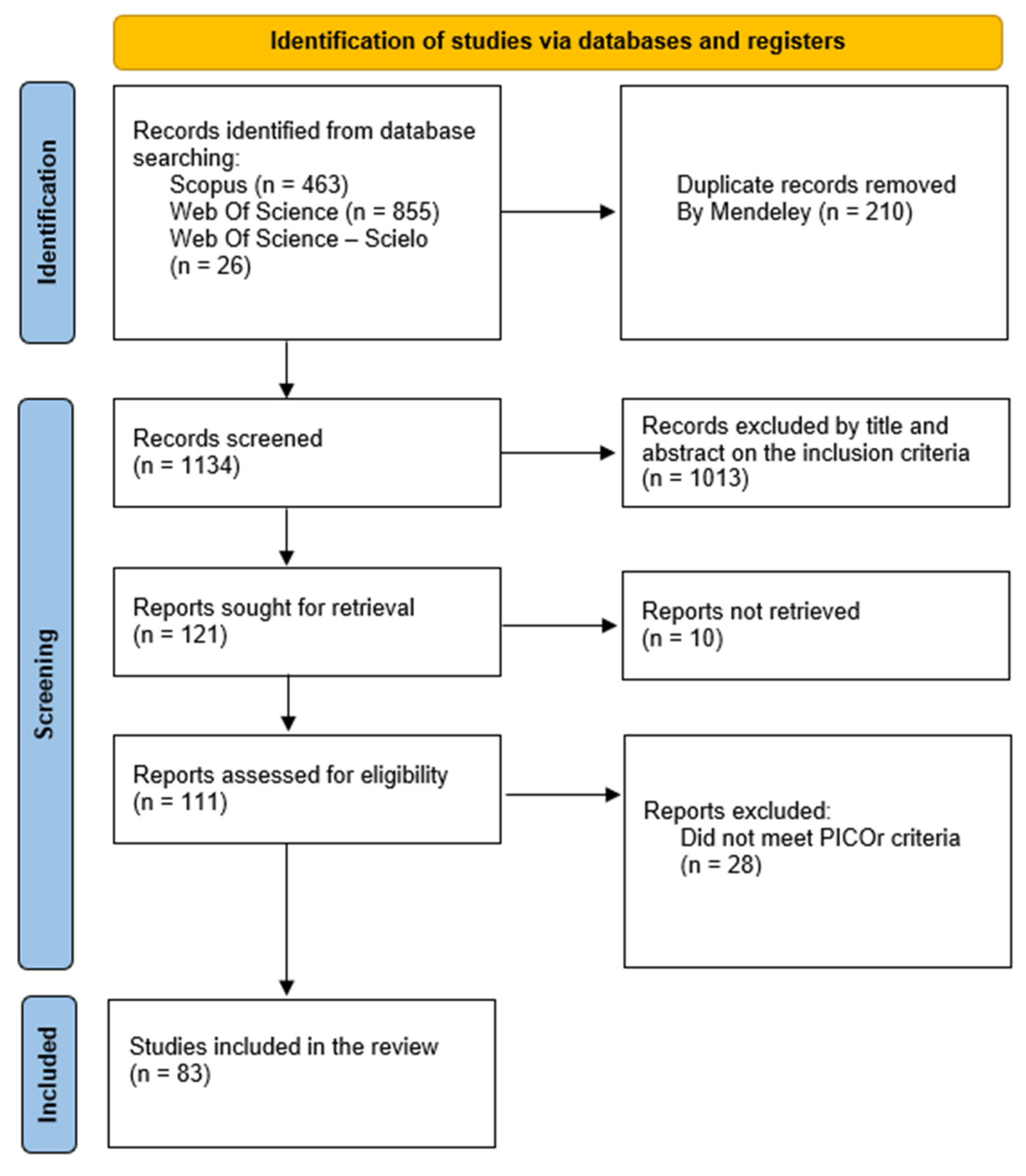
3.1. Database Search and Screening
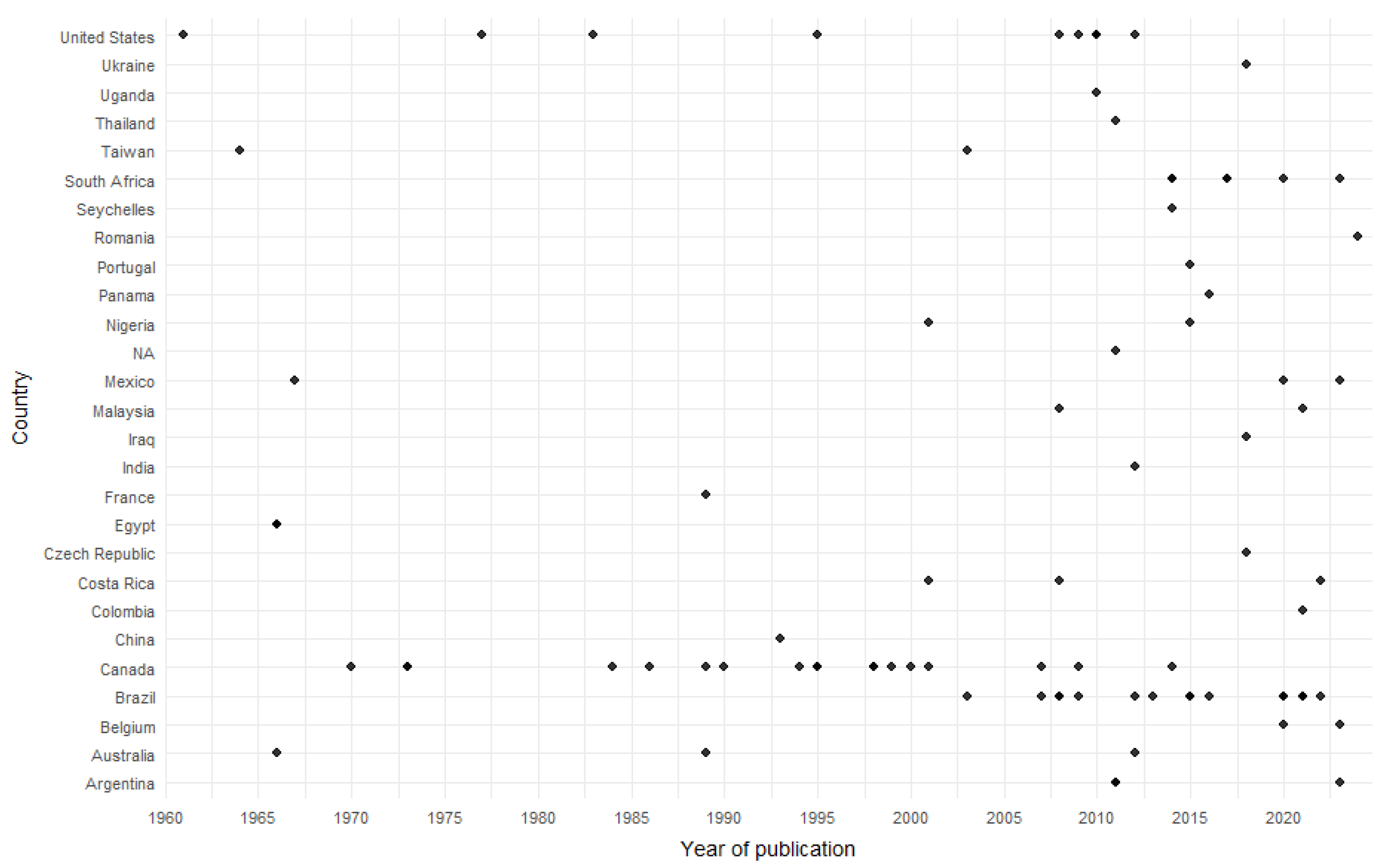
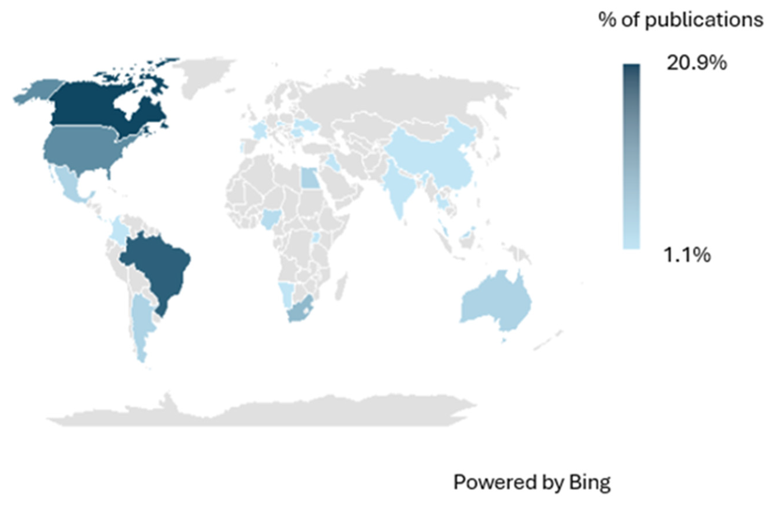
3.2. Publications by Year and Geographic Distribution
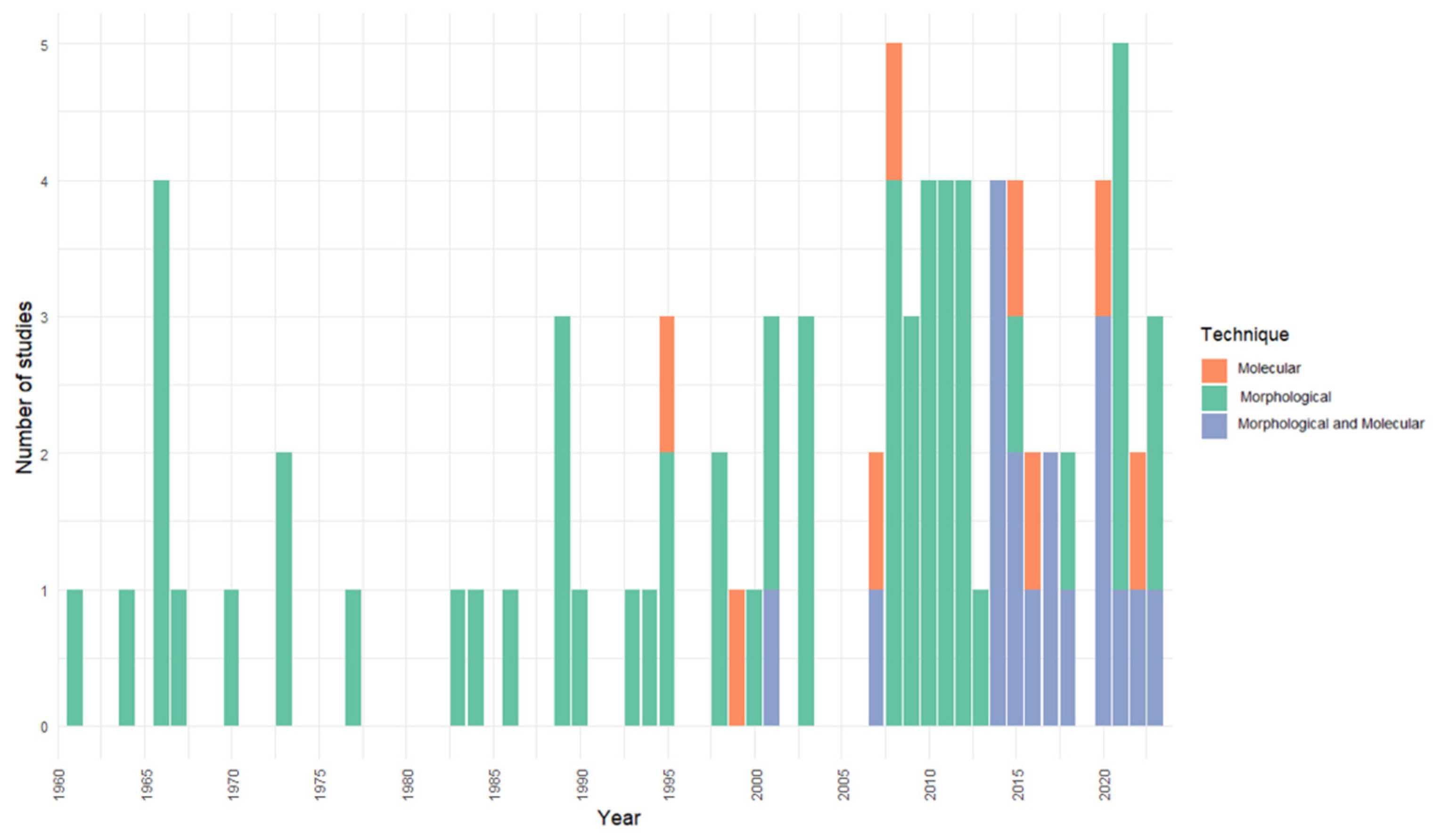
3.3. Diagnostic Techniques
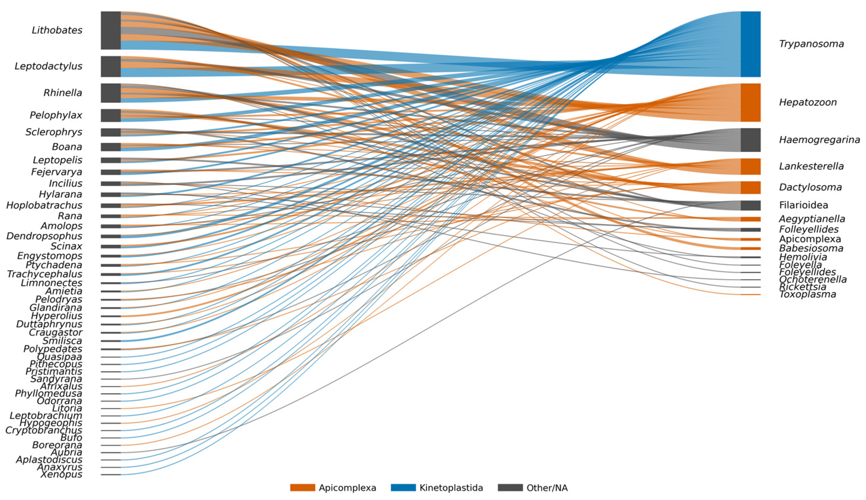
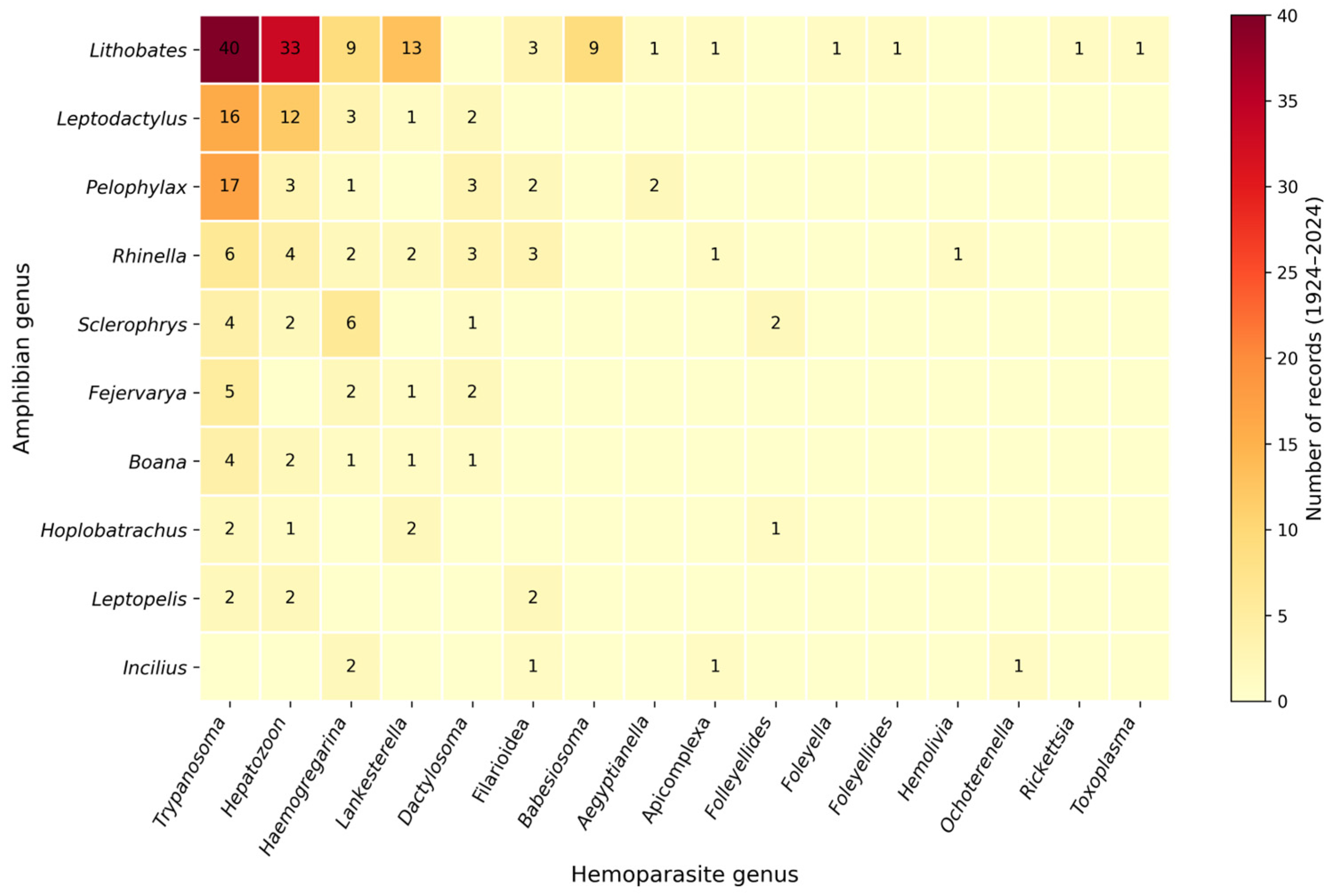
3.4. Hemoparasite Diversity and Amphibian Hosts
3.5. Additional Relevant Literature Published in 2025
4. Discussion
5. Conclusions
Supplementary Materials
Author Contributions
Funding
Institutional Review Board Statement
Informed Consent Statement
Data Availability Statement
Acknowledgments
Conflicts of Interest
References
- Stuart, S.N.; Chanson, J.S.; Cox, N.A.; Young, B.E.; Rodrigues, A.S.L.; Fischman, D.L.; Waller, R.W. Status and trends of amphibian declines and extinctions worldwide. Science 2004, 306, 1783–1786. [Google Scholar] [CrossRef]
- Wake, D.B.; Vredenburg, V.T. Are we in the midst of the sixth mass extinction? A view from the world of amphibians. Proc. Natl. Acad. Sci. USA 2008, 105, 11466–11473. [Google Scholar] [CrossRef] [PubMed]
- Collins, J.P.; Storfer, A. Global amphibian declines: Sorting the hypotheses. Divers. Distrib. 2003, 9, 89–98. [Google Scholar] [CrossRef]
- Blaustein, A.R.; Han, B.A.; Relyea, R.A.; Johnson, P.T.J.; Buck, J.C.; Gervasi, S.S.; Kats, L.B. The complexity of amphibian population declines: Understanding the role of cofactors in driving amphibian losses. Ann. N. Y. Acad. Sci. 2011, 1223, 108–119. [Google Scholar] [CrossRef] [PubMed]
- Daszak, P.; Cunningham, A.A.; Hyatt, A.D. Infectious disease and amphibian population declines. Divers. Distrib. 2003, 9, 141–150. [Google Scholar] [CrossRef]
- Johnson, P.T.J.; Preston, D.L.; Hoverman, J.T.; Richgels, K.L.D. Diversity, decoys and the dilution effect: How ecological communities affect disease risk. J. Exp. Biol. 2013, 216, 961–970. [Google Scholar] [CrossRef]
- Barta, J.R.; Desser, S.S. Blood parasites of amphibians from Algonquin Park, Ontario. J. Wildl. Dis. 1984, 20, 180–189. [Google Scholar] [CrossRef]
- Davies, A.J.; Johnston, M.R.L. The biology of some intraerythrocytic parasites of fishes, amphibia and reptiles. Adv. Parasitol. 2000, 45, 1–107. [Google Scholar] [CrossRef]
- Schall, J.J. Parasite virulence. In The Behavioural Ecology of Parasites; Lewis, E.E., Campbell, J.F., Sukhdeo, M.V.K., Eds.; CABI Publishing: Wallingford, UK, 2002; pp. 283–313. [Google Scholar] [CrossRef]
- Tompkins, D.M.; Dunn, A.M.; Smith, M.J.; Telfer, S. Wildlife diseases: From individuals to ecosystems. J. Anim. Ecol. 2011, 80, 19–38. [Google Scholar] [CrossRef]
- Poynton, S.L.; Whitaker, B.R. Protozoa and metazoa infecting amphibians. In Amphibian Medicine and Captive Husbandry; Wright, K.M., Whitaker, B.R., Eds.; Krieger Publishing Company: Malabar, FL, USA, 2001; pp. 193–221. [Google Scholar]
- Telford, S.R. Hemoparasites of the Reptilia: Color Atlas and Text; CRC Press: Boca Raton, FL, USA, 2009. [Google Scholar] [CrossRef]
- Valkiūnas, G. Avian Malaria Parasites and Other Haemosporidia; CRC Press: Boca Raton, FL, USA, 2005. [Google Scholar] [CrossRef]
- Pérez-Tris, J.; Hellgren, O.; Križanauskienė, A.; Waldenström, J.; Secondi, J.; Bonneaud, C.; Fjeldså, J.; Hasselquist, D.; Bensch, S. Within-host speciation of malaria parasites. PLoS ONE 2007, 2, e235. [Google Scholar] [CrossRef]
- Page, M.J.; McKenzie, J.E.; Bossuyt, P.M.; Boutron, I.; Hoffmann, T.C.; Mulrow, C.D.; Moher, D. The PRISMA 2020 statement: An updated guideline for reporting systematic reviews. BMJ 2021, 372, n71. [Google Scholar] [CrossRef] [PubMed]
- Frost, D.R. Amphibian Species of the World: An Online Reference; Version 6.2; American Museum of Natural History: New York, NY, USA, 2023; Available online: https://amphibiansoftheworld.amnh.org (accessed on 5 April 2025).
- González, L.P.; Vargas-León, C.M.; Fuentes-Rodríguez, G.A.; Calderón-Espinosa, M.L.; Matta, N.E. Do blood parasites increase immature erythrocytes and mitosis in amphibians? Rev. Biol. Trop. 2021, 69, 615–624. [Google Scholar] [CrossRef]
- Matta, N.E.; Gamboa-Suárez, B.A.; Ospina-Rios, A.T.; Fuentes-Rodriguez, G.A.; Jimenez, M.N. From Field to Lab: Giant Toad (Rhinella horribilis) as an Emerging Model for the Study of Amphibian Blood Parasites. Integr. Zool. 2025; ahead of print. [Google Scholar] [CrossRef] [PubMed]
- Votýpka, J.; Jirků, M.; Spodareva, V.; Režnarová, J.; Poloprutská, K.; Pajer, P.; Milner, D.; Richards, T.; Yurchenko, V.; Ibáñez, R.; et al. Trypanosomes in Neotropical frogs: Unveiling hidden megadiversity and complex host–parasite patterns. Open Biol. 2025, 15, 250190. [Google Scholar] [CrossRef]
- Sigl, M.; Virgo, J.; Grafe, U.; Henske, J.; Schweinsberg, M.; Eltz, T. Uninvited guests: Diversity and specificity of Trypanosoma infections in frog-biting midges (Corethrella spp.). Parasites Vectors 2025, 18, 348. [Google Scholar] [CrossRef]
- Bilhalva, L.C.; Peres, L.S.; Soares, J.F.; Santos, A.P.; Dallegrave, E.; Valle, S.d.F. EDTA as a potential obstacle to the detection of trypanosomes in anuran blood samples using Woo’s technique. Vet. Parasitol. Reg. Stud. Rep. 2025, 66, 101386. [Google Scholar] [CrossRef]
- Weng, M.; Liu, X.; Zhang, C.; Shu, R.; Wang, A.; Zhang, H.; Wang, X.; Yang, H.; Zhang, J. A Global Review of the Zoonotic Potential and Disease Risks of Amphibian Parasites in Bullfrog Aquaculture. Rev. Aquac. 2025, 17, e70030. [Google Scholar] [CrossRef]
- Mostafa, O.M.; Morsy, K.; Bin Dajem, S.; Fedda, M.H. A review of parasitic fauna of Egyptian amphibia. Afr. J. Herpetol. 2025, 74, 173–187. [Google Scholar] [CrossRef]
- Huggins, L.G.; Michaels, C.J.; Cruickshank, S.M.; Preziosi, R.F.; Else, K.J. A novel copro-diagnostic molecular method for qualitative detection and identification of parasitic nematodes in amphibians and reptiles. PLoS ONE 2017, 12, e0185151. [Google Scholar] [CrossRef]
- Cunningham, A.A.; Daszak, P.; Wood, J.L.N. One Health, emerging infectious diseases and wildlife: Two decades of progress? Philos. Trans. R. Soc. B 2017, 372, 20160167. [Google Scholar] [CrossRef]
- DeCandia, A.L.; Dobson, A.P.; vonHoldt, B.M. Toward an integrative molecular approach to wildlife disease. Conserv. Biol. 2018, 32, 798–807. [Google Scholar] [CrossRef] [PubMed]
- Bienentreu, J.-F.; Lesbarrères, D. Amphibian disease ecology: Are we just scratching the surface? Herpetologica 2020, 76, 153–166. [Google Scholar] [CrossRef]
- Bower, D.S.; Brannelly, L.A.; McDonald, C.A.; Webb, R.J.; Greenspan, S.E.; Vickers, M.; Gardner, M.G.; Greenlees, M.J. A review of the role of parasites in the ecology of reptiles and amphibians. Austral Ecol. 2019, 44, 433–448. [Google Scholar] [CrossRef]
- Cabrera-Guzmán, E.; Sánchez-Montes, S.; Parra-Olea, G. Research on helminths from Mexican amphibians: Gaps, trends and biases. J. Helminthol. 2021, 95, e81. [Google Scholar] [CrossRef]
- Carlson, C.J.; Dallas, T.A.; Alexander, L.W.; Phelan, A.L.; Phillips, A.J.; Poisot, T. What would it take to describe the global diversity of parasites? Proc. R. Soc. B 2020, 287, 20201841. [Google Scholar] [CrossRef]
- Ellison, G.T.H.; Warner, J.; Robinson, M.W.; Cable, J. Taxonomic and geographic bias in 50 years of research on parasites. PLoS ONE 2021, 16, e0261379. [Google Scholar] [CrossRef]
- Isaak-Delgado, A.B.; López-Díaz, O.; Romero-Callejas, E.; Martínez-Hernández, F.; Muñoz-García, C.I.; Villalobos, G.; Rendón-Franco, E. Morphological and molecular characteristics of hemoparasites in Vaillant’s frogs (Lithobates vaillanti). Parasitol. Res. 2020, 119, 1891–1901. [Google Scholar] [CrossRef]
- Spodareva, V.V.; Grybchuk-Ieremenko, A.; Losev, A.; Votýpka, J.; Lukeš, J.; Yurchenko, V. Diversity and evolution of anuran trypanosomes: Insights from the study of European species. Parasites Vectors 2018, 11, 447. [Google Scholar] [CrossRef]
- Diaz, E.; Hidalgo, A.; Villamarin, C.; Donoso, G.; Barragan, V. Vector-borne zoonotic blood parasites in wildlife from Ecuador: A report and systematic review. Vet. World 2021, 14, 1935–1945. [Google Scholar] [CrossRef]
- Pacheco, M.A.; Escalante, A.A. Origin and diversity of malaria parasites and other Haemosporida. Trends Parasitol. 2023, 39, 501–516. [Google Scholar] [CrossRef]
- O’Donoghue, P. Haemoprotozoa: Making biological sense of molecular and morphological data. Int. J. Parasitol. Parasites Wildl. 2017, 6, 241–256. [Google Scholar] [CrossRef] [PubMed]
- Schmittner, S.M.; McGhee, R.B. The Intra-erythrocytic Development of Babesiosoma stableri n. sp. in Rana Pipiens pipiens. J. Protozool. 1961, 8, 381–386. [Google Scholar] [CrossRef]
- Manwell, R.D. The Genus Dactylosoma. J. Protozool. 1964, 11, 526–530. [Google Scholar] [CrossRef] [PubMed]
- Mohammed, A.H.H.; Mansour, N.S. Development of Haemogregarina boueti in the toad Bufo regularis. J. Protozool. 1966, 13, 259–264. [Google Scholar] [CrossRef] [PubMed]
- Stehbens, W.E. Observations on Lankesterella hylae. J. Protozool. 1966, 13, 59–62. [Google Scholar] [CrossRef]
- Mansour, N.S.; Mohammed, A.H.H. Development of Haemogregarina pestanae in the toad Bufo regularis. J. Protozool. 1966, 13, 265–269. [Google Scholar] [CrossRef]
- Mansour, N.S.; Mohammed, A.H.H. Haemogregarina faiyumensis n. sp. in the toad Bufo regularis in Egypt. J. Protozool. 1966, 13, 269–271. [Google Scholar] [CrossRef]
- Baker, J.R.; Lainson, R. The Fine Structure of the Gametocytes of an Adeleine Haemogregarine. J. Protozool. 1967, 14, 233–238. [Google Scholar] [CrossRef]
- Bardsley, J.E.; Harmsen, R. The trypanosomes of Ranidae. II. The effects of excitation and adrenalin on the peripheral parasitaemia in the bullfrog (Rana catesbeiana Shaw). Can. J. Zool. 1970, 48, 1317–1319. [Google Scholar] [CrossRef]
- Bardsley, J.E.; Harmsen, R. The Trypanosomes of Anura. Adv. Parasitol. 1973, 11, 1–73. [Google Scholar] [CrossRef]
- Desser, S.S.; Weller, I. Structure, Cytochemistry, and Locomotion of Haemogregarina sp. from Rana berlandieri. J. Protozool. 1973, 20, 65–73. [Google Scholar] [CrossRef] [PubMed]
- Levine, N.D.; Nye, R.R. A Survey of Blood and Other Tissue Parasites of Leopard Frogs Rana pipiens in the United States. J. Wildl. Dis. 1977, 13, 17–23. [Google Scholar] [CrossRef] [PubMed]
- Woo, P.T.K. Sensitivity of Diagnostic Techniques in Determining the Prevalence of Anuran Trypanosomes. J. Wildl. Dis. 1983, 19, 24–26. [Google Scholar] [CrossRef] [PubMed]
- Barta, J.R.; Desser, S.S. Light and Electron Microscopic Observations on the Intraerythrocytic Development of Babesiosoma stableri (Apicomplexa, Dactylosomatidae) in Frogs from Algonquin Park, Ontario. J. Protozool. 1986, 33, 359–368. [Google Scholar] [CrossRef]
- Desser, S.S.; Barta, J.R. The Morphological Features of Aegyptianella bacterifera: An Intraerythrocytic Rickettsia of Frogs from Corsica. J. Wildl. Dis. 1989, 25, 313–318. [Google Scholar] [CrossRef]
- Barta, J.R.; Desser, S.S. Development of Babesiosoma stableri (Dactylosomatidae; Adeleida; Apicomplexa) in its Leech Vector (Batracobdella picta) and the Relationship of the Dactylosomatids to the Piroplasms of Higher Vertebrates. J. Protozool. 1989, 36, 241–253. [Google Scholar] [CrossRef]
- Delvinquier, B.L.J. Occurrence of the Protozoans, Lankesterella hylae and Haemogregarina sp., in the Blood of the Green Tree Frog, Litoria caerulea; Memoirs of the Queensland Museum: South Brisbane, Australia, 1989.
- Desser, S.S.; Siddall, M.E.; Barta, J.R. Ultrastructural Observations on the Developmental Stages of Lankesterella minima (Apicomplexa) in Experimentally Infected Rana catesbeiana Tadpoles. J. Parasitol. 1990, 76, 97. [Google Scholar] [CrossRef]
- Werner, J.K. Blood Parasites of Amphibians from Sichuan Province, People’s Republic of China. J. Parasitol. 1993, 79, 356. [Google Scholar] [CrossRef]
- Smith, T.G.; Desser, S.S.; Martin, D.S. The development of Hepatozoon sipedon sp. nov. (Apicomplexa: Adeleina: Hepatozoidae) in its natural host, the Northern water snake (Nerodia sipedon sipedon), in the culicine vectors Culex pipiens and C. territans, and in an intermediate host, the Northern leopard frog (Rana pipiens). Parasitol. Res. 1994, 80, 559–568. [Google Scholar] [CrossRef]
- Desser, S.S.; Hong, H.; Martin, D.S. The Life History, Ultrastructure, and Experimental Transmission of Hepatozoon catesbianae n. comb., an Apicomplexan Parasite of the Bullfrog, Rana catesbeiana and the Mosquito, Culex territans in Algonquin Park, Ontario. J. Parasitol. 1995, 81, 212. [Google Scholar] [CrossRef]
- Clark, C.G.; Martin, D.S.; Diamond, L.S. Phylogenetic Relationships Among Anuran Trypanosomes as Revealed by Riboprinting. J. Eukaryot. Microbiol. 1995, 42, 92–96. [Google Scholar] [CrossRef]
- Herzenberg, A.M.; Barta, J.R.; Desser, S.S. Monoclonal Antibodies Raised against Coccidia and Malarial Parasites Recognize Antigenic Epitopes Found in Lankesterellid and Adeleorin Parasites. J. Parasitol. 1995, 81, 543. [Google Scholar] [CrossRef] [PubMed]
- Kim, B.; Smith, T.G.; Desser, S.S. The Life History and Host Specificity of Hepatozoon clamatae (Apicomplexa: Adeleorina) and ITS-1 Nucleotide Sequence Variation of Hepatozoon Species of Frogs and Mosquitoes from Ontario. J. Parasitol. 1998, 84, 789. [Google Scholar] [CrossRef] [PubMed]
- Smith, T.G.; Desser, S.S. Ultrastructural Features of Cystic and Merogonic Stages of Hepatozoon sipedon (Apicomplexa: Adeleorina) in Northern Leopard Frogs (Rana pipiens) and Northern Water Snakes (Nerodia sipedon) from Ontario, Canada. J. Eukaryot. Microbiol. 1998, 45, 419–425. [Google Scholar] [CrossRef]
- Smith, T.G.; Kim, B.; Desser, S.S. Phylogenetic relationships among Hepatozoon species from snakes, frogs and mosquitoes of Ontario, Canada, determined by ITS-1 nucleotide sequences and life-cycle, morphological and developmental characteristics. Int. J. Parasitol. 1999, 29, 293–304. [Google Scholar] [CrossRef] [PubMed]
- Smith, T.G.; Kim, B.; Hong, H.; Desser, S.S. Intraerythrocytic Development of Species of Hepatozoon Infecting Ranid Frogs: Evidence for Convergence of Life Cycle Characteristics among Apicomplexans. J. Parasitol. 2000, 86, 451. [Google Scholar] [CrossRef]
- Paperna, I.; Martin, C. The development and fine structure of Lankesterella cf. dicroglossi (Apicomplexa: Lankesterellidae) infecting frogs in Niger, West Africa. Folia Parasitol. 2001, 48, 178–186. [Google Scholar] [CrossRef]
- Boulard, Y.; Paperna, I.; Petit, G.; Landau, I. Ultrastructure of developmental stages of Hemolivia stellata (Apicomplexa: Haemogregarinidae) in the cane toad Bufo marinus and in its vector tick Amblyomma rotondatum. Parasitol. Res. 2001, 87, 598–604. [Google Scholar] [CrossRef]
- Desser, S.S. The Blood Parasites of Anurans from Costa Rica with Reflections on the Taxonomy of Their Trypanosomes. J. Parasitol. 2001, 87, 152–160. [Google Scholar] [CrossRef]
- Lainson, R.; Paperna, I.; Naiff, R.D. Development of Hepatozoon caimani (Carini, 1909) Pessôa, De Biasi & De Souza, 1972 in the Caiman Caiman c. crocodilus, the frog Rana catesbeiana and the mosquito Culex fatigans. Mem. Inst. Oswaldo Cruz 2003, 98, 103–113. [Google Scholar] [CrossRef]
- Chang, L.T.; Tsai, S.S.; Tung, M.C. Observations of Hepatozoon (Apicomplexa: Adelenia) in blood of the Taiwan anuran Rana rugulosa Weigmann, 1835. Bull. Eur. Ass. Fish Pathol. 2003, 23, 143. [Google Scholar]
- Ferreira, R.C.; Campaner, M.; Viola, L.B.; Takata, C.S.A.; Takeda, G.F.; Teixeira, M.M.G. Morphological and molecular diversity and phylogenetic relationships among anuran trypanosomes from the Amazonia, Atlantic Forest and Pantanal biomes in Brazil. Parasitology 2007, 134, 1623–1638. [Google Scholar] [CrossRef] [PubMed]
- Boulianne, B.; Evans, R.C.; Smith, T.G. Phylogenetic analysis of Hepatozoon species (Apicomplexa: Adeleorina) infecting frogs of Nova Scotia, Canada, determined by ITS-1 sequences. J. Parasitol. 2007, 93, 1435–1441. [Google Scholar] [CrossRef] [PubMed]
- Stenberg, P.L.; Bowerman, W.J. Hemoparasites in Oregon Spotted Frogs (Rana pretiosa) from Central Oregon, USA. J. Wildl. Dis. 2008, 44, 464–468. [Google Scholar] [CrossRef]
- McKenzie, V.J.; Starks, H.A. Blood Parasites of Two Costa Rican Amphibians with Comments on Detection and Microfilaria Density Associated with Adult Filarial Worm Intensity. J. Parasitol. 2008, 94, 824–829. [Google Scholar] [CrossRef]
- Lemos, M.; Morais, D.H.; Carvalho, V.T.; D’Agosto, M. First Record of Trypanosoma chattoni in Brazil and Occurrence of Other Trypanosoma Species in Brazilian Frogs (Anura, Leptodactylidae). J. Parasitol. 2008, 94, 148–151. [Google Scholar] [CrossRef]
- Wahab, A.R.; Andy, T.W.A.; Intan, S. On the parasitic fauna of two species of anurans collected from Sungai Pinang, Penang Island, Malaysia. Trop. Biomed. 2008, 25, 160–165. [Google Scholar]
- Ferreira, R.C.; De Souza, A.A.; Freitas, R.A.; Campaner, M.; Takata, C.S.A.; Barrett, T.V.; Teixeira, M.M.G. A Phylogenetic Lineage of Closely Related Trypanosomes (Trypanosomatidae, Kinetoplastida) of Anurans and Sand Flies (Psychodidae, Diptera) Sharing the Same Ecotopes in Brazilian Amazonia. J. Eukaryot. Microbiol. 2008, 55, 427–435. [Google Scholar] [CrossRef]
- Leal, D.D.M.; O’dwyer, L.H.; Ribeiro, V.C.; Silva, R.J.; Ferreira, V.L.; Rodrigues, R.B. Hemoparasites of the genus Trypanosoma (Kinetoplastida: Trypanosomatidae) and hemogregarines in Anurans of the São Paulo and Mato Grosso do Sul States—Brazil. Anais Acad. Bras. Ciênc. 2009, 81, 199–206. [Google Scholar] [CrossRef]
- Schotthoefer, A.M.; Bolek, M.G.; Cole, R.A.; Beasley, V.R. Parasites of the Mink Frog (Rana septentrionalis) from Minnesota, USA. Comp. Parasitol. 2009, 76, 240–246. [Google Scholar] [CrossRef]
- Shutler, D.; Smith, T.G.; Robinson, S.R. Relationships between leukocytes and Hepatozoon spp. in green frogs, Rana clamitans. J. Wildl. Dis. 2009, 45, 67–72. [Google Scholar] [CrossRef] [PubMed]
- Stenberg, P.L.; Bowerman, W.J. First report of Hepatozoon sp. in the Oregon Spotted Frog, Rana pretiosa. J. Wildl. Dis. 2010, 46, 956–960. [Google Scholar] [CrossRef] [PubMed]
- Readel, A.M.; Goldberg, T.L. Blood Parasites of Frogs From an Equatorial African Montane Forest in Western Uganda. J. Parasitol. 2010, 96, 448–450. [Google Scholar] [CrossRef] [PubMed]
- Harkness, L.M.; Drohan, A.E.; Dickson, C.M.; Smith, T.G. Experimental Transmission of Hepatozoon clamatae (Apicomplexa: Adeleida) to the Wood Frog, Rana sylvatica, and to the Mosquito Culex pipiens. J. Parasitol. 2010, 96, 434–436. [Google Scholar] [CrossRef]
- Gericota, B.; Garner, M.M.; Barr, B.; Nordhausen, R.; Larsen, R.S.; Lowenstine, L.J.; Murphy, B.G. Morphologic, Immunohistochemical, and Molecular Characterization of a Novel Lankesterella Protozoan in Two White’s Tree Frogs (Litoria caerulea). J. Zoo Wildl. Med. 2010, 41, 242–248. [Google Scholar] [CrossRef]
- Cabagna Zenklusen, M.C.; Lajmanovich, R.C.; Attademo, A.M.; Peltzer, P.M.; Junges, C.M.; Fiorenza Biancucci, G.; Bassó, A. Hematología y citoquímica de las células sanguíneas de Rhinella fernandezae (Anura: Bufonidae) en Espinal y Delta-Islas del río Paraná, Argentina. Rev. De Biol. Trop. 2011, 59, 17–28. [Google Scholar] [CrossRef]
- Shutler, D.; Marcogliese, D.J. Leukocyte Profiles of Northern Leopard Frogs, Lithobates pipiens, Exposed to Pesticides and Hematozoa in Agricultural Wetlands. Copeia 2011, 2011, 301–307. [Google Scholar] [CrossRef]
- Sailasuta, A.; Satetasit, J.; Chutmongkonkul, M. Pathological Study of Blood Parasites in Rice Field Frogs, Hoplobatrachus rugulosus (Wiegmann, 1834). Vet. Med. Int. 2011, 2011, 850568. [Google Scholar] [CrossRef]
- Young, S.; Warner, J.; Speare, R.; Berger, L.; Skerratt, L.F.; Muller, R. Hematologic and plasma biochemical reference intervals for health monitoring of wild Australian tree frogs. Vet. Clin. Pathol. 2012, 41, 478–492. [Google Scholar] [CrossRef]
- Viana, L.A.; Soares, P.; Silva, J.E.; Paiva, F.; Coutinho, M.E. Anurans as paratenic hosts in the transmission of Hepatozoon caimani to caimans Caiman yacare and Caiman latirostris. Parasitol. Res. 2011, 110, 883–886. [Google Scholar] [CrossRef]
- Gupta, D.K.; Gupta, N.; Gangwar, R. Infectivity of Bufo melanostictus (Amphibia: Bufonidae) to Two New Species of Haematozoan Parasites from Rohilkhand, India. Proc. Zool. Soc. 2012, 65, 22–32. [Google Scholar] [CrossRef]
- Lemos, M.; Souza, C.S.F.; da Costa, S.C.G.; Souto-Padrón, T.; D’Agosto, M. Isolation and In Vitro Culture of Trypanosomes From Leptodactylus ocellatus From the Atlantic Forest in a New Experimental Culture Medium. J. Parasitol. 2013, 99, 164–167. [Google Scholar] [CrossRef] [PubMed]
- Davis, A.K.; Hopkins, W.A. Widespread trypanosome infections in a population of eastern hellbenders (Cryptobranchus alleganiensis alleganiensis) in Virginia, USA. Parasitol. Res. 2012, 112, 453–456. [Google Scholar] [CrossRef] [PubMed]
- Netherlands, E.C.; Cook, C.A.; Smit, N.J. Hepatozoon species (Adeleorina: Hepatozoidae) of African bufonids, with morphological description and molecular diagnosis of Hepatozoon ixoxo sp. nov. parasitising three Amietophrynus species (Anura: Bufonidae). Parasites Vectors 2014, 7, 552. [Google Scholar] [CrossRef]
- Leveille, A.; Ogedengbe, M.E.; Hafeez, M.A.; Tu, H.-H.; Barta, J.R. The Complete Mitochondrial Genome Sequence of Hepatozoon catesbianae (Apicomplexa: Coccidia: Adeleorina), a Blood Parasite of the Green Frog, Lithobates (Formerly Rana) clamitans. J. Parasitol. 2014, 100, 651–656. [Google Scholar] [CrossRef]
- Harris, D.J.; Damas-Moreira, I.; Maia, J.P.M.C.; Perera, A. First Report of Hepatozoon(Apicomplexa: Adeleorina) in Caecilians, with Description of a New Species. J. Parasitol. 2014, 100, 117–120. [Google Scholar] [CrossRef]
- Netherlands, E.C.; Cook, C.A.; Smit, N.J.; Du Preez, L.H. Redescription and Molecular Diagnosis of Hepatozoon theileri (Laveran, 1905) (Apicomplexa: Adeleorina: Hepatozoidae), Infecting Amietia quecketti (Anura: Pyxicephalidae). Folia Parasitol. 2014, 61, 293. [Google Scholar] [CrossRef]
- Menezes Leal, D.D.; Dreyer, C.S.; da Silva, R.J.; Ribolla, P.E.M.; dos Santos Paduan, K.; Bianchi, I.; O’Dwyer, L.H. Characterization of Hepatozoon spp. in Leptodactylus chaquensis and Leptodactylus podicipinus from Two Regions of the Pantanal, State of Mato Grosso do Sul, Brazil. Parasitol. Res. 2015, 114, 1541–1549. [Google Scholar] [CrossRef]
- Seabra-Babo, J.; Maia, J.P.; Harris, D.J. Scanning for Apicomplexan Parasites (Suborder Adeleorina) in Five Holarctic Anuran Species. Herpetozoa 2015, 27, 168–172. [Google Scholar]
- Aisien, M.S.O.; Aigbirior, P.O.; Ovwah, E.; Edo-Taiwo, O. Blood Parasites of Some Anurans from Southern Nigeria. Trop. Biomed. 2015, 32, 598–607. [Google Scholar]
- Ferreira, J.I.G.d.S.; da Costa, A.P.; Ramirez, D.; Roldan, J.A.M.; Saraiva, D.; Founier, G.F.R.d.S.; Sue, A.; Zambelli, E.R.; Minervino, A.H.H.; Verdade, V.K.; et al. Anuran Trypanosomes: Phylogenetic Evidence for New Clades in Brazil. Syst. Parasitol. 2015, 91, 63–70. [Google Scholar] [CrossRef] [PubMed]
- Attias, M.; Sato, L.H.; Ferreira, R.C.; Takata, C.S.A.; Campaner, M.; Camargo, E.P.; de Souza, W. Developmental and Ultrastructural Characterization and Phylogenetic Analysis of Trypanosoma herthameyerin sp. of Brazilian Leptodactylidae Frogs. J. Eukaryot. Microbiol. 2016, 63, 610–622. [Google Scholar] [CrossRef] [PubMed]
- Bernal, X.E.; Pinto, C.M. Sexual Differences in Prevalence of a New Species of Trypanosome Infecting Túngara Frogs. Int. J. Parasitol. Parasites Wildl. 2016, 5, 40–47. [Google Scholar] [CrossRef] [PubMed]
- Conradie, R.; Cook, C.A.; du Preez, L.H.; Jordaan, A.; Netherlands, E.C. Ultrastructural Comparison of Hepatozoon ixoxo and Hepatozoon theileri (Adeleorina: Hepatozoidae), Parasitising South African Anurans. J. Eukaryot. Microbiol. 2016, 64, 193–203. [Google Scholar] [CrossRef]
- Netherlands, E.C.; Cook, C.A.; Du Preez, L.H.; Vanhove, M.P.M.; Brendonck, L.; Smit, N.J. Monophyly of the Species of Hepatozoon (Adeleorina: Hepatozoidae) Parasitizing African Anurans, with the Description of Three New Species from Hyperoliid Frogs in South Africa. Parasitology 2017, 145, 1039–1050. [Google Scholar] [CrossRef]
- Al-Khamesi, M.B.; Salman, I.S.; Abid, S.A.; Ibrahim, S.M. Study Prevalence and Effect of Internal Parasites and Histopathological Changes on Common Frogs at Baghdad City. Iraqi J. Agric. Sci. 2018, 49, 445–451. [Google Scholar] [CrossRef]
- Netherlands, E.C.; Cook, C.A.; Du Preez, L.H.; Vanhove, M.P.M.; Brendonck, L.; Smit, N.J. An Overview of the Dactylosomatidae (Apicomplexa: Adeleorina: Dactylosomatidae), with the Description of Dactylosoma kermiti n. sp. Parasitising ptychadena anchietae and Sclerophrys gutturalis from South Africa. Int. J. Parasitol. Parasites Wildl. 2020, 11, 246–260. [Google Scholar] [CrossRef]
- Úngari, L.P.; Netherlands, E.C.; Santos, A.L.Q.; de Alcantara, E.P.; Emmerich, E.; da Silva, R.J.; O’Dwyer, L.H. A New Species, Dactylosoma piperis n. sp. (Apicomplexa, Dactylosomatidae), from the Pepper Frog Leptodactylus labyrinthicus (Anura, Leptodactylidae) from Mato Grosso State, Brazil. Parasite 2020, 27, 73. [Google Scholar] [CrossRef]
- De Abreu Reis Ferreira, D.; Perles, L.; Machado, R.Z.; Prado, C.P.A.; André, M.R. Molecular Detection of Apicomplexan Hemoparasites in Anurans from Brazil. Parasitol. Res. 2020, 119, 3469–3479. [Google Scholar] [CrossRef]
- Coêlho, T.A.; Souza, D.C.; Kawashita-Ribeiro, R.A.; Corrêa, L.L. First Record of Trypanosoma sp. (Kinetoplastea: Trypanosomatidae) Parasitizing Rhinella major in the Brazilian Amazon. An. Acad. Bras. Ciênc. 2021, 93, e20190467. [Google Scholar] [CrossRef]
- Úngari, L.P.; Netherlands, E.C.; Quagliatto Santos, A.L.; Paulino de Alcantara, E.; Emmerich, E.; da Silva, R.J.; O’Dwyer, L.H. New Insights on the Diversity of Brazilian Anuran Blood Parasites: With the Description of Three New Species of Hepatozoon (Apicomplexa: Hepatozoidae) from Leptodactylidae Anurans. Int. J. Parasitol. Parasites Wildl. 2021, 14, 190–201. [Google Scholar] [CrossRef] [PubMed]
- Agustar, H.K.; Jansar, K.M.; Ali, A.H.; Ghani, N.F.A. Abundance of Anuran Parasites and Pesticide Pollution from Lowland and Highland Golf Courses. J. Environ. Biol. 2021, 42, 789–797. [Google Scholar] [CrossRef]
- Coêlho, T.A.; De Souza, D.C.; da Costa Oliveira, E.; Correa, L.L.; Viana, L.A.; Kawashita-Ribeiro, R.A. Haemogregarine of Genus Dactylosoma (Adeleorina: Dactylosomatidae) in Species of Rhinella (Anura: Bufonidae) from the Brazilian Amazon. Acta Parasitol. 2021, 66, 1574–1580. [Google Scholar] [CrossRef] [PubMed]
- Shangi, N.E.; Gardner, K.M.; Mennill, D.J.; Doucet, S.M. Is Color Related to Parasite Load in a Sexually Dichromatic Neotropical Toad? Herpetologica 2022, 78, 235–243. [Google Scholar] [CrossRef]
- Úngari, L.P.; Netherlands, E.C.; Santos, A.L.Q.; de Alcantara, E.P.; Emmerich, E.; da Silva, R.J.; O’dWyer, L.H. Diversity of Haemogregarine Parasites Infecting Brazilian Anurans, with a Description of New Species of Dactylosoma (Apicomplexa: Adeleorina: Dactylosomatidae). Acta Parasitol. 2022, 67, 1740–1755. [Google Scholar] [CrossRef]
- Pollo, F.; Salinas, Z.; Baraquet, M.; Otero, M.A.; Grenat, P.R.; Salas, N.; Martino, A.L.; Sinsch, U. Hemoparasites Do Not Affect Life-History Traits and Cellular Immune Response in Treefrog Hosts Boana cordobae. Animals 2023, 13, 3566. [Google Scholar] [CrossRef]
- Jordaan, B.J.; du Preez, L.H.; Netherlands, E.C. Taxonomic Re-Evaluation of African Anuran Trypanosomes with the Redescription and Molecular Diagnosis of Trypanosoma (Trypanosoma) nelspruitense Laveran, 1904 and Trypanosoma (Haematomonas) grandicolor Pienaar, 1962. Parasitology 2023, 150, 477–487. [Google Scholar] [CrossRef]
- Isaak-Delgado, A.B.; Zavala-Norzagaray, A.A.; Espinoza-Romo, B.A.; Ortega-Anaya, J.G.; Ley-Quiñonez, C.P.; Aguirre, A.; Rendón-Franco, E. Hematologic Parameters and the Effect of Hemoparasites of Wild Anurans in Northern Sinaloa, Mexico. Vet. Clin. Pathol. 2023, 52, 386–395. [Google Scholar] [CrossRef]
- Pavľáková, B.; Pipová, N.; Balogová, M.; Majláth, I.; Mikulíček, P.; Majláthová, V. Blood Parasites of Water Frogs (Pelophylax esculentus Complex) from the Danube Delta, Romania. Parasitol. Int. 2024, 102, 102920. [Google Scholar] [CrossRef]



| Component | Definition | Application in This Study |
|---|---|---|
| Population (P) | Target population | Amphibians (orders Anura, Caudata, and Gymnophiona) |
| Intervention (I) | Exposure/condition of interest | Presence of hemoparasites identified by morphological or molecular techniques 1 |
| Comparison (C) | Factors of comparison | Year of publication, country/region, diagnostic method (morphological vs. molecular) |
| Outcomes (O) | Expected results | Reported prevalence, parasite taxa identified, diagnostic methods used |
Disclaimer/Publisher’s Note: The statements, opinions and data contained in all publications are solely those of the individual author(s) and contributor(s) and not of MDPI and/or the editor(s). MDPI and/or the editor(s) disclaim responsibility for any injury to people or property resulting from any ideas, methods, instructions or products referred to in the content. |
© 2026 by the authors. Licensee MDPI, Basel, Switzerland. This article is an open access article distributed under the terms and conditions of the Creative Commons Attribution (CC BY) license.
Share and Cite
Yepes, P.A.; Barrientos, L.S.; Pulido-Villamarín, A. Anuran amphibian Hemoparasites over the Last Century: Advances, Challenges, and Future Prospects: A Systematic Review. Animals 2026, 16, 847. https://doi.org/10.3390/ani16050847
Yepes PA, Barrientos LS, Pulido-Villamarín A. Anuran amphibian Hemoparasites over the Last Century: Advances, Challenges, and Future Prospects: A Systematic Review. Animals. 2026; 16(5):847. https://doi.org/10.3390/ani16050847
Chicago/Turabian StyleYepes, Paula Andrea, Lucas S. Barrientos, and Adriana Pulido-Villamarín. 2026. "Anuran amphibian Hemoparasites over the Last Century: Advances, Challenges, and Future Prospects: A Systematic Review" Animals 16, no. 5: 847. https://doi.org/10.3390/ani16050847
APA StyleYepes, P. A., Barrientos, L. S., & Pulido-Villamarín, A. (2026). Anuran amphibian Hemoparasites over the Last Century: Advances, Challenges, and Future Prospects: A Systematic Review. Animals, 16(5), 847. https://doi.org/10.3390/ani16050847