Article

Analysis of Transcriptome and Differentially Expressed Genes in Chicken Primordial Germ Cells
by
Anastasiia I. Azovtseva
*,
Anna E. Ryabova
,
Artem P. Dysin
,
Grigoriy K. Peglivanyan
,
Natalia R. Reinbach
,
Alina V. Gabova
,
Olga Y. Barkova
,
Ekaterina A. Polteva
and
Tatiana A. Larkina
*
Russian Research Institute of Farm Animal Genetics and Breeding (RRIFAGB)—Branch of the L.K. Ernst Federal Science Centre for Animal Husbandry, Pushkin, 196601 St. Petersburg, Russia
*
Authors to whom correspondence should be addressed.
Animals 2026, 16(3), 522; https://doi.org/10.3390/ani16030522
Submission received: 19 January 2026 / Revised: 30 January 2026 / Accepted: 4 February 2026 / Published: 6 February 2026
(This article belongs to the Section Animal Genetics and Genomics)

Simple Summary

Successful genome editing using primordial germ cells requires a detailed understanding of their functioning during embryonic development. In this study, we performed, for the first time, a comparative transcriptome analysis between chicken primordial germ cells and adult liver cells. We identified a total of 1909 differentially expressed genes involved in intracellular metabolism and protein biosynthesis, including transcription, translation, and post-translational protein modifications. This expression signature is consistent with the highly active and undifferentiated state of primordial germ cells during early embryogenesis. The findings provide a valuable resource for understanding gene activity within avian germ cells and establish a foundational transcriptomic signature for primordial germ cells. This knowledge could potentially serve as an important tool for maintaining the genetic biodiversity of unique, small, and endangered chicken populations and breeds.

Abstract

Achieving successful primordial germ cell (PGC)-based genome editing requires a deep understanding of their molecular identity. For the first time, a comparative transcriptomic analysis of chicken PGCs and adult liver cells to define their specific gene expression signature was performed. PGCs were isolated from Rhode Island Red chicken embryos, cultured, and subjected to RNA sequencing alongside liver tissue. Differential expression analysis with Benjamini–Hochberg correction identified 1909 differentially expressed genes (DEGs). Functional annotation revealed that PGCs possess a unique transcriptional landscape, characterized not only by enhanced proliferation and metabolic activity but also by a profound molecular convergence with neural crest cells. This is evidenced by the upregulation of gene modules governing long-range migration, neuronal signaling, and specialized “neuro-lipid” metabolism (e.g., sphingolipid and plasmalogen pathways). Additionally, we identified unannotated transcripts linked to immune pathways and ciliary signaling. Our study expands the functional annotation of avian PGCs and reveals an unexpected evolutionary recruitment of conserved morphogenetic programs, providing a refined molecular foundation for advanced germline editing technologies.

1. Introduction

Qualitative breakthroughs in the development of genome editing (GE) tools and technologies allow for precise genomic modifications [1], demonstrating high potential for the development of biomedical, biotechnological and agricultural industries [2,3]. GE-technologies enable targeted GE for both the treatment of human diseases [4] and the improvement in productive traits and the achievement of desired phenotypic functions in livestock, including disease-specific resistance [2,5]. Current mammalian GE-protocols mainly involve embryo editing with its subsequent transplantation to an animal recipient. However, this approach is not feasible in avian species due to their embryogenesis peculiarities [1]. Despite the lack of successful transfer of genetic modifications when editing chicken embryos in ovo [3], studies exploring GE in chickens persisted. A pivotal solution came with the development of the in vitro cultivation protocol for primordial germ cells (PGCs), facilitated by the identification of PGC self-renewal mechanisms [6,7]. PGCs are gamete precursors, the only cell type capable of transmitting genetic and epigenetic information across generations [8,9]. Chicken PGCs originate from the epiblast during early embryogenesis [10], when the germ cell line separates from the somatic cell line [11]. They are first identified in the center of the area pellucida (stage X blastoderm) [12], from where they migrate to the germinal crescent [13]. Subsequently, PGCs enter the vascular system [14] and use it to reach the germinal ridges, where they accumulate as gonadal PGCs or gonocytes [15]. Later, they differentiate into spermatogonia and oogonia [9].
PGCs have great potential for genome editing. Their capacity for in vitro cultivation allows for genetic modification and reproduction in clonal populations, which can be used for creating chimeras capable of producing offspring with a modified germ cell line [1,3]. The advantage of PGCs is their availability at many embryonic stages; they can be isolated by both embryo dissection and blood aspiration [3]. The latter method is feasible because, unlike most mammals, chicken PGCs migrate to the gonadal buds via the vascular system instead of the intestinal epithelium [14,16]. Ultimately, PGCs can significantly improve the quality of agriculture and livestock products by the rapid introduction of desired phenotypic traits, improving disease management, enhancing animal welfare, and ensuring the safety of chicken egg-grown vaccines [1]. A significant contribution can be made to the conservation of global biodiversity. Intensive selection has reduced the genetic diversity of bird species almost by half [6,17], creating an urgent need for effective ex situ conservation strategies [18]. Cryopreservation of germplasm faces a fairly low efficiency in birds due to the polylecithal egg structure and the reduced fertility of cryopreserved semen [19]. In this context, PGCs biobanking is a promising alternative for preserving biomaterial from endangered species for future population recovery [20,21].
However, the successful application of PGC-based genome editing requires substantial information on the embryonic development governing germ cell production from PGCs, alongside knowledge on the peculiarities of PGC functioning. The latter include migration, survival, sexual differentiation, and epigenetic reprogramming [11].
The aim of the present study was to perform a comparative transcriptome analysis of chicken PGCs and adult liver cells. Through differential gene expression analysis, we aimed to identify key genes active in PGCs and to expand the existing knowledge of their functional genomics.

2. Materials and Methods

2.1. Animal Sampling

Biological material. PGCs (n = 2) were obtained from embryos of the Rhode Island Red chicken breed, kept in the Center of Collective Use (CCU) "Genetic Collection of Rare and Endangered Chicken Breeds" (Pushkin, Saint-Petersburg, Russia). As a control, liver cells (n = 2) were obtained from adult roosters of the same breed via post-mortem cell sampling at the age of 52 weeks.
Rationale for control tissue selection. Adult liver was selected as a negative control tissue for transcriptomic comparison. Liver cells represent a terminally differentiated somatic state with a stable metabolic profile. This choice provides a maximal transcriptional contrast to the pluripotent, migratory PGCs, thereby allowing the filtering out of ubiquitous housekeeping gene expression and facilitating the robust identification of genes that are specifically and highly upregulated in the germ cell lineage.

2.2. PGC Culture Medium

Culture medium for PGC cultivation consisted of base medium Opti-MEM (Reduced Serum Medium, GlutaMAXTM Supplement) (GibcoTM, Thermo Fisher Scientific Inc., Waltham, MA, USA) supplemented with sodium pyruvate (Applichem GmbH., Darmstadt, Germany)—1 M; EmbryoMax nucleosides (100×) (MilliporeSigma, Burlington, MA, USA)—up to 1×; chicken serum (GibcoTM, Thermo Fisher)—2%; 2-mercaptoethanol (NF grade, VWR International, Radnor, PA, USA)—0.01%; antibiotic-antimycotic (100×) (GibcoTM, Thermo Fisher)—up to 1×; human Activin A Recombinant Protein (GibcoTM, Thermo Fisher)—25 ng/µL; human FGF-basic (FGF-2/bFGF) Recombinant Protein (GibcoTM, Thermo Fisher)—10 ng/µL.

2.3. Derivation and Culturing of Chicken PGCs

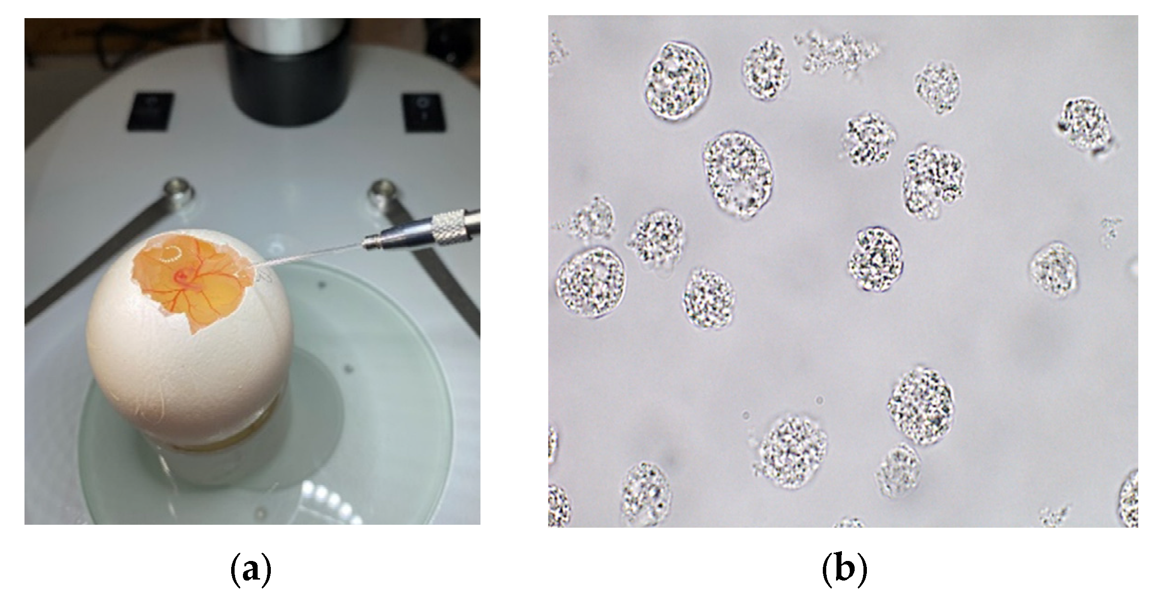
Fertilized eggs from the Rhode Island Red chicken breed were screened and subsequently disinfected. Egg incubation was carried out up to stage 13–14 by Hamburger-Hamilton (approximately 50–52 h post-laying) [22]. At this stage, PGCs are bipotent and have not initiated sex-specific differentiation programs. PGC extraction was performed using embryo blood. Blood samples were collected by egg dissection using a graver (Dremel Inc., Mount Prospect, IL, USA), namely by opening a small window (approximately 10–15 mm in diameter), without damaging the underlying shell membrane. Blood was aspirated from the dorsal aorta of the embryos using a microinjector (model IM-11-2, Narishige Co., Ltd., Tokyo, Japan) (Figure 1a). The collected blood samples were immediately transferred to prepared vials with a culture medium and maintained at +37 °C until plating. PGC cultivation was performed in multiwell plates with supplemented culture medium and growth factors at +37 °C for 21 days (Figure 1b). Quantification of PGC cultures started within 10–12 days of cultivation.
We did not perform genetic sex determination of the pooled PGC population prior to RNA-seq analysis. Therefore, the PGC samples represent a mixed population potentially containing cells from both male and female embryos.

2.4. RNA Extraction and Library Preparation

RNA isolation was performed using the MagMaxTM Total RNA Isolation Kit (Thermo Fisher Scientific Inc., Waltham, MA, USA). RNA quality assessment was performed by quantitative fluorescence and fragment length analysis, yielding the RNA integrity number (RIN) and DV200 index to evaluate the degree of degradation and fragmentation. Sequencing libraries were performed using the TruSeq Stranded mRNA Library Prep Kit (Illumina Inc., San Diego, CA, USA). To minimize batch effects and ensure comparability, all the samples (both PGC and liver) underwent RNA extraction, library preparation, and sequencing in the same batch, under identical protocols and on the same sequencing lane of the NovaSeq 6000 platform.

2.5. Generation of RNA-Seq Data and Bioinformatics Analysis

Sequencing was performed using the NovaSeq 6000 platform (Illumina Inc., San Diego, CA, USA), with read length—2 × 101 b.p. Quality control revealed that 92.06% of the reads had a Phred quality score of Q30 (probability 1 wrong base per 1000 readings). Demultiplexing of the raw data was performed using bcl2fastq2 (v2.20) (Illumina Inc., San Diego, CA, USA), and removal of adapter sequences with Skewer (v0.2.2b) [23]. The trimmed reads were aligned to the reference genome of Red Jungle Fowl (GRCg6a assembly) using the STAR aligner (v2.7.3) [24].

2.6. Transcriptome Comparison and Differential Gene Expression Analysis

Differential expression analysis between groups was performed using the DESeq2 package (v1.24.0) in the R (v4.0.4) [25] programming environment. DESeq2 uses a negative binomial generalized linear model to test for differential expression based on raw gene counts.
According to DESeq2 recommendations, the genes with fewer than two reads were removed from all the samples, as their variance is the highest and they often give false positives. The package internally performs size factor normalization to account for differences in library depth. Using the normalized counts, DESeq2 calculates the log2 fold changes (log2FC) and tests their statistical significance using the Wald test. The resulting p-values were adjusted for multiple testing using the Benjamini–Hochberg correction to control the false discovery rate (FDR) [26]. The genes with log2 FC > 1.5 and an adjusted p-value (padj) < 0.05 were marked as significantly differentially expressed.
To assess the overall sample relationship, we performed hierarchical clustering and principal component analysis (PCA). These visualizations required the application of the variance-stabilizing transformation (VST) to the count data to ensure equal gene contribution in the distance between the samples.
The Gene Ontology (GO) enrichment analysis for biological processes was conducted on the identified DEGs using the GO terms from the GO database (URL: https://geneontology.org/ accessed on 23 September 2025) using the clusterProfiler package (v4.3.1.900) in R (v4.0.4). Enrichment p-values were also corrected for multiple testing using the Benjamini–Hochberg correction. The results were visualized using ggplot2 (v3.4.4) and ggdendro packages (v0.1.22) in R (v4.0.4). Functional annotation of individual genes was performed via the NCBI genome browser.

3. Results

RNA sequencing was conducted on a total of four samples for differential expression analysis. Group 1 contained two liver cell samples, and Group 2—two PGC samples. The quality metrics and output statistics of the obtained raw data are presented in Table 1.
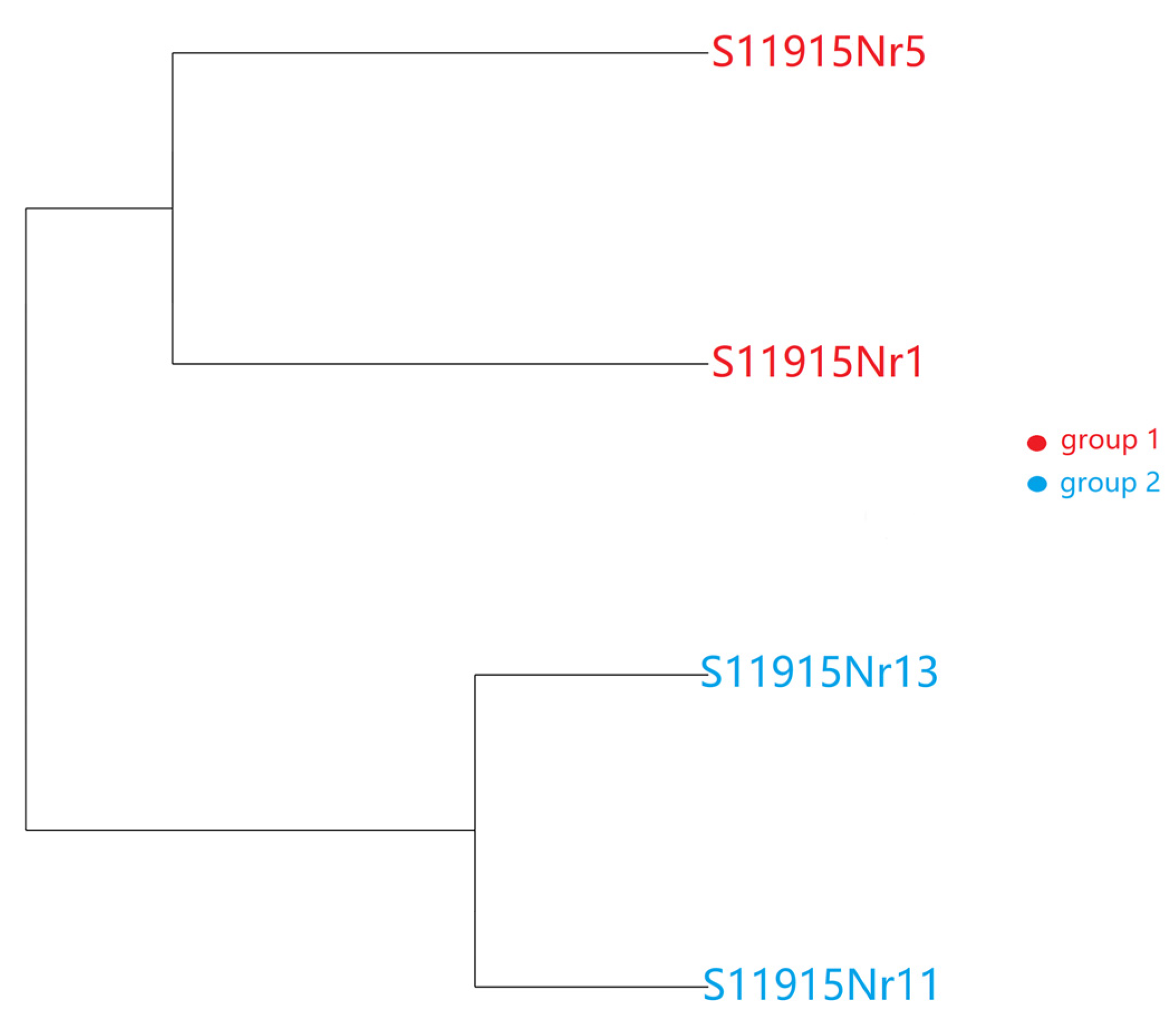
The dendrogram revealed clear separation between the two cell types, with sample clustering exclusively by their origin (Figure 2). This underscores their transcriptomic differences.
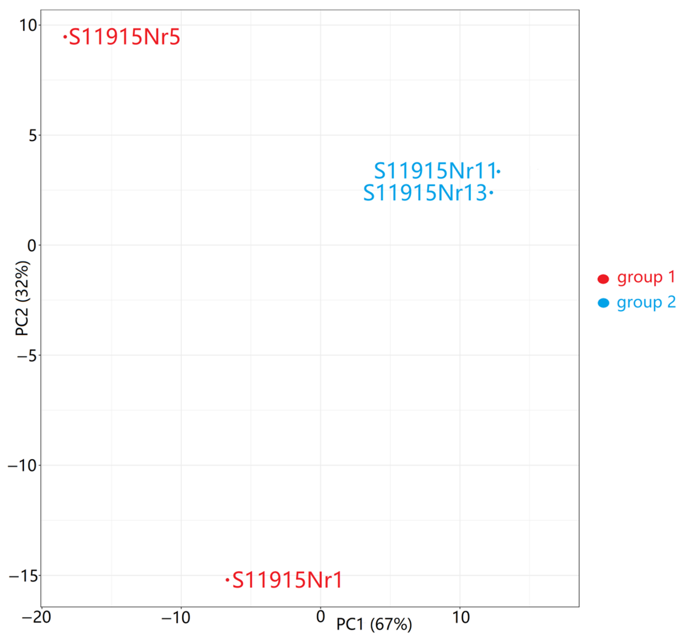
Differential expression analysis between Group 2 (PGCs) and Group 1 (liver cells) was performed on a total of 16,859 genes (Table S1). Principal component analysis (PCA) was applied to visualize the transcriptomic relationships (Figure 3). The first (PC1) and second (PC2) components explained 67% and 32% of the variance between the samples, respectively. The observed PGC sample clustering indicates a high similarity in their sets of differentially expressed genes. In contrast, the liver cell samples did not show any clustering; sample S11915Nr1 had moderate negative loading on PC1 and high negative loading on PC2, whereas sample S11915Nr5 showed high negative loading on PC1 and high positive loading on PC2. The notable heterogeneity among the liver samples most likely reflects the natural physiological variation and complex functional state of the adult liver.
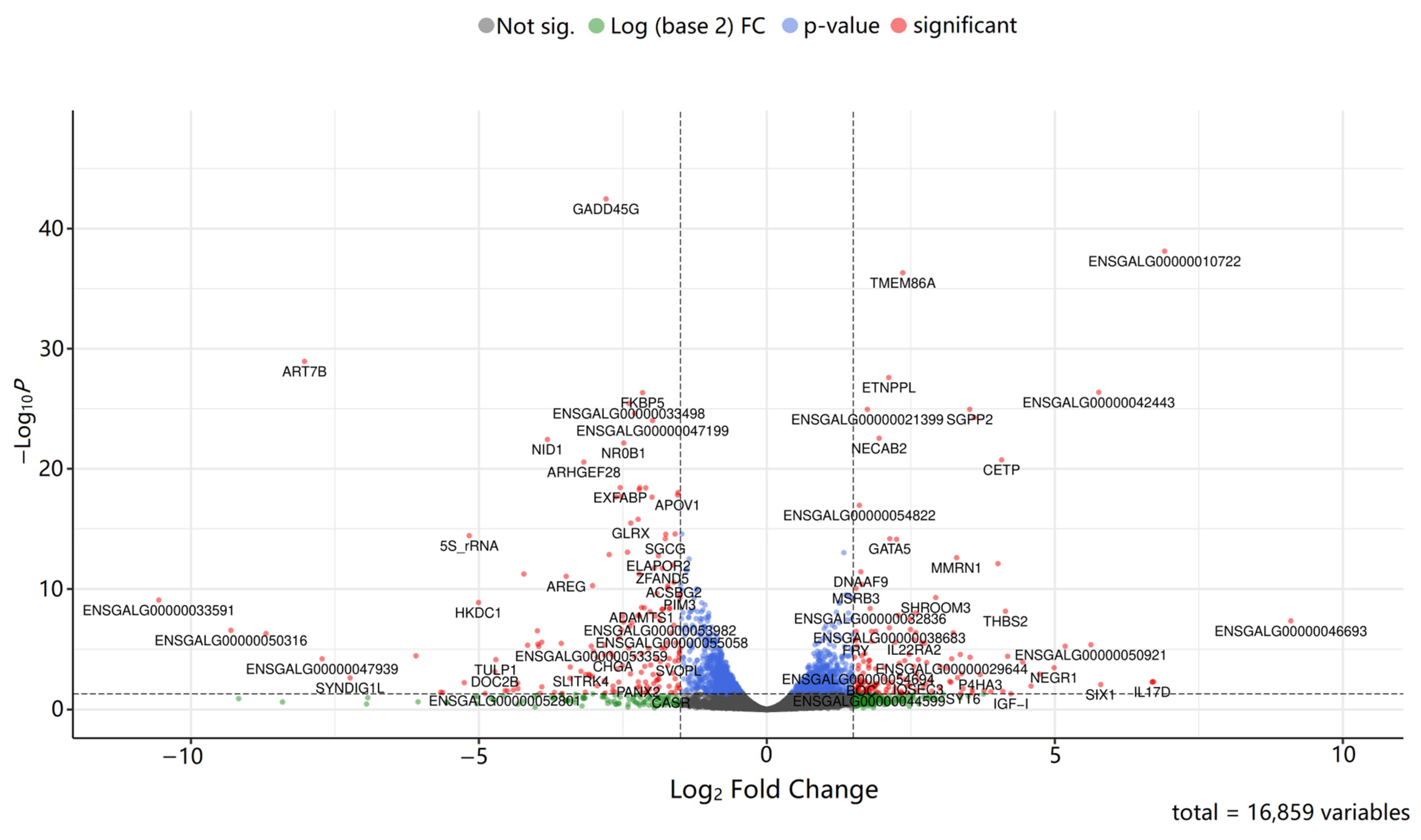
In addition, a volcano plot was constructed to visualize the differential gene expression between groups (Figure 4).
We identified a set of genes whose expression has been previously mentioned in other PGC transcriptomic studies. This includes genes known to be exclusively expressed in PGCs, such as DMRT1, PIWIL1, TDRD5, MAEL, GTSF1, SPAG16, DNAH1, TDRD15, MOV10L1, FKPB6, and ENSGALG00000054787 and ENSGALG00000052964 [19]. Our results matched the findings from Jean C. et al. (2015), confirming the expression of GSTF1, DDX4, PIWIL1, FKBP6, DMRT1, TDRD5, DND1, and others [27]. Similarities were found with the work of Rengaraj D. et al. (2012) on signaling and metabolic pathways in avian PGCs. We detected the expression of key genes highlighted in the aforementioned study, including MAD1L1, RBL1, CCNH, WEE1, CCNE2, DBF4, ANAPC10, TFDP2, and TTK [28]. Besides the ones mentioned above, our study revealed the expression of genes associated with pluripotency in avian PGCs, namely SOX10, DNMT3B, KLF5, and VRTN genes, which are the main pluripotency factors in geese [29]. Finally, we detected the expression of PRDM14, a gene with a crucial role in PGC development and specification [30,31].
Of the 16,859 genes analyzed, only 1909 were considered statistically significant after applying the Benjamini–Hochberg correction (padj < 0.05; Table S2). A selection of these genes overlaps with previous PGC studies (Table 2).
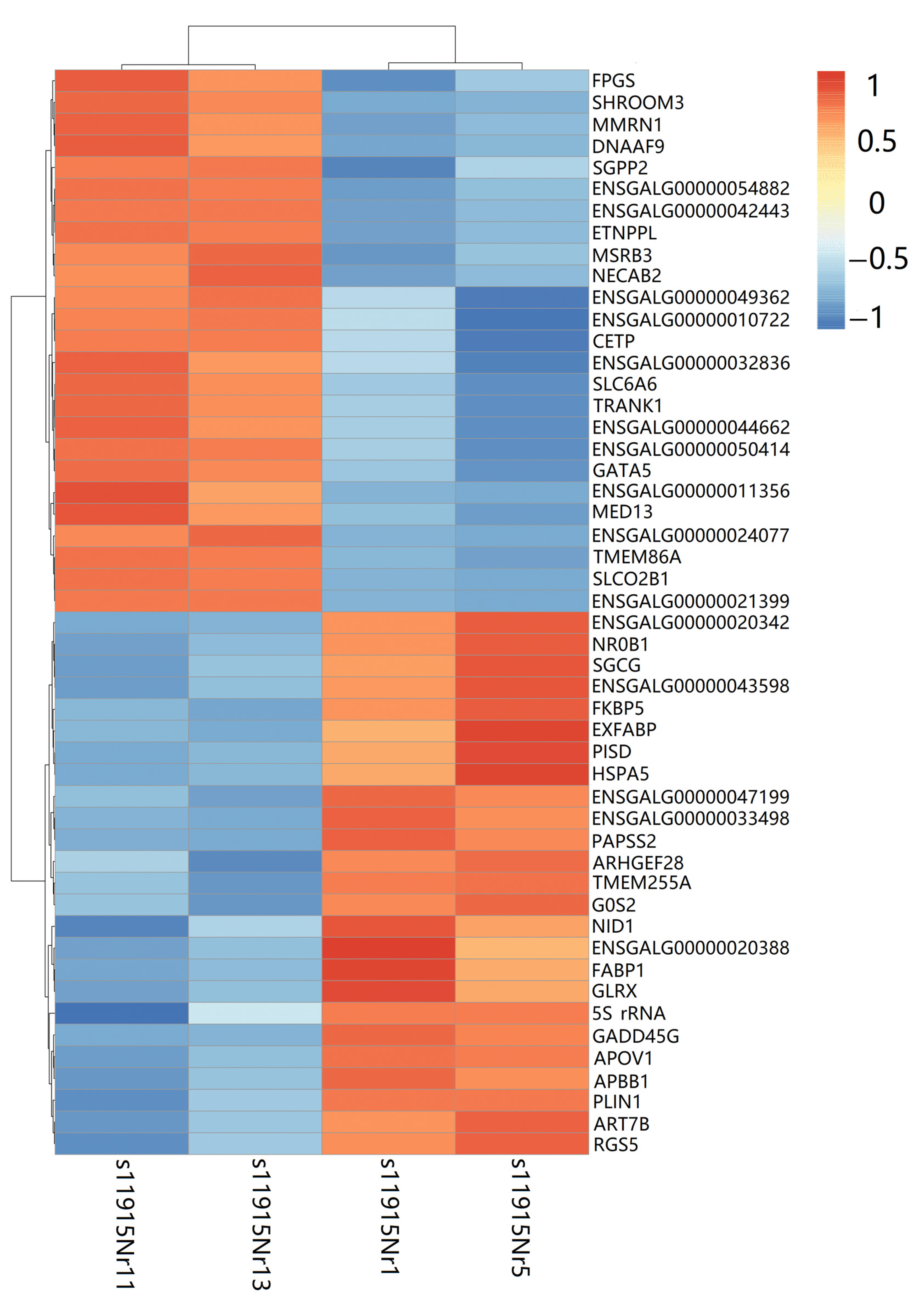
Additionally, a heatmap was constructed to display the expression patterns of the top 25 upregulated and downregulated genes between the sample groups (Figure 5).
Upregulated genes in PGCs included: FPGS, SHROOM3, MMRN1, DNAAF9, SGPP2, ETNPPL, MSRB3, NECAB2, CETP, SLC6A6, TRANK1, GATA5, MED13, TMEM86A, SLCO2B1, and genes with unknown functions—ENSGALG00000054822, ENSGALG00000042443, ENSGALG00000049362, ENSGALG00000010722, ENSGALG00000032836, ENSGALG00000044662, ENSGALG00000050414, ENSGALG00000011356, ENSGALG00000024077, and ENSGALG00000021399 (Figure 5). Downregulated in PGCs were the following genes: NR0B1, SGCG, FKBP5, EXFABP, PISD, HSPA5, PAPSS2, ARHGEF28, TMEM255A, G0S2, NID1, FABP1, GLRX, 5S_rRNA, GADD45G, APOV1, APBB1, PLIN1, ART7B, RGS5, and other genes with unknown functions—ENSGALG00000020342, ENSGALG00000043598, ENSGALG00000047199, ENSGALG00000033498, and ENSGALG00000020388 (Figure 5).
To elucidate the biological relevance of the 1909 DEGs, we performed the GO enrichment analysis, which covered three blocks: molecular function (MF), biological process (BP), and cellular component (CC). To simplify the result interpretation, data redundancy was minimized, i.e., similar GO terms were reduced, taking into account their hierarchical position and parent nodes (Table 3). The full list of significant GO terms with identifiers and the list of corresponding DEGs are provided in the Supplementary Materials (Table S3).
GO enrichment analysis revealed that DEGs were predominantly associated with protein processing, folding, and trafficking within the endoplasmic reticulum (ER), and translation, ribosome assembly, and mitochondrial respiratory chain function. Key cellular components included the ER, ribosomes, coated vesicles, and respiratory complexes. At the molecular level, terms related to ribosomal structure, ATP-dependent chaperone activity, and isomerase function were most significant. The complete lists of GO terms are provided in Supplementary Materials (Table S3).

4. Discussion

A key limitation of our research is the small sample size (n = 2 per group), which reduces the statistical power of the differential expression analysis by increasing the risk of false positive discoveries (FDR) and false negatives, where actual DEGs with modest fold changes may remain undetected. We consciously accepted this limitation due to the pilot-scale nature of our work, which aimed to identify the most prominent transcriptomic differences between chicken PGCs and adult liver cells. To mitigate these issues, we used conservative significance thresholds (log2 FC > |1.5|and padj < 0.05) as a compensatory measure and focused on functional coherence of the DEG subset with the cell type. Furthermore, the overlap of several DEGs with previously reported PGC markers in other studies provides indirect validation. Therefore, the results presented here should be interpreted as a reliable core signature that highlights the biggest transcriptional differences, rather than as a comprehensive catalog of all DEGs. With this framework in mind, we proceed to discuss our results.
As noted in Table 2, several genes from our DEG set have been previously mentioned in other PGC studies. NEGR1, encoding a neuron growth factor, has been proposed as a PGC surface marker [32]. TDRD15, GTSF1, and novel transcripts ENSGALG00000052964 and ENSGALG00000054787 were also identified as germ-cell-exclusive genes in the study by Doddamani et al. (2023) [19]. The same study reported sex-biased expression for a gene subset. GOLPH3, LIFR, DHFR, ARHGEF28, SIGMAR1, GFRA2, VCP, and ENSGALG00000017558 showed increased expression in male PGCs, while ENSGALG00000031327, ENSGALG00000046955, and ENSGALG00000002350 were upregulated in female PGCs. The ENSGALG00000052964 and ENSGALG00000054787 transcripts show homology to TOPAZ1, a vertebrate germ cell-specific gene [33,34]. ENSGALG00000031327 encodes a sequence similar to CHD1W, a W-chromosome gene that can, therefore, be used for sex identification [52].
Functional analysis of DEGs revealed several biological processes by which they can be grouped.
1. Nervous system development. A notable set of PGC DEGs is associated with nervous system development and function. This may be explained by the fact that both PGCs and neurons are characterized by long-range migration and complex cell–cell communication. Besides NEGR1, this set includes GFRA2, NRXN1, NCAM1, GFRA1, and FAM126A. GFRA1 and GFRA2 encode neurotrophin receptors crucial for neuron survival and differentiation [37,53]. NRXN1 is essential for synapse formation [54], NCAM1 for synaptic plasticity and neurogenesis [55], and FAM126A for the myelination of nerve fibers [45].
2. Lipid metabolism. Other DEG set, including GOLPH3, CYP1A1, ALDH1A1, CYP2C18, and BRCA1, points to an active role in lipid metabolism. This finding aligns with established evidence that lipid metabolism is tightly coupled with differentiation and energy homeostasis in male germ cells [40].
3. Genome stability. Another set of DEGs, including VCP, PSMC5, ATM, and BRCA1, is central to DNA damage response and genome integrity. All of them are necessary for DNA repair: ATM acts as a DNA damage sensor [48]; VCP facilitates the DNA repair process [56]; PSMC5 represents a proteasomal component involved in DNA damage response [57]; and BRCA1 acts as a central coordinator of repair pathways, ubiquitination, and transcription regulation to maintain genomic stability [49,51,58].
A closer investigation of the top 25 upregulated DEGs in PGCs revealed both known and novel genes. Among the annotated genes are FPGS, SHROOM3, MMRN1, DNAAF9, SGPP2, ETNPPL, MSRB3, NECAB2, CETP, SLC6A6, TRANK1, GATA5, MED13, TMEM86A, and SLCO2B1. Novel transcripts with unknown functions include ENSGALG00000054822, ENSGALG00000042443, ENSGALG00000049362, ENSGALG00000010722, ENSGALG00000032836, ENSGALG00000044662, ENSGALG00000050414, ENSGALG00000011356, ENSGALG00000024077, and ENSGALG00000021399, highlighting promising targets for future functional characterization (Figure 5).
These genes can be categorized into several groups.
1. Folate metabolism and nucleotide biosynthesis. Expression of the enzymes FPGS and DHFR, both essential for folate metabolism, highlights the increased demand for nucleotide precursors in rapidly proliferating PGCs [36,59]. This upregulation is a distinctive feature of cells with high proliferative potential, consistent with the role of FPGS in the survival of proliferating cells [59].
2. Lipid metabolism. A cluster of DEGs, including SGPP2, ETNPPL, CETP, and TMEM86A, is involved in lipid metabolism. Enzymes coded by SGPP2, ETNPPL, and TMEM86A are essential for processing sphingosine, phosphoethanolamine, and lysoplasmalogens, respectively [60,61,62]. Their substrates and products play crucial roles in myelination, synaptic plasticity, and cell signaling within the nervous system. Specifically, SGPP2 controls the level of sphingosine-1-phosphate (S1P), a key signaling lipid that regulates neuron migration and survival. The involvement of S1P in critical signaling pathways pinpoints the role of SGPP2 in ER stress regulation and cell proliferation [60]. ETNPPL metabolizes phosphoethanolamine, a precursor of ethanolamine plasmalogens vital for antioxidant protection of the brain and neuronal membranes. This function explains both the significant ETNPPL upregulation during embryogenesis [61] and its identification as a primate-specific marker of neural stem cells (NSCs) [63]. Lysoplasmalogens, whose metabolism involves TMEM86A, act as membrane stabilizers due to their antioxidant properties and antagonism to lysolecithin [62]. Finally, CETP participates in lipid transport, which is consistent with its associations with cholesterol levels [64,65]. By transferring cholesterol esters, CETP directly influences the cholesterol availability for membrane construction and repair. In summary, this gene cluster highlights that beyond lipid metabolism, these genes are vital regulators of structural and signaling lipids essential for cellular membranes and neuronal function. The broader list of DEGs further reinforces this, including other lipid-related genes like GOLPH3, CYP1A1, ALDH1A1, and CYP2C18 [38], highlighting the close relationship between lipid metabolism and cell differentiation and proliferation [40]. Moreover, lipid metabolism is closely related to fertility across humans, animals, and plants [66,67], which explains the significant differential expression of these genes in PGCs—the gamete precursors.
3. Protection and cellular homeostasis. The obtained data suggest that PGC development requires robust mechanisms for cellular stress management. MSRB3 acts as a key antioxidant enzyme that reduces oxidized proteins and, hence, protects against oxidative stress. Furthermore, it regulates cell proliferation and apoptosis via p53-p21 and p27 pathways [68,69]. Loss of MSRB3 function causes ER stress and subsequent apoptosis [70]. NECAB2 maintains both calcium and mitochondrial homeostasis, which is consistent with the mitochondrial dysfunction observed upon its knockout [71]. SLC6A6 encodes the taurine transporter, which regulates many processes, including cell proliferation, differentiation, calcification, and apoptosis [72]. SLC6A6 presumably contributes to intracellular signaling via taurine transport [72].
4. Morphogenesis and cell adhesion. Upregulation of the developmental protein, SHROOM3, a key regulator of cell shape during morphogenesis [73,74], suggests its potential role in cytoskeletal rearrangement required for PGC migration and tissue integration. MMRN1, while known for its role in hemostasis, also participates in cell adhesion [75], potentially facilitating the critical adhesive and signaling interactions between PGCs and their surrounding somatic niche, which are vital for guided migration, survival, and eventual colonization of the gonads. However, we acknowledge that its expression could alternatively reflect a low-level persistence of hematopoietic or endothelial cells from the initial blood isolate. This dual possibility highlights the need for future single-cell resolution studies to definitively assign its expression to PGCs.
5. Transcriptional regulation and development. A subset of genes shows its influence on developmental programs in PGC biology. GATA5 encodes a transcription factor essential for cell differentiation, particularly during the development of the vertebrate cardiovascular system [76,77]. MED13, a component of the transcription coactivator complex, is important for vertebrate embryonic development. Its specific role in zygotic genome activation and successful preimplantation development [78] suggests a fundamental function in establishing totipotency, a key feature of early germ cells. Finally, SLCO2B1 mediates the cellular uptake of signaling molecules (prostaglandins and steroid conjugates) [79], thus controlling the availability of ligands for nuclear receptors. The latter suggests its indirect effect on transcriptional programs important for cell differentiation and tissue development, making it a potential regulator of PGC maturation or niche communication.
6. Unexplored functions. The last gene subset includes genes with unclear germ cell functions. It includes DNAAF9, which encodes an uncharacterized protein with a C-terminal helical region, and the brain-expressed TRANK1, the latter being a GWAS candidate for neuropsychiatric disorders [80,81,82].
In summary, the transcriptional signature of the known top 25 DEGs represents PGCs’ highly proliferative cells with adapted metabolic networks, complex stress-response systems, and under precise developmental control, which is consistent with their role as progenitors of the germline.
Novel transcripts were also analyzed, revealing conserved themes in PGC development. Homology searches for the identified transcripts revealed functionally relevant data.
1. Early embryogenesis signaling and morphogenesis. The ENSGALG00000042443 transcript is homologous to VWDE, which is implicated in cell proliferation and differentiation [83]. To date, ENSGALG00000010722 has been linked to SERAF, a gene highly homologous to VWDE [84,85]. Upregulation of SERAF is observed in migrating neural crest cells and in Schwann cell precursors during avian embryogenesis [86]. Notably, SERAF expression is regulated by SOX10, whose expression was detected in our dataset (below the significance threshold). Furthermore, the ENSGALG00000011356 transcript is linked to ERICH3, a protein highly expressed in the central nervous system [87], and a component of primary cilia [88]. Cilia coordinate a number of vital signaling pathways (Wnt, TGF-beta, and Shh), which regulate cell fate, differentiation, and migration [89]. The co-expression of VWDE/SERAF homologs, their regulator SOX10 [85], and a ciliary component (ERICH3) suggests the utilization of evolutionarily conserved molecular programs, namely long-range migration and niche interaction, which are crucial for both neural crest and PGCs.
2. Specialized metabolism and homeostasis. The ENSGALG00000032836 transcript corresponds to MAOB, a key enzyme in amine metabolism. MAOB regulates neurotransmitter and hormone levels in the central nervous system and peripheral tissues [90,91]. Additionally, ENSGALG00000021399 is linked to ABCA8, an ATP-binding cassette transporter that facilitates molecule transport across membranes and contributes to sphingomyelin production in oligodendrocytes, playing a role in myelin formation and maintenance [92]. The upregulation of MAOB and ABCA8 in PGCs pinpoints the necessity for precise control over amine and lipid balance and maintenance of specialized membrane composition during PGC migration and maturation.
3. Immune and hematopoietic pathways. The remaining transcripts are unexpectedly associated with immunity/hematopoiesis. The ENSGALG00000044662 exhibits homology to the LOC107049909 locus, a member of the leukocyte immunoglobulin-like receptor family, indicating its role in the immune system. The ENSGALG00000024077 transcript is currently linked to CRYBG3, a gene implicated in platelet morphology and function [93]. The co-expression of LOC107049909/CRYBG3 may reflect either evolutionarily conserved mechanisms for adhesion, extracellular interaction used by PGC for embryo navigation, or, potentially, a novel immuno-like surveillance or interaction mechanism intrinsic to germline development. The involvement of these genes in PGC development and function requires further research.

5. Conclusions

Our comparative transcriptomic analysis of PGCs and adult liver cells, despite the limited sample size (n = 2 per group), revealed a core PGC-specific transcriptional signature. It is crucial to acknowledge that liver control represents a heterogeneous cellular mixture, including hepatocytes, epithelial, endothelial and immune cells. This means that the identified DEG set reflects differences between a pure PGC population and an average transcriptional profile of a complex somatic organ. Indeed, this comparison may mask signals specific to individual liver cell subtypes, but it also effectively highlights the global features that fundamentally distinguish PGCs from differentiated somatic tissues. The functional coherence of the identified DEG set with known PGC biology confirms the robustness of this core signature.
The analysis delineated two principal axes of the PGC transcriptome. First, it comprises genes providing the fundamental properties of embryonic progenitors: hyperproliferation (folate metabolism, FPGS and DHFR), specialized metabolic and membrane homeostasis (lipid transport, CETP and TMEM86A; detoxification, MAOB; and antioxidant defense, MSRB3), and genome stability maintenance (BRCA1 and ATM). This profile stands in sharp contrast to the metabolic function dominant in differentiated liver cells. Second, unexpectedly, a profound molecular convergence between PGCs and neural crest cells was discovered. This is evidenced by the active expression of a suite of genes governing long-range migration (SHROOM3 and MMRN1), neuronal signaling and adhesion (NEGR1, GFRA1/2, and NCAM1), and lipid metabolism critical for myelination and synaptic plasticity (SGPP2 and ETNPPL). This finding suggests that PGCs, like neural crest cells, recruit evolutionarily conserved modules to accomplish similar morphogenetic tasks. The detection of this shared program against a heterogeneous control background underscores its specificity to these migratory cell types. Thus, despite the methodological constraints, our study demonstrates that PGCs are not merely proliferating cells but a highly specialized population with a unique “neuro-lipid” metabolic profile and complex developmental systems ensuring their migration and fate determination. The unannotated transcripts related to immune pathways and primary cilia signaling (ERICH3), identified here, point to promising new avenues for the functional characterization of avian PGC biology. While our comparative approach provides a robust core signature, future studies using single-cell RNA-seq or fluorescence-activated cell sorting (FACS) with specific surface markers will be essential to definitively assign all of the identified genes to PGCs.

Supplementary Materials

The following supporting information can be downloaded at https://www.mdpi.com/article/10.3390/ani16030522/s1. Table S1: List of all 16,859 analyzed differentially expressed genes.; Table S2: List of statistically significant (1909) genes after applying the Benjamini–Hochberg correction; Table S3: List of reduced GO-terms with identifiers and identified DEGs associated with them.

Author Contributions

Conceptualization, T.A.L. and A.I.A.; methodology, T.A.L. and O.Y.B.; software, A.I.A. and A.E.R.; validation, A.P.D. and G.K.P.; formal analysis, A.I.A., A.P.D., and A.E.R.; investigation, A.I.A.; resources, G.K.P., N.R.R., E.A.P., and A.V.G.; data curation, O.Y.B., N.R.R., A.V.G., and E.A.P.; writing—original draft preparation, A.I.A. and A.P.D.; writing—review and editing, A.I.A., T.A.L., A.E.R., A.P.D., O.Y.B., N.R.R., A.V.G., and E.A.P.; visualization, A.I.A. and A.E.R.; supervision, T.A.L.; project administration, T.A.L.; funding acquisition, T.A.L. All authors have read and agreed to the published version of the manuscript.

Funding

This investigation was financially supported by the Russian Science Foundation (RSF), grant No. 24-16-00133.

Institutional Review Board Statement

The principles of laboratory animal care were followed, and all the procedures were conducted according to the ethical guidelines of the L.K. Ernst Federal Science Center for Animal Husbandry. The protocol was approved by the Commission on the Ethics of Animal Experiments of the L.K. Ernst Federal Science Center for Animal Husbandry on 18 February 2020 (Protocol Number: 2020/2) and the Law of the Russian Federation on Veterinary Medicine No. 4979-1 (14 May 1993).

Informed Consent Statement

Not applicable.

Data Availability Statement

The data presented in this study are included in the article and the Supplementary Materials. Further inquiries can be directed to the corresponding author(s).

Conflicts of Interest

The authors declare no conflicts of interest. The funders had no role in the design of the study; in the collection, analyses, or interpretation of data; in the writing of the manuscript; or in the decision to publish the results.

Abbreviations

The following abbreviations are used in this manuscript:
PGCPrimordial germ cell
DEGDifferentially expressed gene
GEGenome editing
CCUCenter of collective use
GOGene ontology
MFMolecular function
BPBiological process
CCCellular component
EREndoplasmic reticulum
NSCNeural stem cell
IEEInner enamel epithelium
GWASGenome-wide association study
FACSfluorescence-activated cell sorting
FDRFalse discovery rate

References

  1. Cooper, C.A.; Doran, T.J.; Challagulla, A.; Tizard, M.L.V.; Jenkins, K.A. Innovative approaches to genome editing in avian species. J. Anim. Sci. Biotechnol. 2018, 9, 15. [Google Scholar] [CrossRef]
  2. Lee, J.; Kim, D.H.; Lee, K. Current Approaches and Applications in Avian Genome Editing. Int. J. Mol. Sci. 2020, 21, 3937. [Google Scholar] [CrossRef]
  3. Woodcock, M.E.; Idoko-Akoh, A.; McGrew, M.J. Gene editing in birds takes flight. Mamm. Genome 2017, 28, 315–323. [Google Scholar] [CrossRef] [PubMed]
  4. Walsh, R.M.; Hochedlinger, K. A variant CRISPR-Cas9 system adds versatility to genome engineering. Proc. Natl. Acad. Sci. USA 2013, 110, 15514–15515. [Google Scholar] [CrossRef] [PubMed]
  5. Polejaeva, I.A.; Rutigliano, H.M.; Wells, K.D. Livestock in biomedical research: History, current status and future prospective. Reprod. Fertil. Dev. 2016, 28, 112–124. [Google Scholar] [CrossRef] [PubMed]
  6. Whyte, J.; Glover, J.D.; Woodcock, M.; Brzeszczynska, J.; Taylor, L.; Sherman, A.; Kaiser, P.; McGrew, M.J. FGF, Insulin, and SMAD Signaling Cooperate for Avian Primordial Germ Cell Self-Renewal. Stem Cell Rep. 2015, 5, 1171–1182. [Google Scholar] [CrossRef]
  7. Ichikawa, K.; Horiuchi, H. Fate Decisions of Chicken Primordial Germ Cells (PGCs): Development, Integrity, Sex Determination, and Self-Renewal Mechanisms. Genes 2023, 14, 612. [Google Scholar] [CrossRef]
  8. Hancock, G.V.; Wamaitha, S.E.; Peretz, L.; Clark, A.T. Mammalian primordial germ cell specification. Development 2021, 148, dev189217. [Google Scholar] [CrossRef]
  9. Tagami, T.; Miyahara, D.; Nakamura, Y. Avian Primordial Germ Cells. In Avian Reproduction: From Behavior to Molecules; Springer: Singapore, 2017; Volume 1001, pp. 1–18. [Google Scholar] [CrossRef]
  10. Dunislawska, A.; Szczerba, A.; Siwek, M.; Bednarczyk, M. Dynamics of the transcriptome during chicken embryo development based on primordial germ cells. BMC Res. Notes 2020, 13, 441. [Google Scholar] [CrossRef]
  11. Nguyen, D.H.; Jaszczak, R.G.; Laird, D.J. Heterogeneity of primordial germ cells. Curr. Top. Dev. Biol. 2019, 135, 155–201. [Google Scholar] [CrossRef]
  12. Abed Al-Shuwaili, E.H.; Nabipour, A.; Hosseini, A.; Dehghani, H. Ultrastructure characteristics of primordial germ cells in stage X of pheasant (Phasianus colchicus) embryo. Vet. Res. Forum 2022, 13, 521–527. [Google Scholar] [CrossRef] [PubMed]
  13. Tagami, T.; Kagami, H. Developmental origin of avian primordial germ cells and its unique differentiation in the gonads of mixed-sex chimeras. Mol. Reprod. Dev. 1998, 50, 370–376. [Google Scholar] [CrossRef]
  14. Nakamura, Y.; Yamamoto, Y.; Usui, F.; Mushika, T.; Ono, T.; Setioko, A.R.; Takeda, K.; Nirasawa, K.; Kagami, H.; Tagami, T. Migration and proliferation of primordial germ cells in the early chicken embryo. Poult. Sci. 2007, 86, 2182–2193. [Google Scholar] [CrossRef] [PubMed]
  15. Collarini, E.J.; Leighton, P.A.; Van de Lavoir, M.C. Production of Transgenic Chickens Using Cultured Primordial Germ Cells and Gonocytes. Methods Mol. Biol. 2019, 1874, 403–430. [Google Scholar] [CrossRef]
  16. Kanamori, M.; Oikawa, K.; Tanemura, K.; Hara, K. Mammalian germ cell migration during development, growth, and homeostasis. Reprod. Med. Biol. 2019, 18, 247–255. [Google Scholar] [CrossRef]
  17. Rathgeber, B.M.; McCarron, P.; Budgell, K.L. Salmonella penetration through eggshells of chickens of different genetic backgrounds. Poult. Sci. 2013, 92, 2457–2462. [Google Scholar] [CrossRef]
  18. Liu, J.; Cheng, K.M.; Silversides, F.G. Fundamental principles of cryobiology and application to ex situ conservation of avian species. Avian Biol. Res. 2013, 6, 187–197. [Google Scholar] [CrossRef]
  19. Doddamani, D.; Woodcock, M.; Taylor, L.; Nandi, S.; McTeir, L.; Davey, M.G.; Smith, J.; McGrew, M.J. The Transcriptome of Chicken Migratory Primordial Germ Cells Reveals Intrinsic Sex Differences and Expression of Hallmark Germ Cell Genes. Cells 2023, 12, 1151. [Google Scholar] [CrossRef]
  20. Mathan; Zaib, G.; Jin, K.; Zuo, Q.; Habib, M.; Zhang, Y.; Li, B. Formation, Application, and Significance of Chicken Primordial Germ Cells: A Review. Animals 2023, 13, 1096. [Google Scholar] [CrossRef]
  21. Larkina, T.; Krutikova, A.; Peglivanyan, G.; Shcherbakov, Y.; Barkova, O. Development of optimal technological approaches for obtaining PGCs in Pushkin breed chickens for further transformation by the CRISPR/Cas9 system. [Abstract]. FASEB J. 2021, 35, S1. [Google Scholar] [CrossRef]
  22. Hamburger, V.; Hamilton, H.L. A series of normal stages in the development of the chick embryo. Dev. Dyn. 1992, 195, 231–272. [Google Scholar] [CrossRef]
  23. Jiang, H.; Lei, R.; Ding, S.W.; Zhu, S. Skewer: A fast and accurate adapter trimmer for next-generation sequencing paired-end reads. BMC Bioinform. 2014, 15, 182. [Google Scholar] [CrossRef]
  24. Dobin, A.; Davis, C.A.; Schlesinger, F.; Drenkow, J.; Zaleski, C.; Jha, S.; Batut, P.; Chaisson, M.; Gingeras, T.R. STAR: Ultrafast universal RNA-seq aligner. Bioinformatics 2013, 29, 15–21. [Google Scholar] [CrossRef]
  25. Love, M.I.; Huber, W.; Anders, S. Moderated estimation of fold change and dispersion for RNA-seq data with DESeq2. Genome Biol. 2014, 15, 550. [Google Scholar] [CrossRef] [PubMed]
  26. Benjamini, Y.; Hochberg, Y. Controlling the False Discovery Rate: A Practical and Powerful Approach to Multiple Testing. J. R. Stat. Soc. Ser. B Methodol. 1995, 57, 289–300. [Google Scholar] [CrossRef]
  27. Jean, C.; Oliveira, N.M.; Intarapat, S.; Fuet, A.; Mazoyer, C.; De Almeida, I.; Trevers, K.; Boast, S.; Aubel, P.; Bertocchini, F.; et al. Transcriptome analysis of chicken ES, blastodermal and germ cells reveals that chick ES cells are equivalent to mouse ES cells rather than EpiSC. Stem Cell Res. 2015, 14, 54–67. [Google Scholar] [CrossRef]
  28. Rengaraj, D.; Lee, B.R.; Choi, J.W.; Lee, S.I.; Seo, H.W.; Kim, T.H.; Choi, H.J.; Song, G.; Han, J.Y. Gene pathways and cell cycle-related genes in cultured avian primordial germ cells. Poult. Sci. 2012, 91, 3167–3177. [Google Scholar] [CrossRef]
  29. Doddamani, D.; Lázár, B.; Ichikawa, K.; Hu, T.; Taylor, L.; Gócza, E.; Várkonyi, E.; McGrew, M.J. Propagation of goose primordial germ cells in vitro relies on FGF and BMP signalling pathways. Commun. Biol. 2025, 8, 301. [Google Scholar] [CrossRef]
  30. Okuzaki, Y.; Kaneoka, H.; Suzuki, T.; Hagihara, Y.; Nakayama, Y.; Murakami, S.; Murase, Y.; Kuroiwa, A.; Iijima, S.; Nishijima, K.I. PRDM14 and BLIMP1 control the development of chicken primordial germ cells. Dev. Biol. 2019, 455, 32–41. [Google Scholar] [CrossRef]
  31. Doddamani, D.; Carlson, D.F.; McTeir, L.; Taylor, L.; Nandi, S.; Davey, M.G.; McGrew, M.J.; Glover, J.D. PRDM14 is essential for vertebrate gastrulation and safeguards avian germ cell identity. Dev. Biol. 2025, 521, 129–137. [Google Scholar] [CrossRef]
  32. Kim, J.L.; Jung, K.M.; Han, J.Y. Single-cell RNA sequencing reveals surface markers of primordial germ cells in chicken and zebra finch. Mol. Genet. Genom. 2024, 299, 90. [Google Scholar] [CrossRef]
  33. Baillet, A.; Le Bouffant, R.; Volff, J.N.; Luangpraseuth, A.; Poumerol, E.; Thépot, D.; Pailhoux, E.; Livera, G.; Cotinot, C.; Mandon-Pépin, B. TOPAZ1, a novel germ cell-specific expressed gene conserved during evolution across vertebrates. PLoS ONE 2011, 6, e26950. [Google Scholar] [CrossRef] [PubMed]
  34. Luangpraseuth-Prosper, A.; Lesueur, E.; Jouneau, L.; Pailhoux, E.; Cotinot, C.; Mandon-Pépin, B. TOPAZ1, a germ cell specific factor, is essential for male meiotic progression. Dev. Biol. 2015, 406, 158–171. [Google Scholar] [CrossRef] [PubMed]
  35. Gòdia, M.; Casellas, J.; Ruiz-Herrera, A.; Rodríguez-Gil, J.E.; Castelló, A.; Sánchez, A.; Clop, A. Whole genome sequencing identifies allelic ratio distortion in sperm involving genes related to spermatogenesis in a swine model. DNA Res. 2020, 27, dsaa019. [Google Scholar] [CrossRef] [PubMed]
  36. Flickinger, K.M.; Mellado Fritz, C.A.; Huggler, K.S.; Wade, G.M.; Chang, G.R.; Fox, K.C.; Simcox, J.A.; Cantor, J.R. Cytosolic NADK is conditionally essential for folate-dependent nucleotide synthesis. Nat. Metab. 2025, 7, 1150–1167. [Google Scholar] [CrossRef]
  37. Sandmark, J.; Dahl, G.; Öster, L.; Xu, B.; Johansson, P.; Akerud, T.; Aagaard, A.; Davidsson, P.; Bigalke, J.M.; Winzell, M.S.; et al. Structure and biophysical characterization of the human full-length neurturin-GFRa2 complex: A role for heparan sulfate in signaling. J. Biol. Chem. 2018, 293, 5492–5508. [Google Scholar] [CrossRef]
  38. Palm, W. GOLPH3 tunes up glycosphingolipid biosynthesis for cell growth. EMBO J. 2021, 40, e108070. [Google Scholar] [CrossRef]
  39. Li, D.; Wang, M.; Cheng, S.; Zhang, C.; Wang, Y.; Zhang, W.; Zhao, R.; Sun, C.; Zhang, Y.; Li, B. CYP1A1 based on metabolism of xenobiotics by cytochrome P450 regulates chicken male germ cell differentiation. In Vitro Cell Dev. Biol. Anim. 2017, 53, 293–303. [Google Scholar] [CrossRef]
  40. Zuo, Q.; Li, D.; Zhang, L.; Elsayed, A.K.; Lian, C.; Shi, Q.; Zhang, Z.; Zhu, R.; Wang, Y.; Jin, K.; et al. Study on the regulatory mechanism of the lipid metabolism pathways during chicken male germ cell differentiation based on RNA-seq. PLoS ONE 2015, 10, e0109469. [Google Scholar] [CrossRef]
  41. Lucas, D.; Goulitquer, S.; Marienhagen, J.; Fer, M.; Dreano, Y.; Schwaneberg, U.; Amet, Y.; Corcos, L. Stereoselective epoxidation of the last double bond of polyunsaturated fatty acids by human cytochromes P450. J. Lipid Res. 2010, 51, 1125–1133. [Google Scholar] [CrossRef]
  42. Mao, M.; Cheng, Y.; Yang, J.; Chen, Y.; Xu, L.; Zhang, X.; Li, Z.; Chen, C.; Ju, S.; Zhou, J.; et al. Multifaced roles of PLAC8 in cancer. Biomark. Res. 2021, 9, 73. [Google Scholar] [CrossRef] [PubMed]
  43. Korzekwa, A.J.; Kotlarczyk, A.M.; Szczepańska, A.A.; Grzyb, M.; Siergiej, A.; Wocławek-Potocka, I. Antioxidative Potential of Red Deer Embryos Depends on Reproductive Stage of Hind as A Oocyte Donor. Animals 2020, 10, 1190. [Google Scholar] [CrossRef] [PubMed]
  44. Blessing, A.M.; Ganesan, S.; Rajapakshe, K.; Ying Sung, Y.; Reddy Bollu, L.; Shi, Y.; Cheung, E.; Coarfa, C.; Chang, J.T.; McDonnell, D.P.; et al. Identification of a Novel Coregulator, SH3YL1, That Interacts with the Androgen Receptor N-Terminus. Mol. Endocrinol. 2015, 29, 1426–1439. [Google Scholar] [CrossRef] [PubMed]
  45. Baskin, J.M.; Wu, X.; Christiano, R.; Oh, M.S.; Schauder, C.M.; Gazzerro, E.; Messa, M.; Baldassari, S.; Assereto, S.; Biancheri, R.; et al. The leukodystrophy protein FAM126A (hyccin) regulates PtdIns(4)P synthesis at the plasma membrane. Nat. Cell. Biol. 2016, 18, 132–138. [Google Scholar] [CrossRef]
  46. Higashi, T.; Tokuda, S.; Kitajiri, S.; Masuda, S.; Nakamura, H.; Oda, Y.; Furuse, M. Analysis of the ‘angulin’ proteins LSR, ILDR1 and ILDR2--tricellulin recruitment, epithelial barrier function and implication in deafness pathogenesis. J. Cell Sci. 2013, 126, 966–977. [Google Scholar] [CrossRef]
  47. Rengaraj, D.; Won, S.; Jung, K.M.; Woo, S.J.; Lee, H.; Kim, Y.M.; Kim, H.; Han, J.Y. Chicken blastoderms and primordial germ cells possess a higher expression of DNA repair genes and lower expression of apoptosis genes to preserve their genome stability. Sci. Rep. 2022, 12, 49. [Google Scholar] [CrossRef]
  48. Warren, C.; Pavletich, N.P. Structure of the human ATM kinase and mechanism of Nbs1 binding. eLife 2022, 11, e74218. [Google Scholar] [CrossRef]
  49. Feng, L.; Huang, J.; Chen, J. MERIT40 facilitates BRCA1 localization and DNA damage repair. Genes Dev. 2009, 23, 719–728. [Google Scholar] [CrossRef]
  50. Hiraike, H.; Wada-Hiraike, O.; Nakagawa, S.; Koyama, S.; Miyamoto, Y.; Sone, K.; Tanikawa, M.; Tsuruga, T.; Nagasaka, K.; Matsumoto, Y.; et al. Identification of DBC1 as a transcriptional repressor for BRCA1. Br. J. Cancer 2010, 102, 1061–1067. [Google Scholar] [CrossRef]
  51. Moreau, K.; Dizin, E.; Ray, H.; Luquain, C.; Lefai, E.; Foufelle, F.; Billaud, M.; Lenoir, G.M.; Venezia, N.D. BRCA1 affects lipid synthesis through its interaction with acetyl-CoA carboxylase. J. Biol. Chem. 2006, 281, 3172–3181. [Google Scholar] [CrossRef]
  52. England, A.D.; Kheravii, S.K.; Musigwa, S.; Kumar, A.; Daneshmand, A.; Sharma, N.K.; Gharib-Naseri, K.; Wu, S.B. Sexing chickens (Gallus gallus domesticus) with high-resolution melting analysis using feather crude DNA. Poult. Sci. 2021, 100, 100924. [Google Scholar] [CrossRef]
  53. Jmaeff, S.; Sidorova, Y.; Nedev, H.; Saarma, M.; Saragovi, H.U. Small-molecule agonists of the RET receptor tyrosine kinase activate biased trophic signals that are influenced by the presence of GFRa1 co-receptors. J. Biol. Chem. 2020, 295, 6532–6542. [Google Scholar] [CrossRef] [PubMed]
  54. Reissner, C.; Runkel, F.; Missler, M. Neurexins. Genome Biol. 2013, 14, 213. [Google Scholar] [CrossRef] [PubMed]
  55. Atz, M.E.; Rollins, B.; Vawter, M.P. NCAM1 association study of bipolar disorder and schizophrenia: Polymorphisms and alternatively spliced isoforms lead to similarities and differences. Psychiatr. Genet. 2007, 17, 55–67. [Google Scholar] [CrossRef] [PubMed]
  56. Xing, Z.; Cai, X.; He, T.; Li, P.; He, J.; Qiu, Y.; Li, N.; Mi, L.; Li, R.; Zhu, J.; et al. VCP’s nuclear journey: Initiated by interacting with KPNB1 to repair DNA damage. Proc. Natl. Acad. Sci. USA 2025, 122, e2416045122. [Google Scholar] [CrossRef]
  57. Yu, Z.Q.; Carmichael, J.; Collins, G.A.; D’Agostino, M.D.; Lessard, M.; Firth, H.V.; Harijan, P.; Fry, A.E.; Dean, J.; Zhang, J.; et al. PSMC5 insufficiency and P320R mutation impair proteasome function. Hum. Mol. Genet. 2024, 33, 1506–1523. [Google Scholar] [CrossRef]
  58. Wu-Baer, F.; Ludwig, T.; Baer, R. The UBXN1 protein associates with autoubiquitinated forms of the BRCA1 tumor suppressor and inhibits its enzymatic function. Mol. Cell. Biol. 2010, 30, 2787–2798. [Google Scholar] [CrossRef]
  59. Gangjee, A.; Dubash, N.P.; Zeng, Y.; McGuire, J.J. Recent advances in the chemistry and biology of folypoly-gamma-glutamate synthetase substrates and inhibitors. Curr. Med. Chem. Anticancer Agents 2002, 2, 331–355. [Google Scholar] [CrossRef]
  60. Taguchi, Y.; Allende, M.L.; Mizukami, H.; Cook, E.K.; Gavrilova, O.; Tuymetova, G.; Clarke, B.A.; Chen, W.; Olivera, A.; Proia, R.L. Sphingosine-1-phosphate Phosphatase 2 Regulates Pancreatic Islet β-Cell Endoplasmic Reticulum Stress and Proliferation. J. Biol. Chem. 2016, 291, 12029–12038. [Google Scholar] [CrossRef]
  61. White, C.J.; Ellis, J.M.; Wolfgang, M.J. The role of ethanolamine phosphate phospholyase in regulation of astrocyte lipid homeostasis. J. Biol. Chem. 2021, 297, 100830. [Google Scholar] [CrossRef]
  62. Van Wouw, S.A.E.; van den Berg, M.; El Ouraoui, M.; Meurs, A.; Kingma, J.; Ottenhoff, R.; Loix, M.; Hoeksema, M.A.; Prange, K.; Pasterkamp, G.; et al. Sterol-regulated transmembrane protein TMEM86a couples LXR signaling to regulation of lysoplasmalogens in macrophages. J. Lipid Res. 2023, 64, 100325. [Google Scholar] [CrossRef] [PubMed]
  63. Wang, W.; Wang, M.; Yang, M.; Zeng, B.; Qiu, W.; Ma, Q.; Jing, X.; Zhang, Q.; Wang, B.; Yin, C.; et al. Transcriptome dynamics of hippocampal neurogenesis in macaques across the lifespan and aged humans. Cell Res. 2022, 32, 729–743. [Google Scholar] [CrossRef] [PubMed]
  64. Moors, J.; Krishnan, M.; Sumpter, N.; Takei, R.; Bixley, M.; Cadzow, M.; Major, T.J.; Phipps-Green, A.; Topless, R.; Merriman, M.; et al. A Polynesian-specific missense CETP variant alters the lipid profile. HGG Adv. 2023, 4, 100204. [Google Scholar] [CrossRef] [PubMed]
  65. Carlson, J.C.; Weeks, D.E.; Hawley, N.L.; Sun, G.; Cheng, H.; Naseri, T.; Reupena, M.S.; Tuitele, J.; Deka, R.; McGarvey, S.T.; et al. Genome-wide association studies in Samoans give insight into the genetic architecture of fasting serum lipid levels. J. Hum. Genet. 2021, 66, 111–121. [Google Scholar] [CrossRef]
  66. Wan, X.; Wu, S.; Li, Z.; An, X.; Tian, Y. Lipid Metabolism: Critical Roles in Male Fertility and Other Aspects of Reproductive Development in Plants. Mol. Plant 2020, 13, 955–983. [Google Scholar] [CrossRef]
  67. Ryabova, A.E.; Azovtseva, A.I.; Shcherbakov, Y.S.; Dysin, A.P.; Dementieva, N.V. Search for Ancient Selection Traces in Faverolle Chicken Breed (Gallus gallus domesticus) Based on Runs of Homozygosity Analysis. Animals 2025, 15, 1487. [Google Scholar] [CrossRef]
  68. Lim, D.-H.; Han, J.Y.; Kim, J.-R.; Lee, Y.S.; Kim, H.-Y. Methionine sulfoxide reductase B in the endoplasmic reticulum is critical for stress resistance and aging in Drosophila. Biochem. Biophys. Res. Commun. 2012, 419, 20–26. [Google Scholar] [CrossRef]
  69. Ye, X.; Liang, T.; Deng, C.; Li, Z.; Yan, D. MSRB3 promotes the progression of clear cell renal cell carcinoma via regulating endoplasmic reticulum stress. Pathol. Res. Pract. 2020, 216, 152780. [Google Scholar] [CrossRef]
  70. Kwak, G.H.; Kim, H.Y. MsrB3 deficiency induces cancer cell apoptosis through p53-independent and ER stress-dependent pathways. Arch. Biochem. Biophys. 2017, 621, 1–5. [Google Scholar] [CrossRef]
  71. Bueno, D.; Narayan Dey, P.; Schacht, T.; Wolf, C.; Wüllner, V.; Morpurgo, E.; Rojas-Charry, L.; Sessinghaus, L.; Leukel, P.; Sommer, C.; et al. NECAB2 is an endosomal protein important for striatal function. Free Radic. Biol. Med. 2023, 208, 643–656. [Google Scholar] [CrossRef]
  72. Rong, Z.; Li, F.; Zhang, R.; Niu, S.; Di, X.; Ni, L.; Liu, C. Ant-Neointimal Formation Effects of SLC6A6 in Preventing Vascular Smooth Muscle Cell Proliferation and Migration via Wnt/β-Catenin Signaling. Int. J. Mol. Sci. 2023, 24, 3018. [Google Scholar] [CrossRef] [PubMed]
  73. Li, P.; Zhang, T.; Wu, R.; Zhang, J.Y.; Zhuo, Y.; Li, S.G.; Wang, J.J.; Guo, W.T.; Wang, Z.B.; Chen, Y.C. Loss of SHROOM3 affects neuroepithelial cell shape through regulating cytoskeleton proteins in cynomolgus monkey organoids. Zool. Res. 2024, 45, 233–241. [Google Scholar] [CrossRef] [PubMed]
  74. Plageman, T.F., Jr.; Zacharias, A.L.; Gage, P.J.; Lang, R.A. Shroom3 and a Pitx2-N-cadherin pathway function cooperatively to generate asymmetric cell shape changes during gut morphogenesis. Dev. Biol. 2011, 357, 227–234. [Google Scholar] [CrossRef]
  75. Posner, M.G. Multimerin-1 and cancer: A review. Biosci. Rep. 2022, 42, BSR20211248. [Google Scholar] [CrossRef] [PubMed]
  76. Morrisey, E.E.; Ip, H.S.; Tang, Z.; Lu, M.M.; Parmacek, M.S. GATA-5: A transcriptional activator expressed in a novel temporally and spatially-restricted pattern during embryonic development. Dev. Biol. 1997, 183, 21–36. [Google Scholar] [CrossRef]
  77. Wei, D.; Bao, H.; Liu, X.Y.; Zhou, N.; Wang, Q.; Li, R.G.; Xu, Y.J.; Yang, Y.Q. GATA5 loss-of-function mutations underlie tetralogy of fallot. Int. J. Med. Sci. 2013, 10, 34–42. [Google Scholar] [CrossRef]
  78. Miao, Y.L.; Gambini, A.; Zhang, Y.; Padilla-Banks, E.; Jefferson, W.N.; Bernhardt, M.L.; Huang, W.; Li, L.; Williams, C.J. Mediator complex component MED13 regulates zygotic genome activation and is required for postimplantation development in the mouse. Biol. Reprod. 2018, 98, 449–464. [Google Scholar] [CrossRef]
  79. Medwid, S.; Price, H.R.; Taylor, D.P.; Mailloux, J.; Schwarz, U.I.; Kim, R.B.; Tirona, R.G. Organic Anion Transporting Polypeptide 2B1 (OATP2B1) Genetic Variants: In Vitro Functional Characterization and Association with Circulating Concentrations of Endogenous Substrates. Front. Pharmacol. 2021, 12, 713567. [Google Scholar] [CrossRef]
  80. Ambati, A.; Hillary, R.; Leu-Semenescu, S.; Ollila, H.M.; Lin, L.; During, E.H.; Farber, N.; Rico, T.J.; Faraco, J.; Leary, E.; et al. Kleine-Levin syndrome is associated with birth difficulties and genetic variants in the TRANK1 gene loci. Proc. Natl. Acad. Sci. USA 2021, 118, e2005753118. [Google Scholar] [CrossRef]
  81. Lai, J.; Jiang, J.; Zhang, P.; Xi, C.; Wu, L.; Gao, X.; Fu, Y.; Zhang, D.; Chen, Y.; Huang, H.; et al. Impaired blood-brain barrier in the microbiota-gut-brain axis: Potential role of bipolar susceptibility gene TRANK1. J. Cell Mol. Med. 2021, 25, 6463–6469. [Google Scholar] [CrossRef]
  82. Jiang, X.; Detera-Wadleigh, S.D.; Akula, N.; Mallon, B.S.; Hou, L.; Xiao, T.; Felsenfeld, G.; Gu, X.; McMahon, F.J. Sodium valproate rescues expression of TRANK1 in iPSC-derived neural cells that carry a genetic variant associated with serious mental illness. Mol. Psychiatry 2019, 24, 613–624. [Google Scholar] [CrossRef]
  83. Iwata, K.; Kawarabayashi, K.; Yoshizaki, K.; Tian, T.; Saito, K.; Sugimoto, A.; Kurogoushi, R.; Yamada, A.; Yamamoto, A.; Kudo, Y.; et al. von Willebrand factor D and EGF domains regulate ameloblast differentiation and enamel formation. J. Cell Physiol. 2022, 237, 1964–1979. [Google Scholar] [CrossRef]
  84. Sengle, G.; Kobbe, B.; Mörgelin, M.; Paulsson, M.; Wagener, R. Identification and Characterization of AMACO, a New Member of the von Willebrand Factor A-like Domain Protein Superfamily with a Regulated Expression in the Kidney. J. Biol. Chem. 2003, 278, 50240–50249. [Google Scholar] [CrossRef] [PubMed]
  85. Suzuki, T.; Osumi, N.; Wakamatsu, Y. Identification of the neural crest-specific enhancer of Seraf gene in avian peripheral nervous system development. Biochem. Biophys. Res. Commun. 2015, 467, 1103–1109. [Google Scholar] [CrossRef] [PubMed]
  86. Wakamatsu, Y.; Osumi, N.; Weston, J.A. Expression of a novel secreted factor, Seraf indicates an early segregation of Schwann cell precursors from neural crest during avian development. Dev. Biol. 2004, 268, 162–173. [Google Scholar] [CrossRef] [PubMed]
  87. Liu, D.; Zhuang, Y.; Zhang, L.; Gao, H.; Neavin, D.; Carrillo-Roa, T.; Wang, Y.; Yu, J.; Qin, S.; Kim, D.C.; et al. ERICH3: Vesicular association and antidepressant treatment response. Mol. Psychiatry 2021, 26, 2415–2428. [Google Scholar] [CrossRef]
  88. Alsolami, M.; Kuhns, S.; Alsulami, M.; Blacque, O.E. ERICH3 in Primary Cilia Regulates Cilium Formation and the Localisations of Ciliary Transport and Sonic Hedgehog Signaling Proteins. Sci. Rep. 2019, 9, 16519. [Google Scholar] [CrossRef]
  89. Wheway, G.; Nazlamova, L.; Hancock, J.T. Signaling through the Primary Cilium. Front. Cell Dev. Biol. 2018, 6, 8. [Google Scholar] [CrossRef]
  90. Binda, C.; Edmondson, D.E.; Mattevi, A. Crystallization of Human Monoamine Oxidase B. Methods Mol. Biol. 2023, 2558, 115–122. [Google Scholar] [CrossRef]
  91. Tripathi, R.K.P.; Ayyannan, S.R. Monoamine oxidase-B inhibitors as potential neurotherapeutic agents: An overview and update. Med. Res. Rev. 2019, 39, 1603–1706. [Google Scholar] [CrossRef]
  92. Kim, W.S.; Hsiao, J.H.; Bhatia, S.; Glaros, E.N.; Don, A.S.; Tsuruoka, S.; Shannon Weickert, C.; Halliday, G.M. ABCA8 stimulates sphingomyelin production in oligodendrocytes. Biochem. J. 2013, 452, 401–410. [Google Scholar] [CrossRef]
  93. Guo, F.; Ming, L.; Si, R.; Yi, L.; He, J.; Ji, R. A Genome-Wide Association Study Identifies Quantitative Trait Loci Affecting Hematological Traits in Camelus bactrianus. Animals 2020, 10, 96. [Google Scholar] [CrossRef]
Figure 1. Blood sample collection for PGC extraction via microinjector (a) and PGC culture at 21st day of cultivation (b).
Animals 16 00522 g001
Figure 2. The dendrogram from hierarchical cluster analysis of gene expression data. Samples from adult liver (Group 1) and PGCs (Group 2) are indicated.
Animals 16 00522 g002
Figure 3. Principal component analysis (PCA) of transcriptome data. The plot shows the separation between the samples of adult liver (Group 1) and PGCs (Group 2) based on variance-stabilized gene expression values.
Animals 16 00522 g003
Figure 4. Volcano plot of differentially expressed genes in Group 2 (PGCs) relative to Group 1 (liver cells).
Animals 16 00522 g004
Figure 5. Heatmap with top 25 DEGs for all 4 samples.
Animals 16 00522 g005
Table 1. Number of sequenced fragments and bases (RNA).
GroupCell TypeIDNumber of Fragments (In Million)Number of Bases
(In Gb)
1Liver cellsS11915Nr153.06610.702
S11915Nr567.04513.504
2PGCsS11915Nr1158.03411.698
S11915Nr1358.20211.740
Table 2. List of significant differentially expressed genes with known association to PGCs.
Gene IDGeneGGApadj *SourceDescription
ENSGALG00000002350ENSGALG00000002350140.0029Doddamani D. et al., 2023 [19]DNAH3 gene ortholog. DNAH3 encodes a motor protein involved in sperm motility; associated with male infertility.
ENSGALG00000046955ENSGALG00000046955-0.0175Doddamani D. et al., 2023 [19]DNAH3 gene ortholog.
ENSGALG00000031327ENSGALG00000031327W8.8415 × 10−5Doddamani D. et al., 2023 [19]Encodes a sequence similar to LOC374195 and CHD1W. CHD1W is located on the W chromosome in female birds and can be used to determine sex (homolog on Z—CHD1Z).
ENSGALG00000011350NEGR180.0003Kim J.L. et al., 2024 [32];
Jean C. et al., 2015 [27]
Encodes a neuron growth factor involved in neuron growth and cell adhesion. Proposed as a surface marker for PGCs in chickens and zebrafish [32].
ENSGALG00000052964ENSGALG0000005296420.0003Doddamani D. et al., 2023 [19]Encodes a sequence close to the TOPAZ1 gene, which is necessary for normal spermatogenesis and male fertility. Specific to vertebrate germ cells [33,34].
ENSGALG00000000349PSMC5270.0019Doddamani D. et al., 2023 [19]Component of the 26S proteasome. Involved in cell cycle progression, apoptosis, and DNA repair.
ENSGALG00000054787ENSGALG0000005478720.0067Doddamani D. et al., 2023 [19]TOPAZ1 transcript variant, mRNA
ENSGALG00000016492TDRD1530.0093Doddamani D. et al., 2023 [19]Associated with spermatogenesis [35].
ENSGALG00000001109GTSF1270.0124Doddamani D. et al., 2023; [19]
Jean C. et al., 2015 [27]
Participates in cell differentiation and spermatogenesis.
ENSGALG00000026757DHFRZ0.0027Doddamani D. et al., 2023 [19]A key enzyme in folate metabolism [36].
ENSGALG00000003733LIFRZ0.0040Doddamani D. et al., 2023 [19]A member of the cytokine receptor I family, involved in cell differentiation, proliferation, and survival in adult organisms and embryos.
ENSGALG00000032856GFRA2220.0089Doddamani D. et al., 2023 [19]Receptor of neurotrophin, a growth factor that plays an important role in the development and survival of sympathetic neurons [37].
ENSGALG00000028996SIGMAR1Z0.0242Doddamani D. et al., 2023 [19]Encodes a receptor that plays an important role in cellular functions of various tissues associated with the endocrine, immune, and nervous systems.
ENSGALG00000001986VCPZ0.0286Doddamani D. et al., 2023 [19]Plays an important role in protein degradation, intracellular membrane fusion, DNA repair and replication, cell cycle regulation, and NF-kB pathway activation.
ENSGALG00000028267GOLPH3Z0.0347Doddamani D. et al., 2023 [19]Plays a regulatory role in Golgi transport. Regulates cellular sphingolipids, thereby promoting the transmission of growth factor signals and cell proliferation [38].
ENSGALG00000014923ARHGEF28Z2.7824 × 10−21Doddamani D. et al., 2023 [19]Functions as a RhoA-specific guanine nucleotide exchange factor that regulates signaling pathways downstream of integrins and growth factor receptors.
ENSGALG00000017558ENSGALG00000017558Z3.1608 × 10−6Doddamani D. et al., 2023 [19]Encodes a sequence similar to the uncharacterized locus LOC768709.
ENSGALG00000001325CYP1A1100.0075Li D. et al., 2017 [39];
Zuo Q. et al., 2015 [40]
Encodes a cytochrome P450 monooxygenase involved in the metabolism of fatty acids, steroid hormones, and vitamins [41].
ENSGALG00000015147ALDH1A1Z0.0009Zuo Q. et al., 2015 [40]Encodes a cytosolic dehydrogenase involved in lipid metabolism and retinol metabolism.
ENSGALG00000052243CYP2C1860.0012Zuo Q. et al., 2015 [40]Encodes a cytochrome P450 monooxygenase involved in retinoid metabolism.
ENSGALG00000009107NRXN130.0243Jean C. et al., 2015 [27]Neurexin is a membrane protein that plays an important role in the formation and functioning of synapses in the nervous system.
ENSGALG00000009495FGFR260.0009Jean C. et al., 2015 [27]Tyrosine kinase, which acts as a receptor for fibroblast growth factors, plays an important role in regulating cell proliferation, differentiation, migration, and apoptosis, and in regulating embryonic development.
ENSGALG00000027240TSPAN1320.0376Jean C. et al., 2015 [27]Member of tetraspanin family, which mediates signal transduction events important for the regulation of cell development, activation, growth, and motility.
ENSGALG00000038740AMY2A80.0332Jean C. et al., 2015 [27]An amylase isoenzyme is involved in carbohydrate metabolism.
ENSGALG00000007839NCAM1240.0356Jean C. et al., 2015 [27]Cell adhesion protein of the immunoglobulin superfamily. Participates in cell–cell and cell–extracellular matrix interactions during development and differentiation. Plays a role in the development of the nervous system by regulating neurogenesis, neurite growth, and cell migration.
ENSGALG00000005451HELLS60.0235Jean C. et al., 2015 [27]Lymphoid helicase. Developmental protein involved in cell proliferation.
ENSGALG00000011190PLAC840.0013Jean C. et al., 2015 [27]Encodes a protein involved in embryonic development [42]. Involved in the development of internal organs [43].
ENSGALG00000009173GFRA160.0071Jean C. et al., 2015 [27]Receptor for alpha 1 factor associated with glial cell-derived neurotrophic factor (GDNF). Plays a key role in controlling neuron survival and differentiation.
ENSGALG00000016362SH3YL130.0462Jean C. et al., 2015 [27]Encodes an androgen receptor involved in meiosis, cell migration, and hair follicle development [44].
ENSGALG00000032386DCLK243.0129 × 10−8Jean C. et al., 2015 [27]Encodes a member of the protein kinase superfamily and the doublecortin family.
ENSGALG00000010925FAM126A20.0399Jean C. et al., 2015 [27]Component of the complex that acts as a regulator of phosphatidylinositol 4-phosphate synthesis. Participates in the myelination of the central and peripheral nervous systems [45].
ENSGALG00000015136ILDR110.0311 Jean C. et al., 2015 [27]Multidimensional receptor on the cell surface. Supports epithelial barrier function [46].
ENSGALG00000017159ATM10.0034Rengaraj D. et al., 2022 [47] Serine/threonine protein kinase, necessary for cellular response to DNA damage and genome stability [48].
ENSGALG00000002781BRCA1270.0002Rengaraj D. et al., 2022 [47]Encodes a E3 ubiquitin–protein ligase that plays an important role in DNA repair and genome stability [49]. Acts as a transcription activator [50]. Involved in lipid metabolism [51].
* padj—adjusted p-value calculated with Benjamini–Hochberg correction.
Table 3. Statistics of GO terms identified for significant DEGs.
Significant GO Terms
Before reductionAfter reduction
BPMFCCBPMFCC
3574214715
Note: BP—biological process; MF—molecular function; CC—cellular component.
Disclaimer/Publisher’s Note: The statements, opinions and data contained in all publications are solely those of the individual author(s) and contributor(s) and not of MDPI and/or the editor(s). MDPI and/or the editor(s) disclaim responsibility for any injury to people or property resulting from any ideas, methods, instructions or products referred to in the content.