Article

Complete Mitochondrial Genome of Acartiella sinensis Shen & Lee, 1963 (Copepoda, Calanoida) and Its Phylogeny

1
South China Sea Fisheries Research Institute, Chinese Academy of Fishery Sciences, Guangzhou 510300, China
2
The Key Laboratory of Water Environment Simulation and Pollution Control, Ministry of Ecology and Environment of the People’s Republic of China, Guangzhou 510535, China
3
South China Institute of Environmental Science, Ministry of Ecology and Environment of the People’s Republic of China, Guangzhou 510535, China
*
Authors to whom correspondence should be addressed.
Animals 2026, 16(15), 2319; https://doi.org/10.3390/ani16152319
Submission received: 20 June 2026 / Revised: 22 July 2026 / Accepted: 27 July 2026 / Published: 28 July 2026

Simple Summary

Acartiidae are often among the most dominant and ecologically critical copepod groups in estuarine pelagic food webs. Here, we characterized the complete mitochondrial genome of Acartiella sinensis, representing the first mitogenome for the family Acartiidae. The complete mitochondrial genome of A. sinensis is 14,368 bp in length with 79.7% A+T. All genes are encoded on the heavy strand with A/T-biased nucleotide composition and atypical tRNA structures. Compared to 29 copepod mitogenomes, it shows a unique gene arrangement, with the Cytb and NAD3 genes exchanged in position and neighboring tRNA genes locally rearranged. Phylogenetic analyses place A. sinensis as a sister to Labidocera rotunda + Eurytemora affinis, clarifying calanoid relationships and providing key genomic data for estuarine copepod evolution.

Abstract

Acartiidae are often among the most dominant and ecologically critical copepod groups in estuarine pelagic food webs. Here, we sequenced and characterized the complete mitochondrial genome of Acartiella sinensis, representing the first mitogenome for the family Acartiidae. The circular genome is 14,368 bp in length and exhibits a strong A+T bias (79.7%) and a compact organization, with all genes encoded on the heavy strand. Codon usage exhibits a preference for A/U-ending codons, while tRNA secondary structure analysis reveals two serine tRNAs lacking DHU arms and several G–U mismatches. Comparative analysis across 29 copepod mitogenomes indicates a distinctive gene arrangement in A. sinensis, particularly a reversal of Cytb and NAD3 with local tRNA rearrangements. Phylogenetic analyses based on concatenated sequences of 13 protein-coding genes recover A. sinensis as sister to the clade comprising Labidocera rotunda (Pontellidae) and Eurytemora affinis (Temoridae), supporting close evolutionary relationships among these calanoid lineages. Our study provides fundamental genomic information for A. sinensis and contributes to the understanding of mitochondrial genome evolution and phylogenetic relationships within Calanoida.

1. Introduction

Complete mitochondrial genomes provide rich and high-resolution genetic data for phylogenetic and phylogeographic analyses [1,2,3]. In recent years, such data have been extensively applied to phylogenetic and comparative genomic studies of aquatic crustaceans, offering deeper insights into their taxonomy and evolutionary history [4,5,6]. Copepods are among the most diverse and abundant microcrustacean groups and play a crucial role in marine pelagic food webs [7,8]. Given this ecological importance, complete mitochondrial genomes have been sequenced for 28 copepod species, spanning the families Calanidae, Diaptomidae, Pontellidae, Pseudodiaptomidae, Temoridae, Cyclopettidae, Ergasilidae, Lernaeidae, Harpacticidae, Miraciidae, Paramesochridae, Lernanthropidae, Pandaridae, and Pennellidae. However, no mitochondrial genome has yet been reported for the family Acartiidae, despite its broad distribution in estuaries and coastal waters [9,10,11].
Many species within the family Acartiidae exhibit strong adaptability to a wide range of salinity and nutrient conditions, allowing them to dominate copepod communities in estuaries characterized by substantial environmental fluctuations [9,12,13,14]. To date, studies on Acartiidae have focused on morphological classification [15,16], geographic distribution [17,18], and community ecology [9,11]. Molecular investigations, however, remain scarce and have relied exclusively on a limited set of genetic markers, such as COI and 18S rRNA [19,20]. The limited information provided by these short sequences constrains robust inference of phylogenetic relationships within the family.
In this study, we focused on Acartiella sinensis (Shen & Lee, 1963), a species within the family Acartiidae. Since its original description, this species has undergone multiple morphological redescriptions and taxonomic revisions [21,22]. A. sinensis is native to the southeastern coast of China and parts of Southeast Asia [22,23], and has been introduced to the west coast of the United States via human-mediated dispersal [24,25]. In light of its dominance in estuarine copepod communities, A. sinensis is of considerable ecological interest and can serve as a suitable model organism for investigating copepod adaptation to estuarine environments [23,26,27,28].
Here, we report the complete mitochondrial genome of A. sinensis as the first representative of the family Acartiidae. We perform comprehensive analyses of its genome structure, base composition, protein-coding gene characteristics, codon usage bias, tRNA secondary structures, and gene arrangement. Using published mitochondrial genome data from other copepods, we reconstruct phylogenetic relationships based on concatenated sequences of 13 protein-coding genes, employing both maximum likelihood (ML) and Bayesian inference (BI). This study fills the gap in mitochondrial genomic data for Acartiidae and clarifies the phylogenetic position of A. sinensis within Copepoda.

2. Materials and Methods

2.1. Sample Collection, Identification, and Sequencing

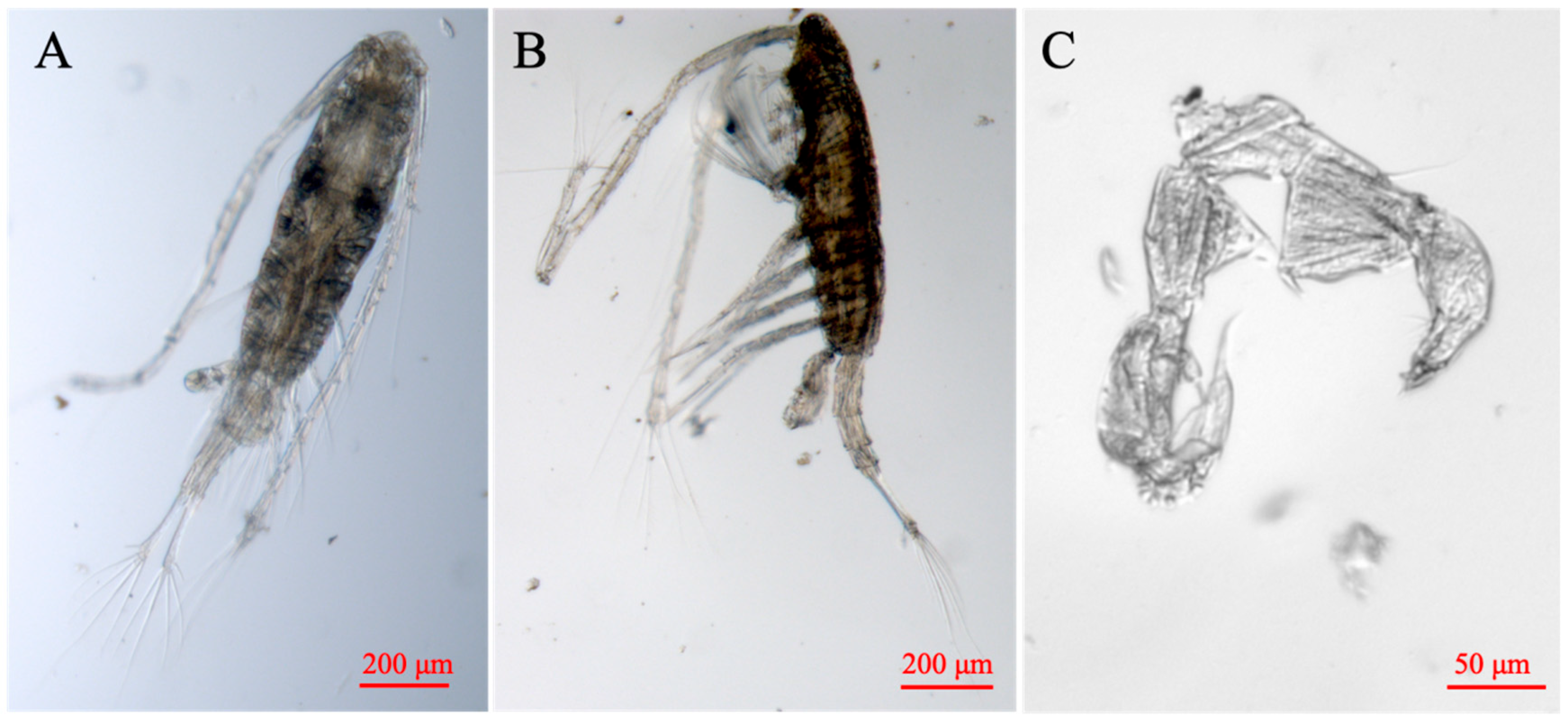
We collected the A. sinensis specimens in the Pearl River Estuary (22.88° N, 113.52° E) during November 2023. After collection, all specimens were immediately fixed in 95% ethanol and stored at −20 °C until further processing. Species identification was based on the morphological characteristics following Shen et al. [21], and representative photographs of the key taxonomic features are shown in Figure 1. A total of 50 intact male individuals were selected and preserved in absolute ethanol for genomic DNA extraction. Genomic DNA was extracted using the Rapid Genomic DNA Extraction Kit (Takara Bio Inc., Beijing, China) according to the manufacturer’s instructions. The quality of the extracted DNA was evaluated by 1% agarose gel electrophoresis. Qualified DNA samples were submitted to Nanjing Personalbio Technology (Nanjing, China) for library preparation and high-throughput sequencing. Paired-end sequencing with a read length of 150 bp was performed on the Illumina NovaSeq 6000 platform (Illumina, San Diego, CA, USA).

2.2. Mitochondrial Genome Assembly and Annotation

Raw reads were processed using fastp v0.23.4 [29] and Trimmomatic v0.39 [30] with default parameters for adapter removal, quality trimming, and length filtering, yielding over 10 GB of high-quality clean reads. We performed de novo assembly of the mitochondrial genome using both GetOrganelle [31] and NOVOPlasty [32], with the COI gene sequence of A. sinensis (GenBank accession no. KF977238.1) used as the seed sequence. In GetOrganelle, we conducted a de Bruijn graph-based assembly with k-mers of 21, 45, 65, 85, and 105 and a maximum of 20 iterations. In NOVOPlasty, we used a seed-extension-circularization strategy with a k-mer size of 33. Both assembly methods produced consistent circular mitochondrial genome sequences.
We performed gene annotation using MITOS2 through the Galaxy Europe platform (usegalaxy.eu, accessed on 19 May 2026) to predict the locations of protein-coding genes (PCGs), transfer RNAs (tRNAs), and ribosomal RNAs (rRNAs) [33]. The preliminary locations of PCGs were predicted by identifying open reading frames (ORFs) using the invertebrate mitochondrial genetic code, and the start and stop codons were verified through multiple sequence alignment with homologous PCGs from other copepod mitogenomes. The exact boundaries of the large and small ribosomal RNA genes (rRNAL and rRNAS) were defined through alignment with homologous sequences. We used MITOS to predict the secondary structures of all 22 tRNA genes for subsequent functional analysis.

2.3. Base Composition, Codon Usage, and Skew Analysis

We analyzed the complete mitochondrial genome of A. sinensis using PhyloSuite v1.2.3 [34]. We calculated nucleotide composition, relative synonymous codon usage (RSCU), and amino acid usage for all protein-coding genes. Strand compositional asymmetry was assessed by AT-skew and GC-skew values using the integrated functions of PhyloSuite.

2.4. Phylogenetic Analysis

Based on the nucleotide sequences of 13 protein-coding genes (PCGs), we conducted phylogenetic analyses to determine the phylogenetic position of A. sinensis within Copepoda. A total of 28 additional complete mitochondrial genomes of Copepoda were retrieved from GenBank (Table S1). The selected taxa included 7 Calanoida species, 10 Cyclopoida species, 7 Harpacticoida species, and 4 Siphonostomatoida species. Two Daphnia species (D. galeata, NC034297; D. pulex, NC000844) were used as outgroups.
We extracted nucleotide sequences of the 13 PCGs using PhyloSuite and performed multiple sequence alignment using MAFFT [35]. The resulting alignments were then refined using MACSE to correct for frameshifts and alignment errors caused by insertions/deletions [36]. We removed non-conserved regions and low-confidence sites using Gblocks [37]. The filtered gene sequences were concatenated into a final data matrix using the Concatenate Sequence function in PhyloSuite.
Maximum likelihood (ML) analyses were performed with IQ-TREE 3 [38]. The best-fit nucleotide substitution model for each partition was selected using ModelFinder, based on the Bayesian Information Criterion (BIC) [39], and partitioning was optimized using the MFP+MERGE strategy. Node support was assessed with 1000 SH-like approximate likelihood ratio tests (SH-aLRT) and 5000 ultrafast bootstrap replicates (UFBoot).
Bayesian inference (BI) analyses were conducted with MrBayes 3.2.7 [40]. Using the 13 partitions determined by IQ-TREE, the GTR+Gamma (GTR+G) model was applied uniformly, allowing independent estimation of base frequencies, substitution rates, and Gamma distribution parameters for each partition. Two independent runs were conducted, each with 4 Markov chains, running for 5,000,000 generations, sampling every 1000 generations, and performing convergence diagnostics every 5000 generations. The first 25% of samples were discarded as burn-in, and the remaining samples were used to construct a 50% majority-rule consensus tree with posterior probabilities (PP) calculated for each node. Convergence was assessed using the average standard deviation of split frequencies (ASDSF), with ASDSF < 0.01 indicating satisfactory convergence.

3. Results

3.1. Mitochondrial Genome Structure

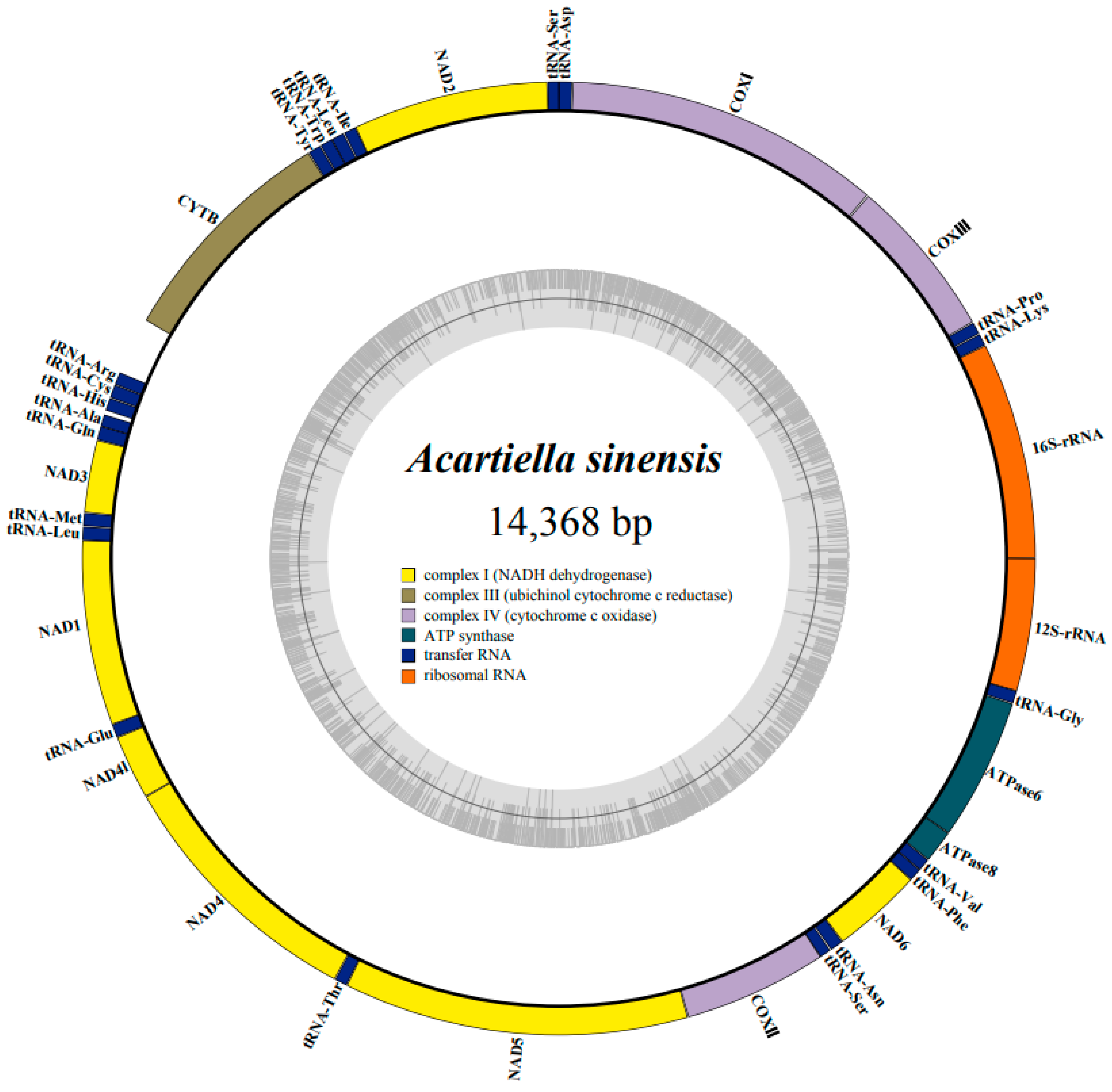
The complete mitochondrial genome of A. sinensis (GenBank accession no. PZ461130) is a circular molecule of 14,368 bp in length (Figure 2). It contained 37 mitochondrial genes including 13 PCGs, 2 rRNAs, and 22 tRNAs, together with a non-coding region (NCR) putatively corresponding to the control region (Figure 2; Table 1). All functional genes were located on the heavy (H-) strand. The mitogenome exhibited a highly compact gene arrangement, with 11 of the 36 adjacent gene pairs directly contiguous (i.e., no intergenic spacers or overlaps; Table 1). Most intergenic spacers were short, ranging from 1 to 4 bp, with the longest spacer of 19 bp located between tRNA-His and tRNA-Ala. Several adjacent gene pairs were directly contiguous, whereas gene overlaps were generally short, usually 1 or 2 bp in length. These overlaps mainly occurred between adjacent tRNA genes or between PCGs and tRNA genes (Table 1).
The overall nucleotide composition of the mitogenome was 35.3% A, 44.4% T, 11.7% G, and 8.6% C, with a high A+T content of 79.7% (Table 2). The AT-skew and GC-skew values were −0.113 and 0.154, respectively, indicating a nucleotide compositional bias toward T over A and G over C. The NCR was 277 bp and was located between Cytb and tRNA-Arg. Its nucleotide composition was 35.0% A, 36.5% T, 20.6% G, and 7.9% C, with an A+T content of 71.5% (Table 2).

3.2. Protein-Coding Genes and Codon Usage

The 13 PCGs comprised a total of 10,959 bp and encoded 3642 amino acids. Among them, NAD5 was the longest PCG with a length of 1683 bp, whereas ATPase8 was the shortest at 162 bp (Table 1 and Table 2). All PCGs started with an ATN codon (ATG, ATT, or ATA). For stop codons, eight genes including COX3, NAD2, and NAD3 terminated with TAA, whereas Cytb and NAD1 terminated with TAG. Incomplete stop codons were also detected, with COX1 and NAD6 ending with TA and COX2 ending with a single T (Table 1).
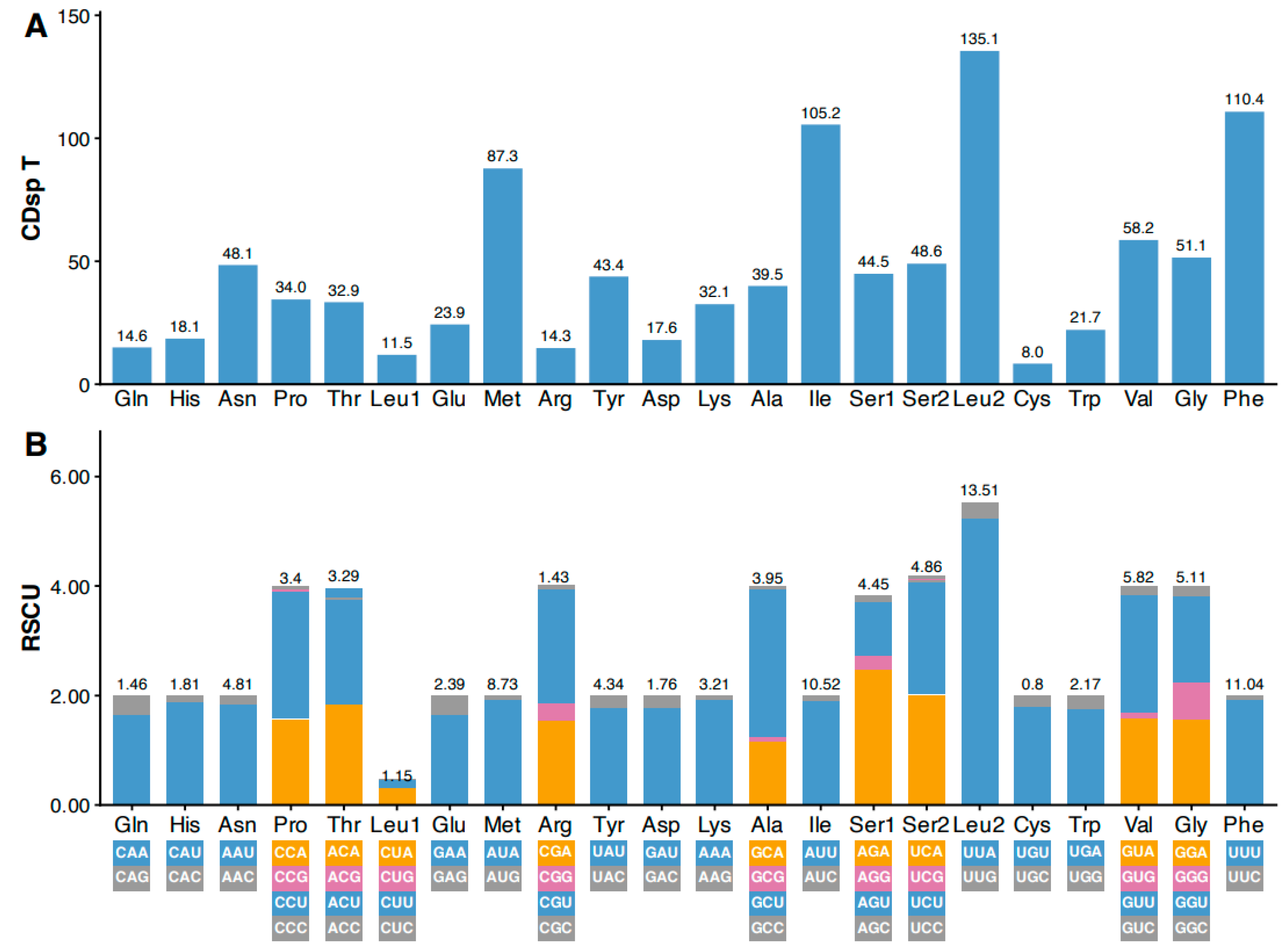
Leucine (Leu) was the most frequently used amino acid in the mitochondrial PCGs of A. sinensis, accounting for 14.66% of total amino acids, followed by phenylalanine (Phe), at 11.04%. In contrast, cysteine (Cys) was the least frequently used amino acid, accounting for only 0.80% (Figure 3A). RSCU analysis revealed a pronounced codon usage bias. Most codons ending with adenine (A) or thymine (T) showed relatively high RSCU values, whereas most codons ending with guanine (G) or cytosine (C) exhibited lower values, consistent with the high A+T content of the mitogenome. Among all codons, UUA (leucine) exhibited the highest RSCU value (5.24), indicating that it was the most preferentially used codon (Figure 3B).

3.3. Ribosomal and Transfer RNA Genes

The two ribosomal RNA genes (12S rRNA and 16S rRNA) comprised 1707 bp and exhibited nucleotide compositions of 44.2% A, 41.2% T, 8.8% G, and 5.8% C, resulting in a high A+T content (Table 2). The AT-skew and GC-skew values were 0.035 and 0.208, respectively. The 12S rRNA (647 bp) and 16S rRNA (1060 bp) were located between tRNA-Gly and tRNA-Lys and arranged in tandem (Table 1).
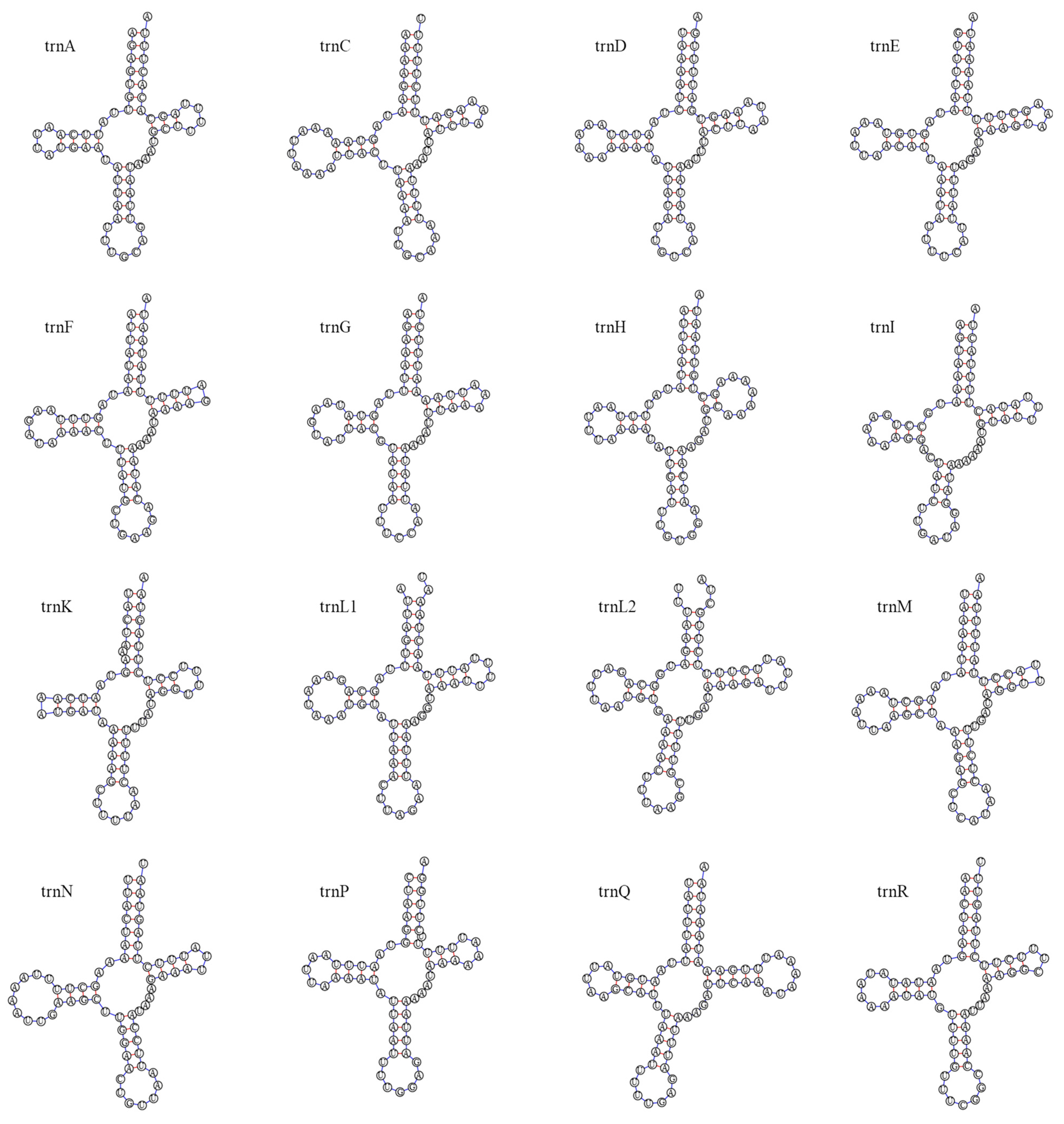
A total of 22 tRNA genes were identified, ranging from 56 bp (tRNA-Ser) to 69 bp (tRNA-Gln), with a total length of 1394 bp (Table 1 and Table 2). Most tRNAs (20 of 22) could be folded into typical cloverleaf secondary structures, whereas two serine tRNAs (tRNA-Ser1 and tRNA-Ser2) lacked a dihydrouridine (DHU) arm, forming atypical structures (Figure 4). The anticodon stems were generally stable, consisting of 3–5 bp, and the acceptor stems were typically 7 bp, with slight variation in several tRNAs. A total of 16 G–U mismatches were detected across the tRNA genes (Figure 4). These mismatches were mainly distributed in the acceptor, DHU, and TΨC arms, whereas no mismatches were observed in the anticodon stems. Among all tRNAs, tRNA-Glu showed the highest number of mismatches (n = 3), followed by tRNA-Leu2 and tRNA-Ser1 (n = 2). Additionally, three tRNAs exhibited bulge structures. Single-nucleotide insertions were observed in tRNA-Lys and tRNA-Pro within the acceptor stem, and in tRNA-Ser1 within the anticodon stem. Furthermore, the acceptor stems of tRNA-Leu1 and tRNA-Leu2 contained unpaired nucleotides at the termini.

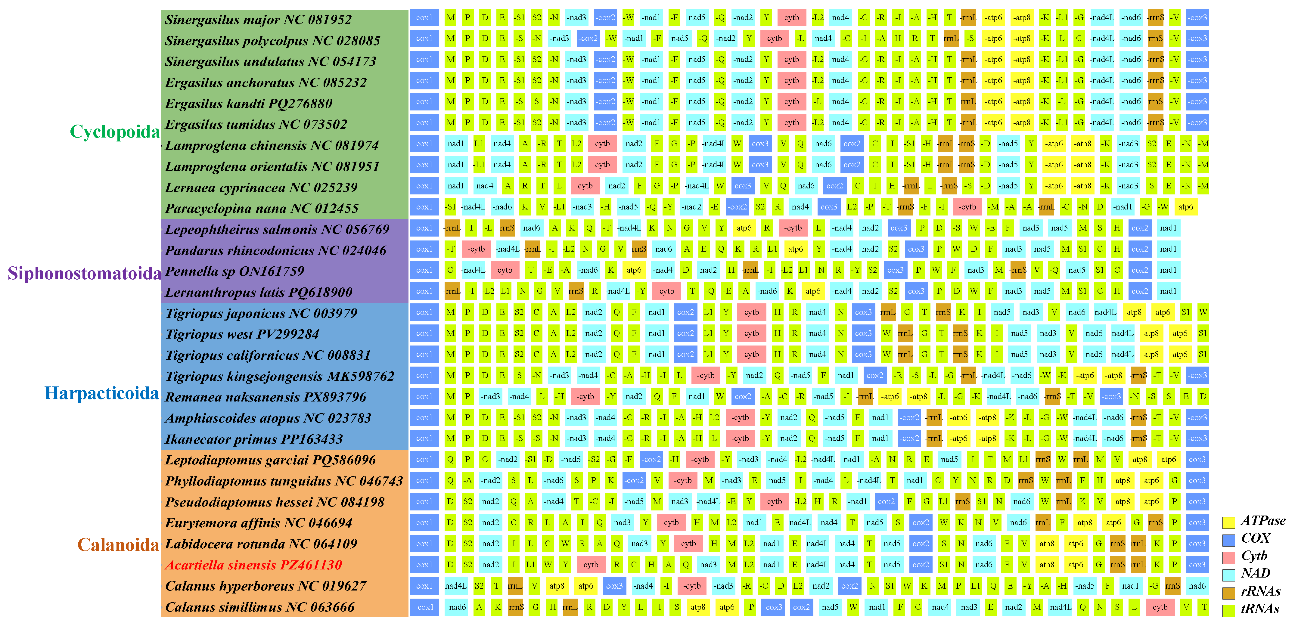
3.4. Comparative Mitochondrial Gene Arrangement in Copepoda

We compared mitochondrial gene arrangements among 29 copepod species representing four orders (Calanoida, Harpacticoida, Cyclopoida, and Siphonostomatoida), with all genomes linearized from the COX1 gene (Figure 5). Gene order was generally conserved within orders but differed substantially among orders. As the first reported mitogenome of Acartiidae, A. sinensis exhibited a gene arrangement distinct from those of other calanoid species. Compared with Labidocera rotunda, the order of most PCGs was conserved, although the relative positions of Cytb and NAD3 were reversed with local rearrangements of neighboring tRNA genes. Compared with the species of Diaptomidae and Pseudodiaptomidae, more extensive rearrangements involving Cytb, NAD1, NAD3, NAD4, NAD4L, NAD5, and adjacent tRNA genes were observed in A. sinensis. The species of Siphonostomatoida displayed gene arrangements markedly different from those of Calanoida. While PCG order was relatively conserved within Siphonostomatoida, the positions of tRNA and rRNA genes, particularly rrnL and rrnS, varied substantially (Figure 5).

3.5. Phylogenetic Relationships Within Copepoda

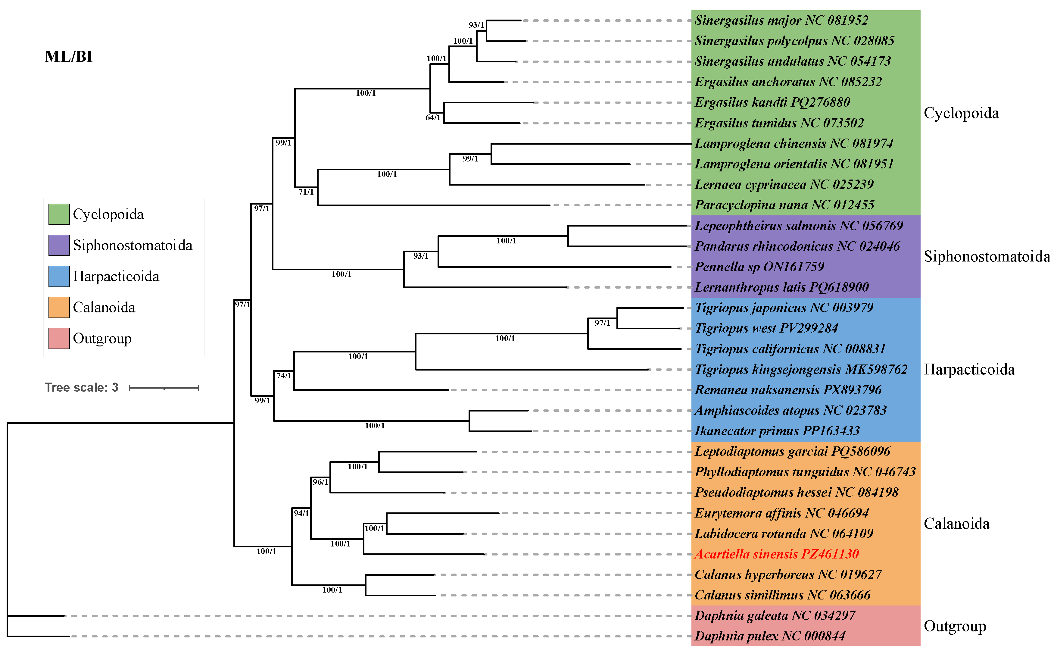
Phylogenetic analyses used ML- and BI-generated identical topologies (Figure 6). All four copepod orders formed well-supported clades. Cyclopoida and Siphonostomatoida were sister groups and together formed a clade sister to Harpacticoida, whereas Calanoida occupied the basal position among the taxa analyzed. Within Calanoida, Calanidae represented the basal lineage, while Diaptomidae and Pseudodiaptomidae clustered as a derived clade. A. sinensis was recovered as sister to the clade comprising L. rotunda and E. affinis.

4. Discussion

Our study showed that A. sinensis exhibits conserved gene content but distinct structural and compositional features, including extreme A+T bias, strand asymmetry, and localized gene rearrangements. These characteristics, combined with phylogenetic evidence, advance our understanding of mitochondrial genome evolution in copepods and provide valuable genomic data for future studies on taxonomy, population genetics, and adaptive evolution within Acartiidae.

4.1. The Mitochondrial Genome of A. sinensis

The mitochondrial genome of A. sinensis contains a set of metazoan mitochondrial genes. This is in agreement with the conserved gene complement observed in most crustaceans [41,42,43]. The A. sinensis genome displays a highly compact organization characterized by extremely short intergenic spacers and occasional gene overlaps, which is typical of compact mitochondrial genomes in invertebrates [44]. Such compaction generally results from the selection pressure toward genome streamlining, leading to reduced non-coding regions and minimized genome size.
All mitochondrial genes of A. sinensis are encoded on the heavy strand. This feature has also been observed in some invertebrate lineages and has been reported in some arthropods such as millipedes [45]. The single-strand encoding pattern may represent a lineage-specific organizational strategy, potentially associated with replication and transcriptional asymmetry. In addition, the A. sinensis mitogenome exhibits a remarkably high A+T content (79.7%) and strong AT- and GC- skews, which are commonly observed in crustacean mitochondrial genomes [46,47]. Similar compositional biases have also been reported in other copepods such as E. affinis and Calanus sinicus [4,42]. These compositional biases may reflect long-term directional mutation pressure and strand-specific replication bias, which may have been influenced by the inversion events of replication origins in crustacean mitochondrial evolution [48].
The mitochondrial PCGs of A. sinensis initiate with ATN start codons, consistent with the mitochondrial translation system of invertebrates [47,49]. Most genes possess complete stop codons (TAA or TAG), whereas several exhibit incomplete termination codons. This is a widespread phenomenon in metazoan mitochondria. The incomplete stop codons are generally completed post-transcriptionally via polyadenylation, which restores functional termination signals while contributing to genome compaction [50]. Codon usage analysis reveals a strong bias toward A/U-ending codons, reflecting mutational pressure rather than translational selection. The leucine codon UUA is the most frequently used codon, indicating marked synonymous codon preference [46,47].
The ribosomal RNA genes of A. sinensis are comparable in length to those of other calanoid species, but their genomic arrangement differs markedly. In A. sinensis, the 12S rRNA and 16S rRNA genes are tandemly located between tRNA-Gly and tRNA-Lys, whereas in other copepods such as C. sinicus and Phyllodiaptomus tunguidus, their positions are highly variable [4,5]. The rearrangements suggest the evolutionary plasticity of mitochondrial genome organization even among closely related taxa.
The 22 tRNA genes in A. sinensis mostly form canonical cloverleaf structures, although two tRNA-Ser genes lack the DHU arm. This structural reduction is considered a tolerated deviation in mitochondrial tRNAs [51,52,53]. Additionally, multiple G–U mismatches were detected across tRNAs, particularly in stem regions excluding anticodon loops. The non-canonical pairings contribute to the structural flexibility while maintaining functional stability [54,55].
Copepod mitochondrial genomes exhibit extensive gene rearrangements, although some conservation is still detectable at the order level [4,5,56]. Our comparative analysis across 29 copepod species confirms strong inter-order divergence but relative intra-order conservation. As the first mitochondrial genome reported for Acartiidae, A. sinensis provides important insight into calanoid genome evolution. Compared with L. rotunda, the overall gene order is largely conserved, except for a localized inversion in the Cytb-NAD3 region accompanied by tRNA rearrangements. Such localized rearrangements may represent hotspots of genomic recombination or replication-related errors [57]. The rearrangements in comparisons with Diaptomidae and Pseudodiaptomidae further demonstrates the lineage divergence within Calanoida [58].

4.2. Phylogenetic Relationships Within Copepoda

Phylogenetic analyses based on concatenated mitochondrial PCGs yielded highly congruent topologies between ML and BI. All major copepod orders formed strongly supported clades [7,59]. Our results recover Calanoida as a basal lineage within Copepoda and confirm the split between Calanoida and Podoplea [4,60].
Within Podoplea, our results support the topology (Calanoida + (Harpacticoida + (Cyclopoida + Siphonostomatoida))), which agree with recent mitogenome-based phylogenies [60,61]. This topology differs from earlier morphological studies and analyses based on one or few rRNA genes, which often recovered Harpacticoida and Siphonostomatoida as sister groups [4,62]. Recent phylogenomic analyses based on hundreds or thousands of nuclear genes also support the monophyly of Podoplea and place Cyclopoida and Harpacticoida as sister groups, suggesting that higher-level copepod relationships are sensitive to molecular marker and dataset size [63]. Together with simulation and empirical studies on copepod mitogenomes, these findings underscore that sparse taxon sampling and short genetic fragments can lead to unstable interordinal relationships, whereas multilocus datasets with extensive sampling generally provide more robust phylogenetic signals [60].
Within Calanoida, our phylogenetic tree recovered Calanidae as a basal lineage and placed Diaptomidae and Pseudodiaptomidae as a derived clade [5,64]. Comparative mitogenomic analyses indicate that Diaptomidae species have relatively conserved PCG arrangements and similar genomic features, revealing the close evolutionary relationships within this lineage [5,43].
The phylogenetic position of Diaptomidae was consistent with previous mitogenomic studies. Specifically, previous analyses based on concatenated mitochondrial protein-coding genes recovered a strongly supported clade comprising L. rotunda and E. affinis, which formed the sister group to Diaptomidae species [57]. This close relationship was also supported by the analyses of concatenated mitochondrial PCGs from multiple copepod mitogenomes [58].

5. Conclusions

This study reports the complete mitochondrial genome of A. sinensis, representing the first mitogenome for Acartiidae. The genome is compact, exhibits a strong AT bias, and comprises the standard set of 37 mitochondrial genes. Its protein-coding genes exhibit pronounced A/U-ending codon preferences, whereas the tRNA set includes two serine tRNAs lacking complete DHU arms and multiple non-canonical base pairs. Comparative analysis revealed extensive variation in mitochondrial gene order across Copepoda and identified a distinctive arrangement in A. sinensis, particularly around the Cytb–NAD3 region. Phylogenetic analyses placed A. sinensis as sister to the clade comprising L. rotunda and E. affinis. These results provide new evidence for mitochondrial genome evolution and phylogenetic relationships within Calanoida and establish a foundation for future taxonomic, population genetic, and adaptive evolution studies of Acartiidae.

Supplementary Materials

The following supporting information can be downloaded at: https://www.mdpi.com/article/10.3390/ani16152319/s1, Table S1: List of species used to construct the phylogenetic tree.

Author Contributions

Conceptualization, Q.T.; investigation, J.N.; data curation, Y.L. and S.L.; writing—original draft preparation, Q.T.; writing—review and editing, Q.T.; visualization, L.W. and D.H.; project administration, J.Z.; funding acquisition, X.W. and F.D. All authors have read and agreed to the published version of the manuscript.

Funding

This research was supported by the Open Fund of The Key Laboratory of Water Environment Simulation and Pollution Control, Ministry of Ecology and Environment of the People’s Republic of China (Grant No. WESPC202406), and Guangdong Basic and Applied Basic Research Foundation [2022B1515250005].

Institutional Review Board Statement

Not applicable.

Informed Consent Statement

Not applicable.

Data Availability Statement

The datasets presented in this study can be found in online repositories. The names of the repository/repositories and accession number(s) can be found below: https://www.ncbi.nlm.nih.gov/genbank/PZ461130 (These data can be accessed after 31 December 2026).

Acknowledgments

We greatly appreciate Lei Xu from the South China Sea Fisheries Research Institute, Chinese Academy of Fishery Sciences, for providing the technical and methodological contributions for this research. We are also grateful to the editor and reviewers for critically evaluating the manuscript and providing constructive comments to improve it.

Conflicts of Interest

The authors declare no conflicts of interest.

References

  1. Cameron, S.L. Insect Mitochondrial Genomics: Implications for Evolution and Phylogeny. Annu. Rev. Entomol. 2014, 59, 95–117. [Google Scholar] [CrossRef] [PubMed]
  2. Chen, X.-D.; Wang, Y.-Y.; Xu, Z.-M.; Xiao, L.; Lu, C.-H.; Sun, C.-H.; Li, C.-Z. Mitogenomic Analysis and Conservation Genetics of the Endangered Oriental Stork (Ciconia boyciana). Animals 2026, 16, 1077. [Google Scholar] [CrossRef] [PubMed]
  3. Shu, J.-Z.; Ma, X.; Zhan, Y.-J.; Chen, X.-D.; Sun, C.-H. Characteristics and Evolutionary Relationships of Two Mitochondrial Genomes of Iguanodectes (Characiformes, Iguanodectidae). Animals 2026, 16, 740. [Google Scholar] [CrossRef] [PubMed]
  4. Minxiao, W.; Song, S.; Chaolun, L.; Xin, S. Distinctive Mitochondrial Genome of Calanoid Copepod Calanus sinicus with Multiple Large Non-Coding Regions and Reshuffled Gene Order: Useful Molecular Markers for Phylogenetic and Population Studies. BMC Genom. 2011, 12, 73. [Google Scholar] [CrossRef]
  5. Zhang, X.-L.; Liu, P.; Xu, S.-L.; Rizo, E.Z.; Zhang, Q.; Dumont, H.J.; Han, B.-P. Geographic Variation of Phyllodiaptomus tunguidus Mitogenomes: Genetic Differentiation and Phylogeny. Front. Genet. 2021, 12, 711992. [Google Scholar] [CrossRef] [PubMed]
  6. Gu, Y.-L.; Sun, C.-H.; Liu, P.; Zhang, X.; Sinev, A.Y.; Dumont, H.J.; Han, B.-P. Complete Mitochondrial Genome of Ovalona pulchella (Branchiopoda, Cladocera) as the First Representative in the Family Chydoridae: Gene Rearrangements and Phylogenetic Analysis of Cladocera. Gene 2022, 818, 146230. [Google Scholar] [CrossRef] [PubMed]
  7. Huys, R.; Boxshall, G.A. Copepod Evolution; The Ray Society: London, UK, 1991. [Google Scholar]
  8. Boxshall, G.; Halsey, S. An Introduction to Copepod Diversity; The Ray Society: London, UK, 2004. [Google Scholar]
  9. Oda, Y.; Nakano, S.; Suh, J.-M.; Oh, H.-J.; Jin, M.-Y.; Kim, Y.-J.; Sakamoto, M.; Chang, K.-H. Spatiotemporal Variability in a Copepod Community Associated with Fluctuations in Salinity and Trophic State in an Artificial Brackish Reservoir at Saemangeum, South Korea. PLoS ONE 2018, 13, e0209403. [Google Scholar] [CrossRef] [PubMed]
  10. Basu, S.; Gogoi, P.; Bhattacharyya, S.; Kumar, L.; Das, S.K.; Das, B.K. Variability in the Zooplankton Assemblages in Relation to Environmental Variables in the Tidal Creeks of Sundarbans Estuarine System, India. Environ. Sci. Pollut. Res. 2022, 29, 45981–46002. [Google Scholar] [CrossRef]
  11. Choi, S.Y.; Lee, E.H.; Jang, M.-C.; Seo, M.H.; Soh, H.Y. Ecological Implications and Seasonal Variability of Grazing by Marine Copepods on Phytoplankton: Comparison between Acartia omorii and A. steueri in Jangmok Bay, Korea. Mar. Biol. 2024, 171, 125. [Google Scholar] [CrossRef]
  12. Tranter, D.J.; Abraham, S. Coexistence of Species of Acartiidae (Copepoda) in the Cochin Backwater, a Monsoonal Estuarine Lagoon. Mar. Biol. 1971, 11, 222–241. [Google Scholar] [CrossRef]
  13. Chen, M.; Kim, D.; Liu, H.; Kang, C.-K. Variability in Copepod Trophic Levels and Feeding Selectivity Based on Stable Isotope Analysis in Gwangyang Bay of the Southern Coast of the Korean Peninsula. Biogeosciences 2018, 15, 2055–2073. [Google Scholar] [CrossRef]
  14. Hahn, A.; Nascimento-Schulze, J.C.; Avgerinou, G.; Bayer, T.; Brennan, R.S. Local Adaptation and Osmoregulatory Mechanisms of the Copepod Acartia tonsa Under Low Salinity Stress. Mol. Ecol. 2026, 35, e70208. [Google Scholar] [PubMed]
  15. Belmonte, G. The Egg Morphology of 7 Acartiidae Species: A Preliminary Survey of the Ootaxonomy of Calanoids. J. Mar. Syst. 1998, 15, 35–39. [Google Scholar] [CrossRef]
  16. Ueda, H. Taxonomic Descriptions of the Brackish-Water Calanoid Copepod Acartia tsuensis Ito from Japan and the Closely Related A. ohnoi n. sp. from the Philippines. Plankton Benthos Res. 2024, 19, 116–124. [Google Scholar] [CrossRef]
  17. Belmonte, G.; Potenza, D. Biogeography of the Family Acartiidae (Calanoida) in the Ponto-Mediterranean Province. Hydrobiologia 2001, 453, 171–176. [Google Scholar] [CrossRef]
  18. Krupa, E.; Aubakirova, M. Checklist and Distribution of Calanoida (Crustacea: Copepoda) in Kazakhstan (Central Asia). Water 2021, 13, 2015. [Google Scholar] [CrossRef]
  19. Figueroa, N.J.; Figueroa, D.F.; Hicks, D. Phylogeography of Acartia tonsa Dana, 1849 (Calanoida: Copepoda) and Phylogenetic Reconstruction of the Genus Acartia Dana, 1846. Mar. Biodivers. 2020, 50, 23. [Google Scholar] [CrossRef]
  20. Mesas, A.; Aguilera, V.M.; González, C.E.; Giesecke, R.; Escribano, R.; Vargas, C.A. Molecular Evidence for a New Endemic Species of Acartia (Copepoda, Calanoida) from the Southeast Pacific Coast. Sci. Rep. 2024, 14, 12366. [Google Scholar] [CrossRef] [PubMed]
  21. Shen, C.J.; Tai, A.Y.; Zhang, Z.Y.; Song, D.X.; Song, G.X.; Chen, G.X. Fauna Sinica, Crustacea: Freshwater Copepoda; Science Press: Peking, China, 1979. [Google Scholar]
  22. Srinui, K.; Ohtsuka, S. Supplementary Description of Three Acartiella Species (Crustacea: Copepoda: Calanoida) from Estuarine Waters in Thailand. Species Divers. 2015, 20, 167–181. [Google Scholar] [CrossRef]
  23. Lin, D.; Li, X.; Fang, H.; Dong, Y.; Huang, Z.; Chen, J. Calanoid Copepods Assemblages in Pearl River Estuary of China in Summer: Relationships between Species Distribution and Environmental Variables. Estuar. Coast. Shelf Sci. 2011, 93, 259–267. [Google Scholar] [CrossRef]
  24. Orsi, J.J.; Ohtsuka, S. Introduction of the Asian Copepods Acartiella sinensis, Tortanus dextrilobatus (Copepoda: Calanoida), and Limnoithona tetraspina (Copepoda: Cyclopoida) to the San Francisco Estuary, California, USA. Plankton Biol. Ecol. 1999, 46, 128–131. [Google Scholar]
  25. Slaughter, A.; Ignoffo, T.; Kimmerer, W. Predation Impact of Acartiella Sinensis, an Introduced Predatory Copepod in the San Francisco Estuary, USA. Mar. Ecol. Prog. Ser. 2016, 547, 47–60. [Google Scholar] [CrossRef]
  26. Shang, X.; Wang, G.; Li, S.; Donghui, G. Preliminary Study of the Retention Mechanism of Planktonic Copepods in the Jiulong Estuary in China. Acta Oceanol. Sin. 2007, 26, 156–163. [Google Scholar]
  27. Xu, X.; Zeng, J.; Zeng, G.; Chen, Q.; Liao, Y.; Gao, A.; Shou, L. Effects of Thermal Water Discharged from Coastal Power Plant on Distribution of Zooplankton. Chin. J. Ecol. 2008, 27, 933–939. [Google Scholar]
  28. Lee, P.-W.; Hsiao, S.-H.; Chou, C.; Tseng, L.-C.; Hwang, J.-S. Zooplankton Fluctuations in the Surface Waters of the Estuary of a Large Subtropical Urban River. Front. Ecol. Evol. 2021, 9, 598274. [Google Scholar] [CrossRef] [PubMed]
  29. Chen, S.; Zhou, Y.; Chen, Y.; Gu, J. Fastp: An Ultra-Fast All-in-One FASTQ Preprocessor. Bioinformatics 2018, 34, i884–i890. [Google Scholar] [CrossRef] [PubMed]
  30. Bolger, A.M.; Lohse, M.; Usadel, B. Trimmomatic: A Flexible Trimmer for Illumina Sequence Data. Bioinformatics 2014, 30, 2114–2120. [Google Scholar] [CrossRef] [PubMed]
  31. Jin, J.-J.; Yu, W.-B.; Yang, J.-B.; Song, Y.; dePamphilis, C.W.; Yi, T.-S.; Li, D.-Z. GetOrganelle: A Fast and Versatile Toolkit for Accurate de Novo Assembly of Organelle Genomes. Genome Biol. 2020, 21, 241. [Google Scholar] [CrossRef] [PubMed]
  32. Dierckxsens, N.; Mardulyn, P.; Smits, G. NOVOPlasty: De Novo Assembly of Organelle Genomes from Whole Genome Data. Nucleic Acids Res. 2017, 45, e18. [Google Scholar] [PubMed]
  33. Bernt, M.; Donath, A.; Jühling, F.; Externbrink, F.; Florentz, C.; Fritzsch, G.; Pütz, J.; Middendorf, M.; Stadler, P.F. MITOS: Improved de Novo Metazoan Mitochondrial Genome Annotation. Mol. Phylogenet. Evol. 2013, 69, 313–319. [Google Scholar] [CrossRef] [PubMed]
  34. Zhang, D.; Gao, F.; Jakovlić, I.; Zou, H.; Zhang, J.; Li, W.X.; Wang, G.T. PhyloSuite: An Integrated and Scalable Desktop Platform for Streamlined Molecular Sequence Data Management and Evolutionary Phylogenetics Studies. Mol. Ecol. Resour. 2020, 20, 348–355. [Google Scholar] [PubMed]
  35. Katoh, K.; Standley, D.M. MAFFT Multiple Sequence Alignment Software Version 7: Improvements in Performance and Usability. Mol. Biol. Evol. 2013, 30, 772–780. [Google Scholar] [CrossRef] [PubMed]
  36. Ranwez, V.; Douzery, E.J.P.; Cambon, C.; Chantret, N.; Delsuc, F. MACSE v2: Toolkit for the Alignment of Coding Sequences Accounting for Frameshifts and Stop Codons. Mol. Biol. Evol. 2018, 35, 2582–2584. [Google Scholar] [CrossRef] [PubMed]
  37. Castresana, J. Selection of Conserved Blocks from Multiple Alignments for Their Use in Phylogenetic Analysis. Mol. Biol. Evol. 2000, 17, 540–552. [Google Scholar] [CrossRef] [PubMed]
  38. Minh, B.Q.; Schmidt, H.A.; Chernomor, O.; Schrempf, D.; Woodhams, M.D.; von Haeseler, A.; Lanfear, R. IQ-TREE 2: New Models and Efficient Methods for Phylogenetic Inference in the Genomic Era. Mol. Biol. Evol. 2020, 37, 1530–1534. [Google Scholar] [CrossRef] [PubMed]
  39. Kalyaanamoorthy, S.; Minh, B.Q.; Wong, T.K.F.; von Haeseler, A.; Jermiin, L.S. ModelFinder: Fast Model Selection for Accurate Phylogenetic Estimates. Nat. Methods 2017, 14, 587–589. [Google Scholar] [CrossRef] [PubMed]
  40. Ronquist, F.; Teslenko, M.; van der Mark, P.; Ayres, D.L.; Darling, A.; Höhna, S.; Larget, B.; Liu, L.; Suchard, M.A.; Huelsenbeck, J.P. MrBayes 3.2: Efficient Bayesian Phylogenetic Inference and Model Choice across a Large Model Space. Syst. Biol. 2012, 61, 539–542. [Google Scholar] [CrossRef] [PubMed]
  41. Kim, S.; Lim, B.-J.; Min, G.-S.; Choi, H.-G. The Complete Mitochondrial Genome of Arctic Calanus hyperboreus (Copepoda, Calanoida) Reveals Characteristic Patterns in Calanoid Mitochondrial Genome. Gene 2013, 520, 64–72. [Google Scholar] [CrossRef] [PubMed]
  42. Choi, B.-S.; Han, J.; Hwang, D.-S.; Souissi, S.; Hagiwara, A.; Lee, J.-S. Complete Mitochondrial Genome of the Calanoid Copepod Eurytemora affinis (Calanoida, Temoridae). Mitochondrial DNA Part B 2019, 4, 2731–2733. [Google Scholar] [CrossRef] [PubMed]
  43. Zhang, X.; Xu, S.; Liu, P.; Huang, Q.; Li, H.; Dumont, H.; Han, B. The Mitochondrial Genome of Phyllodiaptomus tunguidus Shen & Tai, 1964 (Copepoda, Calanoida) and Its Phylogeny. Mitochondrial DNA Part B 2020, 5, 2521–2522. [Google Scholar] [CrossRef] [PubMed]
  44. Noguchi, Y.; Endo, K.; Tajima, F.; Ueshima, R. The Mitochondrial Genome of the Brachiopod Laqueus rubellus. Genetics 2000, 155, 245–259. [Google Scholar] [CrossRef] [PubMed]
  45. Zuo, Q.; Zhang, Z.; Shen, Y. Novel Mitochondrial Gene Rearrangements Pattern in the Millipede Polydesmus sp. GZCS-2019 and Phylogenetic Analysis of the Myriapoda. Ecol. Evol. 2022, 12, e8764. [Google Scholar] [CrossRef] [PubMed]
  46. Zapelloni, F.; Jurado-Rivera, J.A.; Jaume, D.; Juan, C.; Pons, J. Comparative Mitogenomics in Hyalella (Amphipoda: Crustacea). Genes 2021, 12, 292. [Google Scholar] [CrossRef] [PubMed]
  47. Yang, H.-J.; Yang, Z.-H.; Ren, T.-G.; Dong, W.-G. Description and Phylogenetic Analysis of the Complete Mitochondrial Genome in Eulaelaps silvestris Provides New Insights into the Molecular Classification of the Family Haemogamasidae. Parasitology 2023, 150, 821–830. [Google Scholar] [CrossRef] [PubMed]
  48. Jakovlić, I.; Zou, H.; Zhao, X.-M.; Zhang, J.; Wang, G.-T.; Zhang, D. Evolutionary History of Inversions in Directional Mutational Pressures in Crustacean Mitochondrial Genomes: Implications for Evolutionary Studies. Mol. Phylogenet. Evol. 2021, 164, 107288. [Google Scholar] [CrossRef] [PubMed]
  49. Donath, A.; Jühling, F.; Al-Arab, M.; Bernhart, S.H.; Reinhardt, F.; Stadler, P.F.; Middendorf, M.; Bernt, M. Improved Annotation of Protein-Coding Genes Boundaries in Metazoan Mitochondrial Genomes. Nucleic Acids Res. 2019, 47, 10543–10552. [Google Scholar] [CrossRef] [PubMed]
  50. Muhala, V.; Guimarães-Costa, A.; Bessa-Silva, A.R.; Rabelo, L.P.; Carneiro, J.; Macate, I.E.; Watanabe, L.; Balcázar, O.D.; Gomes, G.E.; Vallinoto, M.; et al. Comparative Mitochondrial Genome Brings Insights to Slight Variation in Gene Proportion and Large Intergenic Spacer and Phylogenetic Relationship of Mudskipper Species. Sci. Rep. 2024, 14, 3358. [Google Scholar] [CrossRef] [PubMed]
  51. Benito, J.B.; Porter, M.L.; Niemiller, M.L. Comparative Mitogenomic Analysis of Subterranean and Surface Amphipods (Crustacea, Amphipoda) with Special Reference to the Family Crangonyctidae. BMC Genom. 2024, 25, 298. [Google Scholar] [CrossRef]
  52. Hua, C.; Huang, Y.; Li, H.; Feng, J.; Qiao, J.; Hu, S.; Liu, W.; Shu, X. Characterization of the Complete Mitochondrial Genome of Ergasilus anchoratus Markevich, 1946 (Ergasilidae) and Phylogeny of Copepoda. Syst. Parasitol. 2024, 101, 33. [Google Scholar] [CrossRef] [PubMed]
  53. Ozerova, I.; Fallmann, J.; Mörl, M.; Bernt, M.; Prohaska, S.J.; Stadler, P.F. Aberrant Mitochondrial tRNA Genes Appear Frequently in Animal Evolution. Genome Biol. Evol. 2024, 16, evae232. [Google Scholar] [CrossRef] [PubMed]
  54. Kundu, S.; Kang, H.-E.; Kim, A.R.; Lee, S.R.; Kim, E.-B.; Amin, M.H.F.; Andriyono, S.; Kim, H.-W.; Kang, K. Mitogenomic Characterization and Phylogenetic Placement of African Hind, Cephalopholis taeniops: Shedding Light on the Evolution of Groupers (Serranidae: Epinephelinae). Int. J. Mol. Sci. 2024, 25, 1822. [Google Scholar] [CrossRef] [PubMed]
  55. Yin, J.; Luo, Z.-M.; Li, Y.-H.; Wang, C.-M.; Li, J.; Zhang, R.-Y.; Shan, H.-L.; Wang, X.-Y.; Chen, Y.-Q. Mitochondrial Genome Characteristics and Phylogenetic Analysis of Fulmekiola serrata (Kobus) (Thysanoptera: Thripidae). Int. J. Mol. Sci. 2024, 25, 10431. [Google Scholar] [CrossRef] [PubMed]
  56. Machida, R.J.; Miya, M.U.; Nishida, M.; Nishida, S. Complete Mitochondrial DNA Sequence of Tigriopus japonicus (Crustacea: Copepoda). Mar. Biotechnol. 2002, 4, 406–417. [Google Scholar] [CrossRef]
  57. Jun, J.; Jeong, H.G.; Choi, H.; Woo, H.; Jeon, D.; Seo, Y.-J.; Eyun, S. The Complete Mitochondrial Genome of Labidocera rotunda Mori, 1929 (Copepoda: Calanoida) from Jeju Island, Korea. Mitochondrial DNA Part B 2022, 7, 1702–1703. [Google Scholar] [CrossRef] [PubMed]
  58. Urban-Olivares, J.; Ortega-Mayagoitia, E.; Alcántara-Rodríguez, J.A.; Dimas-Flores, N.; Rougon-Cardoso, A.; Ciros-Pérez, J. The Copepoda Mitogenome as a Dynamic Evolutionary Landscape. PLoS ONE 2026, 21, e0350115. [Google Scholar] [CrossRef] [PubMed]
  59. Eyun, S. Phylogenomic Analysis of Copepoda (Arthropoda, Crustacea) Reveals Unexpected Similarities with Earlier Proposed Morphological Phylogenies. BMC Evol. Biol. 2017, 17, 23. [Google Scholar] [CrossRef] [PubMed]
  60. He, J.; Zhou, Z.; Huang, Y.; Feng, J.; Li, W.; Wang, G.; Hua, C. Evolutionary Rates, Divergence Rates, and Performance of Individual Mitochondrial Genes Based on Phylogenetic Analysis of Copepoda. Genes 2023, 14, 1496. [Google Scholar] [CrossRef] [PubMed]
  61. Huang, Y.; Feng, J.-M.; Liu, W.; Sun, B.-L.; Shu, X.-J.; Li, W.-X.; Wang, G.-T.; Hua, C.-J. Comprehensive Molecular Characterisation of the Complete Mitogenome of Ergasilus tumidus and Phylogenetic Relationships of Copepoda Inferred from Mitogenomes. Folia Parasitol. 2024, 71, 002. [Google Scholar] [CrossRef]
  62. Braga, E.; Zardoya, R.; Meyer, A.; Yen, J. Mitochondrial and Nuclear rRNA Based Copepod Phylogeny with Emphasis on the Euchaetidae (Calanoida). Mar. Biol. 1999, 133, 79–90. [Google Scholar] [CrossRef]
  63. Bernot, J.P.; Khodami, S.; Boyen, J.; De Troch, M.; Boxshall, G.A.; Arbizu, P.M. Copepod Phylogenomics Supports Canuelloida as a Valid Order Separate from Harpacticoida. Mol. Phylogenet. Evol. 2025, 206, 108311. [Google Scholar] [CrossRef] [PubMed]
  64. Blanco-Bercial, L.; Bradford-Grieve, J.; Bucklin, A. Molecular Phylogeny of the Calanoida (Crustacea: Copepoda). Mol. Phylogenet. Evol. 2011, 59, 103–113. [Google Scholar] [CrossRef] [PubMed]
Figure 1. A. sinensis from the Pearl River Estuary, China. (A,B) Male individuals, (C) Fifth thoracic leg.
Animals 16 02319 g001
Figure 2. Mitochondrial genome maps of A. sinensis. The gray part of the inner circle represents GC content.
Animals 16 02319 g002
Figure 3. Amino acid usage and relative synonymous codon usage in the mitochondrial genome of A. sinensis. (A) Amino acid usage frequency in the 13 mitochondrial protein-coding genes. The x-axis represents amino acid types, and the y-axis represents the number of codons per thousand codons (CDspT). (B) Relative synonymous codon usage (RSCU) values of mitochondrial protein-coding genes. Different colors indicate codons with different third-position nucleotides.
Animals 16 02319 g003
Figure 4. Predicted secondary structures of the 22 tRNAs in the mitochondrial genome of A. sinensis.
Animals 16 02319 g004
Figure 5. Comparative mitochondrial gene arrangement of A. sinensis and copepod species. Different colors indicate protein-coding genes, rRNA genes, and tRNA genes. Genes transcribed in the opposite direction are marked with a minus sign. Species are grouped by orders to highlight contrasting patterns of gene rearrangements within versus among orders.
Animals 16 02319 g005
Figure 6. Phylogenetic relationships of 29 copepod species inferred from the nucleotide sequences of 13 mitochondrial protein-coding genes. Maximum likelihood (ML) and Bayesian inference (BI) methods produced identical topologies. Node support values (ultrafast bootstrap for ML and posterior probabilities for BI) are indicated at each node. Colors indicate different copepod orders: Calanoida (orange), Harpacticoida (blue), Cyclopoida (green), Siphonostomatoida (purple), and outgroups (Daphnia species, pink). A. sinensis is highlighted in red text.
Animals 16 02319 g006
Table 1. Features of the mitochondrial genome of A. sinensis.
FeaturePosition LengthAmino AcidCodons AnticodonIntergenic RegionStrand
FromTo StartStop
16S rRNA110601060 H
tRNA-Lys1061112060 TTT0H
tRNA-Pro1123118361 TGG2H
COX311861977792263ATGTAA 2H
COX1198235251544514ATTTA(A) 4H
tRNA-Asp3528359265 GTC2H
tRNA-Ser3591364656 TGA−2H
NAD236474600954317ATATAA 0H
tRNA-Ile4600466162 GAT−1H
tRNA-Leu4664472865 TAG2H
tRNA-Trp4727478963 TCA−2H
tRNA-Tyr4791485464 GTA1H
Cytb485659891134377ATGTAG 1H
NCR59906266277 0H
tRNA-Arg6267633064 TCG0H
tRNA-Cys6331639767 GCA0H
tRNA-His6399646264 GTG1H
tRNA-Ala6482654261 TGC19H
tRNA-Gln6541660969 TTG−2H
NAD366106963354117ATTTAA 0H
tRNA-Met6965702864 CAT1H
tRNA-Leu7031709767 TAA2H
NAD170977999903300ATATAG −1H
tRNA-Glu7999806466 TTC−1H
NAD4L80658379315104ATATAA 0H
NAD4838096571278425ATATAA 0H
tRNA-Thr9659972062 TGT1H
NAD5972111,4031683560ATATAA 0H
COX211,40512,104700233ATTT(AA) 1H
tRNA-Ser12,10512,16258 TCT0H
tRNA-Asn12,16612,23267 GTT3H
NAD612,23312,678446148ATATA(A) 0H
tRNA-Phe12,67812,74063 GAA−1H
tRNA-Val12,74012,80263 TAC−1H
ATPase812,80312,96416253ATTTAA 0H
ATPase612,96413,659696231ATGTAA −1H
tRNA-Gly13,66113,72363 TCC1H
12S rRNA13,72214,368647 −2H
Table 2. Nucleotide composition and skewness of different regions and genes in the mitochondrial genome of A. sinensis.
GeneA%T(U)%G%C%(A+T)%AT-skewGC-skewLength (bp)
Full genome35.344.411.78.679.7−0.1130.15414,368
PCGs33.145.512.29.378.6−0.1580.13710,959
tRNAs4240.510.37.282.50.0170.1801394
rRNAs44.241.28.85.885.40.0350.2081707
ATPase633.843.812.110.377.6−0.1300.077696
ATPase84248.83.75.690.8−0.075−0.200162
COX129.541.81711.771.3−0.1730.1831544
COX232.940.215.211.773.1−0.0990.132700
COX330.442.215.811.672.6−0.1620.152792
Cytb3143.314.111.674.3−0.1650.1001134
NAD131.746.612.29.578.3−0.1910.122903
NAD240.448.65.95.189−0.0930.067954
NAD329.452.89.38.582.2−0.2850.048354
NAD432.649.110.67.881.7−0.2020.1491278
NAD4L35.246.310.87.681.5−0.1360.172315
NAD534.646.110.98.480.7−0.1420.1301683
NAD635.751.39.23.887−0.1800.414446
NCR3536.520.67.971.5−0.0200.443277
16S rRNA46.240.47.85.686.60.0680.1691060
12S rRNA40.842.510.56.283.3−0.0200.259647
Disclaimer/Publisher’s Note: The statements, opinions and data contained in all publications are solely those of the individual author(s) and contributor(s) and not of MDPI and/or the editor(s). MDPI and/or the editor(s) disclaim responsibility for any injury to people or property resulting from any ideas, methods, instructions or products referred to in the content.