1. Introduction
Osteosarcoma (OSA) is the most common primary bone tumour in dogs, accounting for 85–98% of all canine bone tumours [
1,
2]. Appendicular OSA is more common (75%) but can also affect the axial skeleton (24%). The proximal humerus accounts for approximately 25% of all appendicular OSA locations in dogs and is one of the most technically challenging anatomical sites for surgical management [
3,
4]. Total limb amputation allows complete tumour removal through simple, reproducible surgery with generally low morbidity and mortality and acceptable functional outcomes [
5,
6]. However, objective biomechanical data indicate that limb loss can significantly alter load distribution and ground reaction forces during locomotion, contributing to the progression or exacerbation of pre-existing orthopedic conditions [
7,
8]. Limb-sparing surgery may therefore represent a valuable alternative in dogs expected to experience substantial functional impairment after limb amputation [
9,
10,
11].
Several limb-sparing techniques have been described, including the use of commercially available metallic endoprostheses and off-the-shelf endoprosthetic systems [
12,
13,
14,
15]. Recent innovations in additive manufacturing technologies now enable the production of three-dimensional (3D)-printed patient-specific instruments (PSIs) such as patient-specific guides (PSGs) and endoprostheses (PSEs) [
16,
17]. Compared with commercially available implants designed using average reference values, PSIs are designed based on high-resolution computed tomography (CT) scans to reproduce the anatomical, geometric and biomechanical characteristics of the patient, improving surgical accuracy and restoring physiological limb alignment. Other potential advantages of PSI include improved load distribution, reduced intraoperative imaging demand and surgical time [
18,
19,
20,
21]. However, there is limited clinical evidence regarding durability, biomechanical behaviour and revision strategies associated with 3D-printed PSIs, particularly in anatomically challenging regions such as the proximal humerus and using PSEs. Mechanical implant failure related to stress concentration, fatigue loading, or host bone adaptation remains a significant concern that requires further investigation, such as finite element model (FEM) analysis to identify stress-concentration areas, or mechanical and fatigue testing to evaluate construct durability [
22,
23,
24]. There is no documented information on the clinical management of mechanical failure of patient-specific PSEs, nor on modular revision strategies that preserve limb function and avoid immediate amputation.
To the authors’ knowledge, modular revision of a failed patient-specific limb-sparing endoprosthesis has not been previously reported in veterinary patients. The aim of this case report is to describe the use of a 3D-printed titanium scapulo-humeral PSE for limb-sparing reconstruction in a dog with proximal humeral OSA, subsequent medium-term implant failure, revision surgery using a modular PSE designed to incorporate the retained component, and long-term outcomes.
2. Case Description
2.1. Patient History, Clinical and Laboratory Findings
A nine-year-old female mixed-breed dog was referred for a 4-week history of persistent and progressive left forelimb lameness. The patient had no previous history of orthopedic conditions or trauma but had been diagnosed with mild bilateral hip dysplasia at the age of two. The owners reported a gradual onset of lameness, which initially manifested as mild discomfort after exercise, but progressively worsened to persistent weight-bearing lameness. The dog had been treated by the referring veterinarian with carprofen for two weeks, resulting in minimal improvement.
At presentation, the patient was alert and responsive. Vital parameters were within normal limits. The dog weighed 35 kg and had a body condition score of 8/9. A complete orthopaedic examination and lameness assessment were performed at rest and during ambulation. Grade 2/4 left forelimb lameness [
25] was observed, characterised by reduced weight-bearing, shortened stride length and a clear head bob during walking, with reluctance to trot or run. Abnormal weight distribution when standing was also observed, with body weight shifting to the contralateral limb. Palpation of the left forelimb revealed muscle atrophy, predominantly involving the shoulder musculature, particularly the supraspinatus and infraspinatus muscles. A reduction of approximately 1.5 cm in left limb circumference compared to the contralateral limb was measured with a flexible tape at the level of the proximal humerus (scapulo-humeral joint region). Although the skin surface was intact without ulceration or abrasions, marked pain was elicited on deep palpation of the proximal humerus. The range of motion (ROM) of the shoulder joint, measured using a standard plastic goniometer (GIMA S.p.A, Milano, Italy), showed a reduction in both flexion (135° vs. normal 165°) and extension (150° vs. normal 165°) [
26]. No joint effusion or crepitus was detected. The ROM in the elbow and carpus, as well as proprioceptive positioning and withdrawal reflexes, were normal. Neurological examination revealed no abnormalities. Hematological and biochemical analyses, including complete blood count (CBC) (ProCyte Dx* Hematology Analyzer, IDEXX Laboratories Inc., Westbrook, ME, USA), serum biochemistry (Catalyst Dx* Chemistry Analyzer, IDEXX Laboratories Inc., Westbrook, ME, USA) and urinalysis (SediVue Dx*, Urinary Sediment Analyzer, IDEXX Laboratories Inc., Westbrook, ME, USA), were performed and showed values within the reference ranges [
27,
28,
29], which indicated no evidence of specific systemic disease. However, serum alkaline phosphatase was mildly elevated (168 U/L; reference range 20–150 U/L) [
30].
2.2. Radiographic Study, Cytologic Examination and CT Study
After sedation with a combination of intravenous dexmedetomidine (Dexdomitor
®, Vétoquinol Italia S.r.l., Bertinoro, Italy; 0.005 mg/kg) and butorphanol (Dolorex
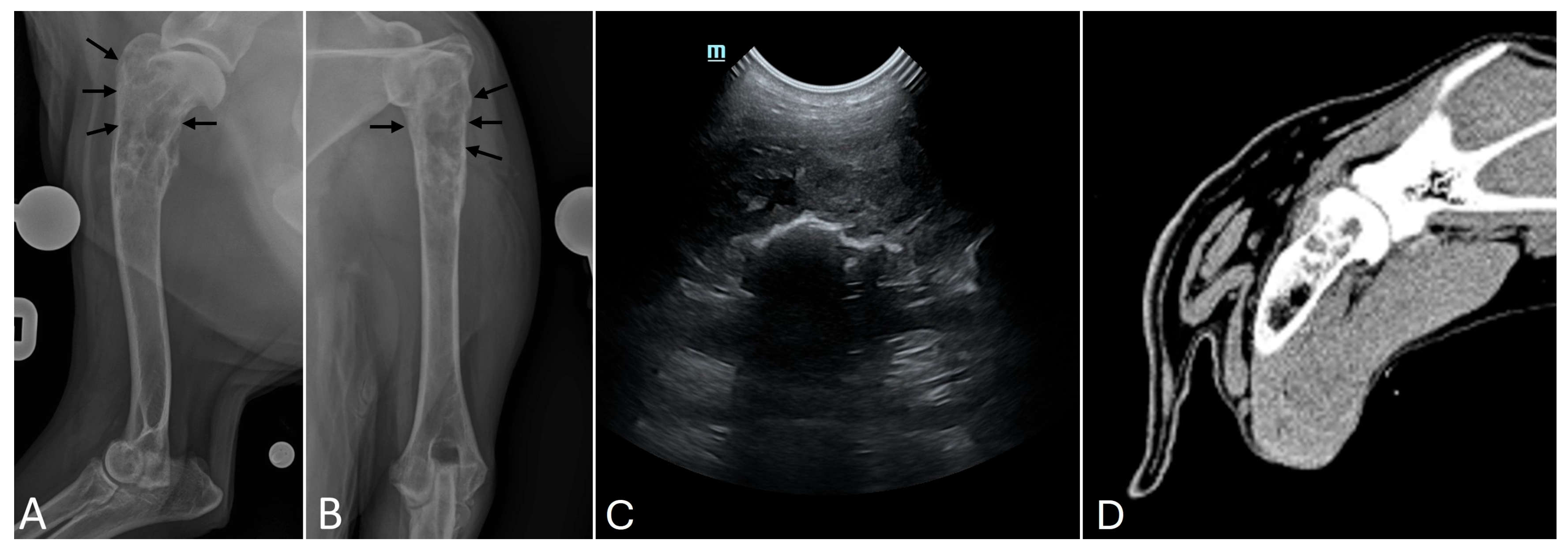
®, MSD Animal Health S.r.l., Milan, Italy; 0.3 mg/kg), radiographs of the left forelimb were obtained in mediolateral and craniocaudal projections with a digital radiography system (VetiX P8, Mindray Animal Medical Technology Co., Ltd., Shenzhen, China; acquisition parameters of 75 kV and 8 mAs). Radiographs revealed significant structural changes in the proximal metaphysis of the left humerus, characterised by a “moth-eaten” pattern of bone lysis, a pronounced and irregular periosteal reaction, and an extensive alteration of the normal medullary canal pattern in a proximal-distal direction, extending up to the middle third of the diaphysis (
Figure 1A,B).
Immediately after the radiographic study and with the patient still sedated, an ultrasound-guided (Mindray Vetus 7, Mindray Animal Medical Technology Co., Ltd., Shenzhen, China) fine needle aspiration cytology (FNAC) was performed (
Figure 1C). The cytological picture was characterized by a large population of predominantly isolated ovoid cells with distinct cytoplasmic borders, abundant basophilic cytoplasm with frequent vacuolization, oval to irregular eccentric nuclei with coarse chromatin, frequent nuclear molding, and one or more prominent nucleoli. Marked anisocytosis and anisokaryosis, multinucleated cells, numerous osteoclast-like giant multinucleated cells, a few segmented neutrophils and eosinophils, and abundant amorphous eosinophilic extracellular material consistent with osteoid matrix were also observed. Cytology results supported the clinical and radiographic presumptive diagnosis of a primary bone tumour consistent with OSA.
A total body CT study was then performed using a 16-slice helical scanner (Somatom Emotion 16; Siemens, Erlangen, Germany) to evaluate local tumour extent and screen for metastatic disease [
31]. While the patient was still sedated, general anesthesia was induced with propofol (Propovet Multidose 10 mg/mL, Zoetis S.r.l., Rome, Italy; 4 mg/kg IV) and maintained with isoflurane (IsoFlo, Zoetis Italia S.r.l., Milan, Italy). Studies were performed with acquisition parameters of 120 kVp and 150 mAs before and after administration of a non-ionic iodinated contrast medium (iohexol, 350 mg iodine/mL; Omnipaque, GE Healthcare Inc., Marlborough, MA, USA; 600 mg/kg, IV) using a power injector. Breath-hold techniques were applied to minimize motion artifacts. A pre-contrast CT acquisition of both forelimbs was performed with a slice thickness of 0.6 mm, extending from the phalanges to the dorsal border of the scapula, and reconstructed using soft tissue and bone algorithms (
Figure 1D). Additionally, a pre-contrast and post-contrast total body CT acquisition was performed with a slice thickness of 1 mm, extending from the nasal planum to the distal aspect of the hind limbs, and reconstructed using standard soft tissue and lung algorithms. The CT scans confirmed a 6.5 × 3 × 3 cm lesion affecting the proximal humeral metaphysis, with no evidence of joint involvement, as the humeral head articular surface and subchondral bone remained intact, the scapulo-humeral joint space and margins were preserved, and no intra-articular extension of the lesion was observed. The cortical bone showed irregular lytic destruction with no associated soft tissue involvement. Regional lymph nodes appeared within normal limits, with no signs of metastatic involvement [
32]. Total body CT scans revealed severe bilateral hip osteoarthrosis (OA). No pulmonary metastases were detected. Limb-sparing surgery with 3D-printed PSI was chosen as an alternative to amputation due to the dog’s overweight status and severe bilateral hip OA. Immediately after the CT study, neoadjuvant chemotherapy was initiated with a first preoperative cycle (carboplatin, 300 mg/m
2 body surface area, IV) [
33,
34]. Preoperative CBC did not show neutropenia or thrombocytopenia, suggesting no signs of myelosuppression [
35,
36,
37].
2.3. VSP and PSI Design
Pre-contrast high-resolution CT scans of the humerus and scapula were reconstructed with a slice thickness of 0.6 mm using a bone tissue reconstruction algorithm and processed with computer-aided design (CAD) software (BonaPlanner v1.1.0, ©Bonabyte, Moscow, Russia). The segmentation process, supervised by a bioengineer, allowed accurate 3D reconstruction of the affected and contralateral forelimbs, manipulation of the bone segment in the axial, sagittal, and frontal planes, and measurement of surgical margins for tumour resection [
38]. Following reconstruction, virtual surgical planning (VSP) was performed using the same CAD software to simulate the limb-sparing surgery, define the osteotomy planes, and optimize both implant placement and surgical accuracy [
39]. The contralateral scapulo-humeral joint was considered an appropriate anatomical reference because no shoulder pathology was detected clinically or on imaging, and bilateral OA was confined to the hip joints. After measuring the contralateral limb scapulohumeral angle, and based on published biomechanical data, a scapulohumeral arthrodesis angle of 125° was selected [
40].
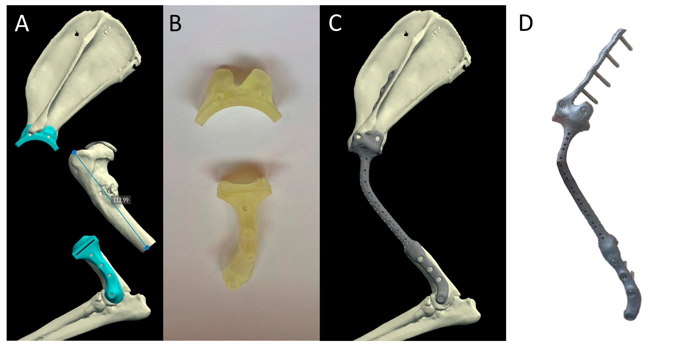
Stereolithographic (SLA) 3D-printing was used to produce PSGs using NextDent Dental SG material. The PSGs were designed according to the surgeon’s blade specifications and the diameters of the selected screws. In both PSGs, the holes were designed for temporary fixation with Steinmann pins (S-pins) and subsequent definitive fixation of the PSE with 3.5 mm screws placed in the same predrilled positions. The humeral guide was contoured to match the lateral aspect of the distal humerus and incorporated four 2.5 mm S-pin holes, with one hole slightly distal and cranial to the lateral epicondyle, used as an easily identifiable surgical landmark. The humeral guide allowed a transverse osteotomy of the distal third of the humeral diaphysis. The scapular guide was contoured to match the lateral scapular surface and incorporated two 2.5 mm S-pin holes just distal to the acromion, used as an easily identifiable surgical landmark. The guide allowed a radial osteotomy of the neck of the scapula (
Figure 2A,B).
The PSE designed to replace the ostectomized bone portion was manufactured using powder bed additive manufacturing (PB-AM) techniques with electron beam melting (EBM), using a medical-grade titanium alloy (Ti-6Al-4V ELI, Grade 23) in accordance with ASTM and US FDA standards for surgical implants [
41,
42] (
Figure 2C,D).
The PSE was designed to fill the bone defect and to incorporate a tubular porous structure (15 mm in diameter and 112 mm in length) with a cell size of 0.1–0.3 mm. The shape of the PSE was defined according to the planned craniolateral surgical approach to the humerus and scapula, avoiding the need for acromial osteotomy. Based on the planned surgical approach and fixation strategy, the bioengineer and surgeon defined the thickness, width, and overall geometry of the implant according to the patient’s size and the position, type, and diameter of the selected screws. Screw trajectories were planned to match the predrilled PSG positions. The 3.5 mm locking screws were planned as four distal humeral screws and six proximal scapular screws. The four distal holes for screws on the humerus were designed on the lateral humeral surface to match the S-pin holes used for humeral guide fixation. Of the six scapular holes, the proximal four were designed with craniocaudal and mediolateral orientations to anchor the PSE to the scapula, while the two distal scapular holes were designed on the neck of the scapula to match the S-pin holes used for scapular guide fixation. The surgeon planned the screw configuration based on pre-operative S-pin guides, with the proximal scapular screws oriented in craniocaudal and mediolateral directions, while the remaining screw holes were aligned with previously placed S-pin positions used for osteotomy guidance, including placement along the lateral surface of the humerus.
2.4. Primary Surgical Procedure, Postoperative Management and Histological Examination
Surgery was performed 21 days after the CT scan. The anesthetic protocol included premedication with acepromazine (Prequillan, Fatro SpA, Bologna, Italy; 0.02–0.04 mg/kg) and methadone (Semfortan, Eurovet Animal Health BV, Bladel, The Netherlands; 0.2 mg/kg), induction with propofol (Proposure, Boehringer Ingelheim Animal Health Italia S.p.A. Milano, Italy; 4 mg/kg IV) and maintenance with isoflurane (IsoFlo, Zoetis Italia S.r.l., Milan, Italy) connected to the anesthesia machine (WATO EX-35 Vet, Mindray Animal Medical Technology Co., Ltd., Shenzhen, China). A constant rate infusion of fentanyl (Fentadon, Eurovet Animal Health BV, Bladel, The Netherlands; 2–4 µg/kg IV) was administered for intraoperative analgesia with a syringe pump (BeneFusion SP1 Vet, Mindray Animal Medical Technology Co., Ltd., Shenzhen, China). Antimicrobial prophylaxis with cefazolin (Cefazolin TEVA, Milan, Italy; 20 mg/kg, IV) was administered 30 min before surgery and repeated at 90 min intraoperatively. Physiological monitoring was carried out with a modular multiparametric monitor (ePM 12M Vet, Mindray Animal Medical Technology Co., Ltd., Shenzhen, China), including electrocardiography, respiratory and heart rate, end-tidal carbon dioxide, blood oxygen saturation and non-invasive blood pressure.
Following aseptic preparation using a hanging-limb technique, a lateral surgical approach to the scapulo-humeral region was performed without acromial osteotomy, combining an approach to the cranio-lateral region of the shoulder joint with an approach to the cranio-lateral region of the mid-shaft of the humerus [
43,
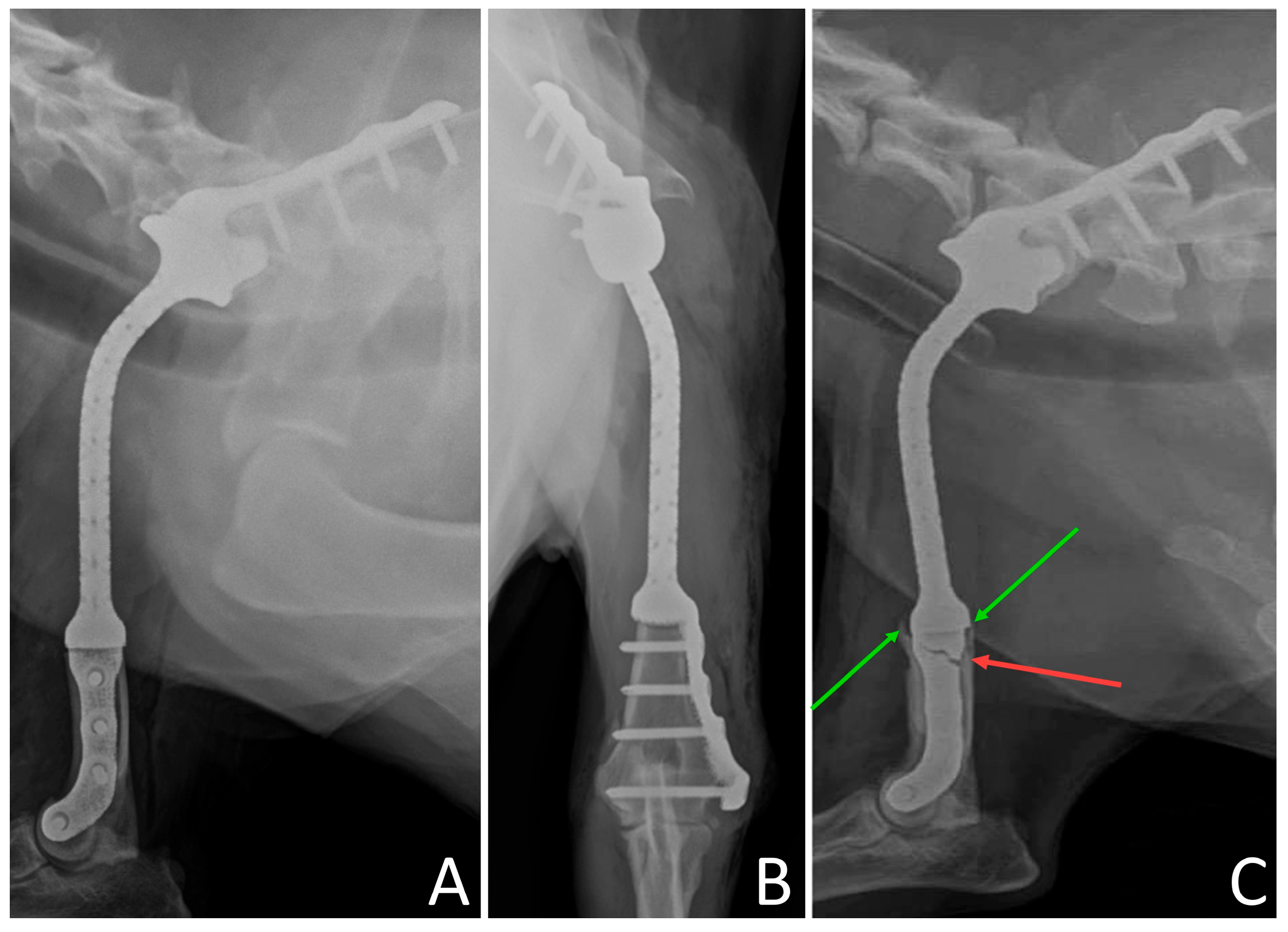
44]. The PSGs were placed on the humeral and scapular sites in a one-way fitting position and secured using 2.5 mm S-pins. The radial osteotomy of the neck of the scapula was performed using a 27 mm crescentic tibial plateau leveling osteotomy (TPLO) saw blade, 1 mm in thickness (DePuy Synthes Vet, West Chester, PA, USA) parallel to the scapular guide. The transverse osteotomy of the distal third of the humeral diaphysis was performed using a 1 mm oscillating saw blade (DePuy Synthes Vet, West Chester, PA, USA) through the lateral slot of the humeral guide. During the osteotomies, the osteotomy sites were flushed with abundant sterile saline solution. The guides were then removed, leaving two parallel S-pins in place for each segment, and the osteotomized segments were removed en bloc. The PSE was positioned using the two parallel S-pins in each segment, in accordance with the drilling guides inserted into the respective holes. After temporary fixation, holes were drilled using a 2.8 mm bit for 3.5 mm locking screws, and the PSE was secured with 3.5 mm titanium locking screws. The surgical site was flushed with 2 L of sterile saline solution, and the soft tissues were sutured to restore anatomical muscle coverage over the porous implant surface, then closed routinely in layers. Skin was closed with a single interrupted pattern. The duration of surgery from surgical approach to skin closure was 96 min. Postoperative radiographs confirmed correct implant placement and restoration of planned limb alignment (
Figure 3A,B).
A spica splint bandage was applied for 48 h to control postoperative swelling, and the patient was hospitalized for monitoring and supportive care. Postoperative therapy included antibiotic therapy with cefazolin (20 mg/kg, IV, q 12 h) and pain management with methadone (0.2 mg/kg, IM, q 12 h) during the first 24 h. Subsequently, the antibiotic treatment was switched to cefadroxil (Cefa-Cure Tabs cpr, MSD Animal Health S.r.l., Milan, Italy; 20 mg/kg, q 24 h for 10 days) and pain management was continued using tramadol (Altadol, Formevet S.r.l., Milan, Italy; 2–4 mg/kg, q 8 h for 10 days). In addition, oral anti-inflammatory therapy with meloxicam (Metacam, Boehringer Ingelheim Animal Health Italia S.p.A., Noventana, Italy; 0.2 mg/kg on the first day, then 0.1 mg/kg, q 24 h for 10 days) and gastroprotective therapy (omeprazole at 1 mg/kg and sucralfate at 50 mg/kg, both orally for 15 days) were administered. The dog was discharged 48 h after surgery with controlled weight-bearing on the operated limb and no major postoperative complications. Following en bloc surgical resection, the excised bone segment was fixed in 10% neutral-buffered formalin and sent to an external diagnostic laboratory (Biocontrol Laboratories, RUDN University, Moscow, Russia) for histopathological examination to confirm the diagnosis, assess tumor type and grade, and evaluate the surgical margins. Histopathological analysis of the resected specimen confirmed OSA with complete surgical resection margins. Postoperative adjuvant chemotherapy was initiated 16 days after surgery and administered at 21-day intervals to complete the planned six-cycle protocol, which included the preoperative cycle, following the same protocol previously administered [
34,
45].
Strict activity restrictions were prescribed, including patient confinement to prevent any high-impact activities, and controlled short-leash walking sessions (five daily sessions of approximately 10 min each) with gradual increase over a three- to four-month rehabilitation period. Adjunct physiotherapy included passive ROM exercises, massage and cryotherapy following splint removal. Follow-up examinations were performed 7 and 14 days postoperatively, and then at 1, 2, 3, and 6 months after surgery. Initial functional recovery was favourable, with progressive return to weight-bearing within 4 weeks. Wound healing was uneventful, and no signs of surgical site infection, implant-related pain, or soft-tissue swelling were observed. Functional assessment from 1 to 6 months postoperatively showed grade 1/4 lameness with no swelling, no pain on both manipulation and palpation, and consistent weight-bearing. At the 8-month follow-up, the dog showed grade 1/4 mechanical lameness but was still fully weight-bearing, with no pain. The owner reported improved patient behavior and return to a normal lifestyle, except for the onset of fatigue after long walks.
2.5. Implant Failure, Revision Surgery, and Long-Term Outcome
At the 9-month follow-up, the patient was presented with acute grade 4/4 non-weight-bearing lameness. Radiographic evaluation revealed a fracture of the PSE at the first proximal screw hole of the distal humeral segment, in the proximo-distal direction, and mild bone resorption of the proximal humerus (
Figure 3C).
The dog was discharged and temporarily managed with a spica splint applied to the left thoracic limb. Grade III–IV lameness persisted during this interval. Anti-inflammatory and analgesic treatment was administered with meloxicam (Metacam, Boehringer Ingelheim Animal Health Italia S.p.A., Noventana, Italy; 0.1 mg/kg, q 24 h orally) and tramadol (Altadol, Formevet S.r.l., Milan, Italy; 4 mg/kg, q 8 h orally). Strict cage rest at home was prescribed to minimize limb loading and uncontrolled activity. Any necessary movement was performed with owner assistance. The bandage was changed every 48 h, and the skin was closely monitored for bandage-related lesions.
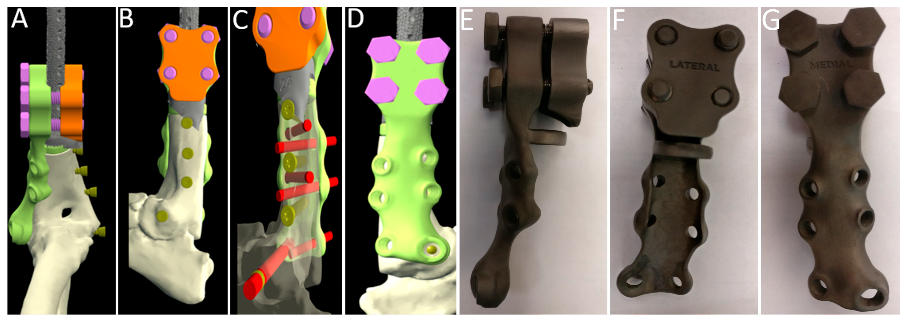
No additional CT examination was performed at the time of implant failure. Revision VSP was based on the original CT dataset used for the primary VSP. The same VSP software (BonaPlanner) was used to plan revision surgery and to design a new two-component modular bridging titanium implant capable of integrating with the retained proximal tubular segment of the original PSE. Following removal of the fractured distal portion, the retained segment served as the proximal anchoring interface for the revision construct. The new implant consisted of medial and lateral components clamped around this interface using bolt-based coupling and multiplanar locking screw fixation. The medial component extended distally as a plate with a redesigned hole configuration arranged in two rows to improve screw distribution and enable both unilateral and biplanar fixation. Three screws were oriented mediolaterally, and three screws were oriented craniocaudally and mediolaterally. The construct included a dedicated distal and cranial hole accommodating a distal 4.5 mm locking screw. The proximal portion of the lateral component was designed to articulate with the medial component and featured a concave groove on its medial surface, matched by a corresponding groove on the lateral surface of the medial component, to seat the retained implant segment between the two components. Each component included four aligned holes, allowing complementary fixation with bolts inserted from medial to lateral. Additionally, the medial component incorporated a proximal ring structure with a central scaffold designed to fill the lytic defect in the proximal humeral segment (
Figure 4A–G).
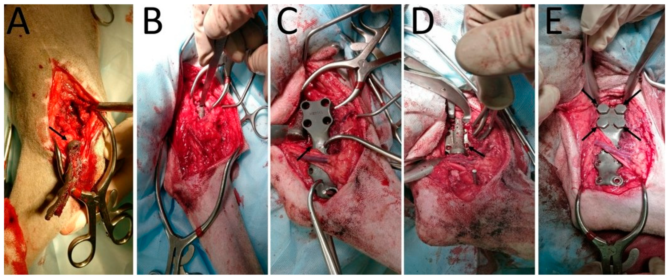
Surgery was performed 14 days after the radiographic evaluation. The patient was premedicated, anesthetized, and surgically prepared for revision surgery following the same protocol previously described for the primary procedure, including intraoperative analgesia and antimicrobial prophylaxis. A lateral approach to the humerus was performed, and the fractured distal portion of the implant was removed (
Figure 5A,B) [
46]. The surgical site was copiously lavaged with 1 L of sterile saline solution and routinely closed. A medial approach to the distal humerus was then performed. The lateral component of the new implant was positioned first, followed by fixation of the medial component to the bone using five 3.5 mm titanium screws, with the most distal and cranial screw consisting of a 4.5 mm locking screw. The implant was then secured proximally to the first prosthetic component by assembling the medial and lateral parts and fixing them together with four bolts inserted in a complementary configuration (
Figure 5C–E).
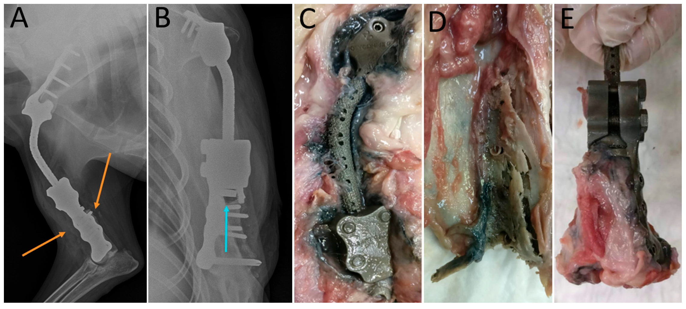
After further lavage with 1 L of sterile saline solution, the surgical wound was closed routinely in layers. Postoperative radiographs confirmed correct implant placement and restoration of planned limb alignment (
Figure 6A,B). A spica splint bandage was applied to the left thoracic limb and maintained for 24 h to control postoperative swelling. The patient was hospitalized for postoperative monitoring and supportive care and was discharged 24 h after surgery with a lameness score of 2/4. At discharge, the patient was prescribed the same postoperative medical protocol adopted following the first surgery. Follow-up examinations performed at 7 and 14 days showed favourable initial functional recovery with progressive return to weight-bearing within 4 weeks. Wound healing was uneventful and no signs of surgical site infection, implant-related pain, or soft-tissue swelling were observed. Functional assessments at 1 and 2 months after revision surgery showed grade 2/4 mechanical lameness with no swelling, no pain on both manipulation and palpation, and consistent weight-bearing. Functional assessments at 3, 4, 5, and 6 months demonstrated restoration of functional limb use with grade 1/4 mechanical lameness, but the patient was still fully weight-bearing, with no pain. At the 15-month follow-up, the patient was euthanized due to suspected pulmonary metastases. Post-mortem examination confirmed multiple pulmonary metastases. The implant site showed stable proximal implant fixation with advanced osseointegration, absence of local tumour recurrence, and evidence of distal stress-shielding (
Figure 6C–E).
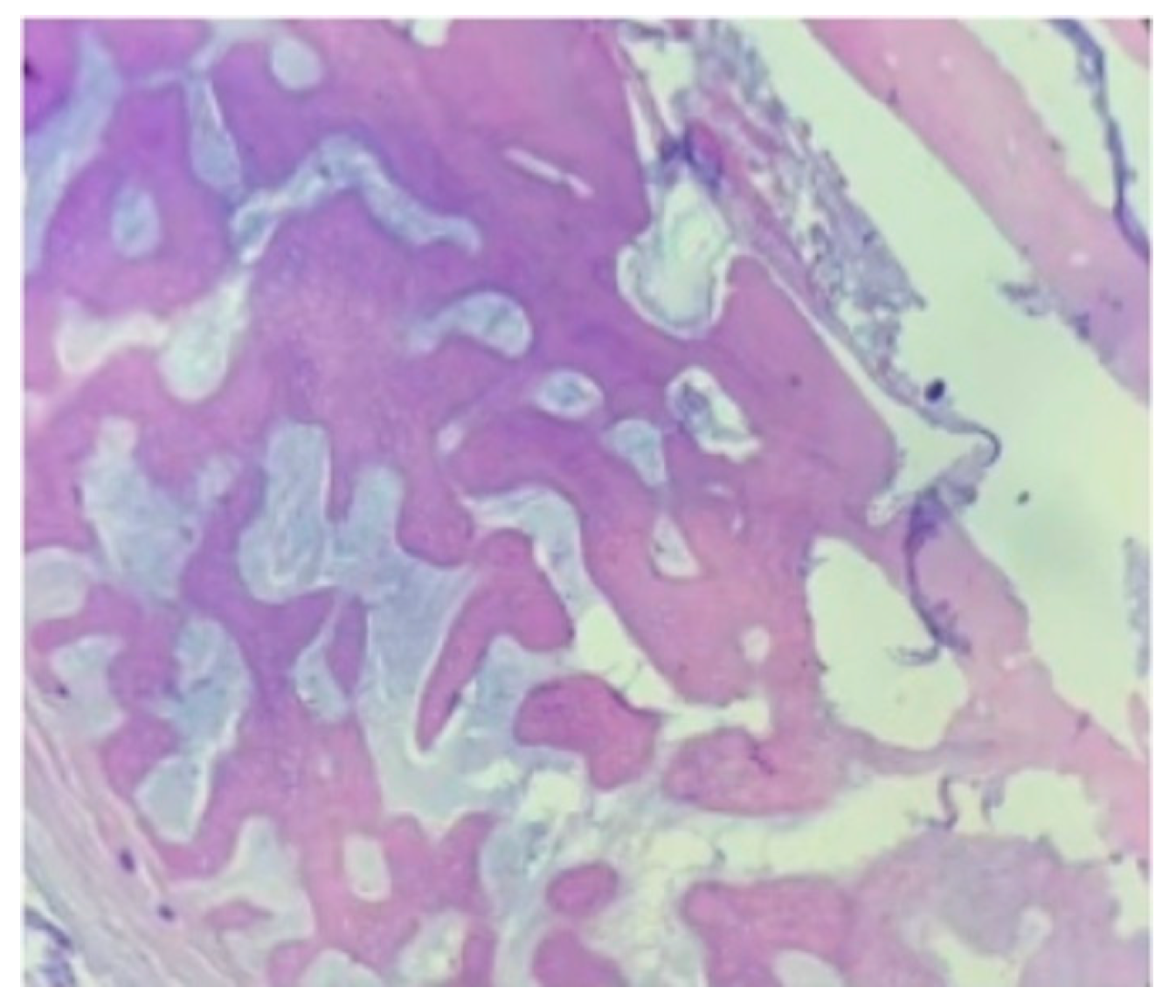
At post-mortem examination, peri-implant tissue samples were collected from the bone–implant interface at the proximal scapular and distal humeral portion of the construct for histological analyses (Biocontrol Laboratories, RUDN University, Moscow, Russia). Histological evaluation of the bone–implant interface revealed extensive mature remodelled bone surrounding the implant bed, with approximately 90% direct bone-to-implant contact visually and semi-quantitatively estimated in the examined sections. Areas of fibrous tissue integration were observed, with no histological evidence of infection or neoplastic infiltration. A mild chronic inflammatory response was present (
Figure 7).
3. Discussion
This report describes the modular revision of a failed 3D-printed titanium scapulo-humeral PSE for limb-sparing treatment of proximal humeral OSA in a dog with concurrent severe bilateral hip OA. Its main clinical relevance lies in documenting mid-term biomechanical PSE behavior, and modular revision after PSE failure with preservation of limb function. The case described contributes to the still limited body of veterinary literature on long-term behaviour of 3D-printed PSEs in dogs with OSA.
Amputation arguably remains the standard of care for OSA in dogs and is generally considered the most effective and reproducible option for local disease control. However, some patients may not be suitable candidates due to marked body size–related biomechanical demands, obesity or other concomitant orthopaedic comorbidities affecting the remaining limbs. Adaptive mechanisms after limb amputation may increase OA incidence in the remaining limbs and exacerbate pre-existing orthopedic conditions such as elbow and hip OA, particularly in large and giant breed dogs [
6,
7,
15,
34,
47,
48]. Owner-related factors may also influence decision-making, since decreased dog’s stamina during exercise, and behavioral changes have been reported after limb amputation [
8,
49]. In this case, severe bilateral hip OA, overweight body condition, and owner concerns regarding postoperative mobility and quality of life (QOL) made limb-sparing surgery a clinically reasonable alternative to amputation.
Limb-sparing surgery involves the removal of the primary bone tumour and the application of internal or external fixation to the remaining bones, with or without segmental bone replacement [
38]. Due to the very limited number of sizes available, prosthetic reconstruction techniques with commercially available metallic endoprostheses may be associated with limited functional outcomes and a high complication rate primarily due to biomechanical failure, infection, and local tumour recurrence [
10,
15,
41]. Prolonged intraoperative contouring required by off-the-shelf systems to fit the remaining bone structure increases intra/extra-corporeal manipulations, extending surgical time and increasing the risk of infection, implant/bone interface biomechanical failure due to screw loosening/fracture, and fatigue-related complications [
12,
13]. In addition, the proximity of the brachial plexus and major vascular structures such as the axillary vessels, and the need to preserve functional load transmission make reconstructive procedures in this region more challenging than in other anatomical sites [
50]. The use of CT-based VSP enabled accurate preoperative 3D visualization of the lesion, definition of resection margins, calculation of the arthrodesis angle, planning of the osteotomies, and design of screw number and trajectories [
16,
41]. The 125° scapulo-humeral angle was selected based on contralateral limb evaluation and available data on canine shoulder arthrodesis, to preserve limb length and reproduce a physiological standing alignment and functional gait [
40]. This is particularly relevant in proximal humeral reconstruction, where even small deviations in alignment may result in abnormal load distribution, reduced function, and increased implant stress. Additive manufacturing techniques allowed PSGs and PSEs to be designed from CT-based reconstruction to fit to the bone surface using reliable anatomic landmarks and enabled reproducible execution of the planned osteotomies. Alignment of the PSG holes with the future PSE screw positions reduced the risk of cumulative error between osteotomy, implant placement, and definitive fixation, avoiding loss of anatomical alignment, which is one of the most common and catastrophic complications during traditional limb-sparing surgery [
15]. The use of a PSE allowed avoidance of implant manipulation since no intraoperative contouring was needed, thus potentially reducing surgical time and the risk of infection, and improving implant fit and load distribution at the bone–implant interface.
Favorable postoperative outcomes with progressively improved weight-bearing within the first postoperative weeks were observed. Serial follow-up showed only mild residual lameness without swelling or pain on manipulation through the first eight months. The early initiation of adjuvant carboplatin chemotherapy without major delay suggests that the complexity of reconstruction did not preclude appropriate systemic oncologic management, which remains a fundamental component of multimodal treatment for canine OSA [
51,
52]. Adjuvant chemotherapy, when following surgery, is considered the standard of care, improving median survival time (MST) by between 8 and 14 months. Many studies have evaluated the use of carboplatin, doxorubicin and cisplatin, either alone or in combination, typically administered in four to six doses. However, carboplatin as a sole agent is still considered the adjuvant chemotherapy of choice. A first cycle of neoadjuvant chemotherapy, taking place before the surgery, was introduced to the treatment protocol to minimize tumour progression and the onset of metastatic disease. Tumour growth during the design and the manufacturing phases of the PSIs could have compromised the use of PSIs. Adjuvant chemotherapy was continued postoperatively every 3 weeks for a further 5 treatments, starting 2 weeks after surgery, as previously reported [
15,
45]. The implant fracture observed radiographically at the level of the first proximal hole of the distal humeral segment nine months after surgery provides useful insight into the mid-term biomechanical behavior of a PSE placed in a possible site of high stress concentration. Any screw hole interrupts material continuity and may act as a stress riser, particularly under repeated cyclic bending [
53]. In the present case, the fracture occurred at the transition zone between the intercalary tubular segment and the distal fixation portion, where changes in implant geometry and the presence of a screw hole may have concentrated mechanical stresses. Repeated cyclic loading during weight-bearing, together with the long lever arm created by the scapulo-humeral arthrodesis construct, the dog’s body weight, and the complex biomechanics of the shoulder region, may have contributed to fatigue failure at this site. In addition, altered load transfer between the implant and the residual humeral bone may have promoted stress shielding-related remodeling, potentially reducing local bone support and further increasing mechanical demand on the implant. This finding is consistent with general biomechanical principles and previous studies on PSIs, which have similarly highlighted the importance of screw distribution, implant thickness, and fatigue resistance for long-term success in load-bearing constructs [
39]. The mild bone resorption observed at the proximal humeral stump at the time of implant failure may be attributed to a multifactorial process involving biologic bone adaptation, altered load transfer, load sharing, or micromotion at the bone–implant interface. Although proximal bone resorption adjacent to the implant suggested possible stress shielding, no dedicated biomechanical or densitometric analysis was performed. Distal stress shielding was suspected based on localized cortical thinning and bone remodeling adjacent to the distal fixation region. This finding should therefore be interpreted as a radiographic suspicion of adaptive bone remodeling rather than a confirmed diagnosis. Importantly, the location and pattern of failure directly influenced the revision strategy, particularly the need to bypass the previous screw-hole transition zone and improve load distribution across the distal humeral fixation region.
The revision concept adopted in this study reflects the modular philosophy widely adopted in human orthopedic oncology to preserve residual bone and delay more radical salvage procedures [
54,
55]. Complete implant replacement was not pursued because the proximal portion of the original PSE appeared stable and well integrated, and its removal would have required a more invasive procedure with greater disruption of the bone–implant interface and surrounding soft tissues. Instead of converting to amputation after primary implant failure, a second modular titanium PSE was designed to preserve and integrate this stable proximal component of the previous implant and bridge the failed distal region. The second PSE incorporated a two-component medial–lateral design, biplanar screw fixation, bolt-based coupling around the retained tubular segment, and a scaffold-containing ring intended to address the lytic defect. This modular medial–lateral configuration was intended to increase construct stability by enclosing the retained tubular segment and transferring load through both sides of the residual humerus rather than through a single lateral fixation plane. The revised screw configuration was designed to distribute mechanical loads over a broader distal humeral segment, increase fixation purchase, reduce stress concentration around individual screw holes, and reduce bending moments, particularly at the transition zone corresponding to the previous failure site, which had been identified as the most vulnerable region of the original construct. The revision surgery described transformed a potentially catastrophic structural complication into a surgically manageable condition and allowed maintenance of four-limb ambulation for an additional six months. Although based on visual semi-quantitative assessment of the examined histological sections rather than on a validated histomorphometric measurement, the reported post-mortem histologic evidence of approximately 90% direct bone-to-implant contact suggests marked osseointegration and supports the high osseointegrative potential of porous titanium structures and further strengthens the revision concept adopted in this study [
42]. The selected porous cell size of approximately 100–300 µm was consistent with pore dimensions reported to support bone and capillary ingrowth in porous Ti6Al4V scaffolds, as pore diameters greater than 100 µm are generally considered favorable for osseointegration. In addition, pore sizes partially overlapping the 200–400 µm range have been associated with osteoblast adhesion, migration, and proliferation, supporting the biological rationale for the porous architecture adopted in this case [
56,
57,
58]. Although conclusions cannot be generalized from a single case, these findings support the use of PSIs with porous architectures in oncologic reconstruction. The absence of local recurrence suggests that the original resection achieved satisfactory oncologic control. The overall survival time of fifteen months falls within the range reported for canine appendicular OSA treated with adjuvant chemotherapy [
33,
51,
52]. The development of pulmonary metastases despite local control is consistent with the biological behavior of canine OSA and highlights the distinction between successful local treatment and systemic disease progression.
Several limitations should be acknowledged. The single-case nature of this report cannot provide robust evidence regarding reproducibility, complication rates, or comparative efficacy. Functional outcome was assessed primarily through clinical examination, owner feedback, and lameness scoring. Objective gait analysis, including force-plate analysis or kinematic evaluation, was not performed, which limits quantitative assessment of limb function. Validated QOL instruments were also not used. Production cost, manufacturing time, and the need for collaboration between surgeons and biomedical engineers may limit the broader applicability of this approach. Dedicated design, manufacturing, and shipping phases for PSI production may result in delays between imaging and surgery and may increase overall treatment costs, potentially limiting accessibility in routine clinical practice. The waiting time usually depends on the company and corresponds to the design, production and shipping phases of the final product. In our study, the expected waiting time for the PSIs to arrive and for the surgery to be performed was 21 days from VSP for the first implant and 14 days for the revision implant, from the radiographic evaluation of the fractured PSE. Because no formal mechanical or fatigue analysis was performed during the initial implant design, implant design in proximal humeral reconstructions should incorporate advanced finite element modelling and fatigue analysis, particularly at screw-hole transition zones and at interfaces between solid and porous segments. Despite these limitations, this report offers key insights for future research. Revision systems should be considered during the primary design, especially for anatomically demanding constructs subjected to high cyclic loads. Multicentric prospective studies are needed to compare limb-sparing reconstructions using PSIs with more traditional techniques in terms of complication rates and functional outcomes.




 ,
,