Article

Marked Antigenic Divergence and Evolutionary Analysis of H5 AIVs from Wild Birds in East China, 2013–2022

1
College of Veterinary Medicine, Yangzhou University, Yangzhou 225000, China
2
Jiangsu Co-Innovation Center for the Prevention and Control of Important Animal Infectious Disease and Zoonoses, Yangzhou 225009, China
3
Jiangsu Research Centre of Engineering and Technology for Prevention and Control of Poultry Disease, Yangzhou 225009, China
*
Authors to whom correspondence should be addressed.
Animals 2026, 16(13), 2109; https://doi.org/10.3390/ani16132109 (registering DOI)
Submission received: 3 June 2026 / Revised: 3 July 2026 / Accepted: 4 July 2026 / Published: 7 July 2026
(This article belongs to the Section Veterinary Clinical Studies)

Simple Summary

Wild birds serve as reservoirs and vectors, playing a critical role in the evolution and spread of H5 avian influenza viruses. Eastern China lies along the East Asian–Australasian Flyway, providing important stopover and wintering sites, while intensive poultry farming and live poultry markets increase contact between wild and domestic poultry. From 2013 to 2022, 16 H5 viruses isolated from wild birds in eastern China were purified, sequenced, and analyzed. These viruses exhibited considerable genetic diversity and carried mammalian adaptation mutations. Most isolates matched the vaccine strains used in China at the time, but potential antigenic mismatches remained. Continuous surveillance in wild birds can therefore provide early warning for domestic poultry, enabling timely detection of variants and vaccine updates.

Abstract

The highly pathogenic H5 subtype avian influenza viruses (AIVs) pose persistent threats to the poultry industry and public health owing to their high lethality and pandemic potential. Migratory wild birds play a pivotal role in the global dissemination and genetic reassortment of the virus, serving as both natural reservoirs and long-distance vectors that drive its spatiotemporal spread. However, the extent and evolutionary drivers of antigenic divergence among H5 AIVs circulating in wild birds in East China remain poorly understood. Here, we aim to characterize the evolutionary dynamics and antigenic divergence of H5 AIVs isolated from wild birds in East China between 2013 and 2022. Whole-genome sequencing and phylogenetic analysis revealed that the isolates belonged to multiple clades, including 2.3.2.1 and 2.3.4.4, and encompassed the H5N1, H5N6, and H5N8 subtypes. Key amino acid site analysis showed that the glycosylation site patterns in the HA and NA proteins varied among clades, with some strains exhibiting gains or losses of glycosylation sites, while certain strains had acquired mutations associated with mammalian adaptation. Cross-hemagglutination inhibition (HI) assays combined with antigenic cartography demonstrated that the majority of the isolates were antigenically well-matched with the contemporaneous vaccine strains used in China, indicating that these vaccines effectively covered the predominant circulating antigenic variants at the time. Nevertheless, potential antigenic mismatches were still observed between some circulating strains and these vaccine strains. These findings suggest that wild birds in East China may contribute to the regional movement and diversification of H5 AIVs, highlighting the value of sustained surveillance for early warning and vaccine strain evaluation.

1. Introduction

Avian influenza virus (AIV), a member of the family Orthomyxoviridae, is a single-stranded, negative-sense RNA virus with a genome composed of eight segments. Based on antigenic differences in the surface glycoproteins hemagglutinin (HA) and neuraminidase (NA), AIVs are currently classified into 18 HA subtypes (H1-H18) and 11 NA subtypes (N1-N11) [1]. Among these, H5 subtype avian influenza viruses, particularly highly pathogenic avian influenza viruses (HPAIVs), pose a serious threat to the poultry industry and can cross the species barrier to infect mammals, including humans [2], thereby presenting a persistent challenge to public health security [3]. Since the first isolation of the A/goose/Guangdong/1/96 (Gs/GD/96) H5N1 virus from geese in Guangdong, China, in 1996, viruses of this lineage have continuously evolved into multiple clades and, through reassortment, have generated various subtypes such as H5Nx, which have gradually spread to many regions worldwide [4,5].
Wild birds are recognized as the natural reservoir of avian influenza viruses. In particular, species belonging to the orders Anseriformes and Charadriiformes play critical roles in the long-term maintenance, transmission, and evolution of these viruses [6]. The migratory behavior of wild birds facilitates the long-distance dissemination of avian influenza viruses across geographical regions, thereby promoting transboundary transmission and genetic reassortment [7]. East China, located at a key node of the East Asian–Australasian Flyway, contains abundant wetland resources and diverse wild bird habitats, making it a crucial region where wild bird migration intersects with poultry production. These ecological conditions provide an ideal setting for the spread, reassortment, and mutation of H5 subtype avian influenza viruses [8]. In recent years, frequent outbreaks of H5 subtype avian influenza viruses in wild birds have suggested that these viruses have established sustained transmission cycles within wild bird populations [9].
Although H5 subtype avian influenza viruses of poultry origin have been extensively studied, long-term systematic surveillance data on the genetic evolution and antigenic variation of H5 viruses in wild birds in East China remain limited. In this study, H5 AIVs isolated from wild birds in East China during 2013–2022 were characterized through whole-genome sequencing, phylogenetic analysis, and antigenic profiling. We hypothesized that these viruses would exhibit genetic heterogeneity, evidence of reassortment, and variable degrees of antigenic relatedness to vaccine strains deployed in China during different periods. This study aimed to elucidate the transmission dynamics and molecular evolutionary trends of H5 AIVs in wild bird populations, thereby providing a scientific basis for early warning of avian influenza outbreaks, vaccine strain selection, and the formulation of region-specific prevention and control strategies.

2. Materials and Methods

2.1. Sample Collection and Virus Isolation

From 2013 to 2022, a total of 27,000 samples were collected from key freshwater lakes and natural wetlands along the East Asian–Australasian Flyway in East China (a detailed breakdown by sampling location and year is provided in Supplementary Table S1). The sampled species spanned multiple orders, predominantly Anseriformes (Tadorna ferruginea, Anas platyrhynchos, Mergus merganser), Charadriiformes (Larus ridibundus, Larus argentatus, Himantopus himantopus), and other waterbirds (Egretta garzetta, Ardea cinerea, Grus grus, Grus japonensis, etc.). After collection, swab samples were placed in viral transport medium, stored in a portable cooler at 4 °C, transported to the laboratory within 24 h, and immediately stored at −80 °C until use. Before virus isolation, the swab samples in viral transport medium were thoroughly mixed, and solid debris was removed by centrifugation at 10,000× g for 10 min. The resulting supernatant was inoculated into 10-day-old specific-pathogen-free (SPF) embryonated chicken eggs at a dose of 0.2 mL per egg, with three eggs used per strain. The inoculated eggs were then incubated at 37 °C for 96 h.

2.2. Plaque Purification of Virus

To ensure genetic homogeneity, three rounds of plaque purification were performed for all 16 isolates in MDCK cells. MDCK cells (ATCC, Manassas, VA, USA; CCL-34) were cultured in Dulbecco’s modified Eagle’s medium (DMEM; Procell, Wuhan, China) supplemented with 10% fetal bovine serum (Gibco, Waltham, MA, USA) at 37 °C under 5% CO2 and seeded into 6-well plates (Corning Incorporated, Corning, NY, USA). Once the cells had formed confluent monolayers, the virus was serially diluted 10-fold in serum-free DMEM and inoculated onto the monolayers. After 1 h of adsorption, the cells were overlaid with a 1:1 mixture of 1.5% agar and 2× high-glucose DMEM (Genom, Hangzhou, China) containing TPCK-treated trypsin (Sigma, St. Louis, MO, USA) at a final concentration of 1 μg/mL. The plates were inverted and incubated at 37 °C with 5% CO2 for 72 h, and plaque formation was monitored daily. Subsequently, 0.33% neutral red staining solution (Sigma, St. Louis, MO, USA) was diluted 1:20 in PBS and applied to the agar overlay. After an additional 12 h of inverted incubation, the plaques were picked from each purification round. The picked plaques were subjected to three freeze–thaw cycles, propagated in 10-day-old specific-pathogen-free (SPF) embryonated chicken eggs, and the resulting viral stocks were stored at −80 °C.

2.3. Whole-Genome Sequencing

Viral RNA was extracted from allantoic fluid using TRIzol reagent (Invitrogen, Carlsbad, CA, USA) according to the manufacturer’s instructions and reverse-transcribed into cDNA, which was subsequently used as the template for PCR amplification. All gene segments were amplified using Hoffman primers [10] and sequenced by GenScript Biotech Corporation. The complete genome sequences of all isolates generated in this study were deposited in GenBank.

2.4. Phylogenetic Analysis

To accurately genotype the viruses and clarify evolutionary relationships, reference sequences were objectively selected based on WHO/WOAH-recommended representative strains and NCBI BLASTN 2.17.0+ homology searches, in addition to the 16 isolates from this study. Sequences were assembled using SnapGene 6.0.2, and phylogenetic analysis was performed with PhyloSuite v1.2.2. The best-fit substitution model was selected using ModelFinder. Maximum-likelihood (ML) phylogenetic trees were then constructed using IQ-TREE, employing the ultrafast bootstrap approximation with 10,000 replicates, a maximum of 1000 iterations, and a minimum correlation coefficient of 0.90. The resulting trees were visualized using the online tool iTOL (https://itol.embl.de/, accessed on 11 April 2026).

2.5. Molecular Characterization

Molecular characterization was performed using MegAlign pro 17.1 and MEGA X to analyze key protein features and mutation sites. Potential glycosylation sites in the HA and NA proteins were predicted using the online bioinformatics tool NetNGlyc 1.0 Server (https://services.healthtech.dtu.dk/services/NetNGlyc-1.0/, accessed on 7 April 2026).

2.6. Selection Pressure Analysis

To evaluate selection pressure on each gene segment, sequences were first aligned using MAFFT, after which stop codons were removed and the aligned sequences were exported in FASTA format. The best-fit substitution model was selected using the Datamonkey web server (http://www.datamonkey.org/, accessed on 3 May 2026). Positive selection sites in the eight gene segments of the 16 isolates were analyzed using four algorithms: fixed effects likelihood (FEL), mixed effects model of evolution (MEME), fast unconstrained Bayesian approximation (FUBAR), and single-likelihood ancestor counting (SLAC). The selection coefficient ω represents the ratio of nonsynonymous to synonymous substitution rates (dN/dS), where ω > 1 indicates positive selection, ω = 1 indicates neutral evolution, and ω < 1 indicates negative (purifying) selection.

2.7. Antigenic Analysis

Antigenic differences among the isolates were analyzed using cross-hemagglutination inhibition (HI) assays in accordance with World Health Organization standard operating procedures. Phylogenetic analysis of the HA gene grouped the 16 isolates into six well-supported clades. To represent the full genetic diversity of the collection without testing highly similar strains redundantly, genetically distant strains were selected from each clade (JYWB4, QP10, GY183, GY999, SSW7, CM120, SH17, DT10, and CIXI20). The nine strains were subsequently used to prepare inactivated oil-emulsion vaccines. For each strain, a group of five 6-week-old SPF chickens (n = 5) was immunized with 0.4 mL of the corresponding vaccine per bird. The vaccines were prepared by emulsifying inactivated whole virus with white oil and Tween-80 adjuvant using a homogenizer. Once satisfactory serum antibody titers had been achieved, serum samples from each group were collected, pooled, and subjected to cross-HI assays. HI assays were performed according to the standard protocol [11]. Viruses were diluted to 4 hemagglutination units (HAU) per 25 μL. In a 96-well V-bottom microtiter plate, 25 μL PBS was added to each well, followed by 25 μL of test serum in the first well. Serial twofold dilutions of serum were made, and then 25 μL of 4-HAU antigen was added to all wells. After incubation at 37 °C for 15 min, 25 μL of 1% chicken red blood cell suspension was added, mixed, and the plate incubated for an additional 15 min before recording the hemagglutination inhibition endpoints. The highest serum dilution that yielded a button-like pellet of erythrocytes was defined as the HI titer. All HI assays were performed in three independent experiments. The resulting HI titers were further analyzed via antigenic cartography (https://www.antigenic-cartography.org/, accessed on 8 April 2026).

3. Results

3.1. Prevalence and Sequencing of H5 Subtype AIV

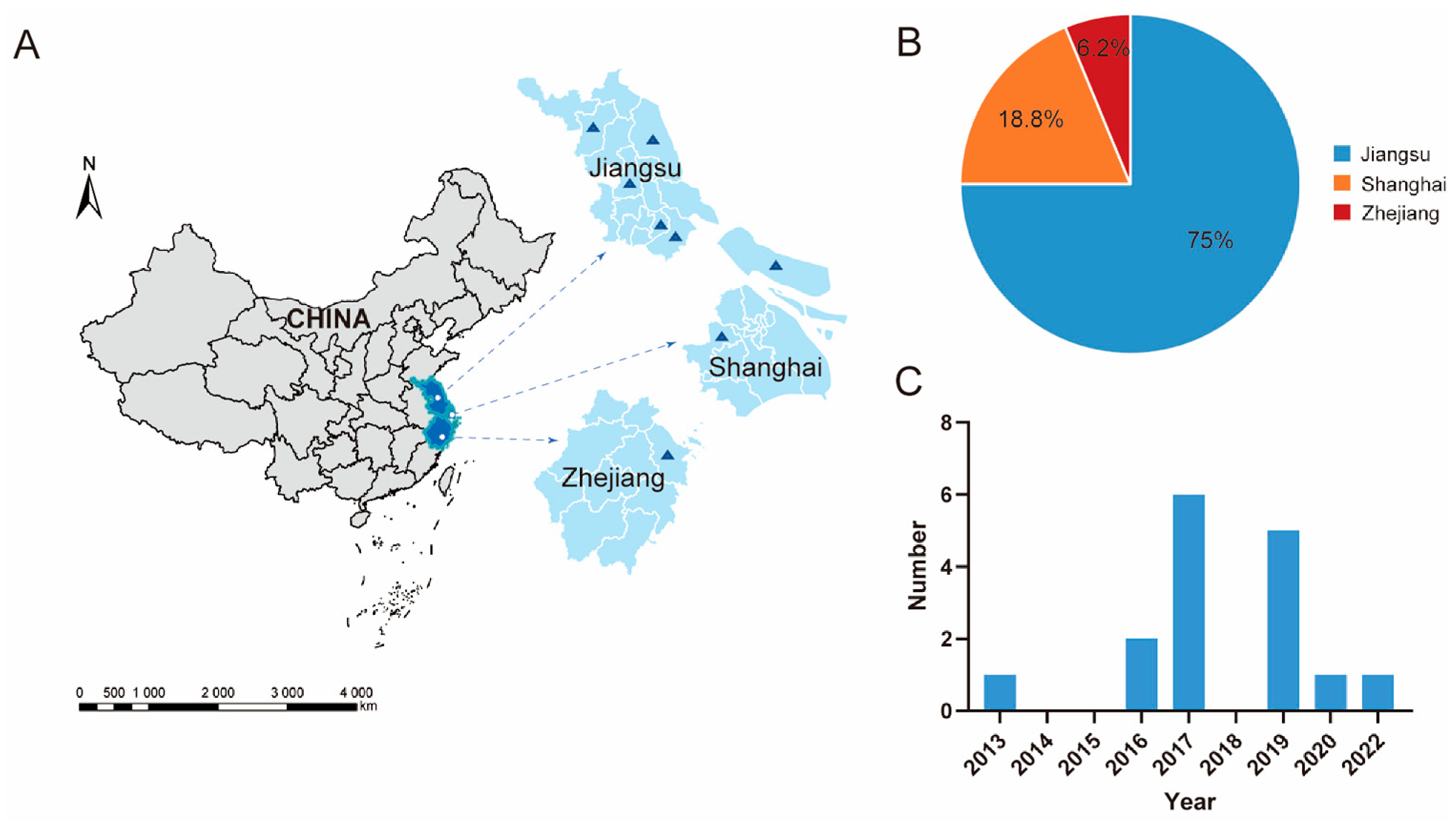
From 2013 to 2022, major freshwater lakes and natural wetlands in East China were selected as sampling sites (Figure 1A), including Lake Taihu, Lake Gaoyou, and Lake Hongze in Jiangsu Province, Hangzhou Bay in Zhejiang Province, and Chongming Island in Shanghai. A total of 27,000 fecal and environmental samples were collected from wild bird habitats. Virus isolation and identification yielded 16 H5 subtype AIVs in 2013, 2016, 2017, 2019, 2020, and 2022 (Table 1; Supplementary Table S2), with an overall isolation rate of 0.6‰. Among these isolates, 12 were obtained from Jiangsu Province, accounting for 75.0% of all positive isolates, whereas three were from Shanghai (18.8%) and one was from Zhejiang Province (6.2%) (Figure 1B,C). After three rounds of plaque purification, the complete genome sequences of all isolates were obtained.

3.2. Phylogenetic Relationships

3.2.1. HA Gene

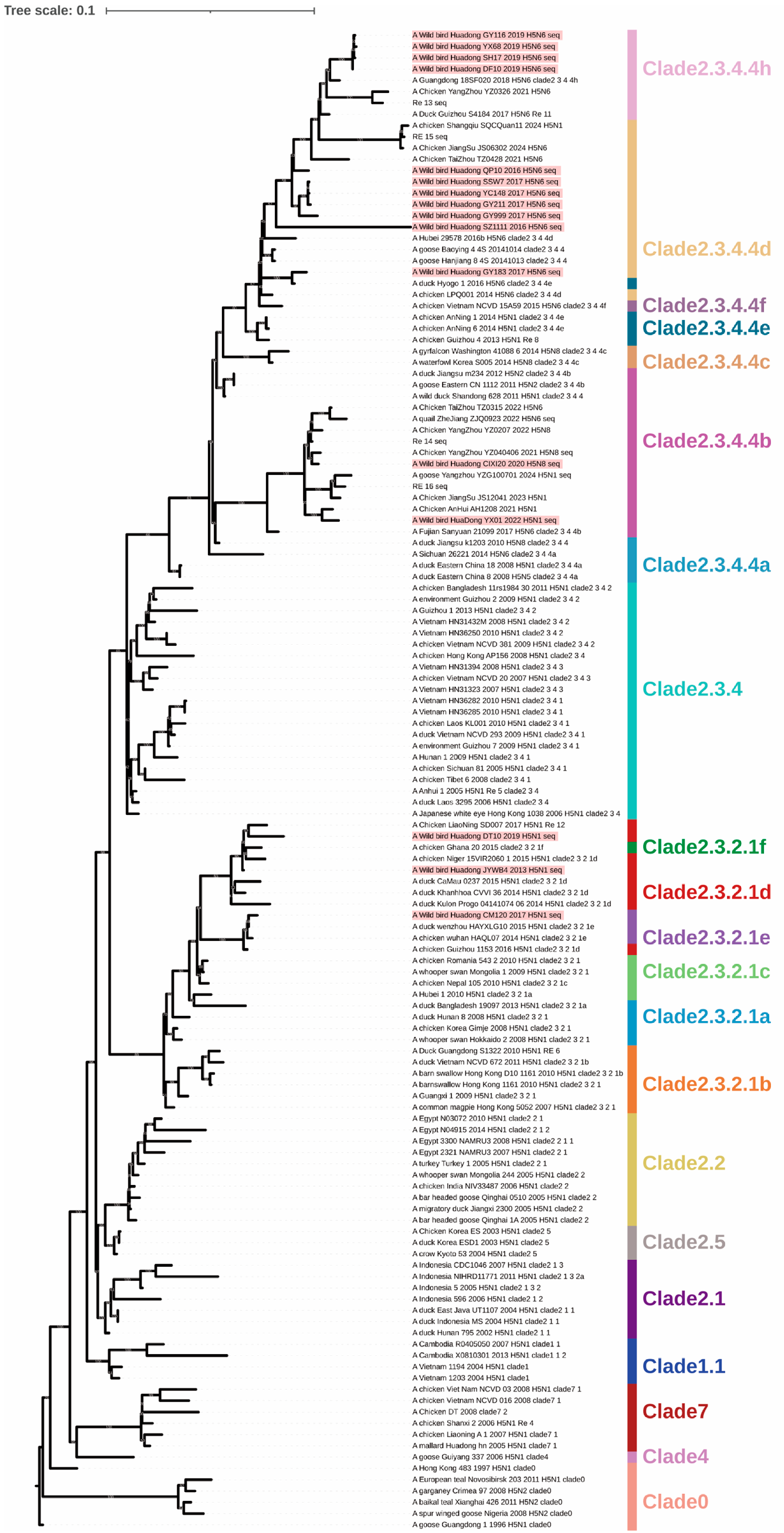
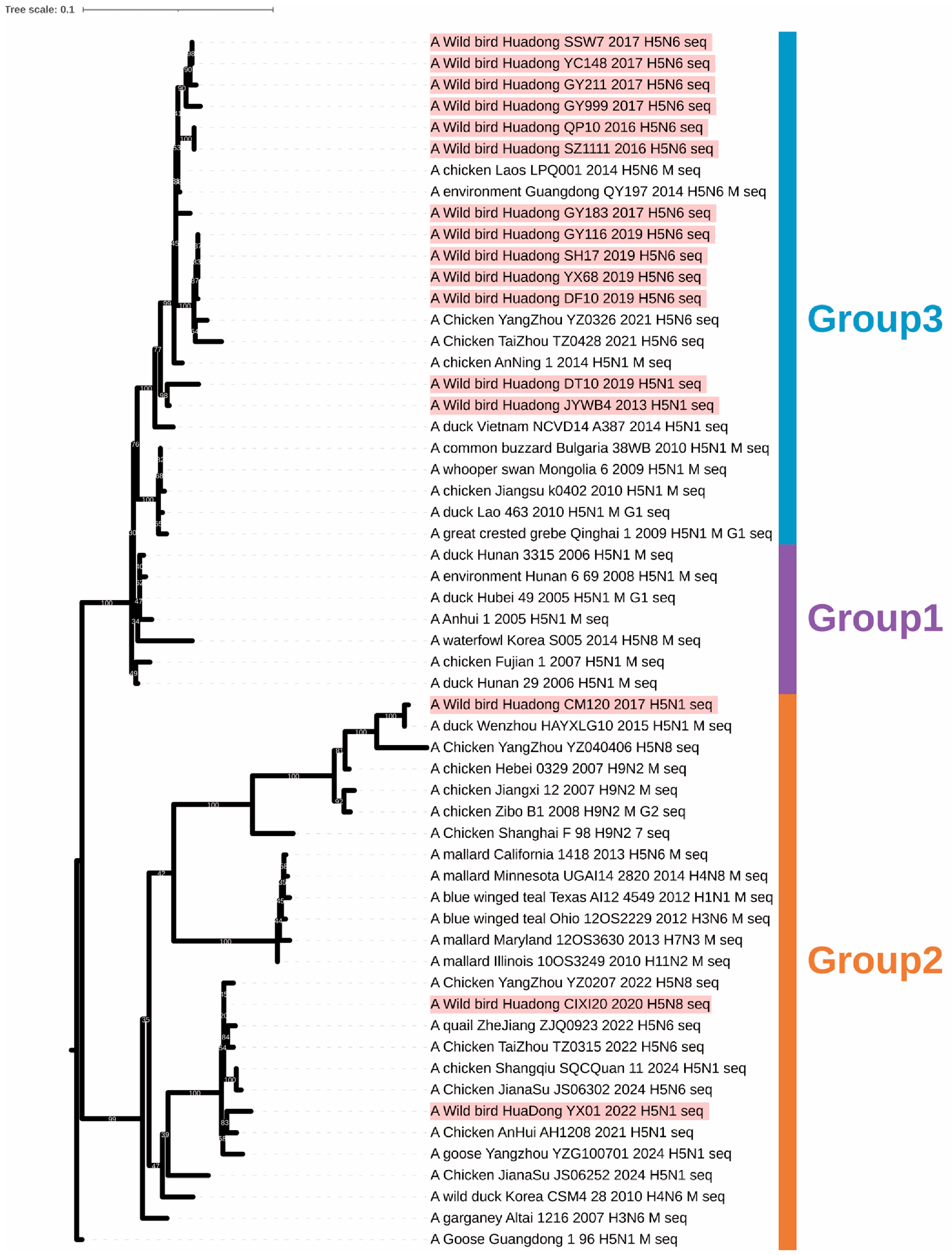
Phylogenetic analysis of the HA gene showed that, among the H5N1 subtype AIVs, CM120 belonged to clade 2.3.2.1e, whereas JYWB4 and DT10 belonged to clade 2.3.2.1d; notably, DT10 was genetically closely related to the Re-12 vaccine strain. YX01 belonged to clade 2.3.4.4b and was genetically closely related to the Re-16 vaccine strain. Among the H5N6 subtype AIVs, GY183 belonged to clade 2.3.4.4e; GY999, GY211, SSW7, YC148, QP10, and SZ1111 belonged to clade 2.3.4.4d; and DF10, GY116, SH17, and YX68 belonged to clade 2.3.4.4h and were genetically closely related to the Re-13 vaccine strain. The H5N8 isolate CIXI20 belonged to clade 2.3.4.4b and was genetically closely related to the Re-14 vaccine strain (Figure 2).

3.2.2. NA Gene

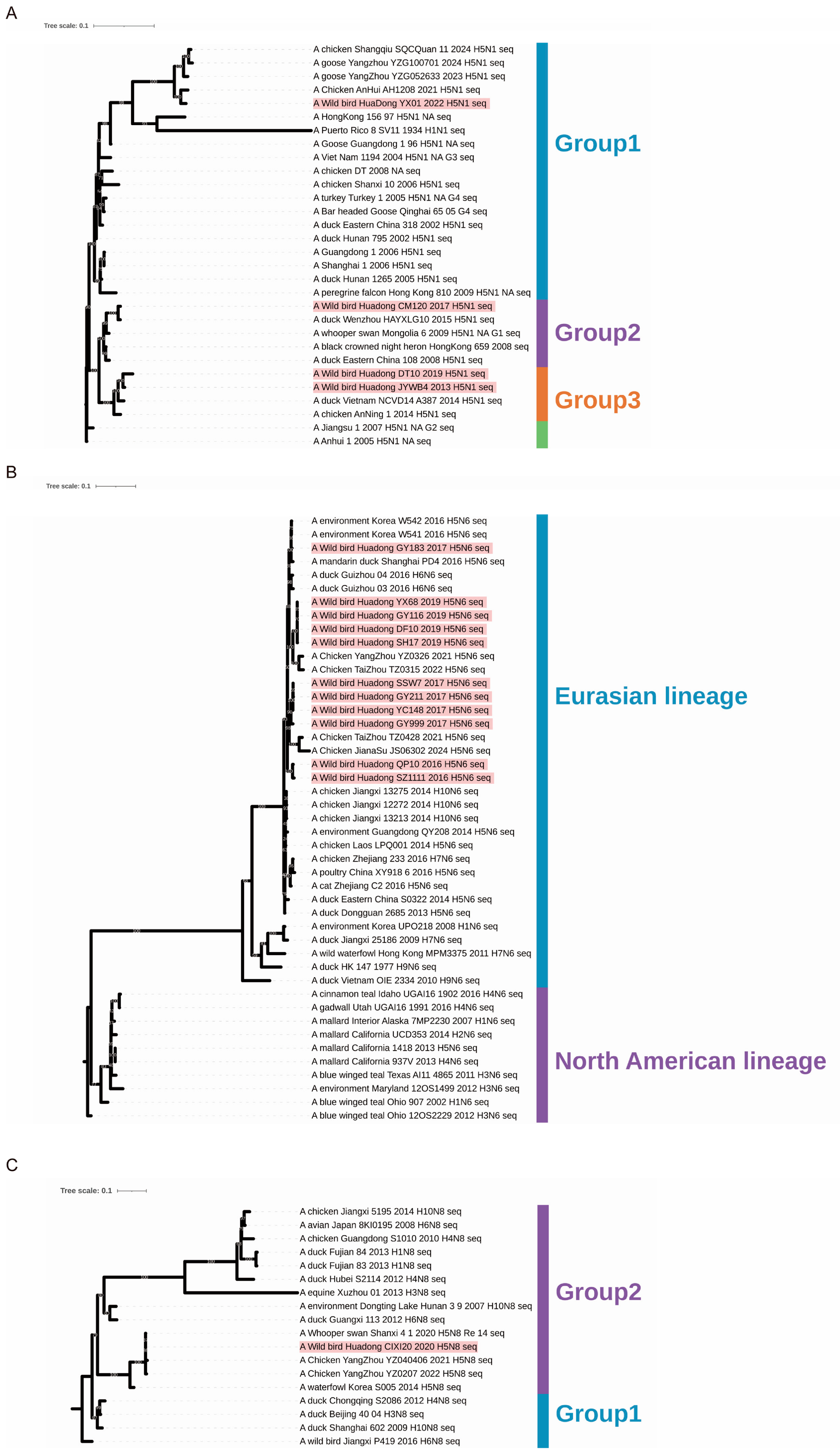
Phylogenetic analysis of the NA gene showed that the isolates comprised four H5N1 strains, eleven H5N6 strains, and one H5N8 strain. The NA genes of the four H5N1 viruses were distributed among groups 1–3, with YX01 being genetically distinct from the other strains. Group 1 evolved from the Gs/GD/96 lineage, whereas groups 2 and 3 represented more recently circulating N1 clades (Figure 3A). Phylogenetic analysis of the N6 gene indicated that all eleven isolates belonged to the Eurasian lineage and were genetically distant from the North American lineage (Figure 3B). Analysis of the N8 gene showed that the NA gene of CIXI20 was genetically closely related to that of the Re-14 vaccine strain (Figure 3C).

3.2.3. PB2 Gene

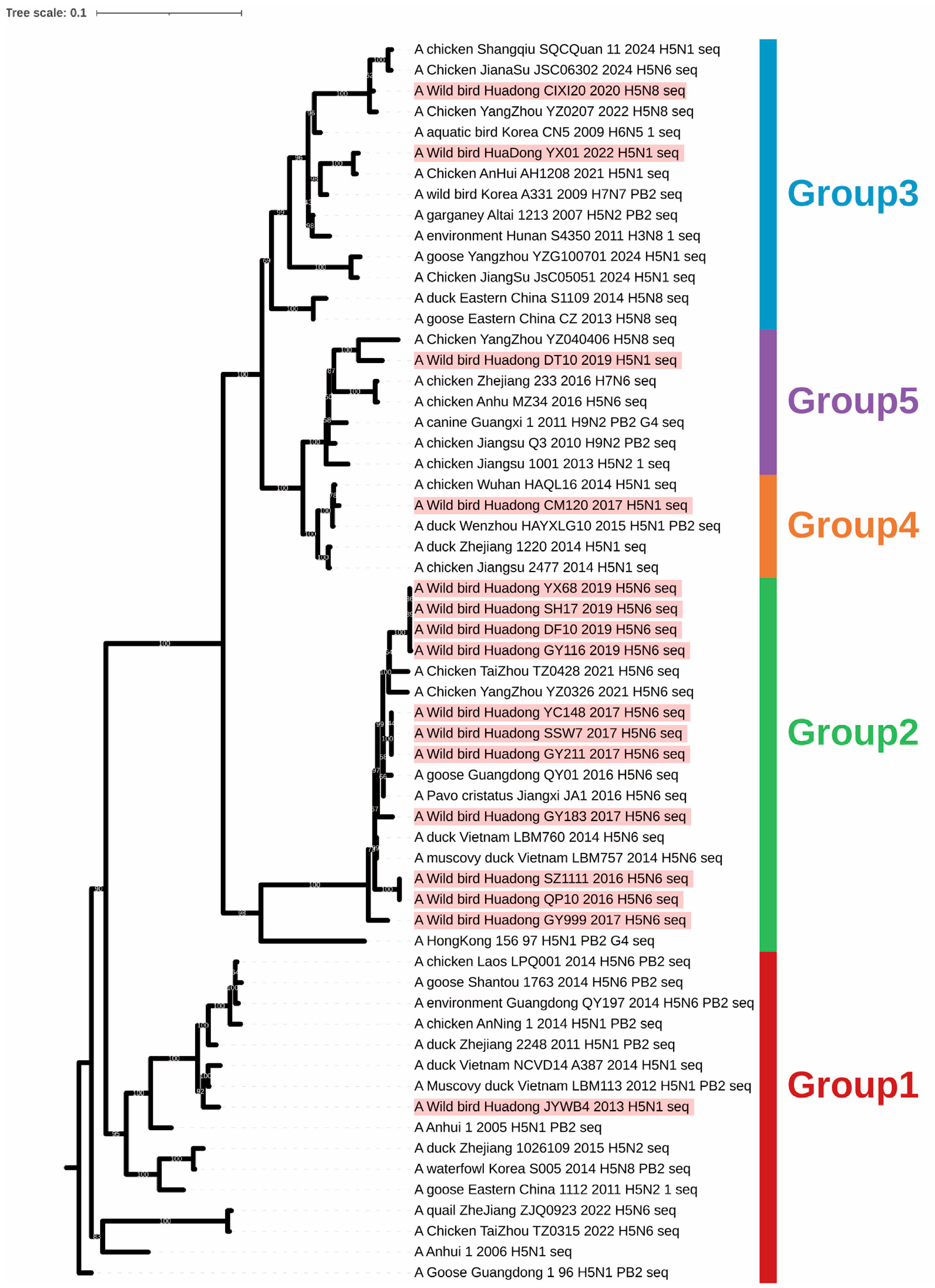
The PB2 genes of the H5 subtype AIVs were mainly distributed among five phylogenetic clades (Figure 4). The PB2 gene of JYWB4 was located in Group 1 and originated from an H5N1 subtype strain (A/goose/GuangDong/1/96); the PB2 genes of the eleven H5N6 subtype AIVs were all located in Group 2 and originated from A/HongKong/156/97; the PB2 gene of CM120 was located in Group 4 (which mainly originated from an H5N1 subtype strain, A/duck/WenZhou/HAYXLG10/15) and was closely related to the current Re-10 epidemic strain in China.

3.2.4. PB1 Gene

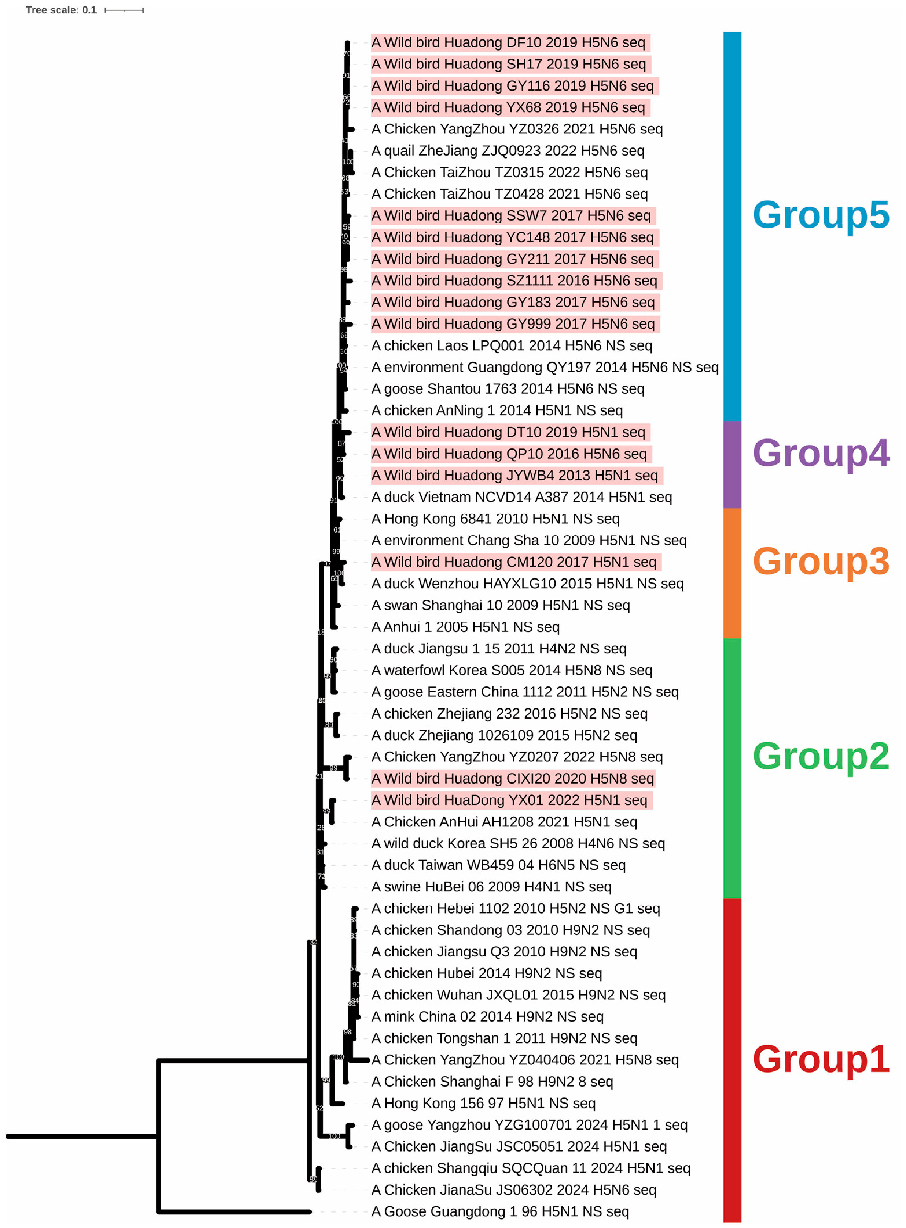
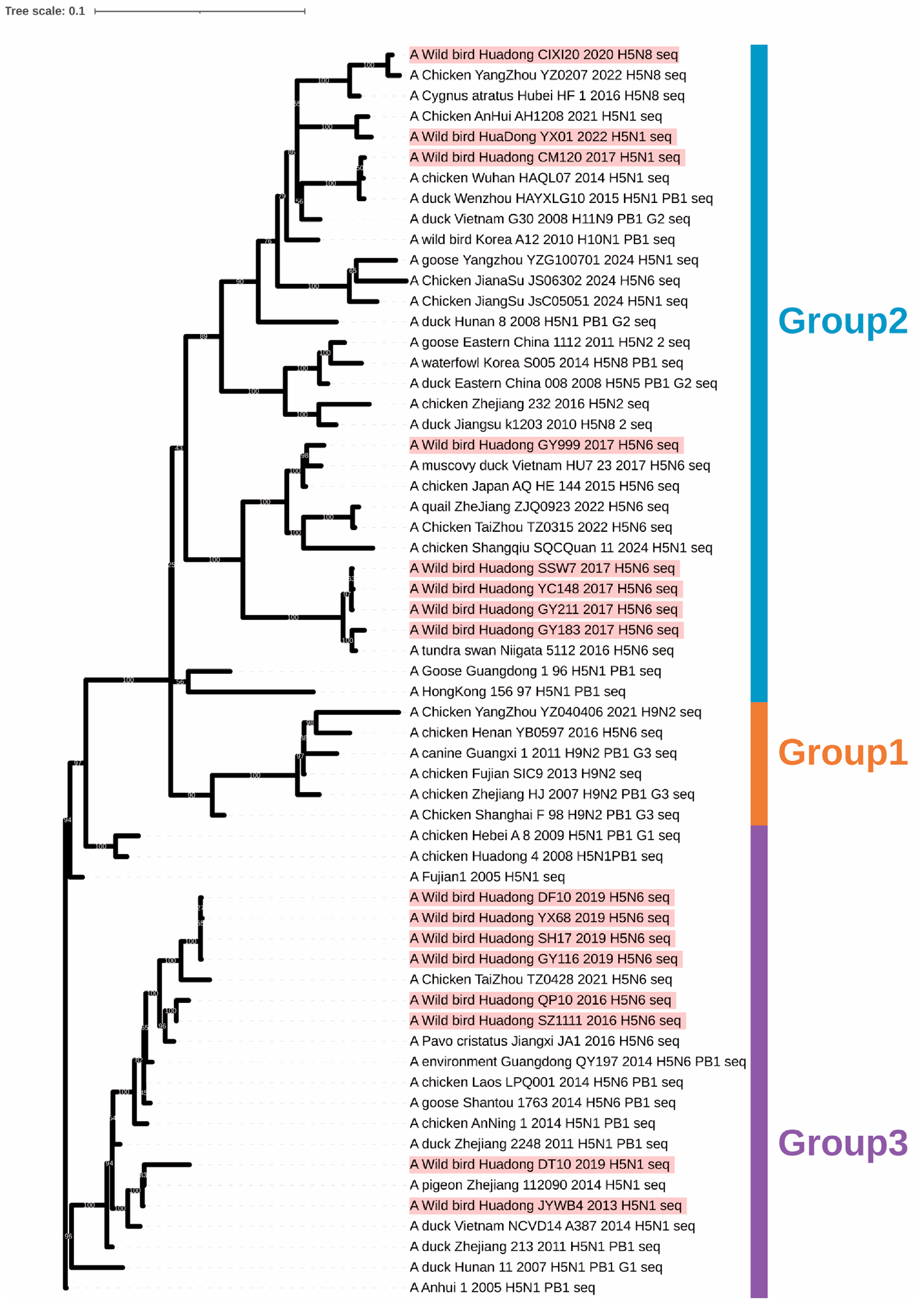
The phylogenetic tree of the PB1 genes (Figure 5) showed that the isolates fell into Groups 2 and 3. Group 2 originated from A/Goose/Guangdong/1/96 and contained most of the clade 2.3.4.4b and 2.3.4.4d isolates. Group 3 contained the Re-5 vaccine strain A/Anhui/1/2005 and the prevalent clade 2.3.4.4d strain A/chicken/LPQ001/14, and included most of the clade 2.3.4.4h isolates.

3.2.5. PA Gene

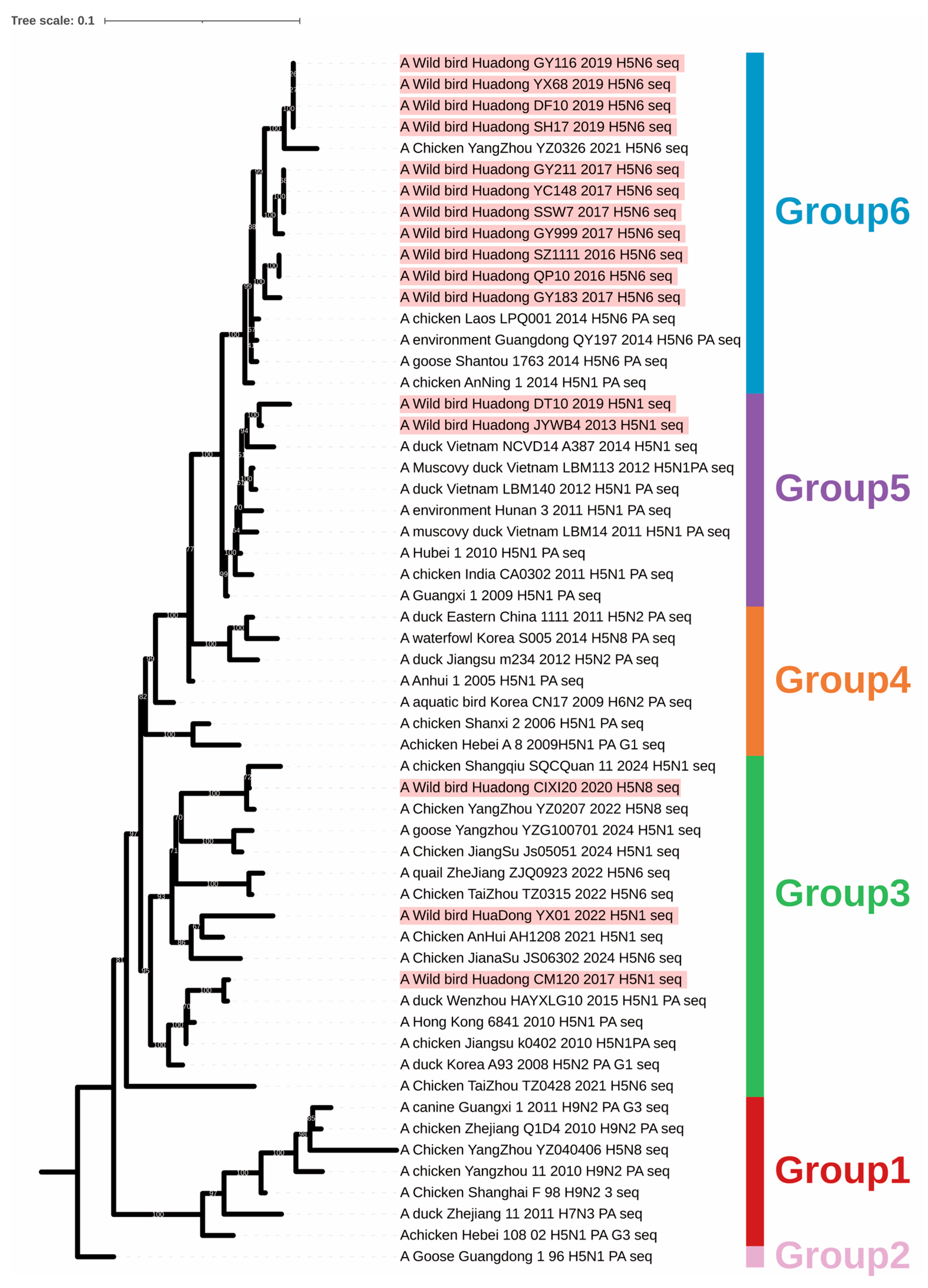
Phylogenetic analysis of the PA genes revealed six evolutionary clades (Figure 6). Group 1 was mainly derived from H9N2 subtype AIVs. The strains in Groups 2–6 all originated from A/goose/Guangdong/1/96. The PA genes of all H5N6 subtype isolates fell into Group 6. In Group 3, one PA gene originated from an H5N8 subtype AIV and two originated from H5N1 subtype AIVs; in Group 5, two PA genes originated from H5N1 subtype AIVs.

3.2.6. NP Gene

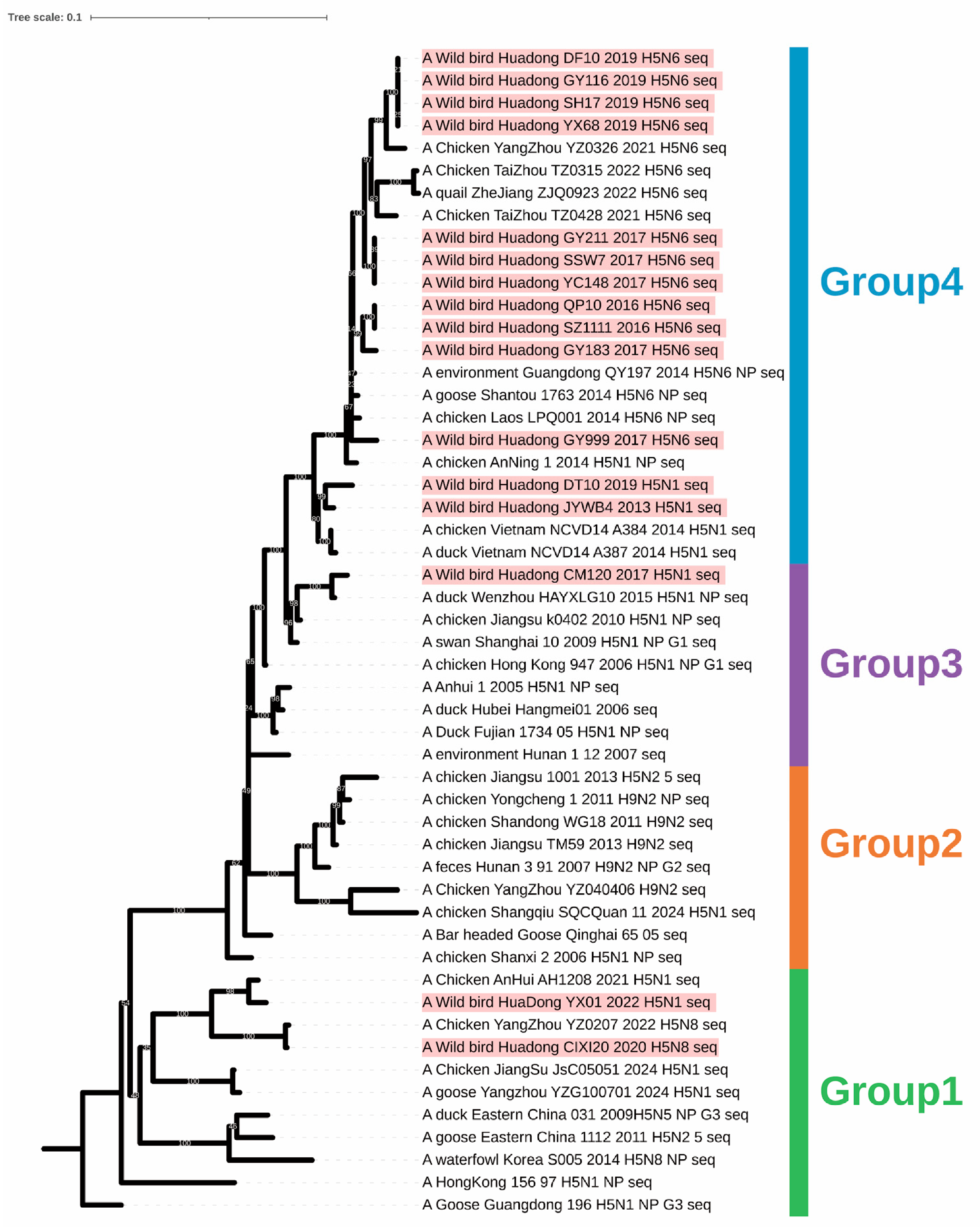
Phylogenetic analysis of the NP genes revealed four evolutionary clades (Figure 7). Group 1 originated from A/goose/Guangdong/1/96, and isolates belonging to clade 2.3.4.4b fell into this group. Group 4 was more closely related to clades 2.3.4.4d and 2.3.4.4h. These results indicated that the NP genes of the H5N6 subtype AIVs were predominantly clustered in Group 4.

3.2.7. M Gene

Phylogenetic analysis of the M genes revealed two major clades (Figure 8): the Eurasian clade and the North American clade. All isolates in this study fell into the Eurasian clade, which was further subdivided into Groups 2 and 3. The M genes in Group 2 originated from H9N2 subtype AIVs prevalent in East China. The M genes of the vast majority of isolates fell into Group 3, and most of the isolates belonging to clades 2.3.4.4d and 2.3.4.4h were located in this group.

3.2.8. NS Gene

Phylogenetic analysis of the NS genes revealed several evolutionary clades (Figure 9). The clade 2.3.4.4b strains fell into Group 2, which originated from A/goose/Guangdong/1/96. Group 3 mainly originated from a clade 2.3.2.1e strain (A/duck/WenZhou/HAYXLG10/15) and included one H5N1 subtype AIV. Group 5 was closely related to a clade 2.3.4.4d strain (A/chicken/LPQ001/14), and most isolates belonging to clades 2.3.4.4d and 2.3.4.4h fell into this group.

3.3. Molecular Profiles

The HA cleavage site motifs of all 16 H5 subtype AIVs were either “PQRERRRKR↓GLF” or “PLRERRRKR↓GLF”, both of which contain multiple basic amino acids, indicating that all strains were highly pathogenic (Table 2). Analysis of the potential glycosylation sites of the HA genes of 16 H5 subtype isolates revealed that there were six conserved glycosylation sites at positions 27 (NSTE), 39 (NVTV), 180 (NNTN), 301 (NSSM), 498 (NGTY), and 557 (NGSL). Variations in glycosylation sites were also observed among different viral strains. A novel glycosylation site, NCSV, was identified at position 70 in Clade 2.3.4.4h isolates. Additional sites included NPTN or NPSN at position 100 and NHTS or NYTS at position 140 in some strains. In contrast, the glycosylation site NPTT at position 208 was absent in certain isolates (Table 3).
Analysis of potential glycosylation sites in the NA protein revealed two conserved sites at positions 146 (NGTV) and 235 (NGSC). The site at position 88 was absent only in the CM120 strain. Novel glycosylation sites emerged at positions 50, 58, 63, and 68 in the YX01 strain (Table 4). All H5N6 subtype avian influenza viruses (AIVs) possessed glycosylation sites at positions 51, 59, 134, 190, and 391, with some viruses acquiring an additional site, NPTT, at position 54. In clade 2.3.4.4h strains, the motif at position 51 changed from NETN to NDTS (Table 5). Glycosylation sites were consistently present at positions 54, 67, 84, and 144 on the NA protein of H5N8 subtype AIVs (Table 6).
In addition, multiple amino acid markers associated with pathogenicity, host adaptation, and antiviral resistance were identified in proteins including NA, PB2, PA, NP, M1, and NS1 (Table 7). The presence of these markers suggests that these wild bird-origin H5 subtype AIVs already possess a certain potential for mammalian adaptation, particularly the mutations in PB2 and NP, which warrant attention. Several pathogenicity-enhancing markers (e.g., PB2 K389R, NP Y52H, M1 N30D/T215A) indicate that these viruses have potential pathogenicity in mammals such as mice.

3.4. Gene Selection Pressure Analysis

Because the HA gene encodes the major surface antigen of influenza virus, directly mediates receptor binding, and plays a key role in immune escape, it is considered one of the gene segments under the strongest host immune selection pressure. Therefore, we performed selection pressure analysis on the HA gene sequences of 16 H5 subtype AIVs isolated from wild birds. The results showed that several sites, including positions 131, 136, 156, and 239, exhibited relatively high dN/dS values (Table 8). Notably, positions 131 and 156 showed relatively strong signals of positive selection in all three methods (MEME, FEL, and FUBAR). The positive selection sites detected in the vaccine strains were relatively limited, mainly sites 131, 136, and 205. In the reference strains, sites such as 10, 136, 156, 157, 171, 172, and 205 showed a certain degree of positive selection signals.

3.5. Antigenic Difference Analysis

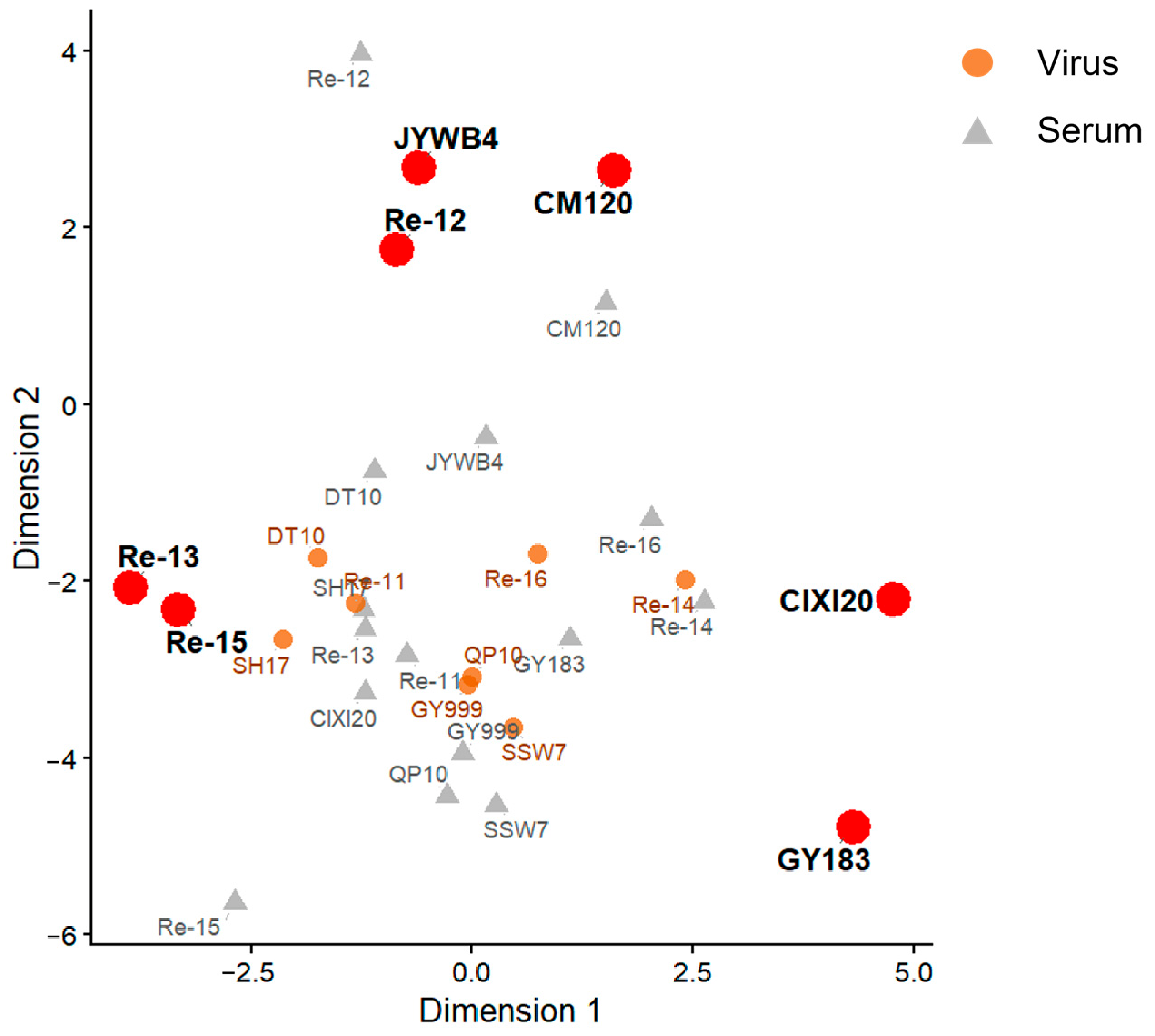
Integrated analysis of the cross-hemagglutination inhibition (HI) assay (Supplementary Table S3) and antigenic cartography (Figure 10) revealed that the isolates and vaccine strains could be classified into multiple antigenic groups. Pairwise antigenic distances calculated from the antigenic map coordinates showed that the greatest distance was observed between GY183 and JYWB4 (8.94 antigenic units), followed by those between CIXI20 and Re-13 (8.65 antigenic units), GY183 and Re-13 (8.63 antigenic units), and GY183 and Re-12 (8.32 antigenic units) (Table 9). Accordingly, the strains exhibiting the most pronounced antigenic divergence were GY183 and CIXI20. Both viruses displayed large antigenic distances from the majority of the other strains, indicating substantial antigenic drift. The early epidemic strain GY183 was antigenically distant from the recent vaccine strains Re-12, Re-13, and Re-15 (ranging from 8.04 to 8.63 antigenic units), demonstrating a clear antigenic gap between the early isolate and the current vaccine strains. In contrast, DT10 and SH17 showed limited antigenic distances from their contemporaneous vaccine strain Re-11, suggesting that these isolates were antigenically well matched to the Chinese vaccine strains of the same period and that the vaccine strains could effectively cover the dominant circulating antigenic types at that time. However, CIXI20 was antigenically distant from its contemporaneous vaccine strain Re-12, Re-13 (6.87 and 8.65 antigenic units), indicating that antigenic mismatch may still exist between some circulating strains and the vaccines currently in use.

4. Discussion

Since the emergence of Gs/GD-lineage H5 viruses, H5 AIVs have diversified through clade turnover and frequent reassortment [22,23,24]. Rather than providing a comprehensive historical reconstruction, the present study offers a regional snapshot of H5 genetic and antigenic diversity in wild birds from East China between 2013 and 2022. The 16 isolates included H5N1, H5N6, and H5N8 viruses belonging to clades 2.3.2.1d, 2.3.2.1e, 2.3.4.4b, 2.3.4.4d, 2.3.4.4e, and 2.3.4.4h. The coexistence of these divergent HA clades, together with the heterogeneous phylogenetic origins of several internal gene segments, suggests that wild birds in this region may be exposed to multiple H5 lineages and reassortant gene pools. This is consistent with previous evidence that migratory wild birds can facilitate the movement and genetic mixing of avian influenza viruses across geographic regions [6,7,8]. These findings contribute to the understanding of H5 evolution in wild birds by showing that the East China segment of the East Asian–Australasian Flyway can serve as an important interface where genetically distinct H5 viruses are detected.
The detection of clade 2.3.4.4b viruses in 2020 and 2022 is noteworthy in the context of recent global H5N1/H5Nx evolution. Since 2020, clade 2.3.4.4b H5 viruses have become dominant in many regions and have been associated with extensive outbreaks in wild birds, poultry, and an increasing range of mammals [3,25,26]. The H5N8 isolate CIXI20/2020 and the H5N1 isolate YX01/2022 identified in this study are consistent with the broader replacement and geographic expansion of clade 2.3.4.4b viruses [25,26]. In contrast, the earlier H5N6 viruses detected in 2016–2019 belonged mainly to clades 2.3.4.4d, 2.3.4.4e, and 2.3.4.4h, reflecting the regional diversity of H5N6 viruses in East Asia before the widespread predominance of clade 2.3.4.4b [23,27] Therefore, our findings are broadly consistent with the global transition from multiple regionally circulating H5Nx lineages toward the increasing predominance of clade 2.3.4.4b.
All isolates contained multiple basic amino acids at the HA cleavage site, consistent with the molecular characteristics of highly pathogenic avian influenza viruses [22,28]. This finding indicates that highly pathogenic H5 viruses can be detected in wild birds in this region and may pose a risk to poultry populations if introduced into farms or live poultry markets. In addition to the cleavage motif, changes in HA and NA glycosylation patterns may influence receptor binding, antigenic exposure, and the balance between HA and NA functions [29,30,31]. The presence or loss of glycosylation sites near antigenic or receptor-binding regions, together with positively selected sites in HA, suggests that immune selection and host adaptation may contribute to the antigenic diversification of these viruses [5,32,33,34,35].
Several molecular markers associated with mammalian adaptation or increased pathogenicity were detected in PB2, PA, NP, M1, NA, and NS1 proteins. These markers have been reported to influence polymerase activity, host restriction, virulence in mammals, or host antiviral responses [12,13,14,15,16,17,18,19,20,21]. Their presence in wild-bird-origin H5 viruses deserves attention, especially in the context of the increasing global detection of clade 2.3.4.4b viruses in mammals [3,25].
Antigenic analysis is a critical approach for evaluating vaccine efficacy and the potential for viral immune escape, providing an antigenic basis for avian influenza vaccine strain selection, optimization of immunization programs, and epidemiological surveillance [36]. In this study, cross-hemagglutination inhibition (HI) assays revealed clear time-dependent antigenic drift among avian influenza isolates from different years: isolates from the same year exhibited limited antigenic distances, whereas those from different years showed pronounced antigenic divergence, a pattern driven primarily by high-frequency mutations in the HA gene and host immune selection [37,38]. The marked antigenic differences between early epidemic strains and recent vaccine strains reflect the continuous updating of vaccine strains in China. The majority of wild bird-derived isolates were antigenically well matched with the contemporary Chinese vaccine strains, indicating that these vaccines could effectively cover the dominant antigenic types circulating at the time. Notably, these vaccine strains not only conferred effective protection against viral circulation in domestic poultry in China but also displayed good antigenic matching with strains circulating in wild birds, underscoring a strong epidemiological linkage between wild bird and domestic poultry viruses [27,39]. Nevertheless, a small number of isolates still showed potential antigenic mismatch with the contemporary vaccine strains [26]. Therefore, continuous surveillance of avian influenza virus circulation in wild birds can provide early warning for domestic poultry, enabling timely detection of antigenic variants and guiding vaccine strain updates. This substantially improves early detection and helps reduce the risk of introduction and spread of the virus from wild birds to domestic poultry.
Several limitations should be considered when interpreting these findings. Although 27,000 samples were collected over a 10-year period, only 16 H5 viruses were isolated, and no isolates were obtained in several years. Therefore, the dataset is insufficient to estimate the true prevalence, annual continuity, or dominant evolutionary pathways of H5 AIVs in wild birds in East China. The antigenic analysis was based on HI assays and antigenic cartography, which provide valuable serological evidence but cannot fully predict vaccine protection in the field. Therefore, our findings should be interpreted as a surveillance-based snapshot of genetic and antigenic diversity rather than definitive evidence that wild birds maintain or drive H5 evolution in this region. Future studies should combine expanded longitudinal sampling, environmental and host-species metadata, poultry outbreak data, and in vivo vaccine-challenge experiments to clarify transmission pathways and vaccine relevance.


5. Conclusions

Our surveillance of wild birds in East China identified H5 subtype AIVs with genetic diversity, antigenic heterogeneity, and several molecular markers associated with mammalian adaptation. Although the number of isolates was limited, the detection of multiple subtypes and clades suggests that wild birds in this region may be involved in the regional movement and genetic diversification of H5 AIVs. These findings support the value of wild bird surveillance as a complementary component of avian influenza early warning and vaccine strain evaluation. However, this study has certain limitations. Although the surveillance spanned a decade, no viruses were isolated in some years, resulting in an uneven annual distribution that may affect the assessment of evolutionary continuity. Furthermore, the antigenic analysis was primarily based on in vitro HI assays and has not yet been validated by in vivo challenge experiments to assess immune protection. Future studies should integrate structural biology, serological surveillance, and cross-species transmission models to better understand the evolutionary dynamics and public health risks of H5 subtype AIVs.

Supplementary Materials

The following supporting information can be downloaded at https://www.mdpi.com/article/10.3390/ani16132109/s1. Table S1: Number of samples collected from each sampling location in East China, 2013–2022; Table S2: GenBank numbers of H5 AIVs isolates in East China during 2013–2022; Table S3: Cross HI titers(log2) of different viruses.

Author Contributions

Conceptualization, X.S., K.C., X.M., H.Y. and S.C.; methodology, X.S., K.C., Y.Z., H.Y. and T.Q.; software, X.S. and X.Z.; validation, K.C. and Y.Z.; formal analysis, Y.G. and X.Z.; investigation, K.C., Y.Z., Y.G. and X.Z.; resources, S.C. and Y.Y.; data curation, X.S., Y.G. and X.Z.; writing—original draft preparation, X.S., K.C., Y.Z., Y.G. and X.Z.; writing—review and editing, Y.Y., X.M., H.Y., T.Q., S.C. and D.P.; visualization, X.S.; supervision, X.M., H.Y., T.Q. and D.P.; project administration, S.C., Y.Y. and D.P.; funding acquisition, Y.Y. and X.S. All authors have read and agreed to the published version of the manuscript.

Funding

This research was funded by the National Key R&D Project (2024YFC2310300) and the Postgraduate Research & Practice Innovation Program of Jiangsu Province (KYCX20_3003).

Institutional Review Board Statement

All animal experiments were approved by the Jiangsu Provincial Experimental Animal Management Committee (License Nos. SYXKSU2021-0027 and SYXKSU-2017-0044) and the Experimental Animal Ethics Committee of Yangzhou University (Approval No. 202202206, Approval date: 28 February 2022). Laboratory animal housing and experiments were conducted in strict accordance with the relevant animal welfare and ethical guidelines. All experiments involving live viruses and animals were performed in the authorized Animal Biosafety Level 3 (ABSL-3) facilities at Yangzhou University.

Informed Consent Statement

Not applicable.

Data Availability Statement

The original data presented in the study are openly available in GenBank under the accession numbers (Supplementary Table S2).

Acknowledgments

We are grateful to all individuals (Zhen Wang, Weihua Chen and Yijun Ren) and institutions who provided assistance with sample collection.

Conflicts of Interest

The authors declare no conflicts of interest.

Abbreviations

The following abbreviations are used in this manuscript:
AIVAvian influenza virus
HIHemagglutination inhibition
HAHemagglutinin
HAUHemagglutination units
NANeuraminidase
SPFSpecific pathogen free

References

  1. Archetti, I.; Horsfall, F.L., Jr. Persistent antigenic variation of influenza A viruses after incomplete neutralization in ovo with heterologous immune serum. J. Exp. Med. 1950, 92, 441–462. [Google Scholar] [CrossRef] [PubMed]
  2. Bhojani, M.S.; Bhojani, M.F. The emerging pandemic threat of H5N1: Evolutionary adaptations for human transmission, zoonotic spillovers and surveillance gaps. Gene 2025, 967, 149723. [Google Scholar] [CrossRef] [PubMed]
  3. Krammer, F.; Hermann, E.; Rasmussen, A.L. Highly pathogenic avian influenza H5N1: History, current situation, and outlook. J. Virol. 2025, 99, e0220924. [Google Scholar] [CrossRef] [PubMed]
  4. Xu, X.; Subbarao; Cox, N.J.; Guo, Y. Genetic characterization of the pathogenic influenza A/Goose/Guangdong/1/96 (H5N1) virus: Similarity of its hemagglutinin gene to those of H5N1 viruses from the 1997 outbreaks in Hong Kong. Virology 1999, 261, 15–19. [Google Scholar] [CrossRef] [PubMed]
  5. Zhang, M.; Qin, L.; Li, Z.; Chen, J.; Tong, J.; Ding, X.; Li, H.; Ma, Y.; Liu, J.; He, Q.; et al. Dominant substitutions underlying the antigenic evolution of H5 influenza virus. Nat. Commun. 2025, 16, 10708. [Google Scholar] [CrossRef] [PubMed]
  6. Olsen, B.; Munster, V.J.; Wallensten, A.; Waldenström, J.; Osterhaus, A.D.; Fouchier, R.A. Global patterns of influenza a virus in wild birds. Science 2006, 312, 384–388. [Google Scholar] [CrossRef] [PubMed]
  7. The Global Consortium for H5N8 and Related Influenza Viruses. Role for migratory wild birds in the global spread of avian influenza H5N8. Science 2016, 354, 213–217. [Google Scholar] [CrossRef] [PubMed]
  8. Yin, S.; Zhang, C.; Teitelbaum, C.S.; Si, Y.; Zhang, G.; Wang, X.; Mao, D.; Huang, Z.Y.X.; Frederik de Boer, W.; Takekawa, J.; et al. Landscape changes elevate the risk of avian influenza virus diversification and emergence in the East Asian-Australasian Flyway. Proc. Natl. Acad. Sci. USA 2025, 122, e2503427122. [Google Scholar] [CrossRef] [PubMed]
  9. Yang, Q.; Wang, B.; Lemey, P.; Dong, L.; Mu, T.; Wiebe, R.A.; Guo, F.; Trovão, N.S.; Park, S.W.; Lewis, N.; et al. Synchrony of Bird Migration with Global Dispersal of Avian Influenza Reveals Exposed Bird Orders. Nat. Commun. 2024, 15, 1126. [Google Scholar] [CrossRef] [PubMed]
  10. Hoffmann, E.; Neumann, G.; Kawaoka, Y.; Hobom, G.; Webster, R.G. A DNA transfection system for generation of influenza A virus from eight plasmids. Proc. Natl. Acad. Sci. USA 2000, 97, 6108–6113. [Google Scholar] [CrossRef] [PubMed]
  11. WHO Global Influenza. Manual for the Laboratory Diagnosis and Virological Surveillance of Influenza; WHO Press: Geneva, Switzerland, 2011. [Google Scholar]
  12. Sorrell, E.M.; Song, H.; Pena, L.; Perez, D.R. A 27-amino-acid deletion in the neuraminidase stalk supports replication of an avian H2N2 influenza A virus in the respiratory tract of chickens. J. Virol. 2010, 84, 11831–11840. [Google Scholar] [CrossRef] [PubMed]
  13. Li, J.; Ishaq, M.; Prudence, M.; Xi, X.; Hu, T.; Liu, Q.; Guo, D. Single mutation at the amino acid position 627 of PB2 that leads to increased virulence of an H5N1 avian influenza virus during adaptation in mice can be compensated by multiple mutations at other sites of PB2. Virus Res. 2009, 144, 123–129. [Google Scholar] [CrossRef] [PubMed]
  14. Hu, M.; Yuan, S.; Zhang, K.; Singh, K.; Ma, Q.; Zhou, J.; Chu, H.; Zheng, B.J. PB2 substitutions V598T/I increase the virulence of H7N9 influenza A virus in mammals. Virology 2017, 501, 92–101. [Google Scholar] [CrossRef] [PubMed]
  15. Yamayoshi, S.; Yamada, S.; Fukuyama, S.; Murakami, S.; Zhao, D.; Uraki, R.; Watanabe, T.; Tomita, Y.; Macken, C.; Neumann, G.; et al. Virulence-affecting amino acid changes in the PA protein of H7N9 influenza A viruses. J. Virol. 2014, 88, 3127–3134. [Google Scholar] [CrossRef] [PubMed]
  16. Pinto, R.M.; Bakshi, S.; Lytras, S.; Zakaria, M.K.; Swingler, S.; Worrell, J.C.; Herder, V.; Hargrave, K.E.; Varjak, M.; Cameron-Ruiz, N.; et al. BTN3A3 evasion promotes the zoonotic potential of influenza A viruses. Nature 2023, 619, 338–347. [Google Scholar] [CrossRef] [PubMed]
  17. Fan, S.; Deng, G.; Song, J.; Tian, G.; Suo, Y.; Jiang, Y.; Guan, Y.; Bu, Z.; Kawaoka, Y.; Chen, H. Two amino acid residues in the matrix protein M1 contribute to the virulence difference of H5N1 avian influenza viruses in mice. Virology 2009, 384, 28–32. [Google Scholar] [CrossRef] [PubMed]
  18. Chen, S.; Miao, X.; Huangfu, D.; Zhao, X.; Zhang, M.; Qin, T.; Peng, D.; Liu, X. H5N1 avian influenza virus without 80-84 amino acid deletion at the NS1 protein hijacks the innate immune system of dendritic cells for an enhanced mammalian pathogenicity. Transbound. Emerg. Dis. 2021, 68, 2401–2413. [Google Scholar] [CrossRef] [PubMed]
  19. Ayllon, J.; Russell, R.J.; García-Sastre, A.; Hale, B.G. Contribution of NS1 effector domain dimerization to influenza A virus replication and virulence. J. Virol. 2012, 86, 13095–13098. [Google Scholar] [CrossRef] [PubMed]
  20. Wang, B.X.; Brown, E.G.; Fish, E.N. Residues F103 and M106 within the influenza A virus NS1 CPSF4-binding region regulate interferon-stimulated gene translation initiation. Virology 2017, 508, 170–179. [Google Scholar] [CrossRef] [PubMed]
  21. Huang, M.T.; Zhang, S.; Wu, Y.N.; Li, W.; Li, Y.C.; Zhou, C.S.; Kang, X.P.; Jiang, T. Dual R108K and G189D Mutations in the NS1 Protein of A/H1N1 Influenza Virus Counteract Host Innate Immune Responses. Viruses 2021, 13, 905. [Google Scholar] [CrossRef] [PubMed]
  22. Luczo, J.M.; Spackman, E. Molecular Evolution of the H5 and H7 Highly Pathogenic Avian Influenza Virus Haemagglutinin Cleavage Site Motif. Rev. Med. Virol. 2025, 35, e70012. [Google Scholar] [CrossRef] [PubMed]
  23. Lee, S.H.; Kwon, J.H.; Youk, S.; Lee, S.W.; Lee, D.H.; Song, C.S. Epidemiology and pathobiology of H5Nx highly pathogenic avian influenza in South Korea (2003-2024): A comprehensive review. Vet. Q. 2025, 45, 23–38. [Google Scholar] [CrossRef] [PubMed]
  24. Antigua, K.J.C.; Choi, W.S.; Baek, Y.H.; Song, M.S. The Emergence and Decennary Distribution of Clade 2.3.4.4 HPAI H5Nx. Microorganisms 2019, 7, 156. [Google Scholar] [CrossRef] [PubMed]
  25. Baxter, J.; Yang, J.; Harvey, W.; Dellicour, S.; Pohlmann, A.; Ma, M.; Liu, W.; Bi, Y.; Digard, P.; Beer, M.; et al. The Winners Take It All? Evolutionary Success of H5Nx Reassortants in the 2020–2024 Panzootic. bioRxiv 2025. bioRxiv:2025.07.19.665680. [Google Scholar] [CrossRef]
  26. Cui, P.; Shi, J.; Wang, C.; Zhang, Y.; Xing, X.; Kong, H.; Yan, C.; Zeng, X.; Liu, L.; Tian, G.; et al. Global dissemination of H5N1 influenza viruses bearing the clade 2.3.4.4b HA gene and biologic analysis of the ones detected in China. Emerg. Microbes Infect. 2022, 11, 1693–1704. [Google Scholar] [CrossRef] [PubMed]
  27. Bi, Y.; Chen, Q.; Wang, Q.; Chen, J.; Jin, T.; Wong, G.; Quan, C.; Liu, J.; Wu, J.; Yin, R.; et al. Genesis, Evolution and Prevalence of H5N6 Avian Influenza Viruses in China. Cell Host Microbe 2016, 20, 810–821. [Google Scholar] [CrossRef] [PubMed]
  28. Wallace, R.G.; Hodac, H.; Lathrop, R.H.; Fitch, W.M. A statistical phylogeography of influenza A H5N1. Proc. Natl. Acad. Sci. USA 2007, 104, 4473–4478. [Google Scholar] [CrossRef] [PubMed]
  29. Kaverin, N.V.; Rudneva, I.A.; Ilyushina, N.A.; Varich, N.L.; Lipatov, A.S.; Smirnov, Y.A.; Govorkova, E.A.; Gitelman, A.K.; Lvov, D.K.; Webster, R.G. Structure of antigenic sites on the haemagglutinin molecule of H5 avian influenza virus and phenotypic variation of escape mutants. J. Gen. Virol. 2002, 83, 2497–2505. [Google Scholar] [CrossRef] [PubMed]
  30. Chen, W.; Zhong, Y.; Qin, Y.; Sun, S.; Li, Z. The evolutionary pattern of glycosylation sites in influenza virus (H5N1) hemagglutinin and neuraminidase. PLoS ONE 2012, 7, e49224. [Google Scholar] [CrossRef] [PubMed]
  31. Chang, D.; Zaia, J. Why Glycosylation Matters in Building a Better Flu Vaccine. Mol. Cell Proteom. 2019, 18, 2348–2358. [Google Scholar] [CrossRef] [PubMed]
  32. Shi, K.; Feng, S.; Zhao, L.; Chen, J.; Song, W.; Jia, Y.; Qu, X.; Liu, Z.; Jia, W.; Du, S.; et al. N-glycosylation on hemagglutinin head reveals inter-branch antigenic variability of avian influenza virus H5-subtypes. Int. J. Biol. Macromol. 2024, 273, 132901. [Google Scholar] [CrossRef] [PubMed]
  33. Luczo, J.M.; Spackman, E. Epitopes in the HA and NA of H5 and H7 avian influenza viruses that are important for antigenic drift. FEMS Microbiol. Rev. 2024, 48, fuae014. [Google Scholar] [CrossRef] [PubMed]
  34. Gu, C.; Zeng, X.; Song, Y.; Li, Y.; Liu, L.; Kawaoka, Y.; Zhao, D.; Chen, H. Glycosylation and an amino acid insertion in the head of hemagglutinin independently affect the antigenic properties of H5N1 avian influenza viruses. Sci. China Life Sci. 2019, 62, 76–83. [Google Scholar] [CrossRef] [PubMed]
  35. Ilyushina, N.A.; Rudneva, I.A.; Gambaryan, A.S.; Tuzikov, A.B.; Bovin, N.V. Receptor specificity of H5 influenza virus escape mutants. Virus Res. 2004, 100, 237–241. [Google Scholar] [CrossRef] [PubMed]
  36. Smith, D.J.; Lapedes, A.S.; de Jong, J.C.; Bestebroer, T.M.; Rimmelzwaan, G.F.; Osterhaus, A.D.; Fouchier, R.A. Mapping the antigenic and genetic evolution of influenza virus. Science 2004, 305, 371–376. [Google Scholar] [CrossRef] [PubMed]
  37. Ferguson, N.M.; Galvani, A.P.; Bush, R.M. Ecological and immunological determinants of influenza evolution. Nature 2003, 422, 428–433. [Google Scholar] [CrossRef] [PubMed]
  38. Shih, A.C.; Hsiao, T.C.; Ho, M.S.; Li, W.H. Simultaneous amino acid substitutions at antigenic sites drive influenza A hemagglutinin evolution. Proc. Natl. Acad. Sci. USA 2007, 104, 6283–6288. [Google Scholar] [CrossRef] [PubMed]
  39. Lycett, S.J.; Duchatel, F.; Digard, P. A brief history of bird flu. Philos. Trans. R. Soc. Lond. B Biol. Sci. 2019, 374, 20180257. [Google Scholar] [CrossRef] [PubMed]
Figure 1. Surveillance profile of H5 AIV isolates from Wild birds in East China during 2013–2022. (A) Distribution of sampling sites. Blue: the sampled provinces; triangle: the specific sampling sites; (B) Virus isolation rates in different sampling regions; (C) Temporal distribution and number of isolated strains.
Animals 16 02109 g001
Figure 2. Phylogenetic tree based on the HA gene sequences of 16 H5 AIV isolates from wild birds in East China, 2013–2022. The isolates are highlighted with a pink background.
Animals 16 02109 g002
Figure 3. Phylogenetic tree based on the NA gene sequences of 16 H5 AIV isolates from wild birds in East China, 2013–2022. (A) Phylogenetic tree of the NA gene of H5N1 subtype AIVs; (B) Phylogenetic tree of the NA gene of H5N6 subtype AIVs; (C) Phylogenetic tree of the NA gene of H5N8 subtype AIVs. The isolates are highlighted with a pink background.
Animals 16 02109 g003
Figure 4. Phylogenetic tree based on the PB2 gene sequences of 16 H5 AIV isolates from wild birds in East China, 2013–2022. The isolates are highlighted with a pink background.
Animals 16 02109 g004
Figure 5. Phylogenetic tree based on the PB1 gene sequences of 16 H5 AIV isolates from wild birds in East China, 2013–2022. The isolates are highlighted with a pink background.
Animals 16 02109 g005
Figure 6. Phylogenetic tree based on the PA gene sequences of 16 H5 AIV isolates from wild birds in East China, 2013–2022. The isolates are highlighted with a pink background.
Animals 16 02109 g006
Figure 7. Phylogenetic tree based on the NP gene sequences of 16 H5 AIV isolates from wild birds in East China, 2013–2022. The isolates are highlighted with a pink background.
Animals 16 02109 g007
Figure 8. Phylogenetic tree based on the M gene sequences of 16 H5 AIV isolates from wild birds in East China, 2013–2022. The isolates are highlighted with a pink background.
Animals 16 02109 g008
Figure 9. Phylogenetic tree based on the NS gene sequences of 16 H5 AIV isolates from wild birds in East China, 2013–2022. The isolates are highlighted with a pink background.
Animals 16 02109 g009
Figure 10. Antigenic cartography of H5 subtype AIVs. Orange: viral antigens; gray: sera; red-highlighted: strains most divergent from the main cluster.
Animals 16 02109 g010
Table 1. Background of H5 AIV isolates in East China during 2013–2022.
Virus IsolateAbbreviationSample SourceSubtypeCladeGenBank Number (HA)
A/Wild bird/Huadong/JYWB4/2013JYWB4Wild birdH5N12.3.2.1dPZ417131
A/Wild bird/Huadong/QP10/2016QP10Wild birdH5N62.3.4.4dPZ417132
A/Wild bird/Huadong/SZ1111/2016SZ1111Wild birdH5N62.3.4.4dPZ417135
A/Wild bird/Huadong/CM120/2017CM120Wild birdH5N12.3.2.1ePZ417125
A/Wild bird/Huadong/GY183/2017GY183Wild birdH5N62.3.4.4ePZ417123
A/Wild bird/Huadong/GY211/2017GY211Wild birdH5N62.3.4.4dPZ417129
A/Wild bird/Huadong/GY999/2017GY999Wild birdH5N62.3.4.4dPZ417130
A/Wild bird/Huadong/SSW7/2017SSW7Wild birdH5N62.3.4.4dPZ417134
A/Wild bird/Huadong/YC148/2017YC148Wild birdH5N62.3.4.4dPZ417138
A/Wild bird/Huadong/DT10/2019DT10Wild birdH5N12.3.2.1dPZ417127
A/Wild bird/Huadong/DF10/2019DF10Wild birdH5N62.3.4.4hPZ417126
A/Wild bird/Huadong/GY116/2019GY116Wild birdH5N62.3.4.4hPZ417128
A/Wild bird/Huadong/SH17/2019SH17Wild birdH5N62.3.4.4hPZ417133
A/Wild bird/Huadong/YX68/2019YX68Wild birdH5N62.3.4.4h PZ417136
A/Wild bird/Huadong/CIXI20/2020CIXI20Wild birdH5N82.3.4.4bPZ417124
A/Wild bird/Huadong/YX01/2022YX01Wild birdH5N12.3.4.4bPZ417137
Table 2. Connecting-peptide at the cleavage site of HA.
VirusCladeConnecting-Peptide
JYWB4/20132.3.2.1dPQRERRRKRGLF
QP10/20162.3.4.4dPQRERRRKRGLF
SZ1111/20162.3.4.4dPLRERRRKRGLF
CM120/20172.3.2.1ePQRERRRKRGLF
GY183/20172.3.4.4ePLRERRRKRGLF
GY211/20172.3.4.4dPLRERRRKRGLF
GY999/20172.3.4.4dPLRERRRKRGLF
SSW7/20172.3.4.4dPLRERRRKRGLF
YC148/20172.3.4.4dPLRERRRKRGLF
DT10/20192.3.2.1dPQRERRRKRGLF
DF10/20192.3.4.4hPLRERRRKRGLF
GY116/20192.3.4.4hPLRERRRKRGLF
SH17/20192.3.4.4hPLRERRRKRGLF
YX68/20192.3.4.4hPLRERRRKRGLF
CIXI20/20202.3.4.4bPLREKRRKRGLF
YX01/20222.3.4.4bPLRERRRKRGLF
Table 3. Predicted N-glycosylation sites in HA protein of H5 subtype viruses.
VirusH5 Subtype
273970100140180208301498557
JYWB4/2013NSTENVTV---NNTNNPTTNSSMNGTYNGSL
QP10/2016NSTENVTV---NNTNNPTTNSSMNGTYNGSL
SZ1111/2016NSTENVTV---NNTNNPTTNSSMNGTYNGSL
CM120/2017NSTENVTV---NNTNNPTTNSSMNGTYNGSL
GY183/2017NSTENVTV---NNTNNPTTNSSMNGTYNGSL
GY211/2017NSTENVTV--NHTSNNTNNPTTNSSMNGTYNGSL
GY999/2017NSTENVTV--NHTSNNTNNPTTNSSMNGTYNGSL
SSW7/2017NSTENVTV--NHTSNNTNNPTTNSSMNGTYNGSL
YC148/2017NSTENVTV--NHTSNNTNNPTTNSSMNGTYNGSL
DT10/2019NSTENVTV---NNTNNPTTNSSMNGTYNGSL
DF10/2019NSTENVTVNCSV-NYTSNNTNNPTTNSSMNGTYNGSL
GY116/2019NSTENVTVNCSV-NYTSNNTNNPTTNSSMNGTYNGSL
SH17/2019NSTENVTVNCSV-NYTSNNTNNPTTNSSMNGTYNGSL
YX68/2019NSTENVTVNCSV-NYTSNNTNNPTTNSSMNGTYNGSL
CIXI20/2020NSTENVTV---NNTNNPTTNSSMNGTYNGSL
YX01/2022NSTENVTV-NPTN-NNTN-NSSMNGTYNGSL
Re-11NSTENVTV-NPSNNHTSNNTNNPTTNSSMNGTYNGSL
Re-12NSTENVTV---NNTNNPTTNSSMNGTYNGSL
Re-13NSTENVTVNCSV NHTTNNTNNPTTNSSMNGTYNGSL
Re-14NSTENVTV---NNTNNPTTNSSMNGTYNGSL
Re-15NSTENVTV--NHTSNNTN-NSSMNGTYNGSL
Re-16NSTENVTV---NNTNNPTTNSSMNGTYNGSL
Table 4. Predicted N-glycosylation sites in NA protein of H5N1 viruses.
VirusH5N1 Subtype
5058636888146235
JYWB4/2013----NSSLNGTVNGSC
CM120/2017-----NGTVNGSC
DT10/2019----NSSLNGTVNGSC
YX01/2022NQSINNTWNQTYNISNNSSLNGTVNGSC
Table 5. Predicted N-glycosylation sites in NA protein of H5N6 viruses.
VirusH5N6 Subtype
515459134190391
QP10/2016NETN-NITNNGTINASANWSG
SZ1111/2016NETN-NITNNGTINASANWSG
GY183/2017NETNNPTTNITNNGTINASANWSG
GY211/2017NETNNPTTNITNNGTINASANWSG
GY999/2017NETNNPTTNITNNGTINASANWSG
SSW7/2017NETNNPTTNITNNGTINASANWSG
YC148/2017NETNNPTTNITNNGTINASANWSG
DF10/2019NDTS-NITNNGTINASANWSG
GY116/2019NDTS-NITNNGTINASANWSG
SH17/2019NDTS-NITNNGTINASANWSG
YX68/2019NDTS-NITNNGTINASANWSG
Table 6. Predicted N-glycosylation sites in NA protein of H5N8 viruses.
VirusH5N8 Subtype
546784144
CIXI20/2020NETVNTSVNNTENGTV
Table 7. Amino acid markers associated with specific phenotypic effects found in the genome of H5Nx viruses from wild birds.
ProteinGenetic MarkerBiological Functions
NAdeletion of 46–65 AAMarkers for enhanced pathogenicity and adaptation of the virus from wild birds to poultry [12]
PB2L89VIncreased polymerase activity in mammalian cell lines and mice [13]
G309DIncreased polymerase activity in mammalian cell lines and mice [13]
K389REnhanced virulence of HPAIV H5 viruses in mice [14]
R477GIncreased polymerase activity in mammalian cell lines and mice [13]
M676TContributed to high replication and pathogenicity of influenza A virus in mammals [13]
PAN409SIncreased polymerase activity in mammalian cell lines [15]
NPY52HConfer resistance to inhibitors of BTN3A3 (butyrophilin subfamily 3 member A3) [16]
M1N30DIncreased virulence in mice [17]
T215A
NS1deletion of 80–84 AADecreased mammalian pathogenicity [18]
W187RDecreased mammalian pathogenicity [19]
L103FIncreased virulence in mice [20]
I106M
N205SDecreased antiviral response in host [21]
Table 8. The selection profile of HA gene of H5 subtype viruses.
VirusSiteMEMEFELFUBARSLAC
dN/dSp ValuedN/dSp ValuedN/dSPosterior ProbabilitydN/dSp Value
virus isolate97.990.01--28.790.92--
1316.300.025.880.02126.350.98--
136----40.650.94--
1563.270.093.270.07133.320.98--
1604.250.06------
1674.000.06------
179----35.180.93--
2393.820.07--43.930.94--
417----30.080.92--
4204.820.04------
vaccine strain88----42.850.94--
1317.970.0088.020.005962.550.993.620.04
1366.280.023.390.06147.620.98--
1493.560.08------
172--2.730.09----
2043.870.07------
205--2.900.0985.580.97--
34210.020.003------
reference strain212.940.0007------
314.570.0003------
109.6370.0045.4590.0252.690.94--
147.750.009------
157.440.01------
5211.960.0013.030.08----
594.730.04------
824.530.05------
1253.950.06------
1366.700.02--42.470.925.480.09
140----33.060.91--
1543.940.07------
1563.740.073.340.07--11.140.08
1575.320.035.260.0234.190.913.000.09
1714.890.044.890.03148.490.984.100.06
1726.720.026.740.01119.430.975.000.02
205--3.050.0835.980.91--
210--2.820.09----
2395.920.02------
2733.420.093.420.06----
3363.450.08------
3483.340.09------
35011.830.001------
4163.430.09------
Table 9. Antigenic distance between different strains (top 20).
Virus 1Virus 2Distance (Antigenic Units)
GY183JYWB48.935648
CIXI20Re-138.64905
GY183Re-138.630393
GY183Re-128.324965
CIXI20Re-158.106754
GY183Re-158.037562
GY183CM1207.911601
JYWB4CIXI207.260305
CM120Re-137.22475
CM120Re-156.997874
SH17CIXI206.935285
CIXI20Re-126.868677
GY183SH176.801959
GY183DT106.794984
DT10CIXI206.543541
SH17CM1206.499903
SSW7JYWB46.421212
SSW7CM1206.406957
Re-13Re-146.293727
GY183Re-116.180426
Disclaimer/Publisher’s Note: The statements, opinions and data contained in all publications are solely those of the individual author(s) and contributor(s) and not of MDPI and/or the editor(s). MDPI and/or the editor(s) disclaim responsibility for any injury to people or property resulting from any ideas, methods, instructions or products referred to in the content.