Quantitative Ultrasonographic Assessment of Supraspinatus Insertion Tendon in Non-Lame Dogs (Thickness and Relative Echogenicity)

 , , , ,
Simple Summary
Abstract
1. Introduction
2. Materials and Methods
2.1. Study Design
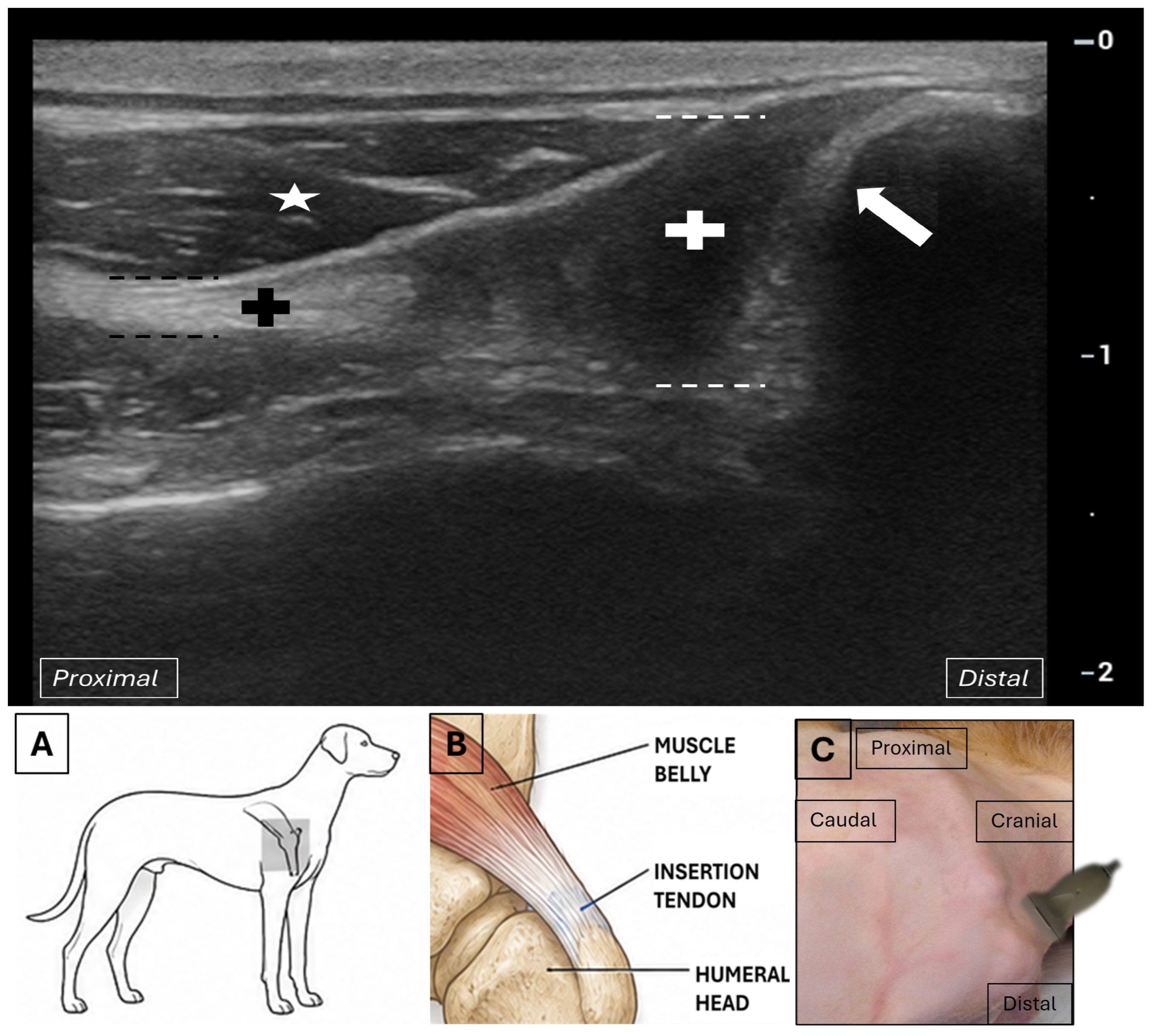
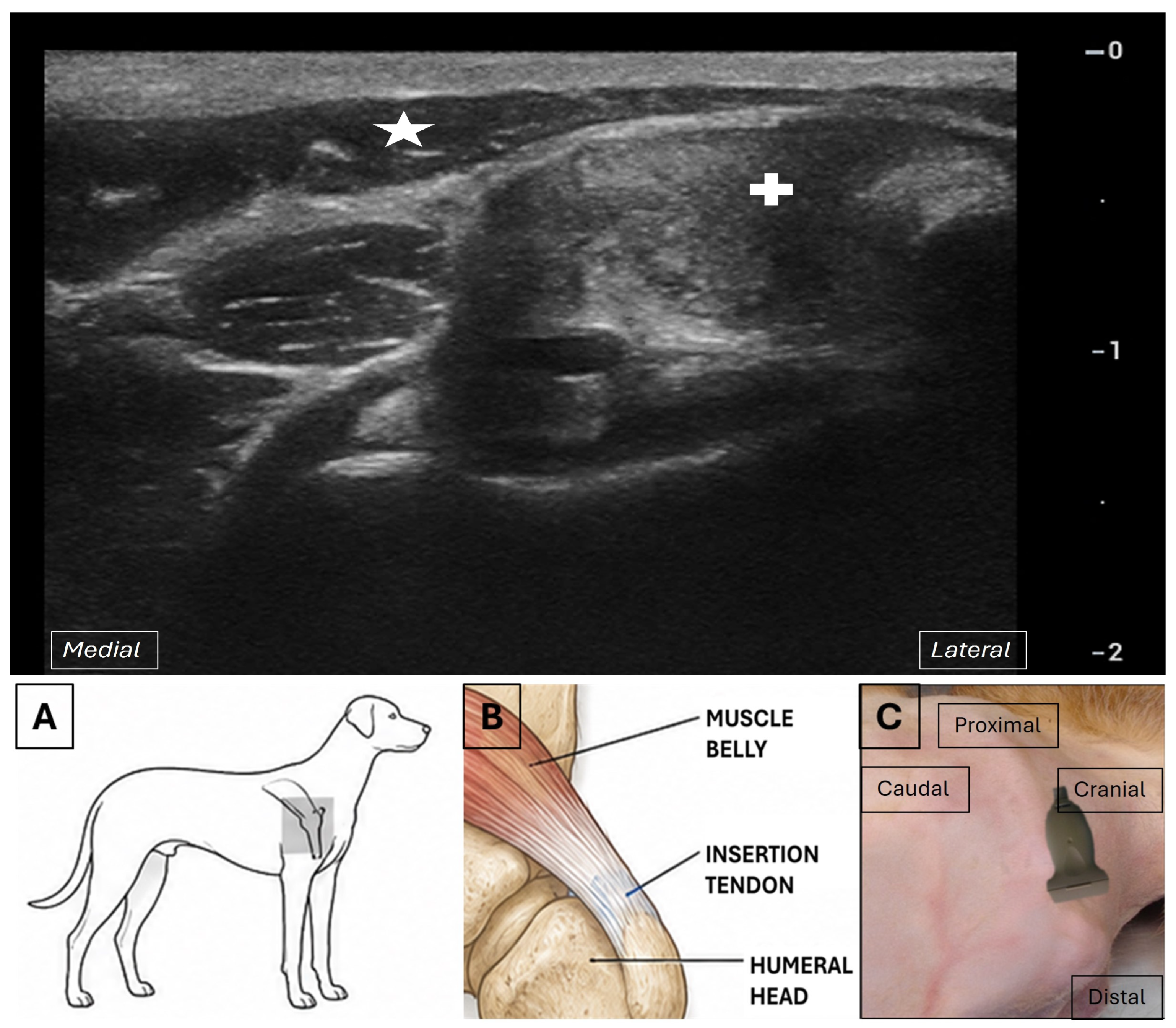
2.2. Ultrasonographic Examination
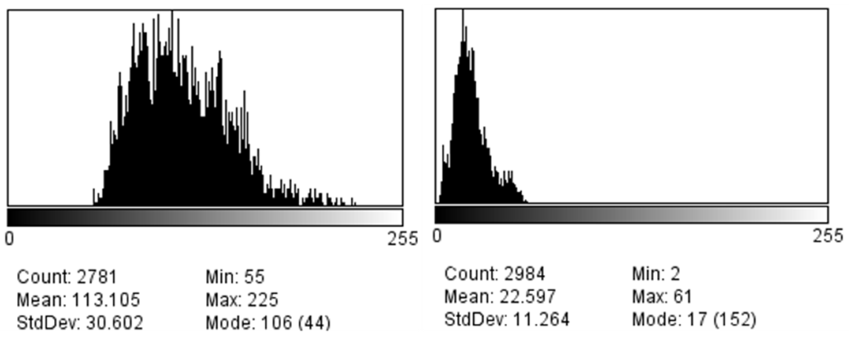
2.3. Image Analysis
2.4. Statistical Analysis
3. Results
3.1. Population Description
3.2. Ultrasonographic Appearance of the Tendon
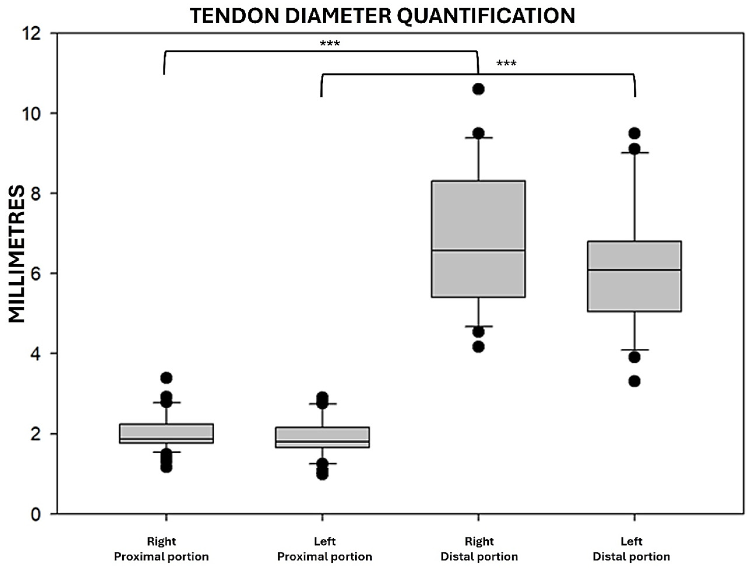
3.3. SIT Thickness Measurements
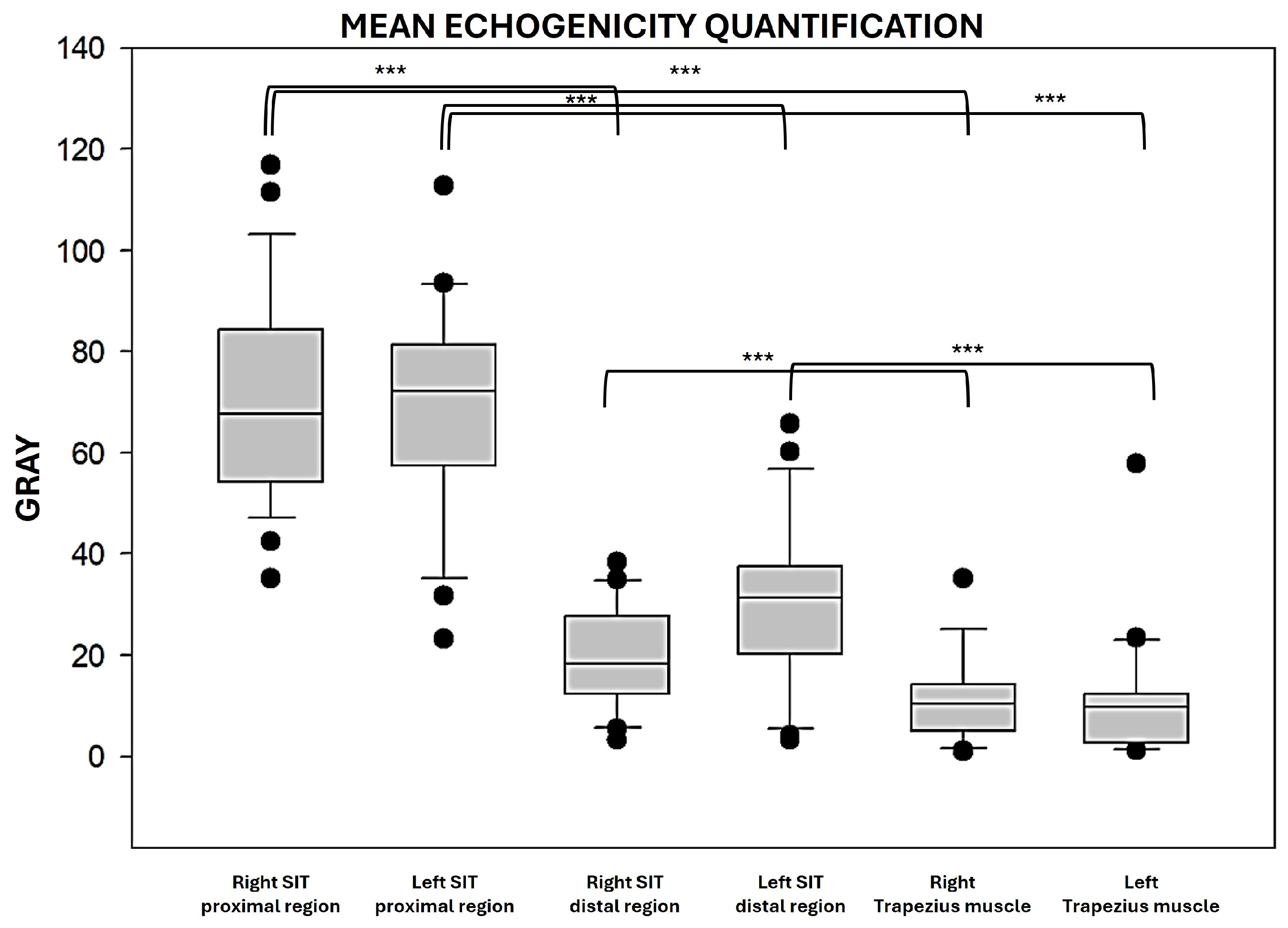
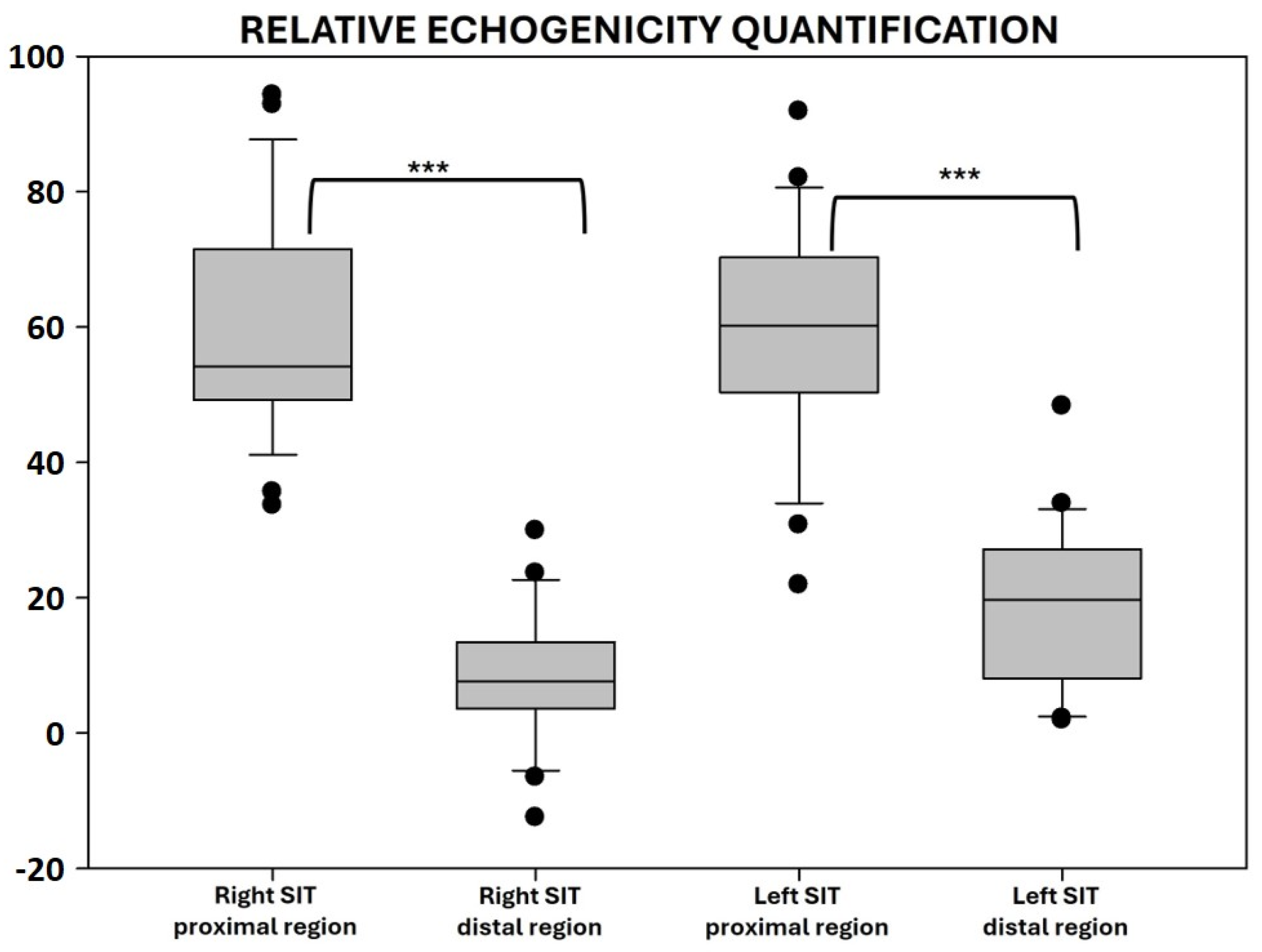
3.4. Echogenicity Analysis
3.5. Relationship Between SIT Measurements and Biological Variables
3.6. Repeatability and Reproducibility Analysis
4. Discussion
4.1. Ultrasonographic Exploration and Image Analysis of the SIT
4.2. Ultrasonographic Appearance and Quantitative Characteristics of the SIT
4.3. Relationships Between Tendon Characteristics and Biological Variables
4.4. Clinical Relevance and Future Directions
4.5. Study Limitations
5. Conclusions
Author Contributions
Funding
Institutional Review Board Statement
Informed Consent Statement
Data Availability Statement
Acknowledgments
Conflicts of Interest
Abbreviations
| CT | Computed tomography |
| BMI | Body mass index |
| MRI | Magnetic resonance imaging |
| ROI | Region of interest |
| SD | Standard deviation |
| SIT | Supraspinatus insertion tendon |
References
- Hermanson, J.W.; de la Hunta, A.; Evans, H.E. Miller and Evans’ Anatomy of the Dog, 5th ed.; W.B. Saunders Company: Philadelphia, PA, USA, 2019; 1004p. [Google Scholar]
- Bardet, J.F. Shoulder diseases in dogs. Vet. Med. 2002, 97, 909–918. [Google Scholar]
- Bardet, J.F. Diagnosis of shoulder instability in dogs and cats: A retrospective study. J. Am. Anim. Hosp. Assoc. 1998, 34, 42–54. [Google Scholar] [CrossRef] [Scilit] [PubMed]
- Cogar, S.M.; Cook, C.R.; Curry, S.L.; Grandis, A.; Cook, J.L. Prospective evaluation of techniques for differentiating shoulder pathology as a source of forelimb lameness in medium and large breed dogs. Vet. Surg. 2008, 37, 132–141. [Google Scholar] [CrossRef] [Scilit] [PubMed]
- McKee, M.; Macias, C. Orthopaedics conditions of the shoulder in the dog. Practice 2004, 26, 118–129. [Google Scholar] [CrossRef] [Scilit]
- Muir, P.; Johonson, K.A. Supraspinatus and biceps brachii tendinopathy in dogs. J. Small Anim. Pract. 1994, 35, 239–243. [Google Scholar] [CrossRef] [Scilit]
- Devitt, C.M.; Neely, M.R.; Vanvechten, B.J. Relationship of physical examination test of shoulder instability to arthroscopic findings in dogs. Vet. Surg. 2007, 36, 661–668. [Google Scholar] [CrossRef] [Scilit] [PubMed]
- Canapp, S.O.; Canapp, D.A.; Carr, B.J.; Cox, C.; Barrett, J.G. Supraspinatus Tendinopathy in 327 Dogs: A Retrospective Study. Vet. Evid. 2026, 1. [Google Scholar] [CrossRef] [Scilit]
- Abbey, R.; Pettitt, R. Prevalence of mineralisation of the tendon of the supraspinatus muscle in non-lame dogs. J. Small Anim. Pract. 2021, 62, 450–454. [Google Scholar] [CrossRef] [Scilit] [PubMed]
- Fransson, B.A.; Gavin, P.R.; Lahmers, K.K. Supraspinatus tendinosis associated with biceps brachii tendon displacement in a dog. J. Am. Vet. Med. Assoc. 2005, 227, 1429–1433. [Google Scholar] [CrossRef] [Scilit] [PubMed]
- Piras, L.A.; Olimpo, M.; Lafuente, P.; Tomba, A.; Del Magno, S.; Lardone, E.; Peirone, B.; Mancusi, D. Surgical Treatment of Nonmineralized Supraspinatus Tendinopathy in Dogs: A Retrospective Long-Term Follow-Up. Animals 2023, 13, 592. [Google Scholar] [CrossRef] [Scilit] [PubMed]
- Redondo-Alonso, L.; Chamorro-Moriana, G.; Jiménez-Rejano, J.J.; López-Tarrida, P.; Ridao-Fernández, C. Relationship between chronic pathologies of the supraspinatus tendon and the long head of the biceps tendon: Systematic review. BMC Musculoskelet. Disord. 2014, 15, 377. [Google Scholar] [CrossRef] [Scilit] [PubMed]
- Radhakrishnan, R.; Goh, J.; Tan, A.H.C. Partial-thickness rotator cuff tears: A review of current literature on evaluation and management. Clin. Shoulder Elb. 2024, 27, 79–87. [Google Scholar] [CrossRef] [Scilit] [PubMed]
- Curtis, A.S.; Burbank, K.M.; Tierney, J.J.; Scheller, A.D.; Curran, A.R. The insertional footprint of the rotator cuff: An anatomic study. Arthroscopy 2006, 22, 609.e1. [Google Scholar] [CrossRef] [Scilit] [PubMed]
- Kim, S.Y.; Boynton, E.L.; Ravichandiran, K.; Fung, L.Y.; Bleakney, R.; Agur, A.M. Three-dimensional study of the musculo-tendinous architecture of supraspinatus and its functional correlations. Clin. Anat. 2007, 20, 648–655. [Google Scholar] [CrossRef] [Scilit] [PubMed]
- Mochizuki, T.; Sugaya, H.; Uomizu, M.; Maeda, K.; Matsuki, K.; Sekiya, I.; Muneta, T.; Akita, K. Humeral insertion of the supraspinatus and infraspinatus. New anatomical findings regarding the footprint of the rotator cuff. J. Bone Jt. Surg. Am. 2008, 90, 962–969. [Google Scholar] [CrossRef] [Scilit] [PubMed]
- Ruotolo, C.; Fow, J.E.; Nottage, W.M. The supraspinatus footprint: An anatomic study of the supraspinatus insertion. Arthroscopy 2004, 20, 246–249. [Google Scholar] [CrossRef] [Scilit] [PubMed]
- Thomazeau, H.; Duval, J.M.; Darnault, P.; Dréano, T. Anatomical relationships and scapular attachments of the supraspinatus muscle. Surg. Radiol. Anat. 1996, 18, 221–225. [Google Scholar] [CrossRef] [Scilit] [PubMed]
- Tnibar, M.A.; Auer, J.A.; Bakkali, S. Ultrasonography of the equine shoulder: Technique and normal appearance. Vet. Radiol. Ultrasound 1999, 40, 44–57. [Google Scholar] [CrossRef] [Scilit] [PubMed]
- Gillis, C.; Meagher, D.M.; Cloninger, A.; Locatelli, L.; Willits, N. Ultrasonographic cross-sectional area and mean echogenicity of the superficial and deep digital flexor tendons in 50 trained thoroughbred racehorses. Am. J. Vet. Res. 1995, 56, 1265–1269. [Google Scholar] [CrossRef] [Scilit] [PubMed]
- Agut, A.; Martínez, M.L.; Sánchez-Valverde, M.A.; Soler, M.; Rodríguez, M.J. Ultrasonographic characteristics (cross-sectional area and relative echogenicity) of the digital flexor tendons and ligaments of the metacarpal region in Purebred Spanish horses. Vet. J. 2009, 180, 377–383. [Google Scholar] [CrossRef] [Scilit] [PubMed]
- Spinella, G.; Britti, D.; Loprete, G.; Musella, V.; Romagnoli, N.; Vilar, J.M.; Valentini, S. Relative Echogenicity of Tendons and Ligaments of the Palmar Metacarpal Region in Foals from Birth to 4 Months of Age: A Longitudinal Study. PLoS ONE 2016, 11, e0159953. [Google Scholar] [CrossRef] [Scilit] [PubMed]
- Corbett, M.P.; Kopec, B.L.; Kent, M.; Rissi, D.R. Encephalic meningioangiomatosis in a cat. J. Vet. Diagn. Investig. 2022, 34, 889–893. [Google Scholar] [CrossRef] [Scilit] [PubMed]
- Novakova, S.S.; Mahalingam, V.D.; Florida, S.E.; Mendias, C.L.; Allen, A.; Arruda, E.M.; Bedi, A.; Larkin, L.M. Tissue-engineered tendon constructs for rotator cuff repair in sheep. J. Orthop. Res. 2018, 36, 289–299. [Google Scholar] [CrossRef] [Scilit] [PubMed]
- Liu, R.W.; Lam, P.H.; Shepherd, H.M.; Murrell, G.A.C. Tape Versus Suture in Arthroscopic Rotator Cuff Repair: Biomechanical Analysis and Assessment of Failure Rates at 6 Months. Orthop. J. Sports Med. 2017, 5, 2325967117701212. [Google Scholar] [CrossRef] [Scilit] [PubMed]
- Barella, G.; Lodi, M.; Faverzani, S. Ultrasonographic findings of shoulder teno-muscular structures in symptomatic and asymptomatic dogs. J. Ultrasound 2018, 21, 145–152. [Google Scholar] [CrossRef] [Scilit] [PubMed]
- Long, C.D.; Nyland, T.G. Ultrasonographic evaluation of the canine shoulder. Vet. Radiol. Ultrasound 1999, 40, 372–379. [Google Scholar] [CrossRef] [Scilit] [PubMed]
- Cook, C.R. Ultrasound Imaging of the Musculoskeletal System. Vet. Clin. N. Am. Small Anim. Pract. 2016, 46, 355–371. [Google Scholar] [CrossRef] [Scilit] [PubMed]
- Kramer, M.; Gerwing, M.; Hach, V.; Schimke, E. Sonography of the musculoskeletal system in dogs and cats. Vet. Radiol. Ultrasound 1997, 38, 139–149. [Google Scholar] [CrossRef] [Scilit] [PubMed]
- Kramer, M.; Gerwing, M.; Sheppard, C.; Schimke, E. Ultrasonography for the diagnosis of diseases of the tendon and tendon sheath of the biceps brachii muscle. Vet. Surg. 2001, 30, 64–71. [Google Scholar] [CrossRef] [Scilit] [PubMed]
- McDougall, R.A.; Canapp, S.O.; Canapp, D.A. Ultrasonographic Findings in 41 Dogs Treated with Bone Marrow Aspirate Concentrate and Platelet-Rich Plasma for a Supraspinatus Tendinopathy: A Retrospective Study. Front. Vet. Sci. 2018, 5, 98. [Google Scholar] [CrossRef] [Scilit] [PubMed]
- Mistieri, M.L.; Wigger, A.; Canola, J.C.; Filho, J.G.; Kramer, M. Ultrasonographic evaluation of canine supraspinatus calcifying tendinosis. J. Am. Anim. Hosp. Assoc. 2012, 48, 405–410. [Google Scholar] [CrossRef] [Scilit] [PubMed]
- Pennick, D.; d’Anjou, M.A. Atlas of Small Animal Ultrasonography, 2nd ed.; Wiley Blackwell: Ames, IA, USA, 2015; 571p. [Google Scholar]
- Piórek, A.; Adamiak, Z. Ultrasonography of the canine shoulder joint and its pathological changes. Pol. J. Vet. Sci. 2010, 13, 193–200. [Google Scholar] [PubMed]
- Spinella, G.; Loprete, G.; Musella, V.; Britti, D.; Vilar, J.M. Cross-sectional area and mean echogenicity of shoulder and elbow tendons in adult German Shepherd dogs. Vet. Comp. Orthop. Traumatol. 2013, 26, 366–371. [Google Scholar] [CrossRef] [Scilit] [PubMed]
- Lassaigne, C.C.; Boyer, C.; Sautier, L.; Taeymans, O. Ultrasound of the normal canine supraspinatus tendon: Comparison with gross anatomy and histology. Vet. Rec. 2020, 186, e14. [Google Scholar] [CrossRef] [Scilit] [PubMed]
- Kern, T.; Manfredi, J.; Tomlinson, J. Ultrasonographic appearance of supraspinatus and biceps tendinopathy improves in dogs treated with low-intensity extracorporeal shock wave therapy: A retrospective study. Front. Vet. Sci. 2023, 10, 1238513. [Google Scholar] [CrossRef] [Scilit] [PubMed]
- Matton, J.S.; Nyland, T.G. Small Animal Diagnostic Ultrasound, 3rd ed.; Elsevier: St. Louis, MO, USA, 2014; 680p. [Google Scholar]
- Kretić, D.; Turk, T.; Rotim, T.; Šarić, G. Reliability of Ultrasound Measurement of Muscle Thickness in Patients with Supraspinatus Tendon Pathology. Acta Clin. Croat. 2018, 57, 335–341. [Google Scholar] [CrossRef] [Scilit] [PubMed]
- Churchill, R.S.; Fehringer, E.V.; Dubinsky, T.J.; Matsen, F.A., 3rd. Rotator cuff ultrasonography: Diagnostic capabilities. J. Am. Acad. Orthop. Surg. 2004, 12, 6–11. [Google Scholar] [CrossRef] [Scilit] [PubMed]
- Drakeford, M.K.; Quinn, M.J.; Simpson, S.L.; Pettine, K.A. A comparative study of ultrasonography and arthrography in evaluation of the rotator cuff. Clin. Orthop. Relat. Res. 1990, 253, 118–122. [Google Scholar] [CrossRef] [Scilit]
- Ferri, M.; Finlay, K.; Popowich, T.; Stamp, G.; Schuringa, P.; Friedman, L. Sonography of full-thickness supraspinatus tears: Comparison of patient positioning technique with surgical correlation. AJR Am. J. Roentgenol. 2005, 184, 180–184. [Google Scholar] [CrossRef] [Scilit] [PubMed]
- Martín-Hervás, C.; Romero, J.; Navas-Acién, A.; Reboiras, J.J.; Munuera, L. Ultrasonographic and magnetic resonance images of rotator cuff lesions compared with arthroscopy or open surgery findings. J. Shoulder Elb. Surg. 2001, 10, 410–415. [Google Scholar] [CrossRef] [Scilit] [PubMed]
- Middleton, W.D.; Teefey, S.A.; Yamaguchi, K. Sonography of the rotator cuff: Analysis of interobserver variability. AJR Am. J. Roentgenol. 2004, 183, 1465–1468. [Google Scholar] [CrossRef] [Scilit] [PubMed]
- O’Connor, P.J.; Rankine, J.; Gibbon, W.W.; Richardson, A.; Winter, F.; Miller, J.H. Interobserver variation in sonography of the painful shoulder. J. Clin. Ultrasound 2005, 33, 53–56. [Google Scholar] [CrossRef] [Scilit] [PubMed]
- Arend, C.F.; Arend, A.A.; da Silva, T.R. Diagnostic value of tendon thickness and structure in the sonographic diagnosis of supraspinatus tendinopathy: Room for a two-step approach. Eur. J. Radiol. 2014, 83, 975–979. [Google Scholar] [CrossRef] [Scilit] [PubMed]
- Huang, S.W.; Wang, W.T. Quantitative diagnostic method for biceps long head tendinitis by using ultrasound. Sci. J. 2013, 2013, 948323. [Google Scholar] [CrossRef] [Scilit] [PubMed]
- Ishigaki, T.; Kouno, M.; Ikebukuro, T.; Kubo, K. Quantification of collagen fiber orientation in human tendons with the coefficient of variation of echogenicity. J. Biomech. 2016, 49, 3923–3927. [Google Scholar] [CrossRef] [Scilit] [PubMed]
- Katayose, M.; Magee, D.J. The cross-sectional area of supraspinatus as measured by diagnostic ultrasound. J. Bone Jt. Surg. Br. 2001, 83, 565–568. [Google Scholar] [CrossRef] [Scilit] [PubMed]
- Nadeau, M.J.; Desrochers, A.; Lamontagne, M.; Larivière, C.; Gagnon, D.H. Quantitative ultrasound imaging of Achilles tendon integrity in symptomatic and asymptomatic individuals: Reliability and minimal detectable change. J. Foot Ankle Res. 2016, 9, 30. [Google Scholar] [CrossRef] [Scilit] [PubMed]
- Tan, S.P.; Fairbairn, K.J.; Kirk, J.E.; Liong, W.C. Shoulder ultrasound. Biomed. Imaging Interv. J. 2006, 2, e58. [Google Scholar] [CrossRef] [Scilit] [PubMed]
- Collinger, J.L.; Fullerton, B.; Impink, B.G.; Koontz, A.M.; Boninger, M.L. Validation of greyscale-based quantitative ultrasound in manual wheelchair users: Relationship to established clinical measures of shoulder pathology. Am. J. Phys. Med. Rehabil. 2010, 89, 390–400. [Google Scholar] [CrossRef] [Scilit] [PubMed]
- Collinger, J.L.; Gagnon, D.; Jacobson, J.; Impink, B.G.; Boninger, M.L. Reliability of quantitative ultrasound measures of the biceps and supraspinatus tendons. Acad. Radiol. 2009, 16, 1424–1432. [Google Scholar] [CrossRef] [Scilit] [PubMed]
- Hsu, J.C.; Chen, P.H.; Huang, K.C.; Tsai, Y.H.; Hsu, W.H. Efficiency of quantitative echogenicity for investigating supraspinatus tendinopathy by the grey-level histogram of two ultrasound devices. J. Med. Ultrason. 2017, 44, 297–303. [Google Scholar] [CrossRef] [Scilit] [PubMed]
- Nielsen, P.K.; Jensen, B.R.; Darvann, T.; Jørgensen, K.; Bakke, M. Quantitative ultrasound tissue characterization in shoulder and thigh muscles—A new approach. BMC Musculoskelet. Disord. 2006, 7, 2. [Google Scholar] [CrossRef] [Scilit] [PubMed]
- Nielsen, P.K.; Jensen, B.R.; Darvann, T.; Jørgensen, K.; Bakke, M. Quantitative ultrasound image analysis of the supraspinatus muscle. Clin. Biomech. 2000, 15, S13–S16. [Google Scholar] [CrossRef] [Scilit] [PubMed]
- Rosskopf, A.B.; Ehrmann, C.; Buck, F.M.; Gerber, C.; Flück, M.; Pfirrmann, C.W. Quantitative Shear-Wave US Elastography of the Supraspinatus Muscle: Reliability of the Method and Relation to Tendon Integrity and Muscle Quality. Radiology 2016, 278, 465–474. [Google Scholar] [CrossRef] [Scilit] [PubMed]
- Yu, T.Y.; Tsai, W.C.; Cheng, J.W.; Yang, Y.M.; Liang, F.C.; Chen, C.H. The effects of aging on quantitative sonographic features of rotator cuff tendons. J. Clin. Ultrasound 2012, 40, 471–478. [Google Scholar] [CrossRef] [Scilit] [PubMed]
- Mercado, K.P.; Helguera, M.; Hocking, D.C.; Dalecki, D. Noninvasive Quantitative Imaging of Collagen Microstructure in Three-Dimensional Hydrogels Using High-Frequency Ultrasound. Tissue Eng. Part C Methods 2015, 21, 671–682. [Google Scholar] [CrossRef] [Scilit] [PubMed]
- Naranjo, A.; Marrero-Pulido, T.; Ojeda, S.; Francisco, F.; Erausquin, C.; Rúa-Figueroa, I.; Rodríguez-Lozano, C.; Hernández-Socorro, C.R. Abnormal sonographic findings in the asymptomatic arthritic shoulder. Scand. J. Rheumatol. 2002, 31, 17–21. [Google Scholar] [CrossRef] [Scilit] [PubMed]
- Hullfish, T.J.; Baxter, J.R. A Reliable Method for Quantification of Tendon Structure Using B-Mode Ultrasound. J. Ultrasound Med. 2018, 37, 2419–2424. [Google Scholar] [CrossRef] [Scilit] [PubMed]
- Tsai, Y.H.; Huang, K.C.; Shen, S.H.; Yang, T.Y.; Huang, T.J.; Hsu, R.W. Quantification of sonographic echogenicity by the grey-level histogram in patients with supraspinatus tendinopathy. J. Med. Ultrason. 2014, 41, 343–349. [Google Scholar] [CrossRef] [Scilit] [PubMed]
- Wall, L.B.; Teefey, S.A.; Middleton, W.D.; Dahiya, N.; Steger-May, K.; Kim, H.M.; Wessell, D.; Yamaguchi, K. Diagnostic performance and reliability of ultrasonography for fatty degeneration of the rotator cuff muscles. J. Bone Jt. Surg. Am. 2012, 94, e83. [Google Scholar] [CrossRef] [Scilit] [PubMed]
- Wong, V.; Spitz, R.W.; Bell, Z.W.; Viana, R.B.; Chatakondi, R.N.; Abe, T.; Loenneke, J.P. Exercise induced changes in echo intensity within the muscle: A brief review. J. Ultrasound 2020, 23, 457–472. [Google Scholar] [CrossRef] [Scilit] [PubMed]
- Pereira, A.Z.; Uezima, C.B.; Zanella, M.T.; Prado, R.R.D.; Gonzalez, M.C.; Zheng, J.; Heymsfield, S.B. Muscle Echogenicity and Changes Related to Age and Body Mass Index. J. Parenter. Enter. Nutr. 2021, 45, 1591–1596. [Google Scholar] [CrossRef] [Scilit] [PubMed]
- Meloni, F.; Milia, F.; Cavazzuti, M.; Doria, C.; Lisai, P.; Profili, S.; Meloni, G.B. Clinical evaluation of sodium hyaluronate in the treatment of patients with supraspinatus tendinosis under echographic guide: Experimental study of periarticular injections. Eur. J. Radiol. 2008, 68, 170–173. [Google Scholar] [CrossRef] [Scilit] [PubMed]
- Burger, I.A.; Vargas, H.A.; Apte, A.; Beattie, B.J.; Humm, J.L.; Gonen, M.; Larson, S.M.; Ross Schmidtlein, C. PET quantification with a histogram derived total activity metric: Superior quantitative consistency compared to total lesion glycolysis with absolute or relative SUV thresholds in phantoms and lung cancer patients. Nucl. Med. Biol. 2014, 41, 410–418. [Google Scholar] [CrossRef] [Scilit] [PubMed]
- Settergren, R. Treatment of supraspinatus tendinopathy with ultrasound guided dry needling. J. Chiropr. Med. 2013, 12, 26–29. [Google Scholar] [CrossRef] [Scilit] [PubMed]
- Sekerci, R.; Ogut, E.; Istil, K.A.; Kilicaslan, O.F.; Acar, N.; Keles-Celik, N. A Time-Dependent Evaluation of Chitosan and Therapeutic Ultrasound Treatments. Bratisl. Med. J. 2025, 126, 1716–1731. [Google Scholar] [CrossRef] [Scilit]



| Mean | SD | Coefficient of Variation | Range | |
|---|---|---|---|---|
| Age (years) | 12.23 | 2.25 | 0.18 | 9 |
| Weight (kg) | 23.23 | 14.70 | 0.63 | 55 |
| Height (cm) | 46.16 | 14.96 | 0.32 | 53 |
| BMI (kg/m2) | 107.55 | 48.41 | 0.45 | 239 |
| Breed (n) | Old English Sheepdog, Boxer, Spanish Mastiff, Catalan Sheepdog, Briard (Brie Sheperd), Scottish Terrier, Dachshund, West Highland White Terrier: 1 Husky, Pointer, Shih Tzu: 2 Fox terrier, Retriever: 3 Poodle: 4 German Shepherd: 5 Cocker Spaniel: 6 Mix breed: 9 | |||
| Mean (mm) | SD (mm) | Coefficient of Variation | Range (mm) | |
|---|---|---|---|---|
| Right proximal tendon region | 2.02 | 0.46 | 0.23 | 2.23 |
| Right distal tendon region | 6.78 | 1.75 | 0.26 | 6.44 |
| Left proximal tendon region | 1.91 | 0.49 | 0.26 | 1.92 |
| Left distal tendon region | 6.13 | 1.61 | 0.26 | 6.2 |
| Mean (Grey) | SD (Grey) | Coefficient of Variation | Range (Grey) | |
|---|---|---|---|---|
| Right SIT proximal region | 71.26 | 21.10 | 0.29 | 81.62 |
| Left SIT proximal region | 69.90 | 20.63 | 0.29 | 89.54 |
| Right SIT distal region | 19.99 | 9.62 | 0.48 | 35.29 |
| Left SIT distal region | 30.62 | 16.36 | 0.53 | 62.43 |
| Right trapezius muscle | 11.80 | 8.54 | 0.72 | 34.13 |
| Left trapezius muscle | 11.35 | 12.40 | 1.09 | 56.79 |
| Mean (Grey) | SD (Grey) | Coefficient of Variation | Range (Grey) | |
|---|---|---|---|---|
| Right SIT proximal region | 59.56 | 16.24 | 0.27 | 60.63 |
| Left SIT proximal region | 58.55 | 16.06 | 0.27 | 69.97 |
| Right SIT distal region | 18.19 | 9.27 | 0.51 | 42.38 |
| Left SIT distal region | 19.26 | 11.89 | 0.62 | 46.41 |
| Variable 1 | Variable 2 | Pearson r | 95% CI Lower | 95% CI Upper | Raw p-Value | Holm-Adjusted p-Value |
|---|---|---|---|---|---|---|
| Mean tendon thickness distal region | Age | −0.002 | −0.356 | 0.353 | 0.9919 | 1 |
| Weight | 0.72 | 0.491 | 0.856 | <0.0001 | 0.0016 | |
| Height | 0.665 | 0.406 | 0.825 | <0.0001 | 0.0016 | |
| BMI | −0.03 | −0.38 | 0.328 | 0.8722 | 1 | |
| Mean tendon thickness proximal region | Age | 0.041 | −0.259 | 0.334 | 0.7899 | 1 |
| Weight | 0.47 | 0.202 | 0.673 | 0.0013 | 0.0182 | |
| Height | 0.47 | 0.202 | 0.673 | 0.0013 | 0.0182 | |
| BMI | −0.055 | −0.346 | 0.246 | 0.7216 | 1 | |
| Mean tendon echogenicity distal region | Age | 0.164 | −0.19 | 0.48 | 0.3614 | 1 |
| Weight | 0.157 | −0.197 | 0.474 | 0.3844 | 1 | |
| Height | 0.271 | −0.08 | 0.562 | 0.1268 | 1 | |
| BMI | −0.135 | −0.457 | 0.218 | 0.4525 | 1 | |
| Mean tendon echogenicity proximal region | Age | −0.157 | −0.475 | 0.197 | 0.3829 | 1 |
| Weight | 0.199 | −0.155 | 0.507 | 0.268 | 1 | |
| Height | 0.122 | −0.231 | 0.447 | 0.4975 | 1 | |
| BMI | 0.071 | −0.279 | 0.405 | 0.6938 | 1 |
Disclaimer/Publisher’s Note: The statements, opinions and data contained in all publications are solely those of the individual author(s) and contributor(s) and not of MDPI and/or the editor(s). MDPI and/or the editor(s) disclaim responsibility for any injury to people or property resulting from any ideas, methods, instructions or products referred to in the content. |
© 2026 by the authors. Licensee MDPI, Basel, Switzerland. This article is an open access article distributed under the terms and conditions of the Creative Commons Attribution (CC BY) license.
Share and Cite
Camara-Serrano, J.A.; Fominaya-Garcia, H.; Rojo-Salvador, C.; Garau-Camacho, A.; Llorens-Pena, P.; Rodriguez-Quiros, J. Quantitative Ultrasonographic Assessment of Supraspinatus Insertion Tendon in Non-Lame Dogs (Thickness and Relative Echogenicity). Animals 2026, 16, 2100. https://doi.org/10.3390/ani16132100
Camara-Serrano JA, Fominaya-Garcia H, Rojo-Salvador C, Garau-Camacho A, Llorens-Pena P, Rodriguez-Quiros J. Quantitative Ultrasonographic Assessment of Supraspinatus Insertion Tendon in Non-Lame Dogs (Thickness and Relative Echogenicity). Animals. 2026; 16(13):2100. https://doi.org/10.3390/ani16132100
Chicago/Turabian StyleCamara-Serrano, Juan Antonio, Hernan Fominaya-Garcia, Concepcion Rojo-Salvador, Angela Garau-Camacho, Pilar Llorens-Pena, and Jesus Rodriguez-Quiros. 2026. "Quantitative Ultrasonographic Assessment of Supraspinatus Insertion Tendon in Non-Lame Dogs (Thickness and Relative Echogenicity)" Animals 16, no. 13: 2100. https://doi.org/10.3390/ani16132100
APA StyleCamara-Serrano, J. A., Fominaya-Garcia, H., Rojo-Salvador, C., Garau-Camacho, A., Llorens-Pena, P., & Rodriguez-Quiros, J. (2026). Quantitative Ultrasonographic Assessment of Supraspinatus Insertion Tendon in Non-Lame Dogs (Thickness and Relative Echogenicity). Animals, 16(13), 2100. https://doi.org/10.3390/ani16132100