Article

Genome-Wide Characterization of the TGF-β Gene Family in Donkey (Equus asinus) Reveals Lineage-Specific Gene Duplications and Deleterious Mutations


1
Department of Genetics & Bioinformatics, Faculty of Animal Production and Technology, Cholistan University of Veterinary and Animal Sciences, Bahawalpur 63100, Pakistan
2
College of Animal Science and Technology, Nanjing Agricultural University, Nanjing 210095, China
3
Department of Clinical Sciences, College of Veterinary Medicine, Qassim University, Buraidah 51452, Saudi Arabia
4
Department of Breeding & Genetics, Faculty of Animal Production and Technology, Cholistan University of Veterinary and Animal Sciences, Bahawalpur 63100, Pakistan
5
Institute of Molecular Biology and Biotechnology, Foundation for Research and Technology-Hellas, 70013 Heraklion, Crete, Greece
6
Department of Physiology & Biochemistry, Faculty of Biosciences, Cholistan University of Veterinary and Animal Sciences, Bahawalpur 63100, Pakistan
7
Department of Medical Biosciences, College of Veterinary Medicine, Qassim University, Buraidah 51452, Saudi Arabia
*
Authors to whom correspondence should be addressed.
These authors contributed equally to this work.
Animals 2026, 16(13), 2028; https://doi.org/10.3390/ani16132028
Submission received: 19 April 2026 / Revised: 19 June 2026 / Accepted: 23 June 2026 / Published: 2 July 2026
(This article belongs to the Special Issue Advances in Genetic Variability and Selection of Equines)

Simple Summary

Donkeys play a significant role in transport, milk generation, and rural living, but their genetic composition has not been as widely studied as that of horses. The transforming growth factor-beta family is an important gene family involved in growth, reproduction, tissue formation, embryonic development, and functions in most species. This study was able to determine and examine every member of this gene family in the donkey genome and compare them to horses and other mammals. We identified 40 highly conserved genes in the evolutionary process, yet we also discovered instances of gene duplication and a variety of genetic alterations, which can affect reproduction and bone formation. It was predicted that some of the mutations would impact biological functions that were crucial, e.g., follicle development and embryogenesis. The findings enhance the comprehension of the genetics of the donkey and offer precious information that can be used in breeding programs in the future, to enhance reproduction and development characteristics.

Abstract

The transforming growth factor-beta (TGF-β) superfamily regulates diverse biological processes, including proliferation, differentiation, apoptosis, tissue remodeling, and reproductive signaling across metazoans. Here, we performed a genome-wide characterization of the TGF-β gene family in donkey (Equus asinus, ASM1607732v2) using comparative genomics and bioinformatics analyses, with horse (Equus caballus, EquCab3.0) as a reference to investigate evolutionary conservation and functional divergence. Genome assemblies and proteomes were retrieved from NCBI, and TGF-β genes were identified using BLASTp and HMMER searches (Pfam PF00019), followed by phylogenetic, conserved motif, synteny, Ka/Ks, mutation prediction, subcellular localization, and tissue-specific expression analyses. We identified 40 TGF-β genes in donkeys, exceeding the numbers reported in several mammals, suggesting possible lineage-specific expansion or differential gene retention within Equidae. Phylogenetic and motif analyses demonstrated strong evolutionary conservation across the two principal clades (TGF-β-like and BMP-like). Four segmental duplications were identified, with Ka/Ks ratios ranging from 0.28 to 0.43, indicating strong purifying selection on duplicated genes. Synteny analysis revealed extensive collinearity with the horse genome, supporting conserved equid genomic architecture. Comparative sequence analysis identified 160 amino acid variants, including 11 predicted deleterious mutations in key genes (GDF6, GDF9, GDF10, BMP15, and RGMA), suggesting potential functional divergence associated with reproductive and developmental pathways. Importantly, transcriptomic validation using publicly available donkey RNA-seq tissue expression data (NCBI BioProject: PRJNA1017964) revealed distinct tissue-specific expression patterns, with reproductive tissues (ovary and uterus) displaying enriched expression of TGF-β/BMP signaling components, particularly TGFBR1, TGFBR2, TGFB1, BMP2, BMP4, and BMP7, while canonical fecundity genes (GDF9 and BMP15) exhibited ovary-associated expression. This receptor-dominant signaling profile may have a coordinated TGF-β regulatory network underlying folliculogenesis, reproductive tissue remodeling, and fertility-related processes in donkeys. Subcellular localization predictions showed that most proteins (22/40) were extracellularly localized, consistent with conserved signaling functions. Together, this study provides the first integrated genomic and tissue-expression atlas of the donkey TGF-β superfamily, offering new insights into equid-specific evolutionary conservation, reproductive signaling, and functional divergence.

1. Introduction

The transforming growth factor-beta (TGF-β) superfamily encompasses a large group of signaling molecules that govern developmental and physiological processes across metazoans. Comprising three canonical isoforms (TGF-β1, TGF-β2, and TGF-β3), these factors form dimeric polypeptides that activate conserved signaling cascades [1,2]. TGF-β family members are evolutionarily conserved across invertebrates and vertebrates and control fundamental developmental events, including dorsoventral patterning, mesoderm induction, limb formation, neurogenesis, and skeletogenesis [3,4,5]. In mammals, TGF-β signaling coordinates both cellular and organismal functions, regulating proliferation, apoptosis, adhesion, migration, angiogenesis, wound healing, and fibrosis [6]. Reproductive tissues are particularly sensitive to TGF-β activity. In cattle, TGF-β isoforms modulate granulosa cell steroidogenesis and follicular maturation, whereas in horses, they facilitate trophoblast differentiation and successful implantation, underscoring their critical role in reproductive physiology [7,8].
TGF-β proteins exert their regulatory functions primarily through the canonical Smad pathway, interacting with transcription factors and coregulators to modulate gene expression [9]. They also influence noncoding RNA networks, including microRNAs (miRNAs) and long noncoding RNAs (lncRNAs), shaping epigenetic landscapes essential for cell fate determination [10]. The superfamily includes more than 30 genes in mammals, such as bone morphogenetic proteins (BMPs), growth differentiation factors (GDFs), activins, inhibins, and nodal proteins, which collectively support diverse developmental and physiological functions [6]. Functional specialization of TGF-β genes has been reported in several domestic species. In buffalo, GDF9 enhances oocyte competence during in vitro maturation [11]. In cattle, BMP1 regulates granulosa cell proliferation and apoptosis, while BMP4/Smad signaling promotes follicular growth and ovulation [12,13]. In horses, MSTN polymorphisms are linked to variation in muscle development and racing performance [14].
Genome-wide identification and comparative genomic analyses have been extensively applied in plants, livestock, birds, and other organisms to characterize gene families, assess genetic diversity, and understand functional variation across species [15,16,17,18,19,20]. These studies typically integrate sequence homology searches (BLASTp), profile-based domain detection (HMMER), phylogenetic reconstruction, synteny analysis, selection pressure estimation (Ka/Ks), motif and domain characterization, and in silico functional prediction tools to provide evolutionary and biological insights into gene family dynamics [21]. Despite the economic importance of donkeys (Equus asinus) in transport, milk production, and rural livelihoods, the donkey genome remains comparatively understudied relative to the horse (Equus caballus) [22]. To date, no genome-wide characterization of the TGF-β superfamily has been reported in Equus asinus, leaving major gaps in our understanding of equid-specific evolutionary diversification, gene duplications, and functional variants associated with reproduction, growth, and developmental adaptation. Furthermore, tissue-level expression evidence supporting the biological relevance of TGF-β family genes in donkey reproductive and non-reproductive tissues remains unexplored. Integrating comparative genomics with transcriptomic validation may therefore provide deeper insights into tissue-specific signaling patterns and the potential functional significance of candidate genes within Equidae.
The objectives of this study were to (i) identify and classify all TGF-β family members in the donkey genome, (ii) perform phylogenetic, structural, synteny, and selection-pressure analyses in comparison with horse and other mammals, (iii) predict functional mutations and subcellular localizations to identify candidate variants with potential impacts on reproductive and developmental pathways, and (iv) evaluate tissue-specific expression profiles of key TGF-β family genes using publicly available donkey RNA-seq transcriptomic datasets (NCBI BioProject: PRJNA1017964) to provide functional support for reproductive and physiological relevance. All analyses were conducted using established comparative genomics and bioinformatics pipelines, including BLASTp and HMMER for gene identification, MEGA7 for phylogeny, MCScanX for synteny and duplication analyses, MEME for conserved motif identification, DnaSP for Ka/Ks estimation, multiple mutation-prediction tools (SIFT, PROVEAN, PolyPhen-2, among others), and WoLF PSORT for subcellular localization. Transcript abundance patterns were further examined across donkey tissues to assess tissue-specific enrichment of reproductive signaling components. This study provides the first integrated genomic and transcriptomic characterization of the donkey TGF-β superfamily and identifies candidate genes potentially involved in reproductive signaling, developmental regulation, and equid-specific evolutionary adaptation.

2. Materials and Methods

2.1. Data Retrieval

Whole-genome assemblies, proteomes, and GFF3 annotation data for donkey (Equus asinus, ASM1607732v2 Equus, RefSeq accession: GCF_016077325.2), horse (Equus caballus, EquCab3.0, RefSeq accession: GCF_002863925.1), cattle (Bos taurus, ARS-UCD1.2, RefSeq accession: GCF_002263795.1), sheep (Ovis aries, Oar_rambouillet_v1.0, RefSeq accession: GCF_002742125.1), goat (Capra hircus, ARS1.2, RefSeq accession: GCF_001704415.2), and human (Homo sapiens, GRCh38.p12, RefSeq accession: GCF_000001405.38) were retrieved on 12 March 2026 from the NCBI Genome database [23]. Non-redundant reference protein sequences for TGF-β family members from horse, cattle, sheep, goat, and human were obtained from UniProt [24]. All genome assembly accession numbers are provided in consistent NCBI format: donkey (ASM1607732v2), horse (EquCab3.0), cattle (ARS-UCD1.2), sheep (Oar_rambouillet_v1.0), goat (ARS1), and human (GRCh38.p12). The following protein accession IDs of the donkey TGF-β superfamily members were analyzed in this study: XP_014683912.1, XP_014705358.1, XP_014696363.1, XP_014705576.1, XP_044624318.1, XP_014699053.2, XP_014683835.1, XP_014702629.1, XP_044635082.2, XP_044610020.1, XP_044604909.1, XP_044629219.2, XP_014697014.3, XP_014706486.2, XP_014706241.1, XP_014703240.3, XP_044627936.1, XP_014712290.3, XP_014697015.2, XP_014712579.2, XP_044629495.2, XP_044622730.1, XP_014684231.2, XP_014691802.1, XP_014720199.1, XP_014707242.1, XP_044629204.2, XP_014687467.1, XP_014692224.1, XP_014682648.1, XP_044626354.2, XP_044626372.2, XP_044615807.1, XP_044622279.1, XP_014685900.2, XP_014700816.2, XP_070357667.1, XP_014697997.1, XP_044635996.1, XP_014702788.1, XP_014714415.1.

2.2. Identification of TGF-β Genes in Donkey

TGF-β protein isoforms in the donkey genome were identified using BLASTp v2.17.0 [25] and HMMER v3.4 [26]. BLASTp searches were performed with UniProt query sequences against the donkey proteome using an e-value threshold of ≤1 × 10⁻⁵, the BLOSUM62 scoring matrix, word size of 6, gap open cost of 11, gap extension cost of 1, and conditional composition score adjustment enabled. HMMER searches used the Pfam TGF-β domain profile (PF00019) [27] with a cutoff e-value of ≤1 × 10⁻⁵. The redundant sequences were removed using CD-HIT v4.8.1 with a 0.95 identity threshold. Candidate sequences were validated for TGF-β family domains using SMART (v8.0) and the NCBI Conserved Domain Database (CDD, accessed on 12 January 2026).

2.3. Characterization of Physicochemical Properties

Physicochemical properties of donkey TGF-β proteins—including amino acid length, molecular weight (MW), theoretical isoelectric point (pI), instability index (II), aliphatic index (AI), and GRAVY (grand average of hydropathicity)—were calculated using ProtParam (https://web.expasy.org/protparam/, accessed 12 March 2026). All FASTA sequences were manually curated prior to analysis.

2.4. Multiple Sequence Alignment

Donkey TGF-β protein sequences and comparative orthologous sequences were aligned using the multiple alignment show tool with MEGA7 [28] and default parameters (gap open penalty = 10; gap extension penalty = 0.2). Alignments were manually inspected to identify conserved regions, insertions, deletions, and amino acid substitutions.

2.5. Structural Analyses (Motifs, Gene Structure, Domains)

Conserved motifs were identified using MEME Suite v5.5.5 [29] with the following parameters: zero or one occurrence per sequence, a maximum of 10 motifs, and motif widths ranging from 6 to 50 amino acids. Gene structures were analyzed using the Gene Structure Display Server (GSDS) [30] by aligning genomic sequences with their corresponding coding sequences. Protein domain architectures were annotated using Pfam [27] and the NCBI Conserved Domain Database (CDD). Gene structures were visualized using TBtools (v2.0) [31].

2.6. Phylogenetic Analysis

Protein sequences from donkey, horse, cattle, sheep, goat, and human were aligned with MEGA7 [20]. A neighbor-joining (NJ) phylogenetic tree in Newick format was constructed using MEGA7 [28] (https://megasoftware.net/, accessed 12 January 2026) with the Poisson substitution model (chosen as the default model for neighbor-joining trees of protein sequences in MEGA7, providing robust distance estimation without assuming complex evolutionary rates), pairwise deletion for gaps/missing data, and 1000 bootstrap replicates to assess node support.

2.7. Synteny and Gene Duplication Analyses

Chromosomal positions of donkey TGF-β genes were extracted from the ASM1607732v2 genome annotations. MCScanX (version 1.1.11) [29] was used to identify collinear blocks and gene duplication events using the following parameters: a minimum of 5 collinear genes (a standard threshold in MCScanX to ensure detection of statistically robust syntenic blocks while minimizing spurious collinearity) and E-value ≤ 1 × 10⁻¹⁰. Donkey–horse synteny plots were visualized using TBtools v2.0 [31]. Homologous gene pairs were realigned using MUSCLE v3.8.31 to confirm duplication relationships.

2.8. Ka/Ks Calculation and Selection Pressure

Synonymous (Ks) and nonsynonymous (Ka) substitution rates for paralogous gene pairs were calculated using DnaSP (v6.12) [30] (http://www.ub.edu/dnasp/, accessed 10 September 2025) with multi-hit correction. Divergence times (T, in million years ago, MYA) were estimated using the equation
T = K s 2 λ
where λ = 1.26 × 10⁻⁸ substitutions per site per year [32].

2.9. SNP Detection and Functional Effect Prediction

Protein sequences of donkey and horse TGF-β genes were aligned using MEGA7 [27], and amino acid variants were visualized using multiple sequence alignment with MEGA7. Functional effects of nonsynonymous mutations were predicted using SIFT (version 6.2.1) [33], PROVEAN (release 1.1.5) [34], PhD-SNP (2010 release) [35], I-Mutant 3.0 (https://busca.biocomp.unibo.it/betaware/, accessed on 12 January 2026), PolyPhen-2 (http://genetics.bwh.harvard.edu/pph2/, accessed on 12 January 2026), and MUpro 9 https://mupro.proteomics.ics.uci.edu/, accessed on 12 January 2026) and others (https://snps.biofold.org/meta-snp/, accessed on 12 January 2026). Default parameters were used unless otherwise indicated. A mutation was classified as deleterious only by consensus (predicted deleterious by at least four of the six tools. Individual tool outputs are provided in Supplementary Tables S1–S40.

2.10. Subcellular Localization Prediction

Subcellular localization of donkey TGF-β proteins was predicted with WoLF PSORT online server (https://wolfpsort.hgc.jp/), accessed on 12 January 2026 [36] using the animal model and k-nearest neighbors (k = 10), the default and recommended setting for animal proteins to maximize prediction accuracy and specificity. Therefore, predicted compartments included extracellular, nuclear, plasma membrane, cytoplasmic, and mitochondrial localizations.

2.11. Tissue-Specific Expression Analysis of TGF-β Genes

To provide transcriptomic support for the identified TGF-β family genes, publicly available donkey RNA-seq expression data were retrieved from the NCBI Sequence Read Archive (SRA) BioProject PRJNA1017964. Expression data were obtained from the donkey transcriptome resource generated by Wang et al. [37] (NCBI BioProject: PRJNA1017964). Tissue-specific transcript abundance data from multiple donkey tissues, including ovary, uterus, pineal gland, spleen, blood, quadriceps femoris, and longissimus dorsi, were used to examine expression patterns of TGF-β family members. Gene expression levels were evaluated as transcripts per million (TPM) to assess tissue-specific enrichment and biological relevance of candidate genes involved in reproductive and developmental signaling pathways.

2.12. Protein–Protein Interaction and KEGG Pathway Enrichment Analysis

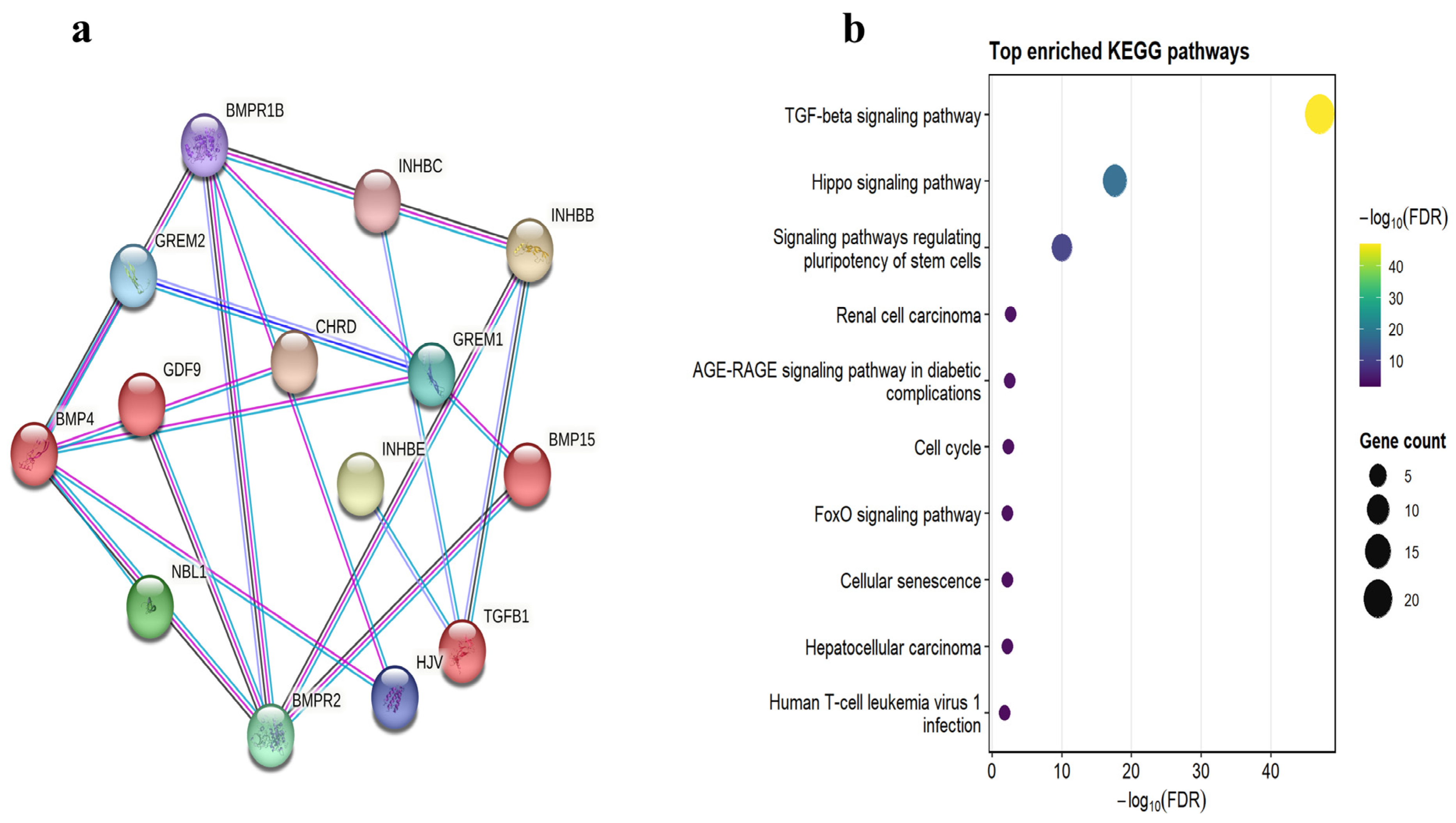
To examine functional interactions of the identified TGF-β superfamily proteins, a protein–protein interaction (PPI) network was drawn with the help of the STRING database (version 11.5). The protein sequences were mapped to their orthologs, and interaction data were retrieved using experimental evidence, curated databases, co-expression, gene neighborhood, gene fusion, and text mining. A confidence score threshold of ≥0.4 was applied to filter interactions. The resulting interaction network was directly visualised with the STRING web interface, with nodes denoting proteins and edges denoting the predicted functional interactions.
To conduct functional characterization, the STRING enrichment module was used to conduct KEGG pathway enrichment analysis. False discovery rate (FDR)-adjusted p-values were used to assess statistical significance. The top enriched pathways were selected based on both statistical significance and biological relevance. The results of the enrichment were plotted in Python (version 3.13) with the Matplotlib (3.10.x) library, with the value of −log10(FDR) indicating the significance of a given pathway and the bubble size denoting the number of genes that participated in this pathway.

3. Results

3.1. Genome-Wide Identification of Donkey TGF-β Genes

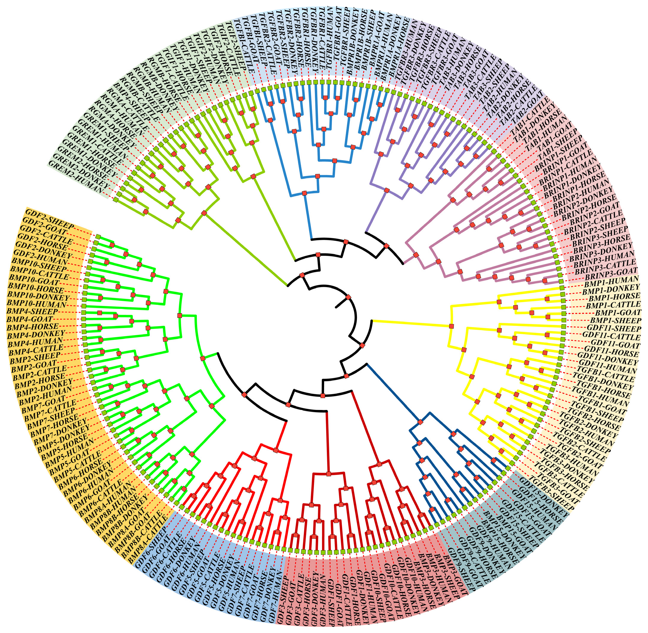
BLAST and HMMER analyses across six mammals (donkey, horse, cattle, sheep, goat, human) identified 205 non-redundant TGF-β-encoded protein sequences. In donkey (Equus asinus, ASM1607732v2), 40 TGF-β genes were identified, exceeding the counts observed in several mammals. This observation may reflect lineage-specific expansion or differential gene retention within Equidae, although additional comparative genomic analyses are required to confirm this evolutionary scenario, which may have contributed to the diversification of growth and reproductive traits in donkeys. Domain annotation using SMART and NCBI-CDD confirmed the presence of canonical TGF-β domains in all candidates, ensuring a high-confidence gene set (Figure 1). This expansion in donkey may be related to other mammals, highlighting potential Equidae-specific evolutionary adaptations in TGF-β signaling.

3.2. Evolutionary Analysis

Phylogenetic analysis, supported by bootstrap values greater than 70%, separated the sequences into two primary clades: TGF-β-like and BMP-like. The TGF-β-like clade was further divided into five subgroups (NODAL, GDF10/BMP3, GDF11/MSTN, TGF-β, and INHIBIN), while the BMP-like clade comprised BMP2/4/6, GDF2, GDF5/6/7, BMP5/6/7/8A/8B, GDF1/3/9/15, and BMP15 (Figure 1). Donkey genes exhibited higher sequence similarity to horse orthologs than to ruminants, consistent with the close evolutionary relationship within Equidae. Notably, expansion in the TGF-β-like subgroups may support species-specific regulatory adaptations in growth and reproductive pathways.
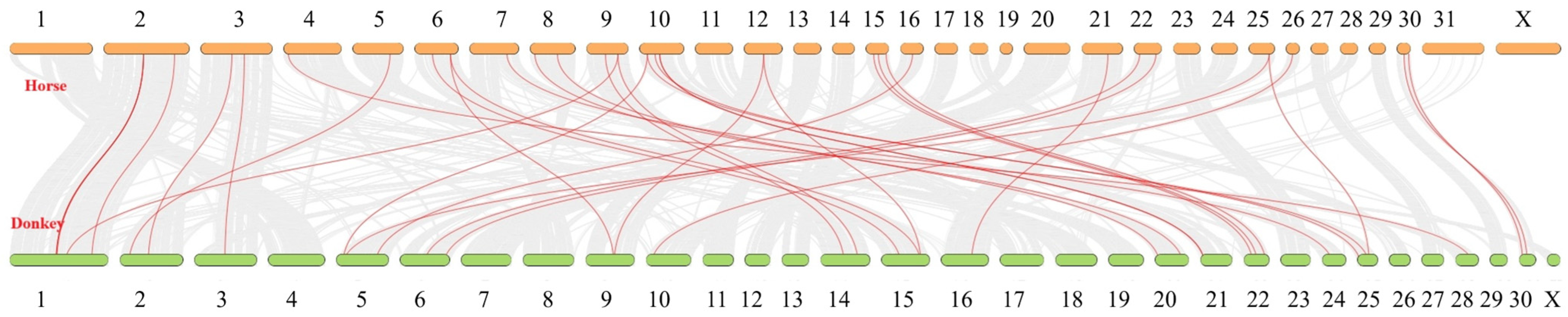
Four segmental duplications were identified. Synteny analysis revealed high collinearity with the horse genome, reflecting conserved equid genomic architecture despite species divergence. Donkey TGF-β genes were mapped to 14 chromosomes, whereas horse genes spanned 16, predominantly at distal chromosomal ends (Figure 2, Table 1). Ka/Ks ratios of 0.28–0.43 indicated strong purifying selection acting on duplicated genes, suggesting that these paralogs have been functionally conserved rather than nonfunctionalized.

3.3. Structural and Physicochemical Characterization

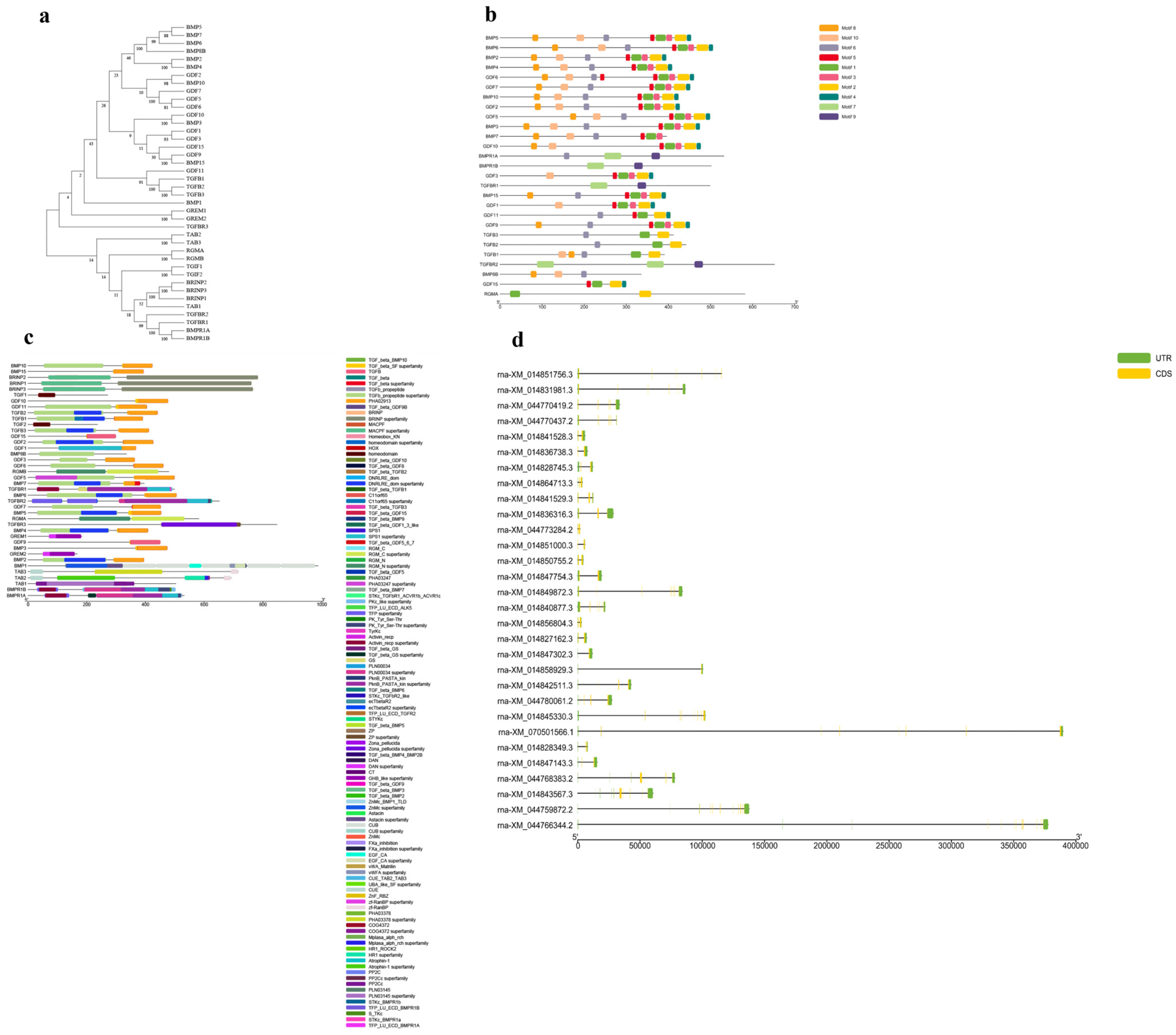
Integration of phylogenetic relationships, motif architecture, conserved domains, and exon–intron organization revealed structural diversity among donkey TGF-β genes, with seven conserved MEME motifs identified (Figure 3a–d). Motifs 1 and 2 corresponded to the canonical TGF-β domains (Figure 3b; Table 2), whereas TGFb_propeptide domains were identified in selected genes (Figure 3c). Variation in exon–intron organization and the diversity of 5′ and 3′ untranslated regions (UTRs) suggest regulatory complexity that may contribute to differential gene expression across tissues and developmental stages (Figure 3d). Furthermore, as shown in Table 3, the predicted molecular weights ranged from 34.5 to 56.2 kDa, while the theoretical isoelectric points (pI) ranged from 4.95 to 10.35. Most proteins were basic, except for TAB1, TGFBR2, TGFBR3, BMP1, and BMP10, which exhibited pI values below 7. High aliphatic index (AI) values (>60) suggested good thermostability, whereas negative GRAVY scores indicated that the analyzed TGF-β family proteins are predominantly hydrophilic (Table 2). Collectively, the coexistence of conserved structural features and lineage-specific variations suggests that the donkey TGF-β superfamily has evolved under strong evolutionary constraints while maintaining regulatory flexibility, thereby preserving essential biological functions and facilitating potential functional divergence of TGF-β signaling in donkeys.

3.4. Mutation Analysis

Comparative sequence analysis uncovered 160 amino acid variants, including 11 predicted deleterious mutations in GDF6 (3), GDF9 (2), GDF10 (4), BMP15 (1), and RGMA (1), highlighting potential functional divergence affecting reproductive and developmental pathways. Protein sequences of donkey and horse TGF-β genes were aligned using ClustalW (v2.1) and visualized in BioEdit (v7.2.5). Functional effects of nonsynonymous mutations were predicted using SIFT (version 6.2.1), PROVEAN (release 1.1.5), PolyPhen-2 (HumVar model, 2013 release), I-Mutant 3.0 (2010 release), Phd-SNP (2010 release), and MUpo (2006 release). Consensus results are presented in Table 4 and Supplementary Tables S1–S40.

3.5. Functional Prediction

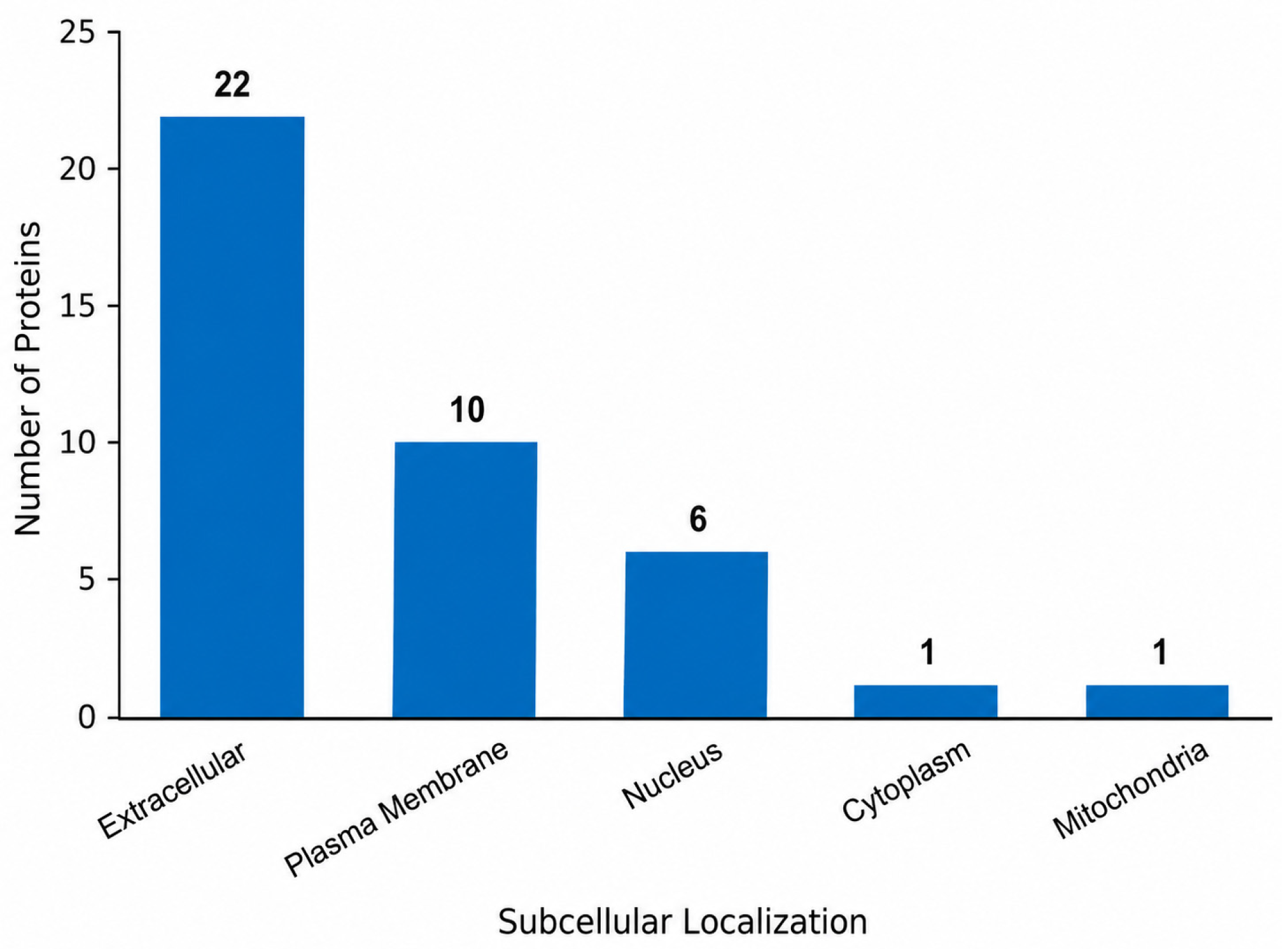
Subcellular localization was predicted using WoLF PSORT, with the majority localized extracellularly, supporting signaling-related functions, with additional nuclear, plasma membrane, cytoplasmic, and mitochondrial distributions. Together, these computational predictions suggest that the identified variants and predicted localizations may be associated with biological functions related to reproduction, growth, and embryogenesis, although experimental validation is required (Figure 4).

3.6. Tissue-Specific Expression Profiling of Donkey TGF-β Family Genes

To assess the potential functional relevance of donkey TGF-β family genes, tissue-specific transcript abundance profiles were examined using publicly available donkey RNA-seq data (NCBI BioProject: PRJNA1017964). Expression analysis across the ovary, uterus, pineal gland, spleen, blood, quadriceps femoris, and longissimus dorsi revealed marked tissue-specific variation in the transcriptional activity of TGF-β signaling components (Table 5). Reproductive tissues, particularly the ovary and uterus, exhibited enriched expression of several TGF-β/BMP pathway genes. In the ovary, TGFBR2 displayed the highest transcript abundance, followed by TGFBR1, TGFB1, BMP2, BMP7, and BMP4, indicating active receptor-mediated signaling. Canonical fertility-associated genes, GDF9 and BMP15, also showed ovary-associated expression, supporting their conserved roles in folliculogenesis, oocyte maturation, and ovulation. Notably, the predominance of receptor transcripts over ligand expression suggests a coordinated and potentially receptor-dominant regulatory environment in donkey ovarian tissue. Similarly, the uterus exhibited strong expression of TGFBR2, BMP4, GDF11, TGFBR1, TGFB2, and BMP7, highlighting an active signaling landscape potentially associated with tissue remodeling and reproductive tract function. Expression of TGF-β signaling genes in the pineal gland further suggested a possible neuroendocrine contribution to reproductive physiology through endocrine signaling pathways.
In contrast, non-reproductive tissues, including spleen, blood, and skeletal muscles (quadriceps femoris and longissimus dorsi), displayed broader but distinct expression profiles. High expression of TGFBR2, TGFB1, BMPR1A, and BMP4 in spleen and muscle tissues supports conserved roles in immune modulation, tissue maintenance, and developmental signaling. Blood tissue showed comparatively restricted expression, dominated by TGFBR1, TGFBR2, and BMP2, indicating systemic signaling responsiveness. Overall, these findings demonstrate tissue-specific enrichment of TGF-β family genes in donkeys and provide transcriptomic support for their involvement in reproductive and developmental processes. The coordinated expression of ligands and receptors in reproductive tissues reinforces the functional significance of TGF-β signaling in equid fertility biology.

3.7. Integrated PPI and KEGG Enrichment Analysis

In order to further clarify the functional interaction and biological role of the identified TGF-β superfamily genes, a combined analysis based on protein–protein interaction (PPI) network and KEGG pathway enrichment was conducted (Figure 5).
STRING-based PPI network predicted that the interaction landscape was highly interconnected, demonstrating that the components of TGF-β signaling are strongly functionally connected (Figure 5a). A number of proteins, such as TGFB1, BMPR1B, BMP4, BMP2, and GDF9, were identified as predicted hub nodes with high connectivity. In particular, TGFB1 was predicted to interact strongly with BMP ligands and receptor proteins (BMPR2 and BMPR1B), highlighting its central role in canonical TGF-β signaling pathways. The BMP and GDF subfamilies (BMP4, BMP15, GDF9, and GDF6) were present in the network as highly linked clusters, indicating some coordinated role in developmental and reproductive events. The interaction between GDF9 and BMP15, as observed, also supports the already established roles of the two in folliculogenesis and oocyte maturation, in line with the ovary-enriched expression patterns of the two in this study. Moreover, the regulatory factors (GREM1, GREM2, and CHRD) were incorporated into the network, which means that they are extracellular antagonists that control BMP signaling activity. Likewise, the core signaling proteins were also linked to inhibin subunits (INHBA, INHBB, and INHBC), which further highlights their role in the regulation of reproductive hormones.
Enrichment analysis of KEGG pathways (Figure 5b) revealed that there is significant enrichment in pathways related to TGF-β signaling, Hippo signaling, and stem cell pluripotency in addition to other regulatory processes that include cell cycle, FoxO signaling, and cellular senescence. The identified gene family is functionally relevant as these pathways are mostly engaged in cell differentiation, proliferation, and developmental regulation. Even though some disease-related pathways were also enriched, they probably represent overlapping molecular signaling pathways, as in most studies of pathway enrichment of conserved signaling networks. In general, the integrated PPI and KEGG results show that the donkey TGF-β gene family is an integrated and coordinated signaling network, with critical roles in developmental control, cellular signaling, and reproductive functions.

4. Discussion

Advances in high-throughput genome sequencing have enabled genome-wide identification of functional gene families and genetic variants across diverse organisms, including livestock, plants, avian species, and pathogenic bacteria [37,38,39]. In livestock, candidate gene studies increasingly integrate genomic resources with transcriptomic evidence to identify functional genes associated with economically important traits, such as disease resistance, reproductive efficiency, and environmental adaptation [40,41]. Comparative genomics between donkeys and horses provides a valuable framework for uncovering species-specific adaptations and the genetic basis of economically important traits [42,43].
In this study, a total of 40 TGF-β genes were identified in the donkey genome (Equus asinus, ASM1607732v2) and classified into TGF-β-like and BMP-like groups. The TGF-β-like group was further divided into five subgroups: NODAL, GDF10/BMP3, GDF11/MSTN, TGF-β, and INHIBIN, whereas the BMP-like group comprised BMP2/4/6, GDF2, GDF5/6/7, BMP5/6/7/8A/8B, GDF1/3/9/15, and BMP15 (Figure 1). This phylogenetic organization is consistent with previous studies demonstrating strong evolutionary conservation of the TGF-β superfamily across vertebrates [44,45,46]. However, the number of TGF-β family members identified in donkeys was slightly higher than that reported in several mammals [47,48], including the closely related horse, suggesting potential lineage-specific expansion or differential retention of duplicated genes. Although additional evolutionary analyses are needed to determine whether these differences represent true biological diversification or annotation variation, the observed expansion may reflect species-specific functional requirements within Equidae.
Structural analyses identified seven conserved motifs, including TGF-β and cystine-knot cytokine domains [45] (Table 1, Figure 3), which are essential for ligand–receptor interactions and downstream signaling [48,49]. Four segmental duplications (GDF2/BMP10, GDF1/GDF3, GDF5/GDF6, and GREM1/GREM2) were identified, with Ka/Ks ratios ranging from 0.28 to 0.43, indicating strong purifying selection (Table 3). High synteny with the horse genome (Figure 2) further supports the conserved genomic architecture of equids, consistent with previous comparative studies [42,43]. Although genomic resources for non-equid perissodactyls such as rhinoceros and tapir remain limited [40,50], the observed genomic conservation appears characteristic of Equidae. Comparative sequence analysis revealed 160 amino acid variants between donkey and horse TGF-β orthologs, including 11 predicted deleterious substitutions in GDF6, GDF9, GDF10, BMP15, and RGMA (Table 4). Notably, some of these variants occurred in genes with established reproductive functions. GDF9 and BMP15 are well-recognized regulators of folliculogenesis, oocyte maturation, and ovulation in mammals [51,52], while GDF6 and GDF10 contribute to developmental signaling and skeletal formation [53,54]. Although the phenotypic effects of these variants remain unresolved in donkeys, their occurrence in functionally relevant genes suggests potential contributions to species-specific reproductive and developmental traits that warrant future experimental investigation.
Importantly, transcriptomic validation using publicly available donkey RNA-seq data (NCBI BioProject: PRJNA1017964) provided additional functional support for the biological relevance of TGF-β family genes. Tissue-specific expression profiling demonstrated that reproductive tissues, particularly the ovary and uterus, exhibited enriched expression of several pathway components, including TGFBR1, TGFBR2, TGFB1, BMP2, BMP4, and BMP7. Canonical reproductive genes such as GDF9 and BMP15 showed ovary-associated expression, consistent with their conserved roles in oocyte–granulosa cell communication and follicular development [7,55]. Interestingly, receptor genes (TGFBR1 and TGFBR2) displayed stronger transcript abundance than several canonical ligands, suggesting that reproductive regulation in donkeys may involve a receptor-dominant TGF-β signaling environment rather than dependence on a limited number of ovary-specific factors alone. Such coordinated expression of ligands and receptors may contribute to follicular maturation, reproductive tissue remodeling, and fertility-related physiology. While the expression patterns in non-reproductive tissues further supported the pleiotropic nature of the TGF-β superfamily. Spleen and muscle tissues showed broader expression of signaling components, including TGFB1, BMP4, BMPR1A, and TGFBR2, consistent with established functions in immune modulation, tissue maintenance, and developmental regulation [56,57]. Likewise, the presence of TGF-β signaling genes in the pineal gland suggests possible neuroendocrine contributions to reproductive physiology, potentially linking endocrine timing with reproductive function in equids. Together, these findings indicate that the donkey TGF-β superfamily exhibits tissue-specific transcriptional specialization while retaining broad physiological functionality.
Subcellular localization predictions suggested that most proteins (22/40) are extracellularly localized (Figure 4), consistent with their established functions as secreted signaling molecules within the TGF-β superfamily [7,55]. Protein–protein interaction analyses further identified hub genes such as TGFB1, BMP4, and GDF9 (Figure 5), mirroring interaction patterns reported in cattle, sheep, and humans [58]. The observed interactions between GDF9 and BMP15 are particularly noteworthy, given their conserved cooperative roles in follicular maturation and female fertility [59,60]. Overall, this study provides the first integrated genomic and transcriptomic characterization of the donkey TGF-β superfamily, revealing conserved evolutionary architecture, duplicated genes under purifying selection, candidate deleterious variants, and tissue-specific expression patterns associated with reproductive and developmental biology. These findings broaden our understanding of equid molecular evolution and provide a foundation for future functional genomics and breeding applications.

Limitations and Future Directions

Although this study provides robust in silico evidence of donkey-specific duplications and deleterious mutations, the findings are based on computational predictions. Functional validation through targeted gene expression analysis, protein interaction assays, CRISPR editing, and phenotypic association studies is required to confirm biological relevance. Data for non-equid perissodactyls remain scarce, limiting broader evolutionary comparisons. Future functional and breeding studies building on this TGF-β atlas will help clarify the role of these variants in donkey reproduction, growth, and resilience, ultimately supporting marker-assisted selection programs. Furthermore, the predicted protein–protein interactions and pathway enrichments are based on database-derived computational models and should not be interpreted as direct experimental evidence in donkey.

5. Conclusions

This study presents the first genome-wide characterization of the TGF-β superfamily in donkey (Equus asinus), identifying 40 TGF-β family genes with strong evolutionary conservation, lineage-specific expansion, and four segmental duplications under purifying selection (Ka/Ks = 0.28–0.43). Comparative analyses identified 11 predicted deleterious variants in key genes (GDF6, GDF9, GDF10, BMP15, and RGMA), suggesting potential functional divergence related to reproductive and developmental processes. Transcriptomic validation using publicly available donkey RNA-seq data further revealed tissue-specific enrichment of TGF-β signaling genes, particularly in reproductive tissues (ovary and uterus), supporting their potential roles in folliculogenesis, reproductive tissue remodeling, and fertility-related signaling. Subcellular localization analysis indicated that most proteins are extracellular, consistent with conserved signaling functions. Finally, this study provides the first integrated genomic and transcriptomic resource for the donkey TGF-β superfamily and offers a valuable foundation for future functional genomics, reproductive biology, and breeding applications in equids.

Supplementary Materials

The following supporting information can be downloaded at https://www.mdpi.com/article/10.3390/ani16132028/s1.

Author Contributions

Methodology, Resources, Data curation, Writing—original draft preparation, T.N.; conceptualization, visualization, M.T. (Muhammad Tariq); methodology, resources, data curation, M.T. (Mohamed Tharwat); conceptualization, writing—review and editing, project administration, supervision: M.S.; investigation, validation, Y.J.; and funding acquisition, writing—review and editing, F.A.A. All authors have read and agreed to the published version of the manuscript.

Funding

The researchers would like to thank the Deanship of Graduate Studies and Scientific Research at Qassim University (www.qu.edu.sa) for financial support (QU-APC-2026).

Institutional Review Board Statement

This study did not involve experiments on live animals and therefore did not require approval from an institutional ethics committee.

Informed Consent Statement

Not applicable.

Data Availability Statement

The original contributions presented in this study are included in the article. Further inquiries can be directed to the corresponding authors.

Conflicts of Interest

The authors declare no conflicts of interest.

Abbreviations

The following abbreviations are used in this manuscript:
AIAliphatic index
BLASTBasic Local Alignment Search Tool
BMPBone morphogenetic protein
CDDConserved Domain Database
GDFGrowth differentiation factor
GFF3General Feature Format version 3
GRAVYGrand average of hydropathicity
HMMHidden Markov model
HMMERHidden Markov Model-based sequence analysis tool
IIInstability index
KaNonsynonymous substitution rate
KsSynonymous substitution rate
lncRNALong noncoding RNA
MCScanXMultiple Collinearity Scan toolkit
MEGAMolecular Evolutionary Genetics Analysis
MEMEMultiple Expectation Maximization for Motif Elicitation
miRNAMicroRNA
MSTNMyostatin
MWMolecular weight
MYAMillion years ago
NCBINational Center for Biotechnology Information
NGSNext-generation sequencing
NJNeighbor-joining
PfamProtein families database
pIIsoelectric point
PROVEANProtein Variation Effect Analyzer
RGMARepulsive Guidance Molecule A
R-SMADReceptor-regulated SMAD
SIFTSorting Intolerant From Tolerant
SMARTSimple Modular Architecture Research Tool
SNPSingle nucleotide polymorphism
SMADSmall Mothers Against Decapentaplegic
TBtoolsToolkit for Biologists
TGF-βTransforming growth factor beta
TGFBRTransforming growth factor beta receptor
UTRUntranslated region
WoLF PSORTWeighted k-nearest neighbor Protein Subcellular Localization Prediction Tool

References

  1. Moses, H.L.; Roberts, A.B.; Derynck, R. The discovery and early days of TGF-β: A historical perspective. Cold Spring Harb. Perspect. Biol. 2016, 8, a021865. [Google Scholar] [CrossRef] [PubMed]
  2. Roth, S.; Gong, W.; Gressner, A.M. Expression of different isoforms of TGF-β and the latent TGF-β binding protein (LTBP) by rat Kupffer cells. J. Hepatol. 1998, 29, 915–922. [Google Scholar] [CrossRef] [PubMed]
  3. Capdevila, J.; Belmonte, J.C.I. Patterning mechanisms controlling vertebrate limb development. Annu. Rev. Cell Dev. Biol. 2001, 17, 87–132. [Google Scholar] [CrossRef] [PubMed]
  4. Rissi, M.; Wittbrodt, J.; Délot, E.; Naegeli, M.; Rosa, F.M. Zebrafish Radar: A new member of the TGF-β superfamily defines dorsal regions of the neural plate and the embryonic retina. Mech. Dev. 1995, 49, 223–234. [Google Scholar] [CrossRef] [PubMed]
  5. Grimaud, E.; Heymann, D.; Rédini, F. Recent advances in TGF-β effects on chondrocyte metabolism: Potential therapeutic roles of TGF-β in cartilage disorders. Cytokine Growth Factor Rev. 2002, 13, 241–257. [Google Scholar] [CrossRef] [PubMed]
  6. Wu, M.Y.; Hill, C.S. TGF-β superfamily signaling in embryonic development and homeostasis. Dev. Cell 2009, 16, 329–343. [Google Scholar] [CrossRef] [PubMed]
  7. Morikawa, M.; Derynck, R.; Miyazono, K. TGF-β and the TGF-β family: Context-dependent roles in cell and tissue physiology. Cold Spring Harb. Perspect. Biol. 2016, 8, a021873. [Google Scholar] [CrossRef] [PubMed]
  8. Kang, J.S.; Liu, C.; Derynck, R. New regulatory mechanisms of TGF-β receptor function. Trends Cell Biol. 2009, 19, 385–394. [Google Scholar] [CrossRef] [PubMed]
  9. Huminiecki, L.; Goldovsky, L.; Freilich, S.; Moustakas, A.; Ouzounis, C.; Heldin, C.-H. Emergence, development and diversification of the TGF-β signalling pathway within the animal kingdom. BMC Evol. Biol. 2009, 9, 28. [Google Scholar] [CrossRef] [PubMed]
  10. Dayal, S.; Chaubey, D.; Joshi, D.C.; Ranmale, S.; Pillai, B. Noncoding RNAs: Emerging regulators of behavioral complexity. Wiley Interdiscip. Rev. RNA 2024, 15, e1847. [Google Scholar] [CrossRef] [PubMed]
  11. Kumar, S.; Chauhan, M.S. Relative expression of the developmentally important candidate genes in immature oocytes and in vitro-produced embryos of buffalo (Bubalus bubalis). Zygote 2022, 30, 509–515. [Google Scholar] [CrossRef] [PubMed]
  12. Cheng, Z.; Fu, L.; Yimamu, M.; Feng, J.; Wu, L.; Hu, Y.; Wu, J.; Xu, X.; Guo, C. Fenofibrate Inhibits Hepatic Stellate Cell Activation and Autophagy in Liver Fibrosis through the Tgfβ1/Smad3 and Pparα/Cgas/Sting Pathways. Pak. Vet. J. 2025, 45, 579–591. [Google Scholar] [CrossRef]
  13. Li, Y.; Jing, J.; Dang, W.; Jia, K.; Guo, X.; Kebreab, E.; Lyu, L.; Zhao, J. Cross-talk between NOTCH2 and BMP4/SMAD signaling pathways in bovine follicular granulosa cells. Theriogenology 2022, 187, 74–81. [Google Scholar] [CrossRef] [PubMed]
  14. Dall’Olio, S.; Fontanesi, L.; Costa, L.N.; Tassinari, M.; Minieri, L.; Falaschini, A. Analysis of horse myostatin gene and identification of single nucleotide polymorphisms in breeds of different morphological types. BioMed Res. Int. 2010, 2010, 542945. [Google Scholar] [CrossRef] [PubMed]
  15. Shehzad, S.; Hafeez, A.; Chang, M.S.; Suthar, V.; Khaskheli, R.A.; Fatima, S.; Binish, S.; Rasool, G.; Mehmood, A.; Ali, A.; et al. Genome-wide identification and functional analysis of Kunitz-Type Trypsin Inhibitor gene family in cotton against pest resistance. Int. J. Agric. Biosci. 2026, 15, 168–174. [Google Scholar] [CrossRef] [PubMed]
  16. Wong, X.J.; Ramaiya, S.D.; Cheng, W.H.; Wang, Z.F.; Hishamuddin, M.S.; Lee, S.Y. Complete plastid genome of Coelostegia griffithii (Malvaceae): Structure, comparative and phylogenetic analysis. Asian J. Agric. Biol. 2025, 2024015. [Google Scholar] [CrossRef]
  17. Budiyanto, A.; Hartanto, S.; Widayanti, R.; Setyawan, E.M.N.; Haryanto, A.; Ibrahim, A.; Pakpahan, S. Genetic diversity of Jawa-Brebes cattle based on reproductive traits markers of the growth hormone gene. Int. J. Vet. Sci. 2025, 14, 351–357. [Google Scholar] [CrossRef]
  18. Fatmona, S.; Sjafani, N.; Sulasmi; Utami, S.; Wahyuni, S.; Sahil, J.; Keintjem, J.R.M. Genetic distance and kinship relationship of Walik Kembang Sula bird (Ptilinopus melanosphila) based on mtDNA CO1 in North Maluku, Indonesia. Int. J. Vet. Sci. 2024, 14, 548–556. [Google Scholar] [CrossRef]
  19. Huang, D.; Wang, R.; Liu, S.; Shao, Y.; Zhang, Z.; Yu, C.; Bao, S.; Cong, Y. Genomic analysis and evaluation of pathogenicity and immunogenicity of Riemerella anatipestifer isolates from chickens in China. Pak. Vet. J. 2025, 45, 1345–1352. [Google Scholar] [CrossRef]
  20. Kun, L.; Hassan, M.; Ahmad, H.I.; Shahzad, A.H.; Raza, S.; Qadeer, I.; Muhammad, S.A.; Liu, W. Comparative genomics analysis of cation channel sperm-associated proteins (CATSPERs) associated with sperm motility in livestock. Pak. Vet. J. 2025, 45, 1157–1167. [Google Scholar] [CrossRef]
  21. Nasir, T.; Safdar, M.; Imran, S.; Tariq, M.; Junejo, Y.; Ozaslan, M.; Younus, M. Genome-wide in silico identification and characterization of the F-box gene family in water buffalo (Bubalus bubalis). Comp. Biochem. Physiol. Part D Genom. Proteom. 2026, 59, 101825. [Google Scholar]
  22. Zhu, Q.; Khan, M.Z.; Jing, Y.; Geng, M.; Zhang, X.; Zheng, Y.; Cao, X.; Peng, Y.; Wang, C. The Donkey Genome: From Evolutionary Insights to Sustainable Breeding Strategies. Animals 2025, 16, 93. [Google Scholar] [CrossRef] [PubMed]
  23. Kitts, P.A.; Church, D.M.; Thibaud-Nissen, F.; Choi, J.; Hem, V.; Sapojnikov, V.; Smith, R.G.; Tatusova, T.; Xiang, C.; Zherikov, A.; et al. Assembly: A resource for assembled genomes at NCBI. Nucleic Acids Res. 2016, 44, D73–D80. [Google Scholar] [CrossRef] [PubMed]
  24. UniProt Consortium. UniProt: A worldwide hub of protein knowledge. Nucleic Acids Res. 2019, 47, D506–D515. [Google Scholar] [CrossRef] [PubMed]
  25. Finn, R.D.; Clements, J.; Eddy, S.R. HMMER web server: Interactive sequence similarity searching. Nucleic Acids Res. 2011, 39, W29–W37. [Google Scholar] [CrossRef] [PubMed]
  26. Potter, S.C.; Luciani, A.; Eddy, S.R.; Park, Y.; López, R.; Finn, R.D. HMMER web server: 2018 update. Nucleic Acids Res. 2018, 46, W200–W204. [Google Scholar] [CrossRef] [PubMed]
  27. El-Gebali, S.; Mistry, J.; Bateman, A.; Eddy, S.R.; Luciani, A.; Potter, S.C.; Qureshi, M.; Richardson, L.J.; Salazar, G.A.; Smart, A.; et al. The Pfam protein families database in 2019. Nucleic Acids Res. 2019, 47, D427–D432. [Google Scholar] [CrossRef] [PubMed]
  28. Kumar, S.; Stecher, G.; Tamura, K. MEGA7: Molecular evolutionary genetics analysis version 7.0 for bigger datasets. Mol. Biol. Evol. 2016, 33, 1870–1874. [Google Scholar] [CrossRef] [PubMed]
  29. Wang, Y.; Tang, H.; DeBarry, J.D.; Tan, X.; Li, J.; Wang, X.; Lee, T.-H.; Jin, H.; Marler, B.; Guo, H.; et al. MCScanX: A toolkit for detection and evolutionary analysis of gene synteny and collinearity. Nucleic Acids Res. 2012, 40, e49. [Google Scholar] [CrossRef] [PubMed]
  30. Rozas, J.; Ferrer-Mata, A.; Sánchez-DelBarrio, J.C.; Guirao-Rico, S.; Librado, P.; Ramos-Onsins, S.E.; Sánchez-Gracia, A. DnaSP 6: DNA sequence polymorphism analysis of large data sets. Mol. Biol. Evol. 2017, 34, 3299–3302. [Google Scholar] [CrossRef] [PubMed]
  31. Chen, C.; Chen, H.; Zhang, Y.; Thomas, H.R.; Frank, M.H.; He, Y.H.; Xia, R. TBtools: An integrative toolkit developed for interactive analyses of big biological data. Mol. Plant 2020, 13, 1194–1202. [Google Scholar] [CrossRef] [PubMed]
  32. Wall, J.D. Estimating ancestral population sizes and divergence times. Genetics 2003, 163, 395–404. [Google Scholar] [CrossRef] [PubMed]
  33. Sim, N.-L.; Kumar, P.; Hu, J.; Henikoff, S.; Schneider, G.; Ng, P.C. SIFT web server: Predicting effects of amino acid substitutions on proteins. Nucleic Acids Res. 2012, 40, W452–W457. [Google Scholar] [CrossRef] [PubMed]
  34. Choi, Y.; Chan, A.P. PROVEAN web server: A tool to predict the functional effect of amino acid substitutions and indels. Bioinformatics 2015, 31, 2745–2747. [Google Scholar] [CrossRef] [PubMed]
  35. Capriotti, E.; Fariselli, P. PhD-SNPg: A webserver and lightweight tool for scoring single nucleotide variants. Nucleic Acids Res. 2017, 45, W247–W252. [Google Scholar] [CrossRef] [PubMed]
  36. Horton, P.; Park, K.-J.; Obayashi, T.; Fujita, N.; Harada, H.; Adams-Collier, C.J.; Nakai, K. WoLF PSORT: Protein localization predictor. Nucleic Acids Res. 2007, 35, W585–W587. [Google Scholar] [CrossRef] [PubMed]
  37. Wang, Y.; Huang, Y.; Zhen, Y.; Wang, J.; Wang, L.; Chen, N.; Wu, F.; Zhang, L.; Shen, Y.; Bi, C.; et al. De novo transcriptome assembly database for 100 tissues from each of seven species of domestic herbivore. Sci. Data 2024, 11, 488. [Google Scholar] [CrossRef] [PubMed]
  38. Kingsley, D.M. The TGF-β superfamily: New members, new receptors, and new genetic tests of function in different organisms. Genes Dev. 1994, 8, 133–146. [Google Scholar] [CrossRef] [PubMed]
  39. Hogan, B.L.M. Bone morphogenetic proteins: Multifunctional regulators of vertebrate development. Genes Dev. 1996, 10, 1580–1594. [Google Scholar] [CrossRef] [PubMed]
  40. Akgun, E.E.; Erdogan, M.; Altunbas, K. BMP-9 and TGF-β3 Synergistically Regulate Chondrogenic Pathways in Bovine Synovial Fluid-Derived Mesenchymal Stem Cells (BSF-MSCs) in Transwell Co-Culture with Chondrocytes. Pak. Vet. J. 2025, 45, 138–148. [Google Scholar] [CrossRef] [PubMed]
  41. Mottershead, D.G.; Ritter, L.J.; Gilchrist, R.B. Signalling pathways mediating specific synergistic interactions between GDF9 and BMP15. Mol. Hum. Reprod. 2012, 18, 121–128. [Google Scholar] [CrossRef] [PubMed]
  42. David, L.; Mallet, C.; Mazerbourg, S.; Feige, J.-J.; Bailly, S. Identification of BMP9 and BMP10 as functional activators of the orphan activin receptor-like kinase 1 (ALK1) in endothelial cells. Blood 2007, 109, 1953–1961. [Google Scholar] [CrossRef] [PubMed]
  43. Kalbfleisch, T.S.; Rice, E.S.; DePriest, M.S., Jr.; Walenz, B.P.; Hestand, M.S.; Vermeesch, J.R.; O’Connell, B.L.; Fiddes, I.T.; Vershinina, A.O.; Saremi, N.F. Improved reference genome for the domestic horse increases assembly contiguity and composition. Commun. Biol. 2018, 1, 197. [Google Scholar] [CrossRef] [PubMed]
  44. Li, S.; Zhao, G.; Han, H.; Li, Y.; Li, J.; Wang, J.; Li, X. Genome collinearity analysis illuminates the evolution of donkey chromosome 1 and horse chromosome 5 in perissodactyls: A comparative study. BMC Genom. 2021, 22, 665. [Google Scholar] [CrossRef]
  45. Massagué, J. TGF-β signal transduction. Annu. Rev. Biochem. 1998, 67, 753–791. [Google Scholar] [CrossRef] [PubMed]
  46. Tanaka, C.; Sakuma, R.; Nakamura, T.; Hamada, H.; Saijoh, Y. Long-range action of Nodal requires interaction with GDF1. Genes Dev. 2007, 21, 3272–3282. [Google Scholar] [CrossRef] [PubMed]
  47. Magadum, S.; Banerjee, U.; Murugan, P.; Gangapur, D.; Ravikesavan, R. Gene duplication as a major force in evolution. J. Genet. 2013, 92, 155–161. [Google Scholar] [CrossRef] [PubMed]
  48. Huang, J.; Zhao, Y.; Bai, D.; Shiraigol, W.; Li, B.; Yang, L.; Wu, J.; Bao, W.; Ren, X.; Jin, B.; et al. Donkey genome and insight into the imprinting of fast karyotype evolution. Sci. Rep. 2015, 5, 14106. [Google Scholar] [CrossRef] [PubMed][Green Version]
  49. Rehman, M.S.-U.; Hassan, F.-U.; Rehman, Z.-U.; Ishtiaq, I.; Rehman, S.U.; Liu, Q. Molecular characterization of TGF-beta gene family in buffalo to identify gene duplication and functional mutations. Genes 2022, 13, 1302. [Google Scholar] [CrossRef] [PubMed]
  50. Conant, G.C.; Wolfe, K.H. Turning a hobby into a job: How duplicated genes find new functions. Nat. Rev. Genet. 2008, 9, 938–950. [Google Scholar] [CrossRef] [PubMed]
  51. Steiner, C.C.; Ryder, O.A. Molecular phylogeny and evolution of the Perissodactyla. Zool. J. Linn. Soc. 2011, 163, 1289–1303. [Google Scholar] [CrossRef]
  52. Otsuka, F.; McTavish, K.J.; Shimasaki, S. Integral role of GDF-9 and BMP-15 in ovarian function. Mol. Reprod. Dev. 2011, 78, 9–21. [Google Scholar] [PubMed]
  53. Belli, M.; Shimasaki, S. Molecular aspects and clinical relevance of GDF9 and BMP15 in ovarian function. Vitam. Horm. 2018, 107, 317–348. [Google Scholar] [CrossRef] [PubMed]
  54. Chen, L.; Deng, C.-X. Roles of FGF signaling in skeletal development and human genetic diseases. Front. Biosci. 2005, 10, 1961–1976. [Google Scholar] [CrossRef] [PubMed]
  55. Fountas, S.; Petinaki, E.; Bolaris, S.; Kargakou, M.; Dafopoulos, S.; Zikopoulos, A.; Moustakli, E.; Sotiriou, S.; Dafopoulos, K. The Roles of GDF-9, BMP-15, BMP-4 and EMMPRIN in Folliculogenesis and In Vitro Fertilization. J. Clin. Med. 2024, 13, 3775. [Google Scholar] [CrossRef] [PubMed]
  56. Asghari, R.; Shokri-Asl, V.; Rezaei, H.; Tavallaie, M.; Khafaei, M.; Abdolmaleki, A.; Seghinsara, A.M. Alteration of TGFB1, GDF9, and BMPR2 Gene Expression in Preantral Follicles of an Estradiol Valerate-Induced Polycystic Ovary Mouse Model Can Lead to Anovulation, Polycystic Morphology, Obesity, and Absence of Hyperandrogenism. Clin. Exp. Reprod. Med. 2021, 48, 245–254. [Google Scholar] [CrossRef] [PubMed]
  57. Settle, S.H., Jr.; Rountree, R.B.; Sinha, A.; Thacker, A.; Higgins, K.; Kingsley, D.M. Multiple joint and skeletal patterning defects caused by single and double mutations in the mouse Gdf6 and Gdf5 genes. Dev. Biol. 2003, 254, 116–130. [Google Scholar] [CrossRef] [PubMed]
  58. Massagué, J. TGFβ signalling in context. Nat. Rev. Mol. Cell Biol. 2012, 13, 616–630. [Google Scholar] [CrossRef] [PubMed]
  59. Carmona Dinau, F.; González-Zambrano, C.M.; Jurado Jimenez, J.; Montoya Florez, L.M.; Oliveira, R.; Sousa Rocha, N. Exploring the dynamics of IL-6, TGF-β1, and CD8+ T cells in the canine transmissible venereal tumor: New perspectives. Pak. Vet. J. 2025, 45, 375–381. [Google Scholar] [CrossRef] [PubMed]
  60. Sanfins, A.; Rodrigues, P.; Albertini, D.F. GDF-9 and BMP-15 Direct the Follicle Symphony. J. Assist. Reprod. Genet. 2018, 35, 1741–1750. [Google Scholar] [CrossRef] [PubMed]
Figure 1. Genome-wide identification and phylogenetic classification of TGF-β genes across six mammals. The bar graph shows the total number of TGF-β genes identified in each species by BLASTp and HMMER searches. The neighbor-joining phylogenetic tree (Poisson model, 1000 bootstrap replicates) classifies donkey TGF-β genes into TGF-β-like and BMP-like clades. Bootstrap support values > 70%. Domain composition of each gene was validated using SMART and NCBI-CDD.
Animals 16 02028 g001
Figure 2. Chromosomal distribution and collinearity of donkey TGF-β genes with the horse genome. Donkey genes are mapped to 14 chromosomes (colored by subfamily). Orthologous relationships are shown as colored lines. Segmental duplication pairs are highlighted in red. TGF-β-like and BMP-like subfamilies are represented using distinct color gradients.
Animals 16 02028 g002
Figure 3. Structural and motif analysis of the 40 donkey TGF-β genes. (a) Phylogenetic tree of donkey TGF-β proteins (same as in Figure 1). (b) Conserved MEME motifs (motifs 1 and 2 correspond to the canonical TGF-β domain). (c) Protein domain architecture identified by NCBI-CDD and Pfam (TGFb_propeptide domains highlighted). (d) Exon–intron organization and 5′/3′ UTRs visualized with GSDS. Color coding in panels (b,c) represents individual motifs and domains, respectively.
Animals 16 02028 g003
Figure 4. Distribution of predicted subcellular localization of donkey TGF-β family proteins using WoLF PSORT. The majority of TGF-β proteins were predicted to localize to the extracellular space, followed by the plasma membrane and nucleus, consistent with their conserved signaling and receptor-mediated functions.
Animals 16 02028 g004
Figure 5. Integrated PPI and KEGG pathway enrichment analysis of TGF-β genes in donkeys. (a) Protein–protein interaction network generated using STRING. Nodes represent proteins and edges indicate functional associations. (b) KEGG pathway enrichment analysis. The x-axis shows −log10(FDR), and bubble size represents gene count. Key enriched pathways include TGF-β signaling, Hippo signaling, and stem cell pluripotency.
Animals 16 02028 g005
Table 1. Segmental duplication events in donkey TGF-β genes, with Ka/Ks ratios.
Gene PairsChromosomeDuplicationKaKsKa/KsMYA
GDF2/BMP10ch2/12SD0.521.900.28146.2
GDF1/GDF3ch10/22SD0.661.540.43118.5
GDF5/GDF6ch15/12SD0.421.060.4081.5
GREM1/GREM2ch2/30SD0.360.930.3971.5
SD (segmental duplication); Ka (non-synonymous substitutions); Ks (synonymous substitutions); MYA (Million Years Ago).
Table 2. The Conserved MEME motifs in donkey TGF-β proteins.
MotifSequenceLengthPfam Domain
1WIIAPKGYEAYYCEGECPFPL21TGF-beta family profile
Transforming growth factor beta-like domain
Cystine-knot cytokines
TGF-beta family signature
2PKPCCVPTKLSPISILYIDENNNVVLKKY29TGF-beta-like domain
TGF-beta family profile
Cystine-knot cytokines
3KNACRRHPLYVSFKDLGWDD20TGF-beta family profile
Cystine-knot cytokines
4ASHLNPTNHAIIQTLVHSMNP21TGF-beta family profile
Consensus disorder prediction
5WLTFDVTAAVRRWLLNRQKNLGLRLSV27TGF-beta propeptide
6ETEIYQTVLLRHENILGFIAADIKGTGSWTQLWLITDYHENGSLYDYLK49TGF-beta receptor type I&II
Protein kinase domain profile
Phosphorylase Kinase; domain 1
Protein kinase-like (PK-like)
7AHRDLKSKNILVKKNGTCCIADLGLAVKFDSDTNEVDIPPNTRVGTKRYM50TGF-beta receptor type I&II
Protein kinase domain profile
Transferase (Phosphotransferase) domain 1
Protein kinase domain, Protein kinase-like (PK-like)
Table 3. Physicochemical properties of donkey TGF-β proteins.
Sr#GeneChr.Exon
Count
A.A.MW
(Da)
pIIIAlGRAVY
1TGFB126739144.078.8346.2989.77−0.255
2TGFB230844250.528.7453.0080.52−0.396
3TGFB37741247.178.1348.5982.06−0.499
4TAB141150454.605.3145.9882.44−0.339
5TAB21969376.518.8358.9663.74−0.750
6TAB3X1371779.048.8178.9357.81−0.836
7TGIF17527229.627.6460.4372.13−0.489
8TGIF215523725.807.7758.8884.35−0.488
9TGFBR1101149955.537.5142.8290.90−0.083
10TGFBR2211065273.875.4444.5678.16−0.351
11TGFBR3161785093.185.7448.5381.13−0.248
12GDF110236839.0410.7971.8490.87−0.050
13GDF22242747.396.3450.5374.89−0.437
14GDF322236440.767.1152.0997.25−0.087
15GDF515249955.289.8746.4371.02−0.617
16GDF612246151.419.1965.4869.65−0.615
17GDF76245246.469.2955.1679.51−0.118
18GDF99245150.718.9257.0878.96−0.366
19GDF102247752.539.5257.2477.38−0.456
20GDF1122240544.928.1860.0280.99−0.303
21GDF1510230032.9310.5661.9193.50−0.270
22BMP132298811.176.2746.6561.84−0.609
23BMP215339544.868.7252.8879.24−0.470
24BMP33347553.459.6259.6779.05−0.519
25BMP47440946.598.5757.8979.80−0.532
26BMP58745451.479.0047.9977.97−0.439
27BMP68750655.788.3859.6571.76−0.468
28BMP715843145.358.3654.4073.69−0.513
29BMP106242448.024.8447.2686.93−0.349
30BMP15X239445.229.6752.8696.19−0.309
31BMP8A5840236.578.7966.9979.97−0.471
32BMP8B5740260.137.4245.5787.97−0.178
33aBMPR1A21353257.248.2351.1584.04−0.327
33bBMPR1B32449188.779.1548.0478.02−0.471
34BRINP110876188.918.4550.3985.71−0.314
35BRINP225878388.188.0658.9783.26−0.335
36BRINP3301076663.289.6966.1466.05−0.586
37RGMA2445551.988.3545.5074.46−0.299
38RGMB9948053.348.2145.2572.94−0.339
39GREM12218420.639.4662.6760.98−0.776
40GREM230316819.259.3655.2377.68−0.486
Molecular weight (MW), isoelectric point (pI), instability index (II), aliphatic index (AI).
Table 4. Amino acid variants in donkey TGF-β proteins relative to horse orthologs. The predicted functional effects of deleterious and neutral variants are indicated.
TAB 1
MutationsMetaSNPMUProPhD-SNPPROVENPolyPhen-2SIFTSNAPI-MutantPANTHEROverall
S260ANEDENENEBENENEDENESYN
TAB 2
S337NNEDENENEBENENEDENESYN
A348TNEDENENEBENENEDENESYN
P353SNEDENENEBENEDIDENESYN
A381SNEDENENEBENENEDENESYN
TGFBR2
I36MNEDENENEBENADIDENASYN
H81RNEDENENEBENANEDENASYN
GDF1
A259PNEDENENEBENENEDENESYN
L184PNEDENENEBENENEDENESYN
GDF2
V135INEDENENEBENENEDENESYN
T182ANEDENENEBENENEDENESYN
A308VNEDENENEBENENEDENASYN
GDF3
A7VNEDENENEBENENEDENASYN
L13MNEDENENEBEDINEDENASYN
GDF6
R73PDIDENENEBENEDIDENENON-SYN
L303PNEDEDINEBENENEDENANON-SYN
V328ANEDENENEBENENEDENANON-SYN
GDF7
E166DNEDENENEBENENEDENESYN
F167SNEDENENEBENENEDEDISYN
P296ANEDENENEBENENEDENASYN
GDF9
R87GDIDEDINEBEDIDIDENENON-SYN
T310RNEDENENEBENENEDENANON-SYN
BMP15
F17YDIDEDINEBENEDIDENENON-SYN
Y150HNEDENENEBENENEDEDISYN
BMP8B
S6GNEDENENEBENENEDENESYN
V141INEDENENEBENENEDENESYN
R188QNEDENENEBENENEDENESYN
C257RNEDENENEBENENEDENASYN
P288SNEDENENEBENENEDENASYN
BMPR1A
S5YNEDENENEBENEDIDENASYN
BMPR1B
L462INEDENENEBENENEDENESYN
BRINP2
G367SNEDENENEBENENEDENESYN
RGMA
P160SNEDENENEBEDIDIDENANON-SYN
GDF10
R28QDIDEDINEBENEDIDENENON-SYN
A225PNEDENENEBENENEDENENON-SYN
F347LNEDENENEBENENEDENANON-SYN
A348PNEDEDINEBENENEDENANON-SYN
NE: neutral effect; DE: deleterious effect; DI: disease-associated prediction; NA: not available; BE: benign; SYN: synonymous variant; NON-SYN: nonsynonymous variant; SNP: single nucleotide polymorphism. Bold gene names indicate gene-specific groups under which the corresponding amino acid variants are listed.
Table 5. Tissue-specific expression (TPM) profile of ovulation- and reproductive-related genes in donkey tissues.
GenesTranscript IDsOvaryUterusPineal GlandSpleenBloodQuadriceps FemorisLongissimus Dorsi
GDF9XM_014856804.30.9320.2960.5460.16200.0990
BMP15XM_014827162.36.1970.03700.08600.0190.097
BMP4XM_014864713.3113.61346.15213.86730.350.36314.07210.257
BMP7XM_014831981.313.1514.45629.08613.790.4182.3771.841
BMPR1BXM_044766344.21.2438.99750.1953.5470.4273.6872.921
TGFB1XM_044746676.225.77111.33813.86730.350.36319.95426.793
GDF10XM_014841529.31.3660.2295.0211.01705.12.696
TGFB2XM_014849872.315.83114.16414.2717.060.1319.0859.261
TGFB3XM_014840877.319.4749.2765.31711.760.7857.7895.245
TGFBR1XM_044779147.230.22718.18623.6117.48243.3728.3489.644
TGFBR2XM_044754085.2137.09767.16667.16339.5961.60353.21235.848
BMPR1AXM_044759872.228.71225.70915.17222.891.7519.57624.253
RGMAXM_014842511.311.2197.9493.40611.23022.31613.281
BMP2XM_044766204.217.38116.86910.4725.58540.0055.4715.418
GDF11XM_014857093.313.26827.79118.0213.4031.0113.6238.502
TPM: Transcripts per million. NCBI SRA BioProject (PRJNA1017964) [37].
Disclaimer/Publisher’s Note: The statements, opinions and data contained in all publications are solely those of the individual author(s) and contributor(s) and not of MDPI and/or the editor(s). MDPI and/or the editor(s) disclaim responsibility for any injury to people or property resulting from any ideas, methods, instructions or products referred to in the content.