Article

M6A Regulates Intramuscular Fat Deposition in Rabbits Through LPL/3-Methyl-L-Histidine/Pathways

1
College of Animal Science, Fujian Agriculture and Forestry University, Fuzhou 350000, China
2
College of Animal Science and Technology, Northwest A&F University, Yangling 712100, China
*
Author to whom correspondence should be addressed.
Animals 2026, 16(11), 1646; https://doi.org/10.3390/ani16111646
Submission received: 12 May 2026 / Revised: 22 May 2026 / Accepted: 23 May 2026 / Published: 28 May 2026
(This article belongs to the Section Animal Products)

Simple Summary

Rabbit meat is known as a health and beauty meat due to its high protein, high lysine, and high phospholipid content, among other characteristics. But the growth rate of rabbit meat consumption has been relatively slow in recent years. Low intramuscular fat and low flavor compounds are one of the main reasons for the low consumption of rabbit meat. M6A is a common RNA modification involved in the regulation of multiple aspects such as gene transcription, translation, and degradation. In this study, we first explored the regulation of m6A modification on LPL gene expression and methylation levels at the cellular level. Subsequently, the regulatory mechanisms in cells were validated in vivo. Meanwhile, we also explored the effects of overexpression of the LPL gene on fatty acids and amino acids in rabbits. Through summary and analysis, we found that m6A can affect the composition of fatty acids and amino acids in rabbit muscles by modifying the LPL gene. This study laid a molecular foundation for the improvement of rabbit meat quality by altering the m6A modification of genes.

Abstract

The flavor of rabbit meat has always been a major factor hindering the development of the rabbit industry. One of the main factors affecting the flavor of rabbit meat is intramuscular fat. N6-methyladenosine (m6A) regulates multiple aspects of the physiology of animals. In this study, qRT-PCR and m6A-qPCR were used to identify genes and methylation levels. AAV virus was used as a vector to overexpress genes. To explore the regulatory mechanism of m6A on intramuscular fat in rabbits, we first explored the regulation of the LPL gene of rabbits by m6A at the cellular level using interfering RNA. Subsequently, we further validated the mechanism and explored the regulation of metabolites by LPL genes in living dorsal muscles. The results demonstrate that METTL3 inhibited LPL expression through m6A modification under the recognition of YTHDF2 in adipocytes and muscles. LPL promotes adipocyte differentiation and intramuscular fat deposition. In addition, LPL regulates intramuscular fat deposition through L-Glutamine/multiple pathways and 3-Methyl-L-histidine. This study confirms that m6A can affect the expression of the LPL gene in rabbits, thereby regulating the IMF of rabbit meat by L-Glutamine/multiple pathways and 3-Methyl-L-histidine. This study lays the molecular foundation for cultivating high-quality rabbit meat.
Keywords:
METTL3; M6A; LPL; Rex rabbits; IMF

1. Introduction

Rabbits are a special economic animal that can convert agricultural and sideline products and weeds into high-quality meat products. Rabbit farming not only contributes to national food security but also effectively reduces environmental pollution. However, the low intramuscular fat content of rabbits reduces the consumption of rabbit meat and slows down the development of the rabbit industry. Fat deposition is characterized as increased cell size (hypertrophy) and increased cell numbers at the cellular level. Adipose tissue deposition is closely related to the meat quality and meat flavor of domestic animals. Drip loss, cooked meat rate, intramuscular fat content (IMF) and the flavor of meat are the main indicators for evaluating meat quality [1]. Amino acids are also important precursors of freshness. Glycine, aspartic acid, arginine, glutamic acid, and alanine are amino acids that belong to the category of freshness, whose composition and content directly affect the quality and freshness of meat [2]. In addition, n-3 polyunsaturated fatty acids play important roles in the taste of meat [2,3]. Improvement of the IMF not only increases sensory and health properties but also improves overall meat quality [4]. A low IMF content and insufficient flavor of rabbit meat have seriously affected the development of the rabbit industry. Therefore, exploring the molecular regulatory mechanisms of rabbit meat quality and flavor is of great economic significance for the development of the rabbit industry.
N6-methylation on adenosine (m6A) is the most prevalent internal mRNA modification in eukaryotes [5,6]. RNA m6A modification can be catalyzed by m6A writer proteins (METTL3/METTL14/WTAP) [7,8], recognized by m6A reader proteins (YTHDF1-3), and removed by m6A eraser proteins (FTO and ALKBH5) [9]. Recently, it has been proposed that m6A regulates adipogenesis through mediating mRNA splicing [10]. Methyltransferase-like 3 (METTL3), a key RNA methyltransferase, has been demonstrated to regulate neurogenesis [11], spermatogenesis [12,13], early embryonic development [14] and stem cell pluripotency in mice [14,15]. In addition, a study shows that METTL3 regulates the differentiation of preadipocytes through m6A modification under the recognition of YTHDF2 [16]. In our previous study, we found multiple m6A modification sites on the LPL gene of rabbits [17].
Lipoprotein lipase (LPL) mainly produces free fatty acids, chylomicrons residues and intermediate density lipoproteins in circulating blood [18,19]. LPL can regulate the selective absorption of lipoproteins related to lipids without the absorption of lipoproteins [20]. According to the current research, the LPL gene can regulate the development of adipose tissue [21]. The study showed that the expression of the LPL gene is consistent with the trend of changes in fat deposition in subcutaneous adipose tissue [22]. In addition, LPL not only regulates the differentiation and maturation of adipocytes but also influences the distribution of TG in fat and muscle [23]. These results indicate that LPL may regulate the meat flavor of animals. However, whether METTL3-mediated m6A modification of mRNA regulates LPL expression has not been investigated. At the same time, LPL can also affect the generation of amino acids and fatty acids in animals’ bodies [24,25]. One study has found that replacing three polar amino acid residues (hstidine46 L-Glutamine50 and L-Glutamine53) in mouse sequences with alanine can eliminate the inhibitory effect of ANGPTL4 on LPL [26]. However, the regulatory role of LPL on A and B is still unclear.
In this study, we explored the regulatory mode of METTL3 on LPL genes and the regulation of LPL on preadipocyte differentiation. Then, we validated the effect of METTL3 on LPL through m6A modification under the recognition of YTHDF2 in the longissimus lumborum muscle of rabbits. Finally, we explored the regulation of the LPL gene on intramuscular fat, amino acids, and fatty acid content. Our study showed that m6A regulates intramuscular fat deposition in rabbits through LPL/3-Methyl-L-histidine/pathways.

2. Materials and Methods

2.1. Ethical Statement

This study was conducted and approved by the Institutional Animal Care and Use Committee of the College of Animal Science and Technology, FAFU, Fuzhou (Permit Number: PZCASFAFU22003).

2.2. Animals and Tissue Collection

Preadipocyte was isolated and cultured from perirenal adipose tissues of 0-day-old Rex rabbits. Longissimus lumborum and blood were collected from six 35-day-old, six 75-day-old and six 165-day-old Rex rabbits. Thirty 123-day-old Rex rabbits were used for overexpression experiments in vivo. All rabbits used in this study were male. (Male rabbits improve a population much faster than females, so male rabbits were used.) All rabbits were taken from the Xinjin Otter Rabbit Farm in Sichuan, and then we obtained 0-day-old rabbits through breeding. All rabbits were housed in single cages and fed regularly under natural ventilation. The temperature during the experiment was between 20 and 30 °C. During the entire experimental period, all rabbits drank standard drinking water and consumed regular commodity diets. Ear vein blood was collected before slaughtering rabbits. All animals were slaughtered in the laboratory of Fujian Agriculture and Forestry University with minimal harm.

2.3. Cell Isolation, Culture, and Induction of Adipogenesis

Perirenal fat was isolated from newborn rabbits and washed in PBS. After the tissue was cut into pieces, we used type 1 collagenase to digest in a 37 °C water bath for about 1 h. We used a growth medium to wash the centrifuge tube and turned liquid into a culture bottle to cultivate cells. The culture bottle was placed in a 37 °C sterile incubator for cultivation, and the solution was changed after 48 h. When the cell growth density reached 80%, we used an induction distribution solution to induce adipocyte differentiation. Cell experiments used repeated techniques.

2.4. Cell Transfection

Lipofectamine 2000 (Invitrogen, Carlsbad, CA, USA) was used to transfect preadipocyte cells. The cells were harvested at different time intervals after transfection and used to study adipogenic differentiation.

2.5. RNA Extraction, cDNA Synthesis and qRT-PCR

Total RNA was extracted using TRIzol reagent. We used the NanoDrop 2000 spectrophotometer (Thermo, Waltham, MA, USA) to detect RNA. cDNA was synthesized by the PrimeScript RT Reagent Kit (Takara, Kusatsu, Japan), and SYBR Premix Ex Taq II (TliRNase H Plus) (Catalog No. RR820A; Takara) was performed qPCR. Primers are in Table 1.

2.6. m6A-qPCR

We used the Magna MeRIP m6A Kit (Millipore, Billerica, MA, USA) to examine m6A modifications on individual genes. Briefly, total RNA was fragmented by metal–ion-induced fragmentation. Then, RNA fragments were incubated with m6A antibody (#MABE1006, included in the kit)-conjugated Protein A/G magnetic beads. Methylated RNAs were eluted and were analyzed by qPCR along with the MeRIPed RNAs.

2.7. Oil Red O Staining and Determination of Triglyceride Content

Adipocytes were washed with phosphate-buffered saline (PBS) and fixed in 4% paraformaldehyde for 30 min. Cells were stained with Oil Red O and observed under a phase contrast microscope. Intracellular triglyceride (TG) content was quantified with the TG Assay Kit (Applygen, Beijing, China).

2.8. Measurement of LPL in Blood

Concentrations of LPL were determined using LPL ELISA kits (USCN Life Science Inc., Wuhan, China). The actual sensitivities of LPL are typically <0.1 ng/mL. The mean intra- and inter-assay coefficient of variation for each LPL was <10 and <15%, respectively. Serial dilutions of plasma samples showed good linearity (differed by 20% or less) in the calculated concentrations of LPL. Each LPL had no significant cross-reactivity or interference with respective analogs.

2.9. Measurement of IMF

IMF was measured using ether. Briefly, the meat sample was chopped, dehydrated, and crushed. About 1.0 g of treated sample (mf) was accurately weighed into a filter paper cylinder and then dehydrated to a constant amount in an oven at 105 °C so that the total mass was mf1. Subsequently, the sample was treated in a Soxhlet extractor and was dried in an oven at 105 °C to a constant amount (mf2). The formula of intramuscular fat content (w) is as follows: w = (mf1 − mf2)/mf × 100%.

2.10. Recombinant AAV Injection

AAV9 was selected as the vector for this study. Recombinant AAV formation requires vector construction, packaging, collection, purification, titer determination, and cell infection testing before it can be used in animal experiments (Hanheng Biotechnology corporation, Shanghai, China). The carrier was digested by a restriction enzyme and then recovered and purified by agarose gel. PCR obtains suitable target fragments, which are then ligated to a vector and cultured. Finally, monoclonal traveling colonies were used for validation and sequencing to establish overexpression vector plasmids. In this study, we synthesized recombinant AAVs to overexpress METTL3, YTHDF2, and LPL genes using AAV9. Each rabbit was selected to inject recombinant AAV at 5 fixed points, and 10 μL (1 × 10¹²) per point was injected. The virus was injected into the rabbit’s longest dorsal muscle at a distance of 1 cm from the spine and 2 cm from the buttocks. Each session lasted for 21 days, with 2 samples taken after each session. We grouped 6 rabbits and injected each rabbit twice (123-day-olds and 144-day-olds).

2.11. Determination of Amino Acid and Fatty Acid Content

We measured the content of amino acids and fatty acids by targeting metabolomics. The sample was added to a centrifuge tube containing steel balls and 1 mL of a solution (methanol, acetonitrile, and water) for 30 s. Then, we repeated the steps of 45 Hz homogenization for 4 min and ultrasound for 5 min under ice-water-bath conditions three times. The supernatant was obtained after being left to stand, centrifuged, and filtered. UHPLC-MS/MS was used to analyze the supernatant of metabolites. The SCIEX Analyst Work Station Software (Version 1.7.2) and Sciex OS 2.0.1 were employed for MRM data acquisition and processing. Metabolomics measurement and analysis were conducted at Baimaike Biotechnology Co., Ltd. (Beijing, China). It is generally believed that metabolites with VIP ≥ 1 have significant differences. The confidence interval is 95%. When Q2Y > 0.5, it can be considered an effective model, and when Q2Y > 0.9, it is an excellent model. The difference in metabolites between the control group and the experimental group was more than 1-fold, with a fold change difference ≥ 1 considered significant. p ≤ 0.05 is considered significant.

2.12. Statistical Analysis

The data was analyzed by GraphPad Prism7 (GraphPad Software, La Jolla, CA, USA). We used two-tail Student’s t-test and one-way analysis of variance in this study. p < 0.05 and p < 0.01 were deemed to be significant and highly significant, respectively.

3. Results

3.1. The Expression of METTL3 and LPL at Different Stages and the Methylation Modification of the LPL Gene in Different Tissues

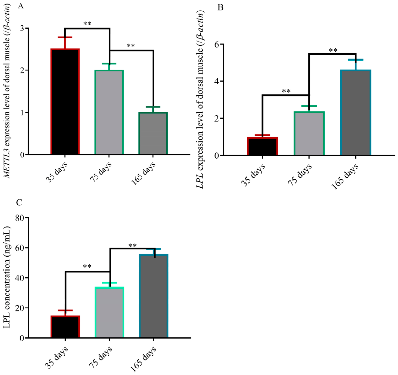
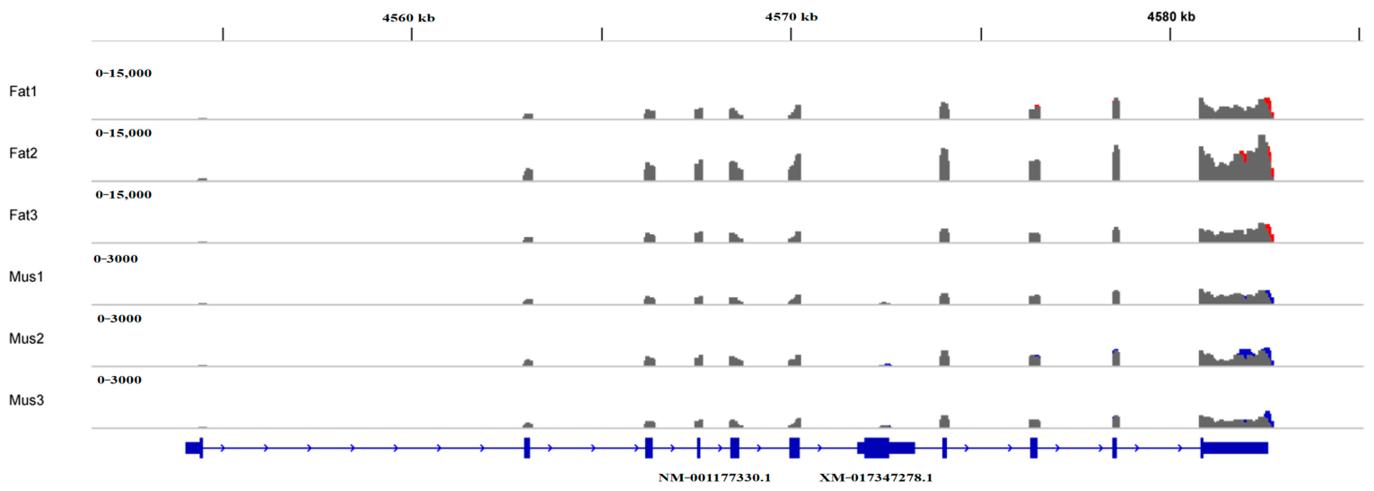
To explore the mechanism of intramuscular fat deposition, we detected the expression of METTL3 and LPL at different ages. We found the expression level of METTL3 in muscles at 35 days of age to be significantly higher than that of muscles at 75 days of age, and the expression level of METTL3 in muscles at 75 days of age is significantly higher than that of muscles at 165 days of age (p < 0.01) (Figure 1A). On the contrary, the expression level of LPL in muscles and the content of LPL in the blood at 35 days of age is significantly lower than those at 75 days of age, and the expression level of LPL in muscles and the content of LPL in the blood at 75 days of age is significantly lower than those at 165 days of age (p < 0.01) (Figure 1B,C). As shown in Figure 2, we found multiple methylation modification sites on the mRNA of the LPL gene, and there are differences between muscle and fat tissues.

3.2. METTL3 and YTHDF2 Synergistically Regulate the Expression and Methylation Level of LPL

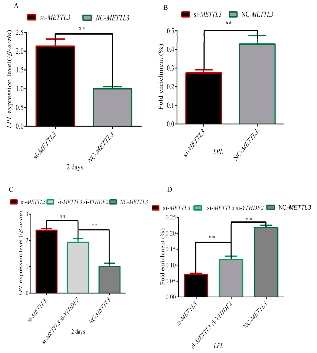
In order to explore the regulation mechanism of the METTL3 gene on LPL, we detected the expression level of LPL after interfering with the METTL3 gene. LPL expression significantly increased after interference with METTL3 (p < 0.01) (Figure 3A). However, the methylation level of the LPL gene decreased after interference with METTL3 (p < 0.01) (Figure 3B). Our results demonstrate that loss of YTHDF2 decreased mRNA levels of LPL in METTL3-depleted cells (p < 0.01) (Figure 3C). In addition, interference of YTHDF2 could partially increase methylation of LPL caused by METTL3 interference (p < 0.01) (Figure 3D).

3.3. Downregulation of LPL Expression Inhibited Rabbit Preadipocyte Differentiation

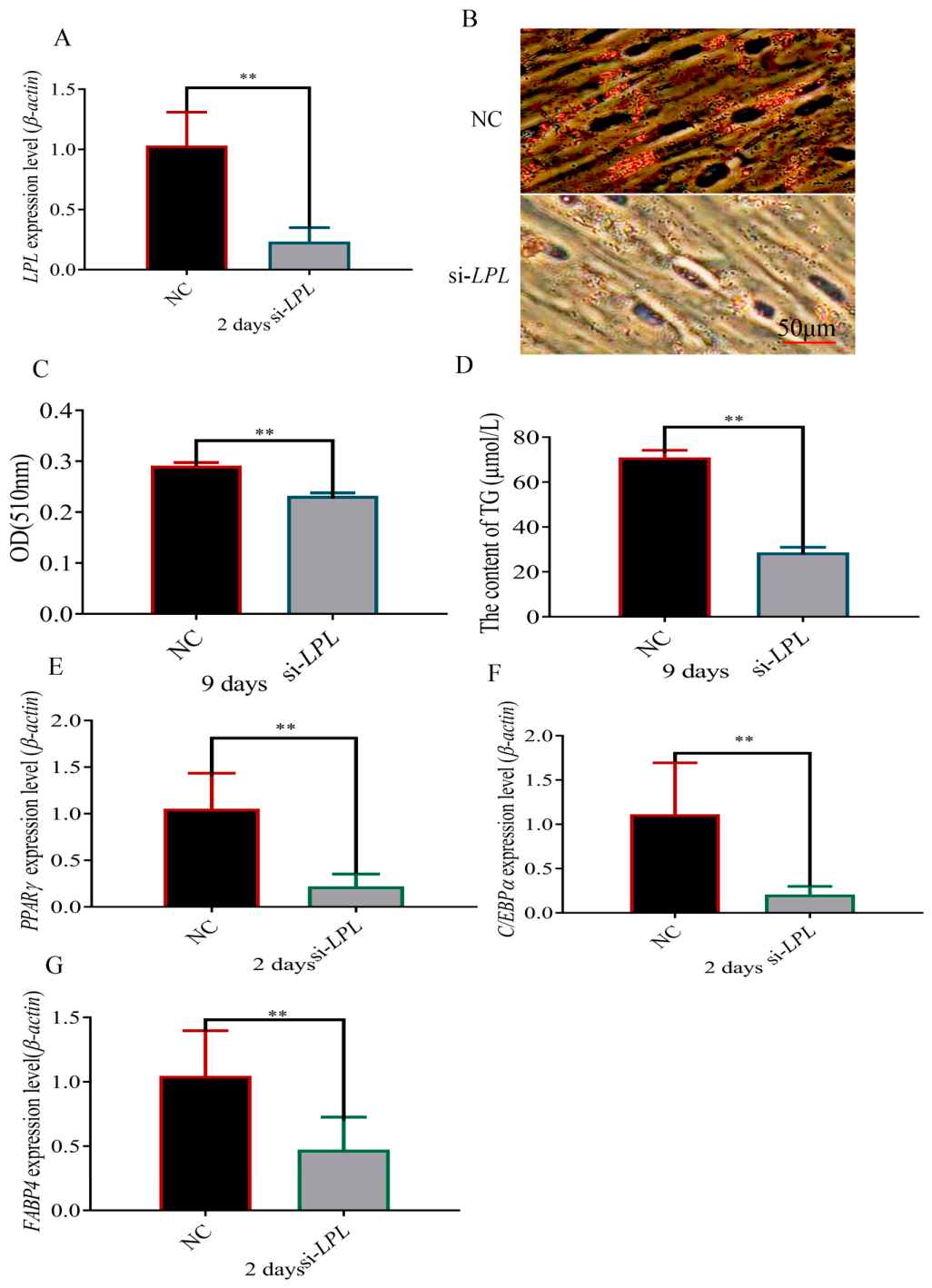
To further confirm whether LPL is involved in the differentiation of rabbit preadipocytes, si-LPL was utilized to cause interference with endogenous LPL. After interfering with the LPL gene, we found that the mRNA and protein levels of the LPL gene were significantly decreased after adipogenic induction differentiation for 2 days (p < 0.01) (Figure 4A). Then, we detected fat droplet accumulation by the oil red O staining assay after adipogenic induction differentiation for 9 days, and the results show that the fat droplet accumulation was reduced in the si-LPL group (p < 0.01) (Figure 4B,C). In addition, TG significantly decreased after interfering with LPL (p < 0.01) (Figure 4D). When LPL was interfered with, the mRNA levels of PPARγ, C/EBPα and FABP4 rapidly decreased on the second day after transfection (p < 0.01) (Figure 4E–G).

3.4. METTL3 Mediates Lipogenesis in an m6A-YTHDF2-Dependent Manner in Rex Rabbits In Vivo

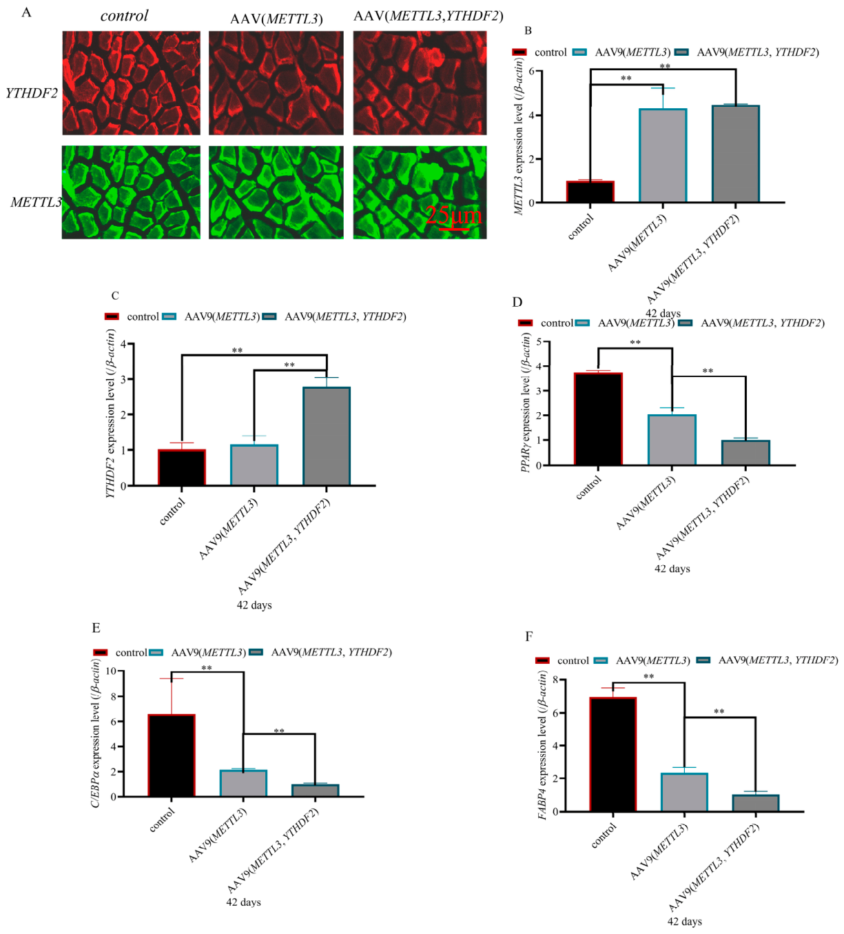
To ascertain whether YTHDF2 is a major contributor to the function of METTL3 in adipogenesis, we overexpressed METTL3 and YTHDF2 in vivo. As shown in Figure 5A, the fluorescence carried by both the METTL3 and YTHDF2 genes is fully stimulated in muscle tissues. The mRNA expression levels of METTL3 and YTHDF2 indicated that METTL3 and YTHDF2 were successfully overexpressed in muscles (Figure 5B,C). However, the expression levels of PPARγ, C/EBPα and FABP4 were significantly reduced after overexpression of METTL3 (p < 0.01) (Figure 5D–F). In addition, FABP4, C/EBPα and PPARγ expressions after overexpressing METTL3 and YTHDF2 simultaneously were lower than those of overexpressing METTL3 (p < 0.01) (Figure 5D–F).

3.5. METTL3 Downregulated the Expression of LPL in an m6A-YTHDF2-Dependent Manner in Rex Rabbits In Vivo

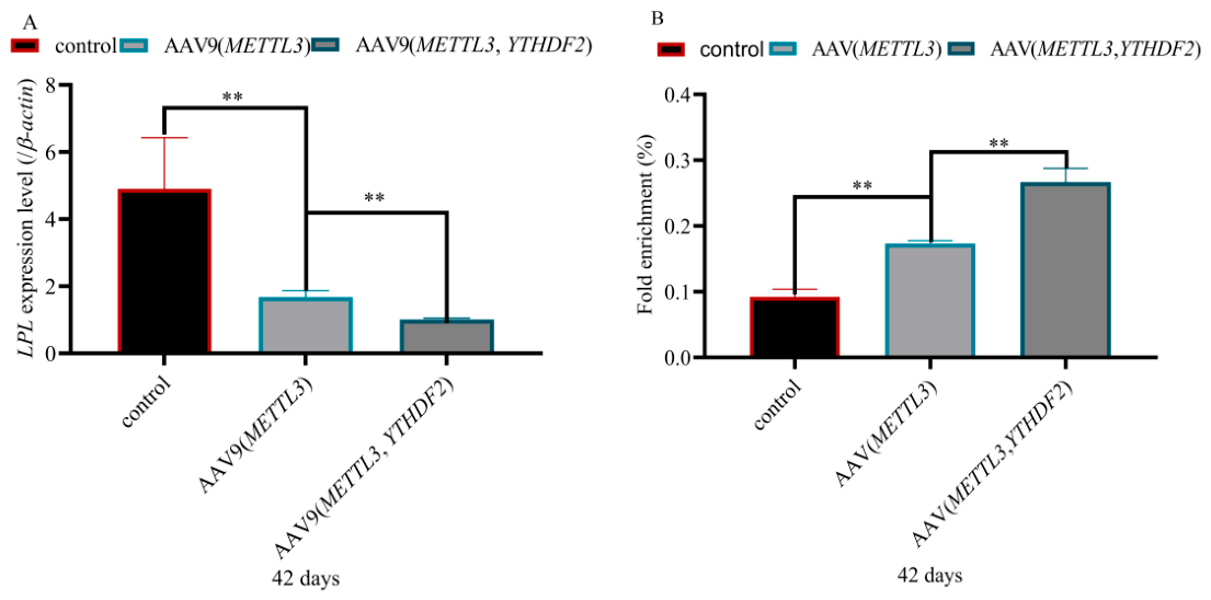
In the living dorsal muscles of Rex rabbits, the mRNA expression of the LPL gene is downregulated after overexpression of METTL3 and is lowest when both METTL3 and YTHDF2 are overexpressed simultaneously (Figure 6A). On the contrary, the methylation level of the LPL gene increased after overexpression of METTL3, and the level further increased after simultaneous overexpression of METTL3 and YTHDF2 (Figure 6B).

3.6. LPL Upregulated Lipogenesis in Rex Rabbits In Vivo

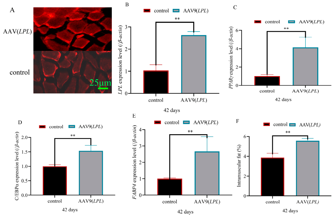
To determine the regulation of the LPL gene on adipogenesis in vivo, we overexpressed the LPL gene in the longissimus lumborum muscle of Rex rabbits. The excitation of red fluorescence indicated that LPL had been expressed in the dorsal muscles of Rex rabbits (Figure 7A). The mRNA level of the LPL gene was significantly increased after overexpression of LPL gene (p < 0.01) (Figure 7B). FABP4, C/EBPα and PPARγ expression were significantly increased after overexpression of LPL (p < 0.01) (Figure 7C–E).

3.7. LPL Regulated Composition of Amino Acid and Fatty Acid Lipogenesis in Rex Rabbits In Vivo

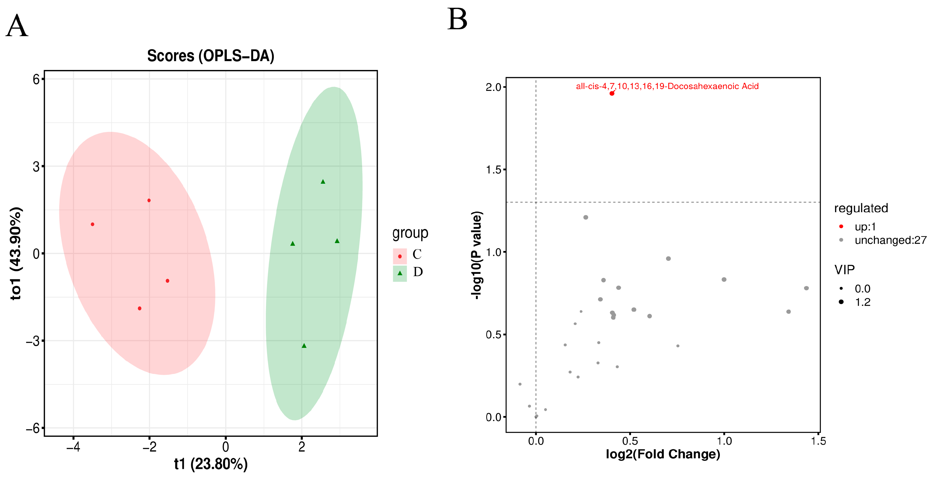
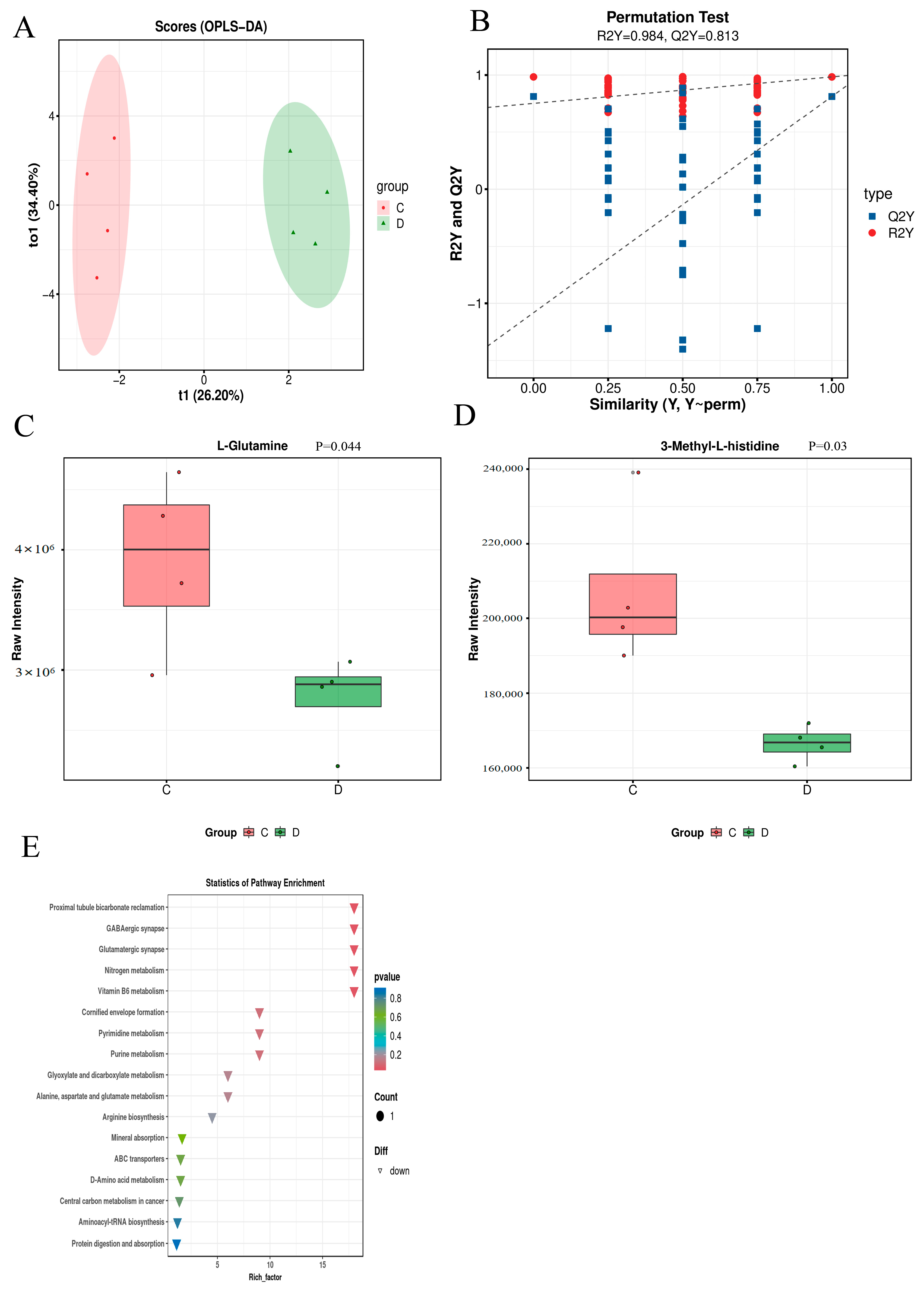
To explore the regulation of LPL on fatty acids in rabbits, we measured the content of fatty acids in muscle tissues after overexpression of the LPL gene in rabbits. As shown in Figure 8A, we found a significant difference between control group and experimental group samples. The results of fatty acid content indicate that all-dis-4,7,10,13,16.19-Deoosahebonoie Acid is upregulated after overexpression of the LPL gene (Figure 8B). At the same time, we found that the analysis results of intra-group repeatability and inter-group differences in amino acid determination samples were also good (Figure 9A,B). In addition, we found that the content of L-Glutamine and 3-Methyl-L-histidine was significantly downregulated in the experimental group (p < 0.05) (Figure 9C,D). Through enrichment of signaling pathways, it was found that L-Glutamine is involved in the regulation of 17 signaling pathways, among which Vitamin B6 metabolism and D-Amino acid metabolism, Arginine biosynthesis, alanine, aspartate and glutamate metabolism, and Glutamatergic synapse play important roles in the process of fat deposition.

3.8. LPL Regulated IMF Through Metabolites

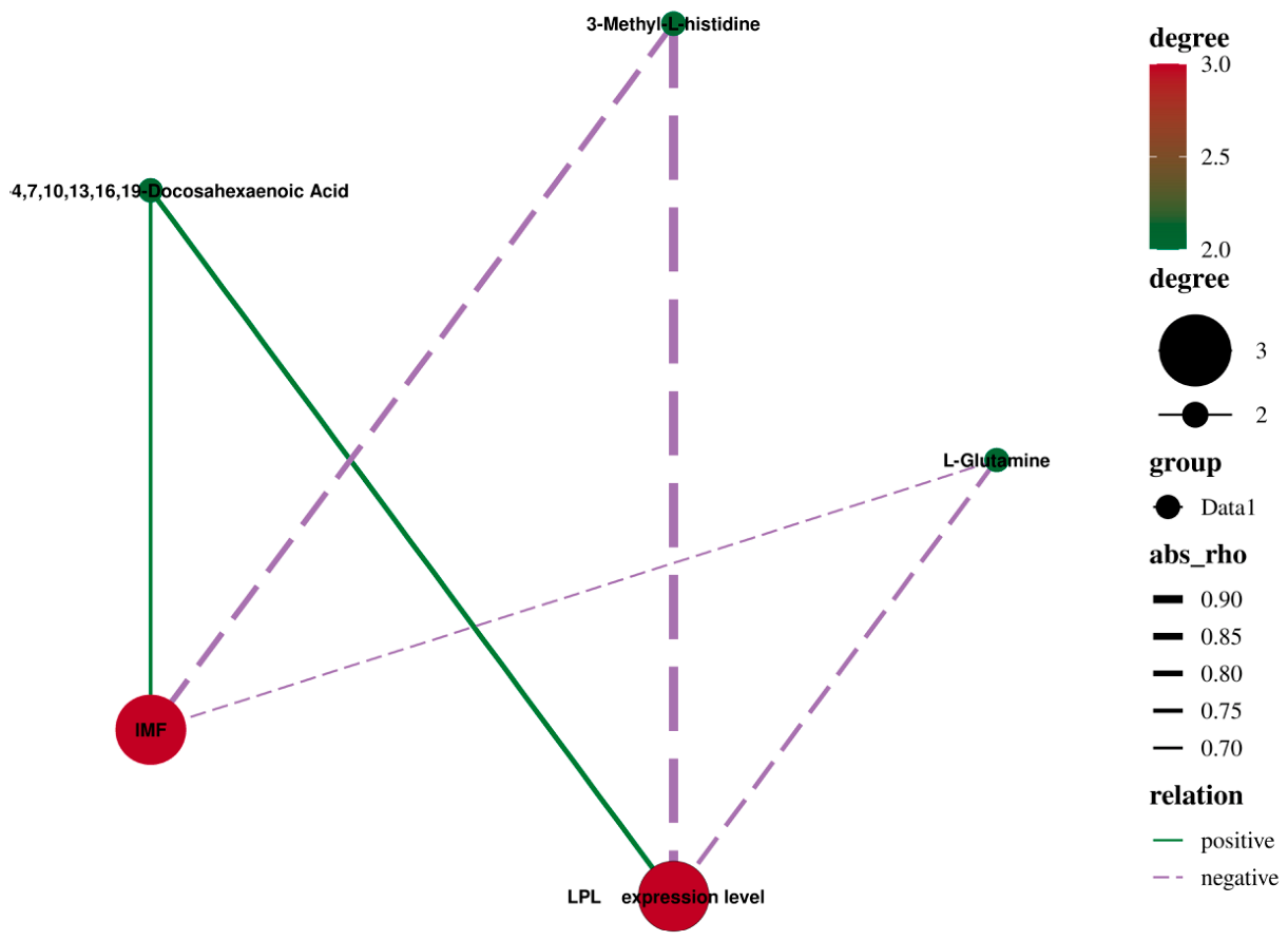
As shown in Figure 10, correlation analysis results show that the expression level of LPL was significantly positively correlated with all-dis-4,7,10,13,16.19-Deoosahebonoie Acid but significantly negatively correlated with L-Glutamine (p < 0.05). In addition, the expression of LPL was significantly negatively correlated with the content of 3-Methyl-L-histidine (p < 0.01). Meanwhile, we also found a significant negative correlation between the content of 3-Methyl-L-histidine and the content of IMF (p < 0.05). So, we speculate that LPL may regulate the content of IMF through 3-Methyl-L-histidine.

4. Discussion

METTL3, as a methyltransferase, catalyzes the formation of m6A and plays important roles in various biological processes [7,13]. Studies reported that METTL3 interacts with translation initiation machinery to promote translation of a subset of m6A containing mRNAs independently of its methyltransferase activity and m6A readers [27]. However, whether METTL3 plays roles in adipogenesis and the underlying mechanisms are still largely unknown. In this study, METTL3 expression decreased during the growth process of rabbits. Studies showed that the content of IMF increased during the growth process of rabbits [28]. In summary, METTL3 may regulate the deposition of IMF through methylation modification. In our previous study, we found that the LPL gene was modified by m6A in both rabbit muscle and adipose tissue [17]. At the same time, we found that the expression of the LPL gene in muscles and the content of LPL in blood increased with an increase in rabbit age.
In order to explore the regulation of LPL by METTL3, we found that interference with METTL3 during preadipocyte differentiation resulted in a decrease in LPL gene expression but an increase in methylation levels. These results indicate that METTL3 can regulate the expression of LPL genes through m6A modification. YTHDF2 is a methylation recognition enzyme, and METTL3 can regulate the expression of the PCK2 gene through YTHDF2’s recognition [16]. Similarly, we also found that YTHDF2 can alter the regulation of METTL3 on LPL gene methylation and expression levels by identifying the m6A site in this study.
To explore the relationship between the LPL gene and adipocyte differentiation, we interfered with the expression of the LPL gene. The triglyceride content was found to be less, and lipid droplets were smaller after transfection with si-LPL. The downregulation of LPL decreased the mRNA expression of FABP4, PPARγ, and CEBPα. At the same time, the protein level of PPARγ also decreased significantly. The role of the lipoprotein lipase (LPL) gene in adipogenesis has been previously reported, and expression of LPL messenger RNA reached a stable level in mature adipocytes in the early stages of adipogenesis, which indicates that LPL is one of the critical factors in the process of adipogenic differentiation [29]. Studies have also shown that LPL plays an important role in lipid metabolism, obesity, insulin effect and weight regulation, whose mRNA expression level, protein concentration and catalytic activity can be used as markers of adipocyte differentiation [29]. These results indicate that the LPL gene can promote adipocyte differentiation.
Adipocyte differentiation and proliferation are the main causes of fat deposition. In order to explore the regulatory mechanism of the LPL gene on fat in muscles, we overexpressed METTL3 and LPL genes in living rabbits and found that they can both promote fat deposition. In addition, METTL3 can also regulate LPL gene expression through YTHDF2 in living rabbits. These results indicate that METTL3, LPL, and YTHDF2 have the same regulatory mechanism for fat deposition in preadipocytes and intramuscular fat.
LPL participates in the PPAR signaling pathway and is associated with lipid transfer and metabolism [30]. In addition, the expression of the LPL gene is positively correlated with intramuscular fat content in pig muscle tissues [31]. Studies have shown that increasing IMF content can improve meat quality, including color, tenderness, flavor, and juiciness [32,33,34]. Amino acids and fatty acids are important precursors of muscle flavor, of which composition and content not only directly affect the taste and flavor of meat but also have a close relationship with human health [35,36]. Amino acid composition in meat is an important indicator for evaluating the nutritional value and quality of meat [37]. In this study, we found that overexpression of the LPL gene upregulated the content of all-dis-4,7,10,13,16.19- Deoosahebonoie Acid but downregulated the content of L-Glutamine and 3-Methyl-L-histidine. In addition, L-Glutamine participated in Vitamin B6 metabolism and D-Amino acid metabolism, Arginine biosynthesis, alanine, and aspartate and glutamate metabolism signaling pathways. Vitamin B6 alters blood lipid abnormalities in mice through the SIRT1/SREBP-1c pathway [38]. Arginine [39] and aspartate [40] can regulate fat deposition in animals. These results indicate that the LPL gene can regulate fat deposition in rabbits through L-Glutamine/multiple pathways. The results of the correlation analysis also indicate a significant negative correlation between the expression of the LPL gene and 3-Methyl-L-histidine content. The study also found that LPL affects the activity of histidine [41]. 3-Methyl-L-histidine is significantly negatively correlated with IMF. Histidine can promote the absorption and utilization of zinc in animals [42]. Zinc affects lipid metabolism and can reduce liver lipid deposition through autophagy [43,44]. These results indicate that the LPL gene may regulate IMF in rabbits through L-Glutamine, 3-Methyl-L-histidine and multiple pathways.

5. Conclusions

In summary, we found that METTL3 suppresses LPL expression in skeletal muscle and reduces circulating LPL levels during rabbit growth. During adipocyte differentiation, METTL3 modulates LPL gene expression via YTHDF2-mediated recognition, thereby inhibiting the differentiation of preadipocytes. In vivo experiments further demonstrated that METTL3 regulates LPL expression through YTHDF2 in muscle tissues. Concurrently, LPL potentially mediated intramuscular fat by L-Glutamine, 3-Methyl-L-histidine and multiple pathways. This study lays the molecular theoretical foundation for cultivating high-quality meat rabbits.

Author Contributions

Conceptualization, Z.R. and G.L.; methodology, J.L. and G.L.; software, J.L. and G.L.; validation, J.L. and G.L.; formal analysis, G.L.; investigation, T.X.; data curation, J.L., X.M. and T.X.; writing-original draft preparation, J.L. and G.L.; writing—review and editing, X.M., Z.R., T.X. and G.L.; visualization, G.L.; project administration, J.L.; funding acquisition, G.L. All authors have read and agreed to the published version of the manuscript.

Funding

This work was supported by the Key Project of Fujian Province Young and Middle-aged Teacher Education Research Project in Fujian Province (JZ230012) and the Fujian Provincial Natural Science Foundation (General Project) (2024J01386).

Institutional Review Board Statement

This study was conducted and approved by the Institutional Animal Care and Use Committee of the College of Animal Science and Technology, FAFU, Fuzhou (Permit Number: PZCASFAFU22003).

Informed Consent Statement

Written informed consent has been obtained from the owner of the animals involved in this study.

Data Availability Statement

The data sets that support the findings of this study are available from the corresponding author.

Acknowledgments

We thank the staff at our laboratory for their ongoing assistance.

Conflicts of Interest

The authors declare no conflicts of interest.

References

  1. Zotte, A.D. Perception of rabbit meat quality and major factors influencing the rabbit carcass and meat quality. Livest. Prod. Sci. 2002, 75, 11–32. [Google Scholar] [CrossRef]
  2. Watkins, P.J.; Frank, D.; Singh, T.K.; Young, O.A.; Warner, R.D. Sheepmeat flavor and the effect of different feeding systems: A review. J. Agric. Food Chem. 2013, 61, 3561–3579. [Google Scholar] [CrossRef]
  3. Erasmus, S.W.; Muller, M.; Hoffman, L.C. Authentic sheep meat in the european union: Factors influencing and validating its unique meat quality. J. Sci. Food Agric. 2017, 97, 1979–1996. [Google Scholar] [CrossRef] [PubMed]
  4. Hocquette, J.F.; Gondret, F.; Baéza, E.; Médale, F.; Jurie, C.; Pethick, D.W. Intramuscular fat content in meat-producing animals: Development, genetic and nutritional control, and identification of putative markers. Anim. Int. J. Anim. Biosci. 2010, 4, 303–319. [Google Scholar] [CrossRef]
  5. Fu, Y.; Dominissini, D.; Rechavi, G.; He, C. Gene expression regulation mediated through reversible m6a rna methylation. Nat. Rev. Genet. 2014, 15, 293–306. [Google Scholar] [CrossRef]
  6. Wu, R.; Jiang, D.; Wang, Y.; Wang, X. N (6)-methyladenosine (m(6)a) methylation in mrna with a dynamic and reversible epigenetic modification. Mol. Biotechnol. 2016, 58, 450–459. [Google Scholar]
  7. Liu, J.; Yue, Y.; Han, D.; Wang, X.; Fu, Y.; Zhang, L.; Jia, G.; Yu, M.; Lu, Z.; Deng, X.; et al. A mettl3-mettl14 complex mediates mammalian nuclear rna n6-adenosine methylation. Nat. Chem. Biol. 2014, 10, 93–95. [Google Scholar]
  8. Ping, X.-L.; Sun, B.-F.; Wang, L.; Xiao, W.; Yang, X.; Wang, W.-J.; Adhikari, S.; Shi, Y.; Lv, Y.; Chen, Y.-S.; et al. Mammalian wtap is a regulatory subunit of the rna n6-methyladenosine methyltransferase. Cell Res. 2014, 24, 177–189. [Google Scholar] [CrossRef] [PubMed]
  9. Yang, Y.; Hsu, P.J.; Chen, Y.-S.; Yang, Y.-G. Dynamic transcriptomic m6a decoration: Writers, erasers, readers and functions in rna metabolism. Cell Res. 2018, 28, 616–624. [Google Scholar] [CrossRef]
  10. Zhao, X.; Yang, Y.; Sun, B.-F.; Shi, Y.; Yang, X.; Xiao, W.; Hao, Y.-J.; Ping, X.-L.; Chen, Y.-S.; Wang, W.-J.; et al. Fto-dependent demethylation of n6-methyladenosine regulates mrna splicing and is required for adipogenesis. Cell Res. 2014, 24, 1403–1419. [Google Scholar] [PubMed]
  11. Ma, C.; Chang, M.; Lv, H.; Zhang, Z.-W.; Zhang, W.; He, X.; Wu, G.; Zhao, S.; Zhang, Y.; Wang, D.; et al. Rna m(6)a methylation participates in regulation of postnatal development of the mouse cerebellum. Genome Biol. 2018, 19, 68. [Google Scholar] [PubMed]
  12. Lin, Z.; Hsu, P.; Xing, X.; Fang, J.; Lu, Z.; Zou, Q.; Zhang, K.-J.; Zhang, X.; Zhou, Y.; Zhang, T.; et al. Mettl3-/mettl14-mediated mrna n6-methyladenosine modulates murine spermatogenesis. Cell Res. 2017, 27, 1216–1230. [Google Scholar] [CrossRef]
  13. Xu, K.; Yang, Y.; Feng, G.-H.; Sun, B.-F.; Chen, J.-Q.; Li, Y.-F.; Chen, Y.-S.; Zhang, X.-X.; Wang, C.-X.; Jiang, L.-Y.; et al. Mettl3-mediated m6a regulates spermatogonial differentiation and meiosis initiation. Cell Res. 2017, 27, 1100–1114. [Google Scholar] [CrossRef]
  14. Geula, S.; Moshitch-Moshkovitz, S.; Dominissini, D.; Mansour, A.A.; Kol, N.; Salmon-Divon, M.; Hershkovitz, V.; Peer, E.; Mor, N.; Manor, Y.S.; et al. M6a mrna methylation facilitates resolution of nave pluripotency toward differentiation. Science 2015, 347, 1002–1006. [Google Scholar] [CrossRef]
  15. Chen, T.; Hao, Y.-J.; Zhang, Y.; Li, M.-M.; Wang, M.; Han, W.; Wu, Y.; Lv, Y.; Hao, J.; Wang, L.; et al. M(6)a rna methylation is regulated by micrornas and promotes reprogramming to pluripotency. Cell Stem Cell 2015, 16, 289–301. [Google Scholar]
  16. Luo, G.; Zhu, T.; Ren, Z. Mettl3 regulated the meat quality of rex rabbits by controlling pck2 expression via a ythdf2-n6-methyladenosine axis. Foods 2022, 11, 1549. [Google Scholar] [CrossRef] [PubMed]
  17. Luo, G.; Wang, S.; Ai, Y.; Li, J.; Ren, Z. N6-methyladenosine methylome profiling of muscle and adipose tissues reveals methylase–mrna metabolic regulatory networks in fat deposition of rex rabbits. Biology 2022, 11, 944. [Google Scholar]
  18. Aaltonen, T.; Adelman, J.; Akimoto, T.; Albrow, M.G.; González, B.Á.; Amerio, S.; Amidei, D.; Anastassov, A.; Annovi, A.; Antos, J.; et al. First measurement of w boson production in association with a single charm quark in pp collisions at s = 1.96 tev. Phys. Rev. Lett. 2008, 100, 091803. [Google Scholar] [PubMed]
  19. Ira, J.G. Lipoprotein lipase: Physiology, biochemistry, and molecular biology. Front. Biosci. 2001, 6, d388. [Google Scholar]
  20. Goldberg, I.J. Lipoprotein lipase and lipolysis: Central roles in lipoprotein metabolism and atherogenesis. J. Lipid Res. 1996, 37, 693–707. [Google Scholar] [CrossRef]
  21. Enerbäck, S.; Gimble, J.M. Lipoprotein lipase gene expression: Physiological regulators at the transcriptional and post-transcriptional level. Biochim. Biophys. Acta Lipids Lipid Metab. 1993, 1169, 107–125. [Google Scholar] [CrossRef]
  22. Vincent, G.; Isabelle, L.; Jacques, M.; Le, D.J. Lipogenic enzyme activities in subcutaneous adipose tissue and skeletal muscle from neonatal pigs consuming maternal or formula milk. Reprod. Nutr. Dev. 2000, 40, 103–112. [Google Scholar] [CrossRef]
  23. Sorisky, A. From preadipocyte to adipocyte: Differentiation-directed signals of insulin from the cell surface to the nucleus. Crit. Rev. Clin. Lab. Sci. 1999, 36, 1–34. [Google Scholar] [CrossRef]
  24. Costa, D.C.; Takata, R.; Silva, W.D.S.E.; Bessonart, M.; Kennedy, L.R. Description of amino acid and fatty acid content during initial development of Lophiosilurus alexandri (siluriformes: Pseudopimelodidae), a carnivorous freshwater catfish. Neotrop. Ichthyol. 2018, 16, e180014. [Google Scholar] [CrossRef]
  25. Karpe, F.; Olivecrona, T.; Walldius, G.; Hamsten, A. Lipoprotein lipase in plasma after an oral fat load: Relation to free fatty acids. J. Lipid Res. 1992, 33, 975–984. [Google Scholar] [CrossRef] [PubMed]
  26. Yau, M.H.; Wang, Y.; Lam, K.S.L.; Zhang, J.; Wu, D.; Xu, A. A highly conserved motif within the nh2-terminal coiled-coil domain of angiopoietin-like protein 4 confers its inhibitory effects on lipoprotein lipase by disrupting the enzyme dimerization. J. Biol. Chem. 2009, 284, 11942–11952. [Google Scholar] [CrossRef]
  27. Lin, S.; Choe, J.; Du, P.; Triboulet, R.; Gregory, R.I. The m(6)a methyltransferase mettl3 promotes translation in human cancer cells. Mol. Cell. Biol. 2016, 62, 335–345. [Google Scholar] [CrossRef]
  28. Luo, G.; Wang, L.; Hu, S.; Du, K.; Wang, J.; Lai, S. Association of leptin mrna expression with meat quality trait in tianfu black rabbits. Anim. Biotechnol. 2020, 33, 480–486. [Google Scholar] [CrossRef] [PubMed]
  29. Wang, H.; Eckel, R.H. Lipoprotein lipase: From gene to obesity. Am. J. Physiol. Endocrinol. Metab. 2009, 297, E271–E288. [Google Scholar] [CrossRef]
  30. Wang, W.; Xue, W.; Xu, X.; Jin, B.; Zhang, X. Correlations of genes expression in ppar signalling pathway with porcine meat quality traits. Czech. J. Anim. Sci. 2016, 61, 333–339. [Google Scholar] [CrossRef]
  31. Wang, G.; Zeng, Y.; Wu, Y.; Wei, S.; Bao, X.; Liu, C.; Sun, Y. The developmental changes of lpl mrna expression in muscle and their association with intramuscular fat for pigs. Acta Vet. Zootech. Sin. 2007, 3, 253–257. [Google Scholar]
  32. Frank, D.; Watkins, P.; Ball, A.; Krishnamurthy, R.; Piyasiri, U.; Sewell, J.; Ortuño, J.; Stark, J.; Warner, R. Impact of brassica and lucerne finishing feeds and intramuscular fat on lamb eating quality and flavor. A cross-cultural study using chinese and non-chinese australian consumers. J. Agric. Food Chem. 2016, 64, 6856–6868. [Google Scholar] [CrossRef] [PubMed]
  33. Ge, K.; Ye, P.; Yang, L.; Kuang, J.; Chen, X.; Geng, Z. Comparison of slaughter performance, meat traits, serum lipid parameters and fat tissue between chaohu ducks with high- and low-intramuscular fat content. Anim. Biotechnol. 2020, 31, 245–255. [Google Scholar] [CrossRef] [PubMed]
  34. Starkey, C.P.; Geesink, G.H.; Collins, D.; Oddy, V.H.; Hopkins, D.L. Do sarcomere length, collagen content, ph, intramuscular fat and desmin degradation explain variation in the tenderness of three ovine muscles. Meat Sci. 2016, 113, 51–58. [Google Scholar] [CrossRef] [PubMed]
  35. Ning, W.; Chu, T.J.; Li, C.J.; Choi, A.M.K.; Peters, D.G. Genome-wide analysis of the endothelial transcriptome under short-term chronic hypoxia. Physiol. Genom. 2004, 18, 70–78. [Google Scholar] [CrossRef]
  36. Yin, L.; Xu, M.; Huang, Q.; Zhang, D.; Lin, Z.; Wang, Y.; Liu, Y. Nutrition and flavor evaluation of amino acids in guangyuan grey chicken of different ages, genders and meat cuts. Animals 2023, 13, 1235. [Google Scholar] [CrossRef]
  37. Chen, G.S.; Liu, M.Y. Analysis of nutritional characteristics of wild crossbred pig muscle. Pig Breed. 2004, 1, 24–27. [Google Scholar]
  38. Zhang, Q.; Zhou, X.; Zhang, J.; Li, Q.; Qian, Z. Selenium and vitamin b(6) cosupplementation improves dyslipidemia and fatty liver syndrome by sirt1/srebp-1c pathway in hyperlipidemic sprague-dawley rats induced by high-fat diet. Nutr. Res. 2022, 106, 101–118. [Google Scholar] [CrossRef]
  39. Kang, L.; Li, X.; Zhao, X.; Liu, T.; Jin, Y.; Duan, Y. Effects of l-arginine supplementation on fat deposition and meat quality in growing lambs: Interactions with gut microbiota and metabolic signalling pathways. Food Chem. 2025, 479, 143677. [Google Scholar] [CrossRef]
  40. Zhang, X.; Ma, G.; Wang, B.; Liu, R.; Guo, Y. Dietary supplemental l-ornithine l-aspartate reduced lipid deposition and alleviated mitochondrial oxidative stress and inflammatory responses in the liver of broiler chicken. Poult. Sci. 2026, 105, 106655. [Google Scholar] [CrossRef]
  41. Maier, M.; Wei, L.; Zeh, N.; Schmieder-Todtenhaupt, V.; Dehghani, A.; Felix, M.N.; Heinzelmann, D.; Lindner, B.; Schmidt, M.; Studts, J.; et al. Illuminating a biologics development challenge: Systematic characterization of cho cell-derived hydrolases identified in monoclonal antibody formulations. mAbs 2024, 16, 2375798. [Google Scholar] [CrossRef]
  42. Glover, C.N.; Bury, N.R.; Hogstrand, C. Zinc uptake across the apical membrane of freshwater rainbow trout intestine is mediated by high affinity, low affinity, and histidine-facilitated pathways. Biochim. Biophys. Acta (BBA)-Biomembr. 2003, 1614, 211–219. [Google Scholar] [CrossRef]
  43. Huang, L.; Yu, Y.Y.; Kirschke, C.P.; Gertz, E.R.; Lloyd, K.K.C. Znt7 (slc30a7)-deficient mice display reduced body zinc status and body fat accumulation. J. Biol. Chem. 2007, 282, 37053–37063. [Google Scholar] [CrossRef]
  44. Wei, C.; Luo, Z.; Hogstrand, C.; Xu, Y.; Wu, L.; Chen, G.; Pan, Y.; Song, Y. Zinc reduces hepatic lipid deposition and activates lipophagy via Zn2+/MTF-1/PPARα and Ca2+/CaMKKβ/AMPK pathways. FASEB J. 2018, 32, 6666–6680. [Google Scholar] [CrossRef]
Figure 1. Expression of LPL and METTL3 genes in Rex rabbits at different stages. (A) The expression of METTL3 in muscle tissues; (B) the expression of LPL in muscle tissues; (C) content of LPL in blood. (“**”, p < 0.01).
Animals 16 01646 g001
Figure 2. Modification sites of m6A on the LPL gene in fat and muscle tissues [17].
Animals 16 01646 g002
Figure 3. METTL3 downregulates the expression of LPL through YTHDF2 during the differentiation of preadipocytes. (A) The mRNA expression of LPL after interfering with METTL3; (B) methylation level of LPL gene after interfering with METTL3; (C) the mRNA expression of LPL after interfering with METTL3 and YTHDF2; (D) methylation level of LPL gene after interfering with METTL3 and YTHDF2 (“**”, p < 0.01).
Animals 16 01646 g003
Figure 4. LPL upregulates differentiation of preadipocytes. (A) The mRNA expression of LPL after interfering with LPL; (B) oil red staining after interfering with LPL; (C,D) fat droplets and triglyceride content after interfering with LPL; (EG) the mRNA expression of PPARγ, C/EBPα and FABP4 after interfering with LPL (“**”, p < 0.01).
Animals 16 01646 g004
Figure 5. METTL3 downregulates adipogenesis through YTHDF2. (A) Slicing of muscle tissues after overexpression of METTL3 and YTHDF2; (B) the mRNA expression of METTL3 after overexpression of METTL3 and YTHDF2; (C) the mRNA expression of YTHDF2 after overexpression of METTL3 and YTHDF2; (DF) the mRNA expression of PPARγ, C/EBPα and FABP4 after overexpression of METTL3 and YTHDF2 (“**”, p < 0.01).
Animals 16 01646 g005
Figure 6. METTL3 downregulates LPL expression through YTHDF2. (A) The mRNA expression of LPL after overexpression of METTL3 and YTHDF2; (B) methylation level of LPL after overexpression of METTL3 and YTHDF2 (“**”, p < 0.01).
Animals 16 01646 g006
Figure 7. LPL upregulates adipogenesis. (A) Slicing of muscle tissues after overexpression of LPL; (B) the mRNA expression of LPL after overexpression of LPL; (CE) the mRNA expression of PPARγ, C/EBPα and FABP4 after overexpression of LPL; (F) IMF content after overexpression of LPL (“**”, p ≤ 0.01).
Animals 16 01646 g007
Figure 8. Overview of differential fatty acids. (A) Results of orthogonal partial least squares discriminant analysis; (B) volcanic diagram of differential fatty acids.
Animals 16 01646 g008
Figure 9. Overview of differential amino acids. (A) Results of orthogonal partial least squares discriminant analysis; (B) results of orthogonal partial least squares discriminant analysis (permutation); (C) box line diagram of L-Glutamine; (D) box line diagram of 3-Methyl-L-histidine; (E) signal pathway enrichment map.
Animals 16 01646 g009
Figure 10. Correlation network diagram of LPL expression–fatty acids and amino acids–IMF.
Animals 16 01646 g010
Table 1. Primers in this study.
Gene NamePrimer Sequence (5′-3′)(Tm/°C)(Product Size/bp)
METTL3CCCACCTCAGTGGATCTGTT60189
ACCCAGAGGAAGAGAAAGCC
LPLGACATTGGGGAGTTGCTGAT60214
ACTTGTCGTGGCATTTCACA
YTHDF2CAGACACAGCCATTGCCTCCAC60122
CCGTTATGACCGAACCCACTGC
β-actinGGAGATCGTGCGGGACAT61.4318
GTTGAAGGTGGTCTCGTGGAT
PPARγGAGGACATCCAGGACAACC61168
GTCCGTCTCCGTCTTCTTT
FABP4GGCCAGGAATTTGATGAAGTC61.4140
AGTTTATCGCCCTCCCGTT
C/EBPαGCGGGAACGAACAACAT64172
GGCGGTCATTGTCACTGGTC
si-METTL3UCAAGGAACAACAGAGCAATT
UUGCUCUGUUGUUCCUUAGTT
si-YTHDF2CAUGAAUACUAUAGACCAATT
UUGGUCUAUAGUAUUCAUGTT
si-LPLCCUCGACAUCGAAACUAAATT
UUUAGUUUCGAUGUCGAGGTT
Negative ControlUUCUCCGAACGUGUCACGUTT
ACGUGACACGUUCGGAGAATT
Disclaimer/Publisher’s Note: The statements, opinions and data contained in all publications are solely those of the individual author(s) and contributor(s) and not of MDPI and/or the editor(s). MDPI and/or the editor(s) disclaim responsibility for any injury to people or property resulting from any ideas, methods, instructions or products referred to in the content.