3. Results
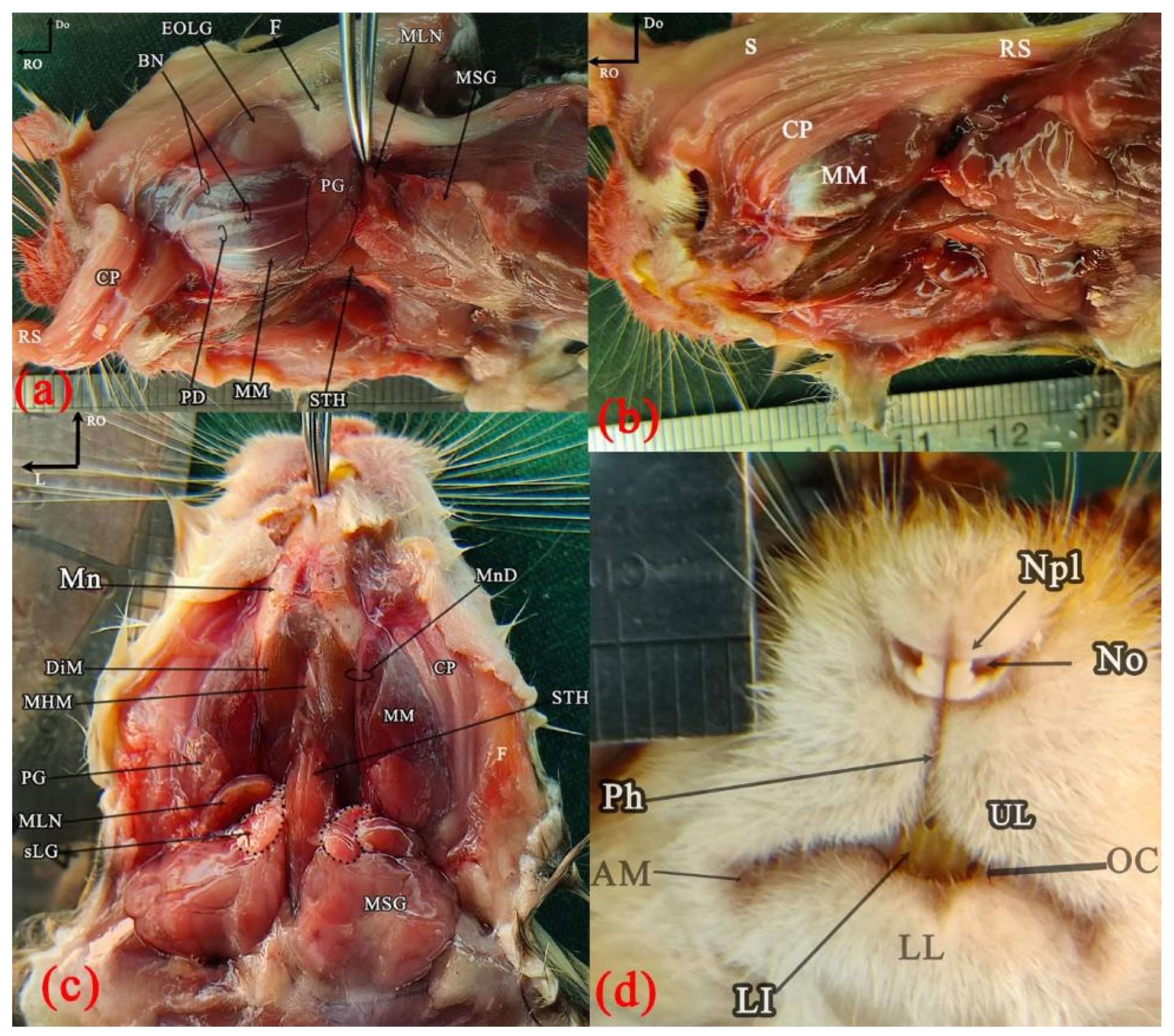
Superficial head structures were demonstrated in the ventral and left lateral views to identify the major salivary glands and lymph nodes, as well as surface features of the muzzle, cheek pouch, and extraorbital lacrimal gland (
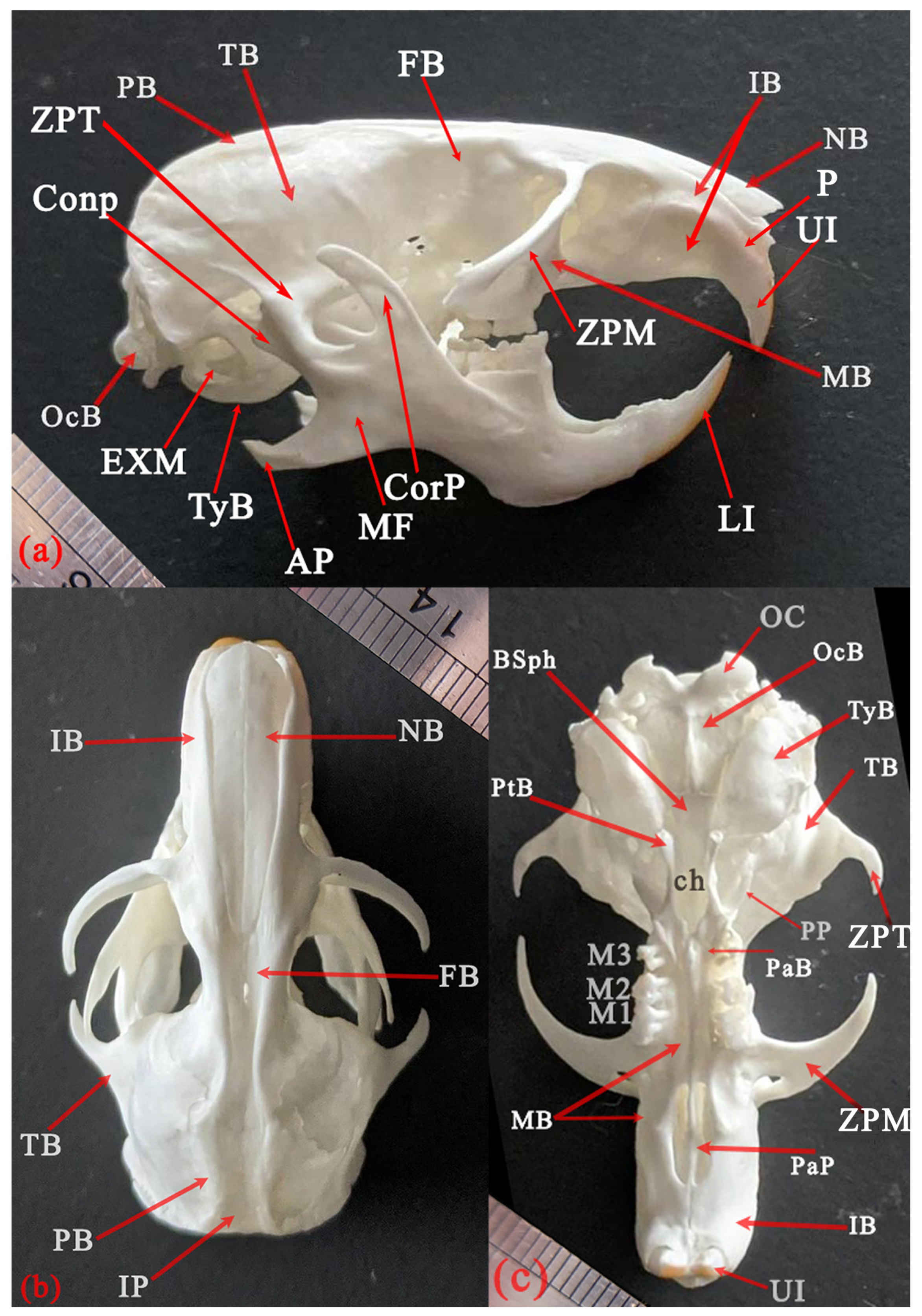
Figure 1). Skull osteology was presented in the lateral, dorsal, and ventral aspects to evaluate the main external bony features (
Figure 2 and
Figure 3).
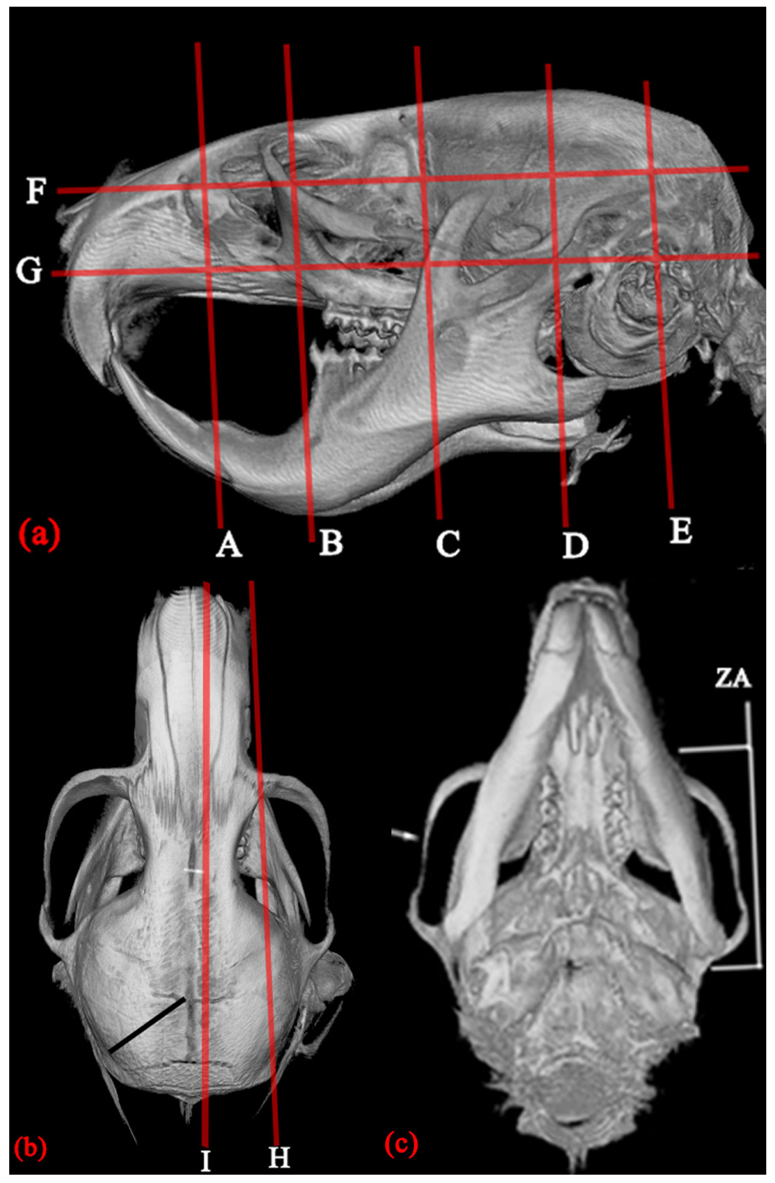
The approximate levels of the dorsal, sagittal, and transverse sectional planes at which images were obtained were illustrated in the lateral (a) and dorsal (b) views of the 3D-reformatted MCT images (
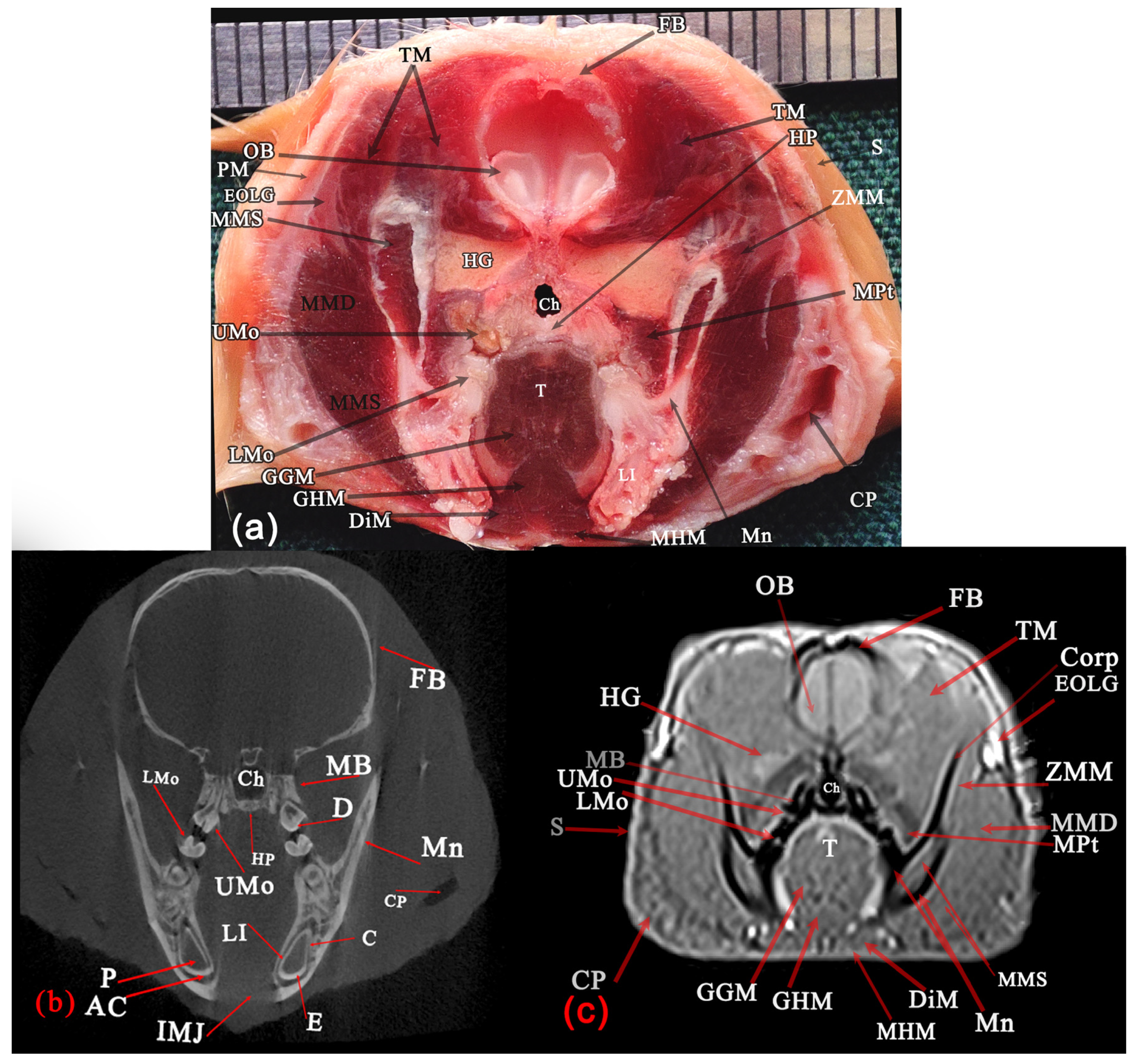
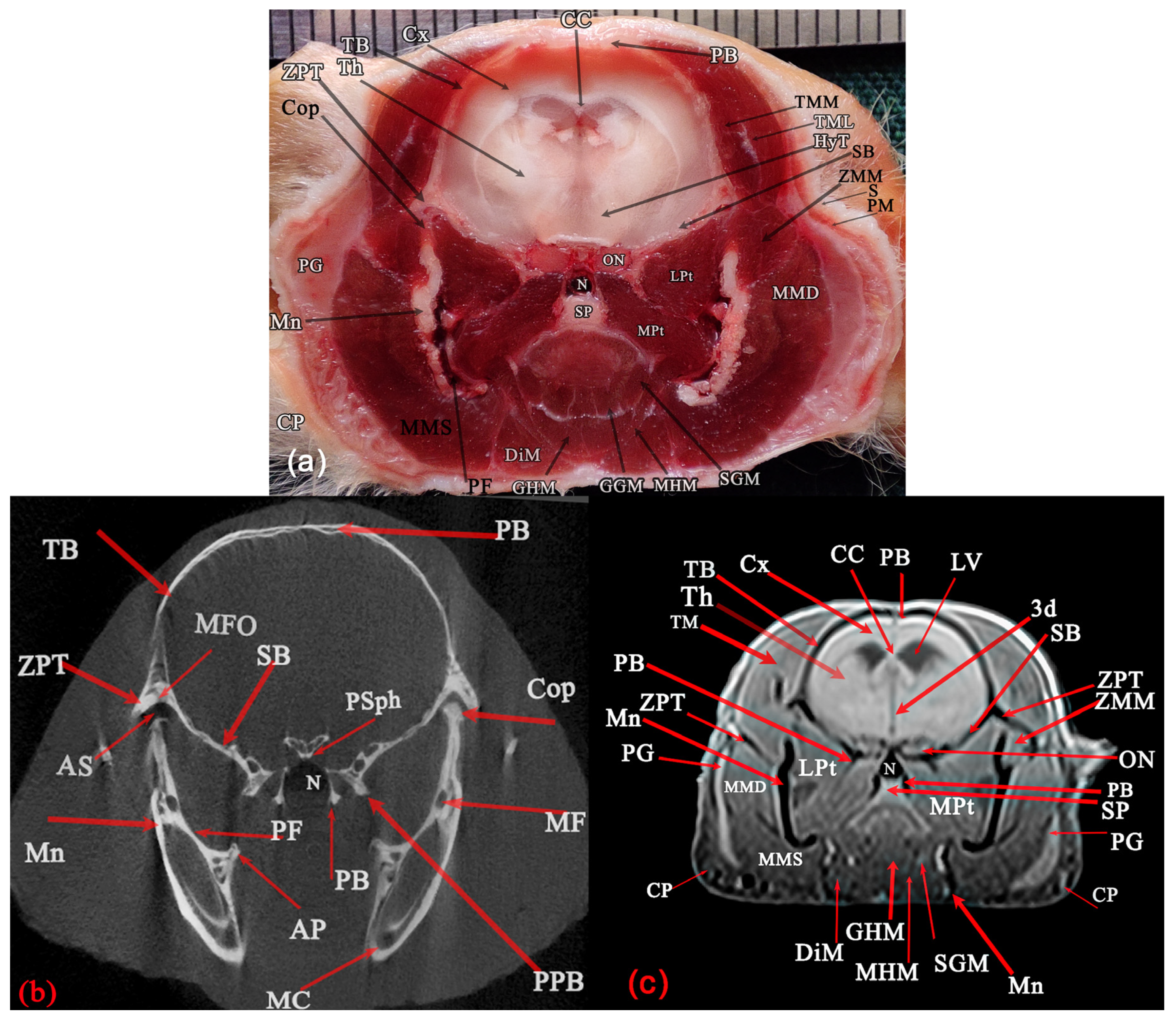
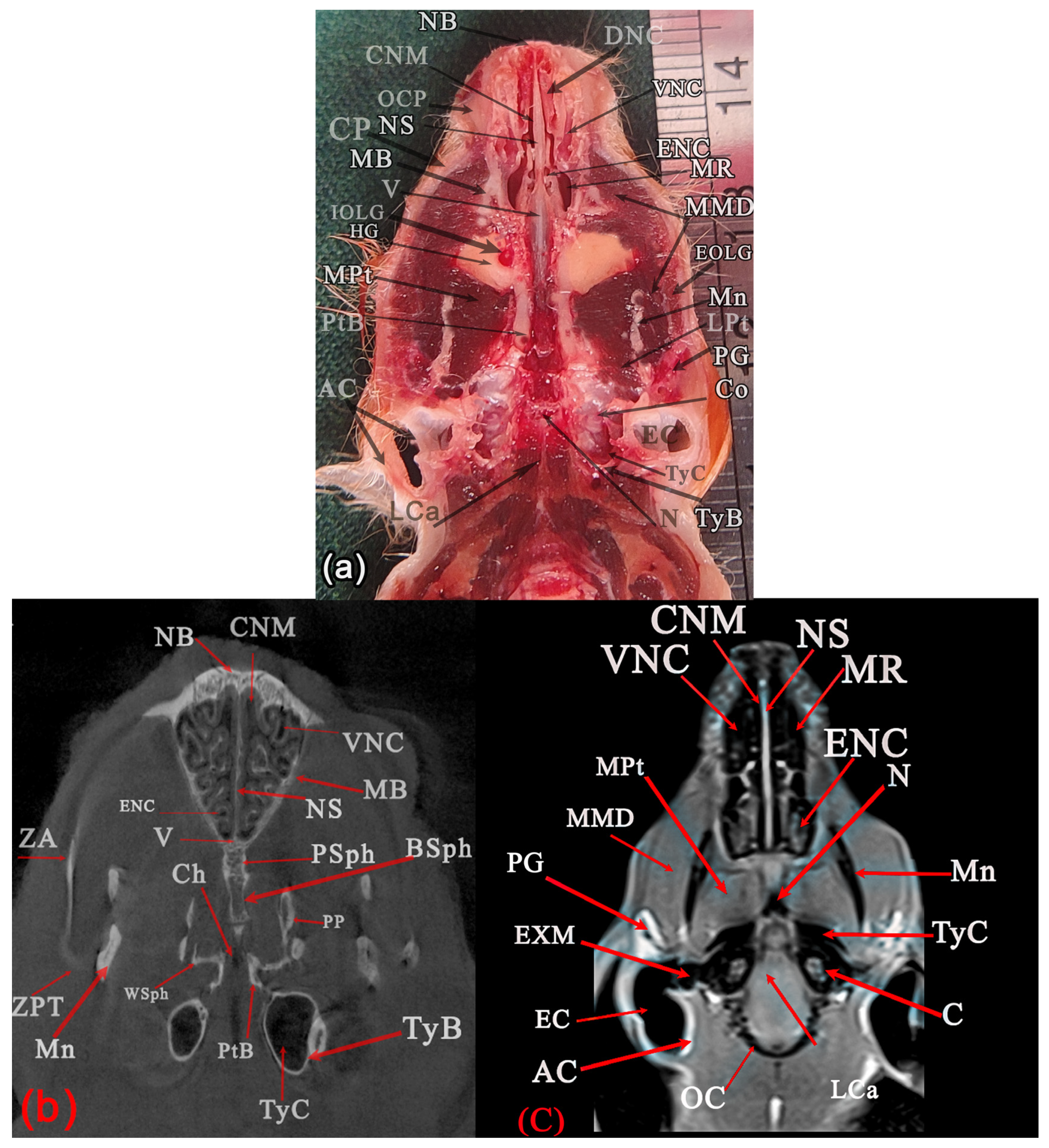
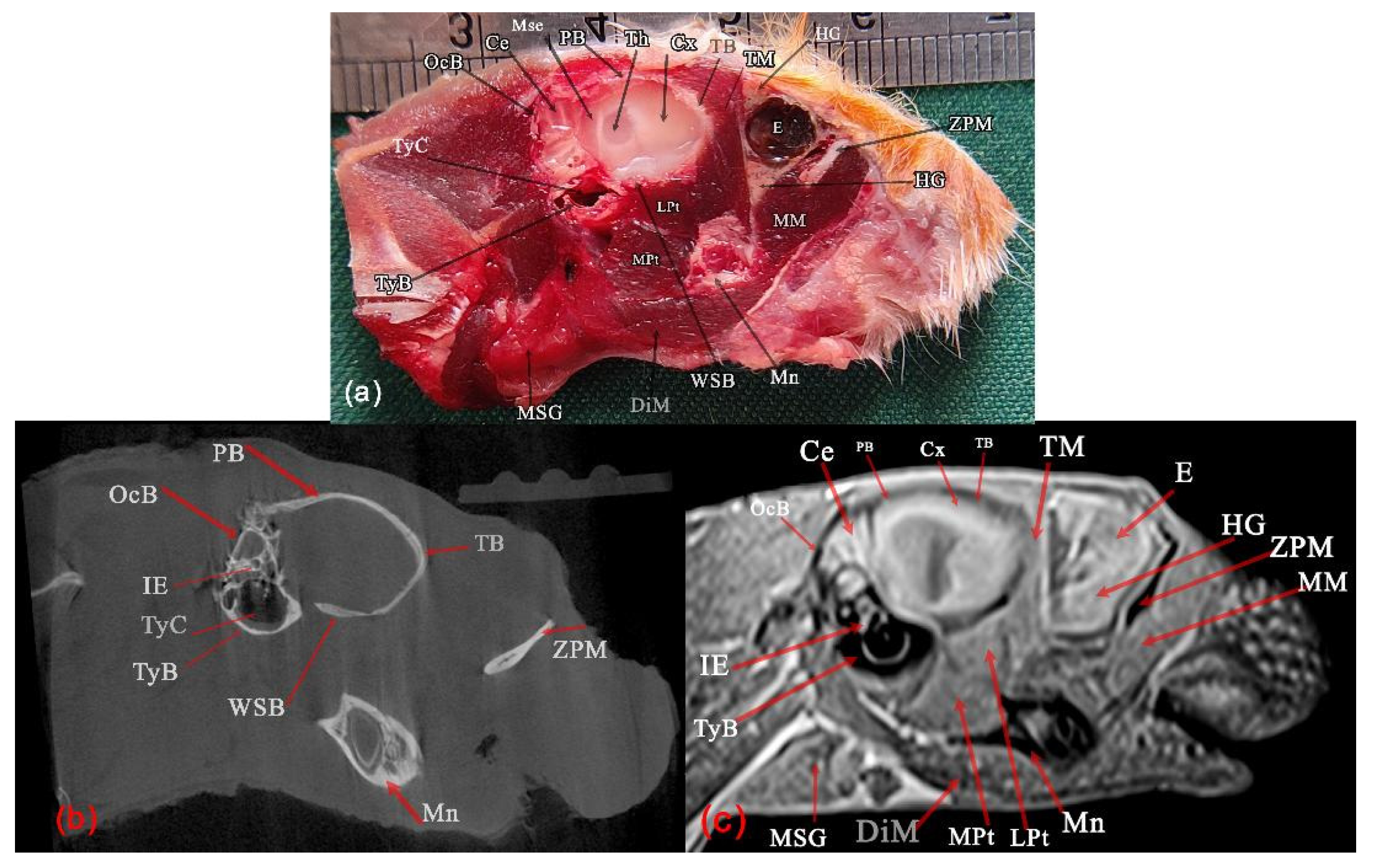
Figure 3). A total of twelve sectional planes were presented, including five serial transverse sections (
Figure 4,
Figure 5,
Figure 6,
Figure 7 and
Figure 8), two dorsal sections (
Figure 9 and
Figure 10), and two sagittal sections (
Figure 11 and
Figure 12). The following anatomical landmarks were used to define the reference lines for these sectional planes: the level of the maxillary incisor teeth (
Figure 4); the caudal part of the diastema, just rostral to the first molar (
Figure 5); a level adjacent to the caudal margin of the orbital cavity (
Figure 6); the level of the temporomandibular joint (
Figure 7); the level of the external acoustic meatus (
Figure 8); a level just dorsal to the occipital condyle (
Figure 9); the level of the external acoustic meatus (
Figure 10); the level of the orbital cavity (
Figure 11); and the midline (
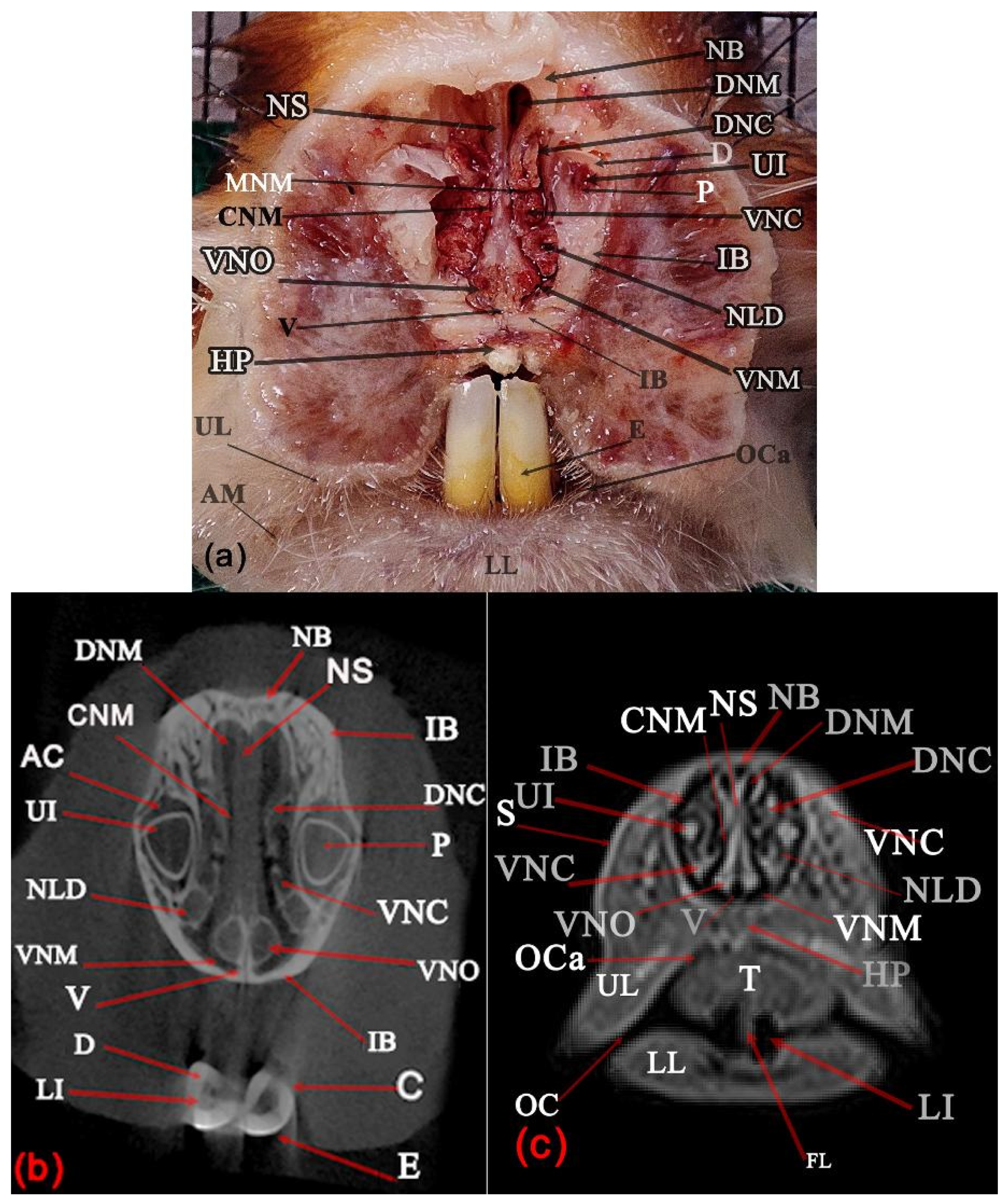
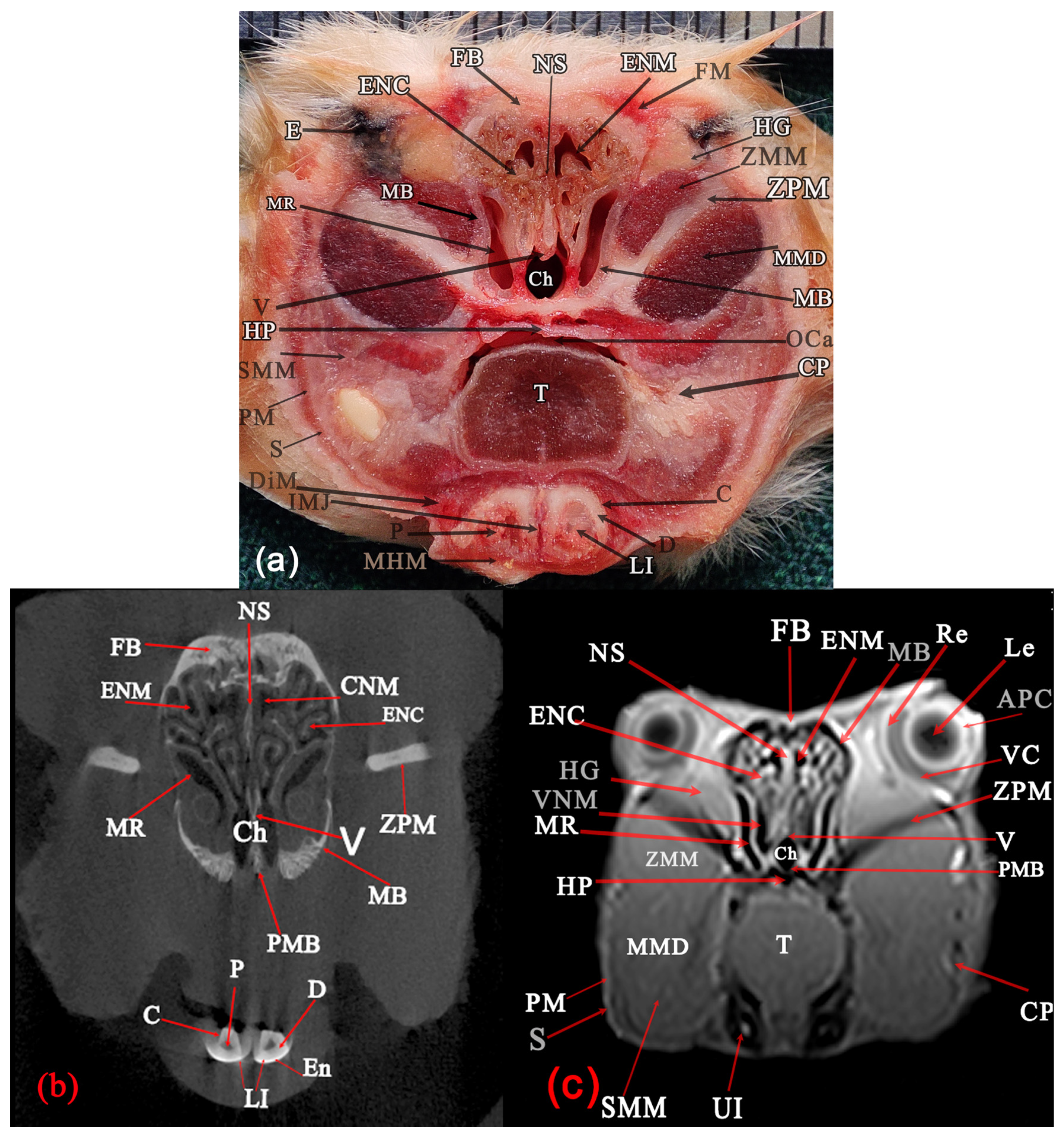
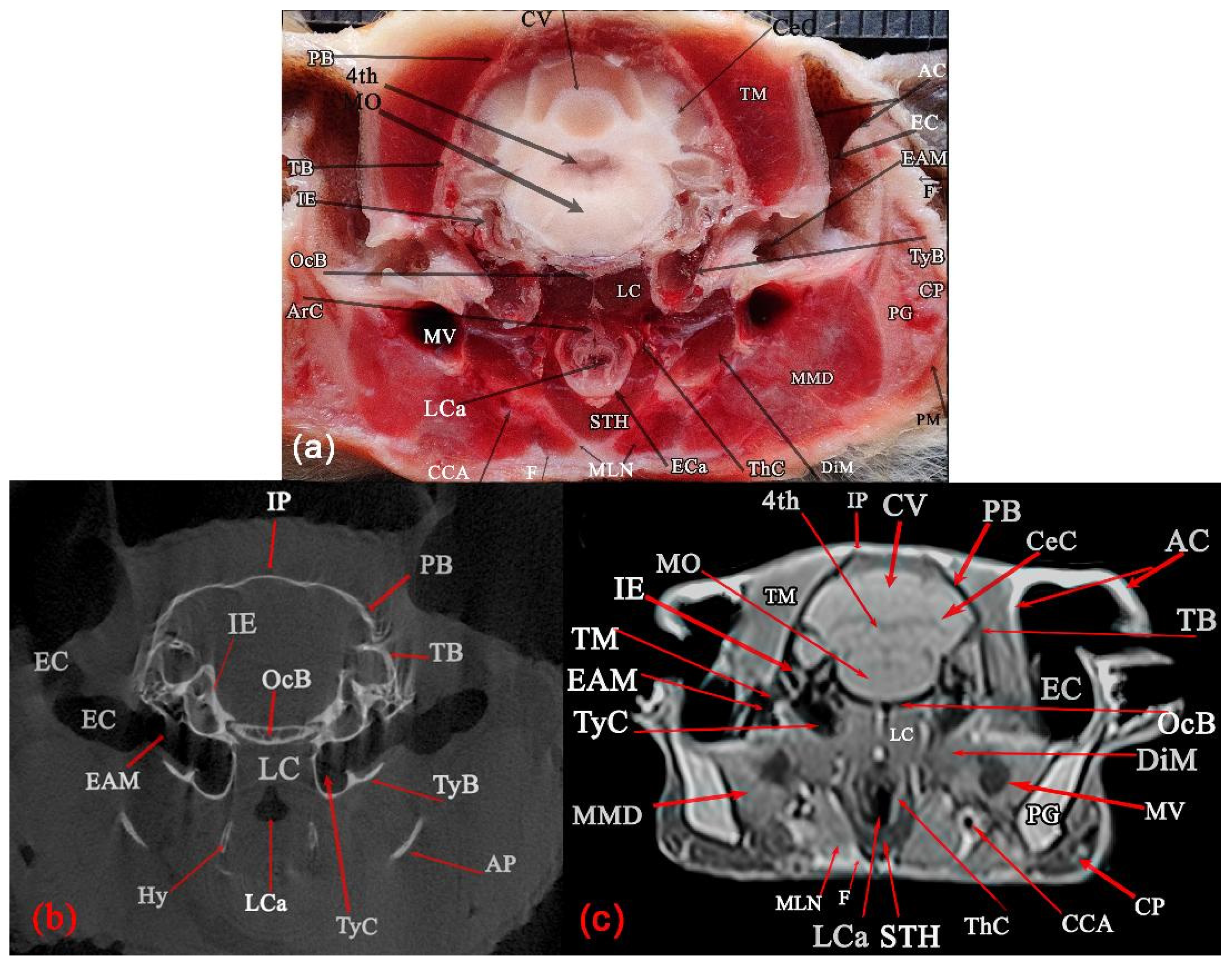
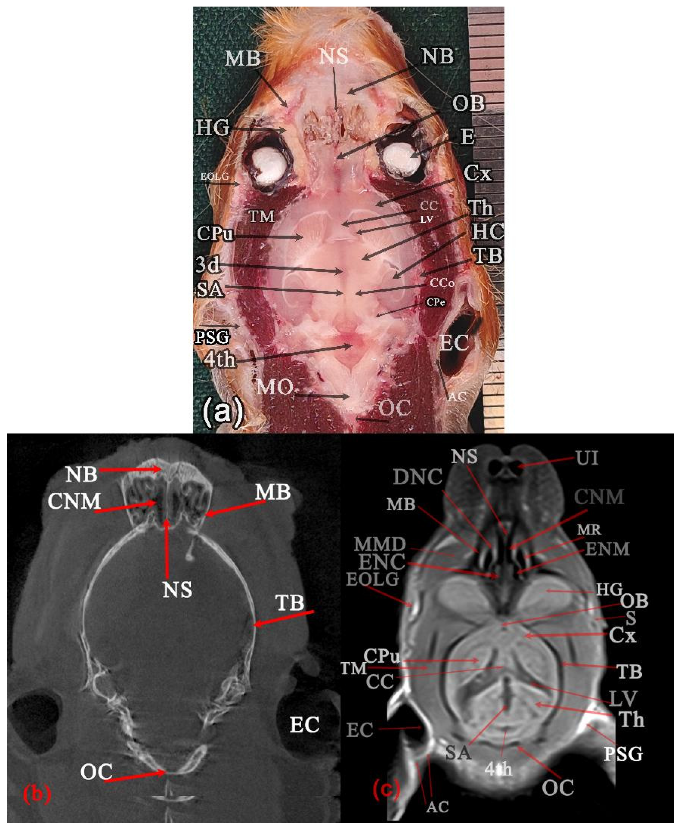
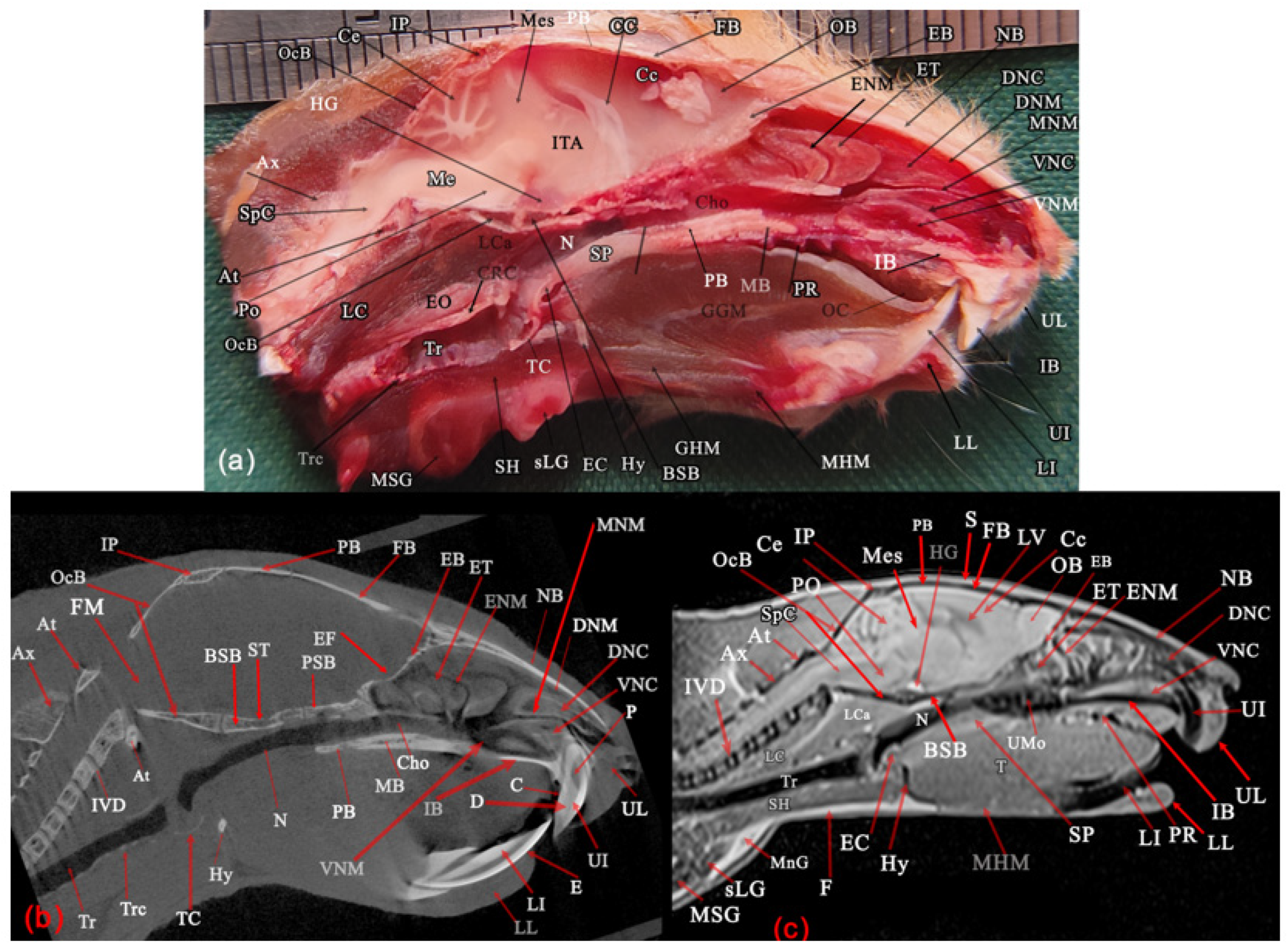
Figure 12). Each figure was composed of a gross anatomical slice (a), an MCT image (b), and an MR image (c). All transverse sections were viewed from the rostral aspect. All clinically relevant structures of the Syrian hamster head were identified on these sections. T1-weighted images were found to provide superior clarity and tissue differentiation compared with T2-weighted images and were therefore considered the optimal modality for analysis in this study. In our specimens, the cerebrospinal fluid (CSF) within the ventricular system and subarachnoid space, as well as the parotid and mandibular salivary glands, the extraorbital lacrimal gland, and the harderian gland, were more clearly distinguishable from the surrounding structures on T1-weighted images.
Figure 1.
Superficial structures of a male fresh cadaver of the Syrian hamster head and cranial end of the neck: left lateral (a,b), ventral (c), and rostral (d) views. The retractor sacculi have been transacted caudally and displaced rostrally to expose the masseter muscle, parotid and mandibular salivary glands, and parotid duct (a). AM: Angle of the mouth, BN: Buccal nerve, CP: Cheek pouch, DiM: Digastric muscle, EOLG: Extraorbital lacrimal gland, F: Fat tissue, LI: Lower incisor teeth, LL: Lower lip, MHM: Mylohyoid muscle, MLN: Mandibular lymph node, MM: Masseter muscle, Mn: Mandible, MnD: Mandibular duct, MSG: Mandibular salivary gland, No: Nostril, Npl: Nasal palate, OC: Oral cleft, PD: Parotid duct, PG: Parotid salivary gland, Ph: Philtrum, RS: Retractor sacculi muscle, S: Skin, STH: Sternothyrohyoid muscle, sLG: Sublingual salivary gland, UL: Upper lip, RO: Rostral, Do: Dorsal, and L: Left. Scale: 1 mm.
Figure 1.
Superficial structures of a male fresh cadaver of the Syrian hamster head and cranial end of the neck: left lateral (a,b), ventral (c), and rostral (d) views. The retractor sacculi have been transacted caudally and displaced rostrally to expose the masseter muscle, parotid and mandibular salivary glands, and parotid duct (a). AM: Angle of the mouth, BN: Buccal nerve, CP: Cheek pouch, DiM: Digastric muscle, EOLG: Extraorbital lacrimal gland, F: Fat tissue, LI: Lower incisor teeth, LL: Lower lip, MHM: Mylohyoid muscle, MLN: Mandibular lymph node, MM: Masseter muscle, Mn: Mandible, MnD: Mandibular duct, MSG: Mandibular salivary gland, No: Nostril, Npl: Nasal palate, OC: Oral cleft, PD: Parotid duct, PG: Parotid salivary gland, Ph: Philtrum, RS: Retractor sacculi muscle, S: Skin, STH: Sternothyrohyoid muscle, sLG: Sublingual salivary gland, UL: Upper lip, RO: Rostral, Do: Dorsal, and L: Left. Scale: 1 mm.
Figure 2.
Skull of a Syrian hamster. Right lateral (a), dorsal (b), and ventral (c) views. AP: Angular process, BSph: Basisphenoid bone, ch: Choanae, Conp: Condylar process of mandible, CorP: Coronoid process of mandible, EXM: External acoustic meatus, FB: Frontal bone, IB: Incisive bone, IP: Interparietal bone, MB: Maxillary bone (palatine process in ventral view), MF: Masseteric fossa, M 1–3: First molar tooth to third molar tooth, LI: Lower incisor tooth, NB: Nasal bone, OC: Occipital condyle, OcB: Occipital bone (basilar part in ventral view), P: Prominence of alveolar crown of tooth, PaB: Horizontal palate of the palatine bone, PaP: Palatine process of incisive bone, PB: Parietal bone, PP: Vertical palate of the palatine bone, PtB: Pterygoid bone, TB: Temporal bone, TyB: Tympanic bulla, UI: Upper incisor, ZPM: Zygomatic process of maxilla, and ZPT: Zygomatic process of temporal; Scale: 1 mm.
Figure 2.
Skull of a Syrian hamster. Right lateral (a), dorsal (b), and ventral (c) views. AP: Angular process, BSph: Basisphenoid bone, ch: Choanae, Conp: Condylar process of mandible, CorP: Coronoid process of mandible, EXM: External acoustic meatus, FB: Frontal bone, IB: Incisive bone, IP: Interparietal bone, MB: Maxillary bone (palatine process in ventral view), MF: Masseteric fossa, M 1–3: First molar tooth to third molar tooth, LI: Lower incisor tooth, NB: Nasal bone, OC: Occipital condyle, OcB: Occipital bone (basilar part in ventral view), P: Prominence of alveolar crown of tooth, PaB: Horizontal palate of the palatine bone, PaP: Palatine process of incisive bone, PB: Parietal bone, PP: Vertical palate of the palatine bone, PtB: Pterygoid bone, TB: Temporal bone, TyB: Tympanic bulla, UI: Upper incisor, ZPM: Zygomatic process of maxilla, and ZPT: Zygomatic process of temporal; Scale: 1 mm.
Figure 3.
Volume rendered 3D-reformatted MCT images of the left lateral (
a), dorsal (
b), and ventral (
c) views of a male Syrian hamster head. In (
a,
b), lines and large alphabetic letters indicate the approximate levels of the transverse (A–E), dorsal (F and G), and sagittal (I and H) gross, MCT, and MRI sectional planes included in
Figure 4,
Figure 5,
Figure 6,
Figure 7,
Figure 8,
Figure 9,
Figure 10,
Figure 11 and
Figure 12. ZA: Zygomatic arch. White arrow: Zygomatic bone. Black line: Cranial suture.
Figure 3.
Volume rendered 3D-reformatted MCT images of the left lateral (
a), dorsal (
b), and ventral (
c) views of a male Syrian hamster head. In (
a,
b), lines and large alphabetic letters indicate the approximate levels of the transverse (A–E), dorsal (F and G), and sagittal (I and H) gross, MCT, and MRI sectional planes included in
Figure 4,
Figure 5,
Figure 6,
Figure 7,
Figure 8,
Figure 9,
Figure 10,
Figure 11 and
Figure 12. ZA: Zygomatic arch. White arrow: Zygomatic bone. Black line: Cranial suture.
Figure 4.
Transverse planes of a male Syrian hamster head: anatomical (a), MCT (b), and T1-weighted MR (c) images, corresponding to line A in (a). Rostral view: AC: Alveolar cavity of the tooth, AM: Angle of mouth, C: Cementum, CNM: Common nasal meatus, D: Dentin, DNC: Dorsal nasal concha, DNM, Dorsal nasal meatus, E: Enamel, FL: Frenulum linguae, HP: Hard palate, IB: Incisive bone, LI: Lower incisor, LL: Lower lip, MNM: Middle nasal meatus, NB: Nasal bone, NLD: Nasolacrimal duct, NS: Nasal septum, OC: Oral cleft, OCa: Oral cavity, P: Pulp cavity of maxillary incisor tooth, S: Skin, T: Tongue, UI: Upper incisor, UL: Upper lip, V: Vomer, VNC: Ventral nasal concha, VNM: Ventral nasal meatus, and VNO: Vomeronasal organ. Scale: 1 mm in (a).
Figure 4.
Transverse planes of a male Syrian hamster head: anatomical (a), MCT (b), and T1-weighted MR (c) images, corresponding to line A in (a). Rostral view: AC: Alveolar cavity of the tooth, AM: Angle of mouth, C: Cementum, CNM: Common nasal meatus, D: Dentin, DNC: Dorsal nasal concha, DNM, Dorsal nasal meatus, E: Enamel, FL: Frenulum linguae, HP: Hard palate, IB: Incisive bone, LI: Lower incisor, LL: Lower lip, MNM: Middle nasal meatus, NB: Nasal bone, NLD: Nasolacrimal duct, NS: Nasal septum, OC: Oral cleft, OCa: Oral cavity, P: Pulp cavity of maxillary incisor tooth, S: Skin, T: Tongue, UI: Upper incisor, UL: Upper lip, V: Vomer, VNC: Ventral nasal concha, VNM: Ventral nasal meatus, and VNO: Vomeronasal organ. Scale: 1 mm in (a).
Figure 5.
Transverse planes of a male Syrian hamster head: anatomical (a), MCT (b), and T1-weighted MR (c) images, corresponding to line B in (a). Rostral view: APC: Anterior and posterior chamber of eye, C: Cementum, Ch: Choanae, CNM: Common nasal meatus, CP: Cheek pouch, D: Dentin, DiM: Digastric muscle, E: Eye, En: Enamel, ENC: Ethmoidal nasal concha, ENM: Ethmoidal nasal meatus, FB: Frontal bone, FM: Frontal muscle, HG: Harderian gland, HP: Hard palate, IMJ: Intermandibular joint, Le: Lens, LI: Lower incisor, MB: Maxilla, MHM: Mylohyoid muscle, MMD: Deep part of masseter muscle, MR: Maxillary recess, NS: Nasal septum, OCa: Oral cavity, P: Pulp cavity of tooth, PM: Platysma muscle, PMB: Palatine process of maxilla, Re: Retina, S: Skin, SMM: Superficial part of masseter muscle, T: Tongue, UI: Upper incisor, V: Vomer, VC: Vitreous chamber, VNM: Ventral nasal meatus, ZPM: Zygomatic process of maxilla, and ZMM: Zygomaticomandibularis muscle. Scale: 1 mm in (a).
Figure 5.
Transverse planes of a male Syrian hamster head: anatomical (a), MCT (b), and T1-weighted MR (c) images, corresponding to line B in (a). Rostral view: APC: Anterior and posterior chamber of eye, C: Cementum, Ch: Choanae, CNM: Common nasal meatus, CP: Cheek pouch, D: Dentin, DiM: Digastric muscle, E: Eye, En: Enamel, ENC: Ethmoidal nasal concha, ENM: Ethmoidal nasal meatus, FB: Frontal bone, FM: Frontal muscle, HG: Harderian gland, HP: Hard palate, IMJ: Intermandibular joint, Le: Lens, LI: Lower incisor, MB: Maxilla, MHM: Mylohyoid muscle, MMD: Deep part of masseter muscle, MR: Maxillary recess, NS: Nasal septum, OCa: Oral cavity, P: Pulp cavity of tooth, PM: Platysma muscle, PMB: Palatine process of maxilla, Re: Retina, S: Skin, SMM: Superficial part of masseter muscle, T: Tongue, UI: Upper incisor, V: Vomer, VC: Vitreous chamber, VNM: Ventral nasal meatus, ZPM: Zygomatic process of maxilla, and ZMM: Zygomaticomandibularis muscle. Scale: 1 mm in (a).
Figure 6.
Transverse planes of a male Syrian hamster head: anatomical (a), MCT (b), and T1-weighted MR (c) images, corresponding to line C in (a). Rostral view: AC: Alveolar cavity of teeth, C: Cementum, Ch: Choanae, Corp: Coronoid process of mandible, CP: Cheek pouch, D: Dentin, DiM: Digastric muscle, E: Enamel, EOLG: Extraorbital lacrimal gland, FB: Frontal bone, GGM: Genioglossus muscle, GHM: Geniohyoid muscle, HG: Harderian gland, HP: Hard palate, IMJ: Intermandibular joint, LI: Lower incisor, LMo: Lower molar tooth, MB: Maxillary bone, MHM: Mylohyoid muscle, MMD: Deep part of masseter muscle, MMS: Superficial part of masseter muscle, MPt: Medial pterygoid muscle, Mn: Ramous of Mandible, OB: Olfactory bulb, P: Pulp of tooth, PM: Platysma muscle, S: Skin, TM: Temporal muscle, T: Tongue, UMo: Upper molar tooth, and ZMM: Zygomaticomandibularis muscle. Scale: 1 mm in (a).
Figure 6.
Transverse planes of a male Syrian hamster head: anatomical (a), MCT (b), and T1-weighted MR (c) images, corresponding to line C in (a). Rostral view: AC: Alveolar cavity of teeth, C: Cementum, Ch: Choanae, Corp: Coronoid process of mandible, CP: Cheek pouch, D: Dentin, DiM: Digastric muscle, E: Enamel, EOLG: Extraorbital lacrimal gland, FB: Frontal bone, GGM: Genioglossus muscle, GHM: Geniohyoid muscle, HG: Harderian gland, HP: Hard palate, IMJ: Intermandibular joint, LI: Lower incisor, LMo: Lower molar tooth, MB: Maxillary bone, MHM: Mylohyoid muscle, MMD: Deep part of masseter muscle, MMS: Superficial part of masseter muscle, MPt: Medial pterygoid muscle, Mn: Ramous of Mandible, OB: Olfactory bulb, P: Pulp of tooth, PM: Platysma muscle, S: Skin, TM: Temporal muscle, T: Tongue, UMo: Upper molar tooth, and ZMM: Zygomaticomandibularis muscle. Scale: 1 mm in (a).
Figure 7.
Transverse planes of a male Syrian hamster head: anatomical (a), MCT (b), and T1-weighted MR (c) images, corresponding to line D in (a). Rostral view: 3d: Third ventricle, AP: Angular process, AS: Articular space of temporomandibular joint, CC: Corpus callosum, Cop: Condyloid process, CP: Cheek pouch, Cx: Cerebral cortex, DiM: Digastric muscle, GGM: Genioglossus muscle, GHM: Geniohyoid muscle, HyT: Hypothalamus, LPt: Lateral pterygoid muscle, LV: Lateral ventricle, MC: Mandibular canal, MF: Mandibular foramen, MFO: Mandibular fossa, MHM: Mylohyoid muscle, Mn: Ramus of Mandibular bone, MMD: Deep part of the masseter muscle, MMS: Superficial part of masseter muscle, MPt: Medial pterygoid muscle, N: Nasopharynx, ON: Optic nerve, PB: Pterygoid bone, PF: Pterygoid fossa, PG: Parotid salivary gland, PM: Platysma, PPB: Perpendicular process of palatine bone, PSph: Presphenoid bone, S: Skin, SB: Wing of presphenoid bone, SGM: Styloglossus muscle, SP: Soft palate, TB: Temporal bone, Th: Thalamus, TM: Temporal muscle, TML: Lateral part of the temporal muscle, TMM: Medial part of temporal muscle, ZMM: Zygomaticomandibular muscle, and ZPT: Zygomatic process of temporal bone. Scale: 1 mm in (a).
Figure 7.
Transverse planes of a male Syrian hamster head: anatomical (a), MCT (b), and T1-weighted MR (c) images, corresponding to line D in (a). Rostral view: 3d: Third ventricle, AP: Angular process, AS: Articular space of temporomandibular joint, CC: Corpus callosum, Cop: Condyloid process, CP: Cheek pouch, Cx: Cerebral cortex, DiM: Digastric muscle, GGM: Genioglossus muscle, GHM: Geniohyoid muscle, HyT: Hypothalamus, LPt: Lateral pterygoid muscle, LV: Lateral ventricle, MC: Mandibular canal, MF: Mandibular foramen, MFO: Mandibular fossa, MHM: Mylohyoid muscle, Mn: Ramus of Mandibular bone, MMD: Deep part of the masseter muscle, MMS: Superficial part of masseter muscle, MPt: Medial pterygoid muscle, N: Nasopharynx, ON: Optic nerve, PB: Pterygoid bone, PF: Pterygoid fossa, PG: Parotid salivary gland, PM: Platysma, PPB: Perpendicular process of palatine bone, PSph: Presphenoid bone, S: Skin, SB: Wing of presphenoid bone, SGM: Styloglossus muscle, SP: Soft palate, TB: Temporal bone, Th: Thalamus, TM: Temporal muscle, TML: Lateral part of the temporal muscle, TMM: Medial part of temporal muscle, ZMM: Zygomaticomandibular muscle, and ZPT: Zygomatic process of temporal bone. Scale: 1 mm in (a).

Figure 8.
Transverse planes of a male Syrian hamster head: anatomical (a), MCT (b), and T1-weighted MR (c) images, corresponding to line E in (a). Rostral view: 4th: Forth ventricle, AC: Auricular cartilage, AP: Angular process, ArC: Arytenoid cartilage, CCA: Common carotid artery, Cec: Cerebellum hemisphere, CP: Cheek pouch, CV: Vermis of cerebellum, DiM: Digastric muscle, EAM: External acoustic meatus, EC: Ear canal, ECa: Epiglottic cartilage, F: Fat tissue, Hy: Thyrohyoid bone, IE: Inner ear, LC: Longus capitis muscle, LCa: Laryngeal cavity, IP: Interparietal bone, MLN: Mandibular lymph node, MMD: Deep part of the masseter muscle, MO: Medulla oblongata, MV: Maxillary vein, OcB: Occipital bone, PB: Parietal bone, PG: Parotid salivary gland, PM: Platysma, STH: Sternothyrohyoid muscle, TB: Temporal bone, ThC: Thyroid cartilage, TM: Temporal muscle, TyB: Tympanic bulla, and TyC: Tympanic cavity. Scale: 1 mm in (a).
Figure 8.
Transverse planes of a male Syrian hamster head: anatomical (a), MCT (b), and T1-weighted MR (c) images, corresponding to line E in (a). Rostral view: 4th: Forth ventricle, AC: Auricular cartilage, AP: Angular process, ArC: Arytenoid cartilage, CCA: Common carotid artery, Cec: Cerebellum hemisphere, CP: Cheek pouch, CV: Vermis of cerebellum, DiM: Digastric muscle, EAM: External acoustic meatus, EC: Ear canal, ECa: Epiglottic cartilage, F: Fat tissue, Hy: Thyrohyoid bone, IE: Inner ear, LC: Longus capitis muscle, LCa: Laryngeal cavity, IP: Interparietal bone, MLN: Mandibular lymph node, MMD: Deep part of the masseter muscle, MO: Medulla oblongata, MV: Maxillary vein, OcB: Occipital bone, PB: Parietal bone, PG: Parotid salivary gland, PM: Platysma, STH: Sternothyrohyoid muscle, TB: Temporal bone, ThC: Thyroid cartilage, TM: Temporal muscle, TyB: Tympanic bulla, and TyC: Tympanic cavity. Scale: 1 mm in (a).
Figure 9.
Dorsal planes of a male Syrian hamster head: anatomical (a), MCT (b), and T1-weighted MR (c) images, corresponding to line F in (a). Ventral segment: 3d: Third ventricle, 4th: Fourth ventricle, AC: Auricular cartilage, CC: Corpus callosum, CCo: Caudal colliculus, CNM: Common nasal meatus, CPu: Caudoputamen, CPe: Cerebral peduncle, Cx: Cerebral cortex, DNC: Dorsal nasal concha, E: Eye, EC: Ear canal, ENC: Ethmoidal nasal concha, ENM: Ethmoidal nasal meatus, EOLG: Extraorbital lacrimal gland, HC: Hippocampus, HG: Harderian gland, LV: Lateral ventricle, MB: Maxillary bone, MMD: Deep part of the masseter muscle, MO: Medulla oblongata, MR: Maxillary recess, NB: Nasal bone, NS: Nasal septum, OB: Olfactory bulb, OC: Occipital bone, PSG: Parotid salivary gland, S: Skin, SA: Sylvius aqueduct, TB: Temporal bone, Th: Thalamus, TM: Temporal muscle, and UI: Upper incisor teeth. Scale: 1 mm in (a).
Figure 9.
Dorsal planes of a male Syrian hamster head: anatomical (a), MCT (b), and T1-weighted MR (c) images, corresponding to line F in (a). Ventral segment: 3d: Third ventricle, 4th: Fourth ventricle, AC: Auricular cartilage, CC: Corpus callosum, CCo: Caudal colliculus, CNM: Common nasal meatus, CPu: Caudoputamen, CPe: Cerebral peduncle, Cx: Cerebral cortex, DNC: Dorsal nasal concha, E: Eye, EC: Ear canal, ENC: Ethmoidal nasal concha, ENM: Ethmoidal nasal meatus, EOLG: Extraorbital lacrimal gland, HC: Hippocampus, HG: Harderian gland, LV: Lateral ventricle, MB: Maxillary bone, MMD: Deep part of the masseter muscle, MO: Medulla oblongata, MR: Maxillary recess, NB: Nasal bone, NS: Nasal septum, OB: Olfactory bulb, OC: Occipital bone, PSG: Parotid salivary gland, S: Skin, SA: Sylvius aqueduct, TB: Temporal bone, Th: Thalamus, TM: Temporal muscle, and UI: Upper incisor teeth. Scale: 1 mm in (a).
Figure 10.
Dorsal planes of a male Syrian hamster head: anatomical (a), MCT (b), and T1-weighted MR (c) images, corresponding to line G in (a). Ventral segment: AC: Auricular cartilage, BSph: Body of Basisphenoid bone, C: Cochlea, Ch: Choana, CNM: Common nasal meatus, Co: Cochlea, CP: Check pouch, DNC: Dorsal nasal concha, EC: Ear canal, ENC: Ethmoidal nasal concha, EXM: External acoustic meatus, EOLG: Extraorbital lacrimal gland, HG: Harderian gland, IOLG: Intraorbital lacrimal gland, LCa: Longus capitis muscle, LPt: Lateral pterygoid muscle, MB: Maxillary bone, MMD: Deep part of the masseter muscle, Mn: Ramus of Mandible, MPt: Medial pterygoid muscle, MR: Maxillary recess, N: Nasopharynx, NB: Nasal bone, NS: Nasal septum, OC: Occipital bone, OCP: Orifice of the cheek pouch, PG: Parotid salivary gland, PP: Perpendicular palate of palatine bone, PSph: Body of Presphenoid bone, PtB: Pterygoid bone, TyB: Tympanic bulla, TyC: Tympanic cavity, V: Vomer bone, VNC: Ventral nasal conchae, WSph: Wings of sphenoid bone, ZA: Zygomatic arch, and ZPT: Zygomatic process of temporal bone. Scale: 1 mm in (a).
Figure 10.
Dorsal planes of a male Syrian hamster head: anatomical (a), MCT (b), and T1-weighted MR (c) images, corresponding to line G in (a). Ventral segment: AC: Auricular cartilage, BSph: Body of Basisphenoid bone, C: Cochlea, Ch: Choana, CNM: Common nasal meatus, Co: Cochlea, CP: Check pouch, DNC: Dorsal nasal concha, EC: Ear canal, ENC: Ethmoidal nasal concha, EXM: External acoustic meatus, EOLG: Extraorbital lacrimal gland, HG: Harderian gland, IOLG: Intraorbital lacrimal gland, LCa: Longus capitis muscle, LPt: Lateral pterygoid muscle, MB: Maxillary bone, MMD: Deep part of the masseter muscle, Mn: Ramus of Mandible, MPt: Medial pterygoid muscle, MR: Maxillary recess, N: Nasopharynx, NB: Nasal bone, NS: Nasal septum, OC: Occipital bone, OCP: Orifice of the cheek pouch, PG: Parotid salivary gland, PP: Perpendicular palate of palatine bone, PSph: Body of Presphenoid bone, PtB: Pterygoid bone, TyB: Tympanic bulla, TyC: Tympanic cavity, V: Vomer bone, VNC: Ventral nasal conchae, WSph: Wings of sphenoid bone, ZA: Zygomatic arch, and ZPT: Zygomatic process of temporal bone. Scale: 1 mm in (a).

Figure 11.
Paramedian planes of a female Syrian hamster head: anatomical (a), MCT (b), and T1-weighted MR (c) images, corresponding to line H in (b). Left medial view: Ce: Cerebellum, Cx: Cerebral cortex, DiM: Digastric muscle, E: Eye, HG: Hypophysial gland, IE: Inner ear, LPt: Lateral pterygoid muscle, MM: Deep part of the masseter muscle, Mn: Mandible, MPt: Medial pterygoid muscle, Mse: Mesencephalon, MSG: Mandibular salivary gland, OcB: Occipital Bone, PB: Parietal bone, Th: Thalamus, TM: Temporalis muscle, TB: Temporal bone, TyC: Tympanic cavity, TyB: Tympanic bulla, WSB: Wing of presphenoid Bone, ZPM: Zygomatic process of maxilla. Scale: 1 mm in (a).
Figure 11.
Paramedian planes of a female Syrian hamster head: anatomical (a), MCT (b), and T1-weighted MR (c) images, corresponding to line H in (b). Left medial view: Ce: Cerebellum, Cx: Cerebral cortex, DiM: Digastric muscle, E: Eye, HG: Hypophysial gland, IE: Inner ear, LPt: Lateral pterygoid muscle, MM: Deep part of the masseter muscle, Mn: Mandible, MPt: Medial pterygoid muscle, Mse: Mesencephalon, MSG: Mandibular salivary gland, OcB: Occipital Bone, PB: Parietal bone, Th: Thalamus, TM: Temporalis muscle, TB: Temporal bone, TyC: Tympanic cavity, TyB: Tympanic bulla, WSB: Wing of presphenoid Bone, ZPM: Zygomatic process of maxilla. Scale: 1 mm in (a).
Figure 12.
Median planes of a female Syrian hamster head: anatomical (a), MCT (b), and T1-weighted MR (c) images, corresponding to line I in (b). Left medial view: At: Atlas, Ax: Axis, BSB: Body of Basisphenoid Bone, C: Cementum, CC: Corpus callosum, Ce: Vermis of cerebellum, Cc: Cerebral cortex, Cho: Choanae, CRC: Cricoid cartilage, D: Dentine, DNC: Dorsal nasal concha, DNM: Dorsal nasal meatus, EC: Epiglottic cartilage, EB: Cribriform palate of Ethmoidal bone, EF: Ethmoid Fossa, E: Enamel, ENM: Ethmoidal nasal meatus, EO: Esophagus, ET: Ethmoidal nasal concha, F: Fat tissue, FB: Frontal Bone, FM: Foramen magnum, GHM: Geniohyoid muscle, GGM: Genioglossus muscle, HG: Hypophysial gland, Hy: Basihyoid bone, IB: Incisive bone, IP: Interparietal bone, ITA: Interthalamic adhesion, IVD: Intervertebral disk, LC: Longus colli muscle, LCa: Longus capitis muscle, LI: Lower Incisor, LL: Lower Lip, LV: Lateral ventricle, MB: Maxilla, Me: Medulla oblongata, Mes: Mesencephalon, MHM: Mylohyoid muscle, MnG: Mandibular lymph node, MNM: Middle nasal meatus, MSG: Mandibular salivary gland, N: Nasopharynx, NB: Nasal bone, OB: Olfactory bulb, OC: Oral cavity, OcB: Occipital bone, P: Pulp of tooth, PB: Parietal Bone, Po: Pons, PR: Palatine ridge, PSB: Body of Presphenoid bone, S: skin, SH: Sternohyoid muscle, slG: sublingual salivary gland, SP: Soft palate, SpC: Spinal cord, ST: Sella Turcica, T: Tongue, TC: Thyroid cartilage, Tr: Trachea, Trc: Trachea cartilage, UI: Upper incisor tooth, UL: Upper lip, UMo: Upper molar teeth, VNC: Ventral nasal concha, and VNM: Ventral nasal meatus. Scale: 1 mm in (a).
Figure 12.
Median planes of a female Syrian hamster head: anatomical (a), MCT (b), and T1-weighted MR (c) images, corresponding to line I in (b). Left medial view: At: Atlas, Ax: Axis, BSB: Body of Basisphenoid Bone, C: Cementum, CC: Corpus callosum, Ce: Vermis of cerebellum, Cc: Cerebral cortex, Cho: Choanae, CRC: Cricoid cartilage, D: Dentine, DNC: Dorsal nasal concha, DNM: Dorsal nasal meatus, EC: Epiglottic cartilage, EB: Cribriform palate of Ethmoidal bone, EF: Ethmoid Fossa, E: Enamel, ENM: Ethmoidal nasal meatus, EO: Esophagus, ET: Ethmoidal nasal concha, F: Fat tissue, FB: Frontal Bone, FM: Foramen magnum, GHM: Geniohyoid muscle, GGM: Genioglossus muscle, HG: Hypophysial gland, Hy: Basihyoid bone, IB: Incisive bone, IP: Interparietal bone, ITA: Interthalamic adhesion, IVD: Intervertebral disk, LC: Longus colli muscle, LCa: Longus capitis muscle, LI: Lower Incisor, LL: Lower Lip, LV: Lateral ventricle, MB: Maxilla, Me: Medulla oblongata, Mes: Mesencephalon, MHM: Mylohyoid muscle, MnG: Mandibular lymph node, MNM: Middle nasal meatus, MSG: Mandibular salivary gland, N: Nasopharynx, NB: Nasal bone, OB: Olfactory bulb, OC: Oral cavity, OcB: Occipital bone, P: Pulp of tooth, PB: Parietal Bone, Po: Pons, PR: Palatine ridge, PSB: Body of Presphenoid bone, S: skin, SH: Sternohyoid muscle, slG: sublingual salivary gland, SP: Soft palate, SpC: Spinal cord, ST: Sella Turcica, T: Tongue, TC: Thyroid cartilage, Tr: Trachea, Trc: Trachea cartilage, UI: Upper incisor tooth, UL: Upper lip, UMo: Upper molar teeth, VNC: Ventral nasal concha, and VNM: Ventral nasal meatus. Scale: 1 mm in (a).

3.1. Gross Anatomy
3.1.1. Superficial Structures of the Head and Cranial End of the Neck
Cheek Pouch
The cheek pouches were described as paired, thin-walled, and distensible evaginations of the oral cavity, forming double-walled epithelial sacs. They were extended dorsocaudally along the lateral mandible from the mouth to the shoulder region, as well as from the ventral region to the ears, and were positioned between the skin and masticatory muscles (
Figure 2a,b). The pouches were located over the parotid salivary gland, masseter muscle, and extraorbital lacrimal gland, but were not visible externally. The medial surface of each pouch was not covered by skin. The retractor sacculi was attached to the caudal pouch and was passed under its dorsomedial surface over the masseter to be inserted at the angle of the mouth (
Figure 2a,b).
Major Salivary Glands
The major salivary glands identified in the Syrian hamster included the parotid, mandibular, and sublingual glands. Adjacent to the glands, lobulated brown fat structures were observed, which could be mistaken for lymph nodes or salivary glands (
Figure 1a,c). The flat parotid glands were lobulated and extended from the ventral aspect of the ear base to the caudal margin of the mandibular angle, lying over the masseter muscle. Laterally, the gland was covered by the cheek pouch and surrounded by adipose tissue (
Figure 1a,c). The parotid duct emerged from the medial surface of the caudal third of the gland and, together with the ventral buccal branch of the facial nerve, coursed laterally across the masseter (
Figure 1a,c).
Upon removal of the skin, the mandibular salivary gland was observed in the ventral neck region. It exhibited a distinctly lobulated structure, partially covering the lateral neck and extending caudally toward the thoracic inlet. The gland was in rostrolateral contact with the mandibular lymph node, and the mandibular glands of both sides were in contact along the midline (
Figure 1a,c).
The small sublingual gland was closely attached to the cranial–medial portion of the mandibular gland. It exhibited lighter coloration compared to the mandibular gland and had a uniform, non-lobulated structure. Laterally, it was situated adjacent to the mandibular lymph node (
Figure 1a,c).
Extraorbital Lacrimal Glands
The oval, pinkish extraorbital lacrimal glands were subcutaneous and under the cheek pouch and over the deep part of the masseter and temporalis muscles. The gland was located caudoventral to the eye and rostrodorsal to the parotid salivary gland (
Figure 1a).
Skull and Teeth
The skull was pyramidal in general form, with the face blending smoothly into the cranium without a dorsal “stop.” The skull was characterized by large orbits, large temporal fossae, incomplete postorbital bars, absence of an external sagittal crest, well-developed incisive bone, and prominent tympanic bullae. The dorsal cranial surface was almost flat, facial length was similar to cranial length, and cranial height was greater than facial height (
Figure 1 and
Figure 3). The zygomatic bone was a fine spicule positioned between the zygomatic processes of the maxilla and temporal bones to form the zygomatic arch, but was not involved in forming the orbital cavity or face. It was not always ossified in the examined animals. In macerated skulls, the zygomatic bone was absent, but it was visible in reconstructed 3D micro-CT images. Zygomatic arches were laterally curved and almost dorsally straight, with the long temporal process of the zygomatic bone joining the short zygomatic process of the temporal bone (
Figure 1 and
Figure 3). The incisive bone was well developed, forming the lateral face, with the infraorbital foramen located rostroventrally to the orbit (
Figure 1 and
Figure 3). The ventral border of the mandibular body was convex, containing a deep concavity, and the angular process projected caudally. The articular surface of the condyloid process was oriented rostrocaudally, and a prominent three-sided masseteric fossa was noted (
Figure 1 and
Figure 3). The dental formula was 2 (I 1/1, C 0/0, P 0/0, M 3/3) = 16, with incisors and molars separated by a relatively large diastema (
Figure 1). The maxillary diastema was larger than the mandibular diastema. Large maxillary incisors were strongly curved, while smaller mandibular incisors were mildly curved. Tooth size decreased from rostral to caudal, with the first mandibular and maxillary molars being the largest (
Figure 1 and
Figure 3).
3.1.2. Anatomical, MCT, and MRI Sections
Bony structures of the head were identified, including the nasal bone (
Figure 4a–c,
Figure 9a,b,
Figure 10a,b and
Figure 12a–c), vomer (
Figure 4a–c,
Figure 5a–c and
Figure 10a–c), nasal septum (
Figure 4a–c,
Figure 5a–c,
Figure 9a–c and
Figure 10a–c), incisive bone (
Figure 4a–c and
Figure 12a–c), frontal bone (
Figure 5a–c and
Figure 12a–c), intermandibular joint (
Figure 5a,c and
Figure 6b), palatine process of maxilla (
Figure 5b,c), zygomatic process of maxilla (
Figure 5a–c and
Figure 11a–c), mandible (
Figure 11a–c), maxilla (
Figure 5a–c,
Figure 6a–c,
Figure 9a–c,
Figure 10a–c and
Figure 12a–c), ramus of mandible (
Figure 5a–c,
Figure 7a–c and
Figure 10a–c), condylar process of mandible (
Figure 7a,b), presphenoid bone (
Figure 7a and
Figure 10b), articular space of temporomandibular joint, mandibular canal, and pterygoid fossa (
Figure 7b), pterygoid bone (
Figure 7b,c and
Figure 10a,b), perpendicular palate of palatine bone (
Figure 7b and
Figure 10b), zygomatic arch (
Figure 10b), zygomatic process of temporal bone (
Figure 7a–c and
Figure 10b), angular process, and thyrohyoid bone (
Figure 8a), interparietal bone (
Figure 8a–c and
Figure 12a–c), external acoustic meatus (
Figure 8a–c and
Figure 10c), tympanic cavity, and tympanic bulla (
Figure 8a–c,
Figure 10a–c and
Figure 11a–c), temporal bone (
Figure 8a–c,
Figure 9a–c and
Figure 11a–c), parietal bone (
Figure 8a–c,
Figure 11a–c and
Figure 12a–c), occipital bone (
Figure 8,
Figure 9,
Figure 11a–c and
Figure 12a–c), wing of presphenoid bone (
Figure 10b and
Figure 11a,b), body of basisphenoid bone (
Figure 10b and
Figure 12a–c), body of presphenoid bone (
Figure 10b and
Figure 12b), ethmoid fossa, sella turcica, and foramen magnum (
Figure 12b), intervertebral disk (
Figure 12b,c), cribriform palate of ethmoidal bone, atlas, axis, and basihyoid bone (
Figure 12a–c).
In micro-CT images, bony structures appeared radiopaque (bright), while air-filled regions were hypodense (dark or black) due to low X-ray attenuation. In micro-CT images, the articular space of the temporomandibular joint appeared as a hypodense gap between bones, while the articular disk was not visualized. The auditory ossicles were not delineated. In micro-CT images, the intermandibular joint (mandibular symphysis) appeared cartilaginous as a narrow radiolucent gap between the mandibular halves. In micro-CT images, the cheek pouches were identifiable laterally as air-filled, radiolucent cavities. On T1-weighted MRI, bony and air-filled structures generally appeared hypointense (dark). Meanwhile, some parts of the bones located within the nasal cavity, including the nasal septum, were easy to assess because of the distinct mucosal covering.
Tooth structures were identified including alveolar cavity of the tooth (
Figure 4b and
Figure 6b), cementum, dentin, enamel, pulp cavity of tooth (
Figure 4a,b,
Figure 5b,
Figure 6b and
Figure 12b), upper incisor (
Figure 4a–c,
Figure 8c and
Figure 12a–c), lower incisor (
Figure 4b,c,
Figure 5a–c,
Figure 6a,b and
Figure 12a–c), lower molar tooth (
Figure 6a–c), and upper molar (
Figure 6a–c and
Figure 12c).
On the MCT images, dental tissues showed distinct patterns: enamel was the brightest, dentin light gray, and cementum a thin, slightly less dense layer, often hard to distinguish from dentin. The pulp cavity appeared darkest in micro-CT due to the lack of mineral content, but hyperintense on T1-weighted images due to high water content. On T1-weighted MRI, tooth structures generally appeared hypointense (dark). Differentiation of enamel, dentin, and cementum on T1-weighted images was not possible.
The muscular and integumentary of systems of the head were identified, including the longus capitis muscle (
Figure 8a,c,
Figure 10a,c and
Figure 12a,c), sternohyoid muscle (
Figure 12a,c), digastric muscle (
Figure 5a,c,
Figure 6a,c,
Figure 7a,c,
Figure 8a,c and
Figure 11a,c), medial pterygoid muscle (
Figure 6a,c,
Figure 7a,c,
Figure 10a,c and
Figure 11a,c), lateral pterygoid muscle (
Figure 7a,c,
Figure 10a,c and
Figure 11a,c), longus colli muscle (
Figure 12a,c), skin (
Figure 4c,
Figure 5a,c,
Figure 6a,c,
Figure 7a,c and
Figure 9c), superficial part of masseter muscle (
Figure 6a,c and
Figure 7a,c), zygomaticomandibularis muscle (
Figure 5a,c,
Figure 6a,c and
Figure 7a,c), deep part of the masseter muscle (
Figure 5a,c,
Figure 6a,c,
Figure 7a,c,
Figure 8a,c,
Figure 9c,
Figure 10a,c and
Figure 11a,c), sternothyrohyoid muscle (
Figure 8a,c and
Figure 12a,c), platysma (
Figure 7a and
Figure 8a), temporal muscle (
Figure 6c,
Figure 7c,
Figure 8a,c,
Figure 9a,c and
Figure 11a,c), genioglossus muscle (
Figure 6a,c and
Figure 7a,c), lateral and medial parts of the temporal muscle (
Figure 6a and
Figure 7a), geniohyoid muscle (
Figure 6a,c,
Figure 7a,c and
Figure 12a), styloglossus muscle (
Figure 7a,c), mylohyoid muscle (
Figure 7a,c and
Figure 12a,c), and fat tissue (
Figure 8a,c and
Figure 12c). Muscles and fats were poorly visible or radiolucent in non-contrast micro-CT and typically indistinguishable without contrast enhancement. On T1-weighted MRI, head muscles showed intermediate signal intensity—brighter than bone but darker than fat. Fat appeared hyperintense, and skin was slightly brighter than muscle.
Nervous structures of the head were identified, including the olfactory bulb (
Figure 6a,c,
Figure 9a,c and
Figure 12a,c), cerebral cortex (
Figure 6,
Figure 7a,c,
Figure 9a,c,
Figure 11a,c and
Figure 12a,c), hypothalamus (
Figure 7a), thalamus, (
Figure 7a,c,
Figure 9a,c and
Figure 11a), lateral ventricle (
Figure 7a,c and
Figure 9c), optic nerve (
Figure 7a,c), Third ventricle (
Figure 7a,c and
Figure 9a,c), forth ventricle (
Figure 8a,c and
Figure 9a,c), cerebellum hemisphere (
Figure 8a,c and
Figure 11a,c), vermis of cerebellum (
Figure 8a,c and
Figure 12a,c), hypophysial gland (
Figure 12a,c), mesencephalon (
Figure 11a and
Figure 12a,c), thalamus, (
Figure 9a,c and
Figure 11a), interthalamic adhesion (
Figure 12a,c), cerebral hemisphere (
Figure 12a,c), corpus callosum (
Figure 7a,c,
Figure 9a,c and
Figure 12a,c), medulla oblongata (
Figure 8a,c,
Figure 9a and
Figure 12a,c), spinal cord (
Figure 12a,c), sylvius aqueduct (
Figure 9a,c), caudal colliculus (
Figure 9a), caudoputamen (
Figure 9a,c), cerebral peduncle (
Figure 9a), and hippocampus (
Figure 9a).
Nervous tissues were poorly visible or radiolucent in non-contrast micro-CT and typically indistinguishable without contrast enhancement. The contrast between gray and white matter was low in the T1-weighted MRI and showed similar signal intensities. In the transverse T1-weighted image, the cerebellum (mostly gray matter) had intermediate signal intensity and was darker than the medulla oblongata (mostly white matter), which had high signal intensity (
Figure 8c). On the T1-weighted MRI, cerebrospinal fluid in the brain ventricles and subarachnoid space also appeared black, reflecting its very low signal.
Respiratory structures of the head were identified, including the choanae (
Figure 5a–c,
Figure 6a–c,
Figure 10b and
Figure 12a,b), dorsal nasal concha, and dorsal nasal meatus (
Figure 4a–c,
Figure 9b,c and
Figure 12a–c), middle nasal meatus (
Figure 4c and
Figure 12a,b), ventral nasal meatus (
Figure 4a–c,
Figure 5c and
Figure 12a,b), ventral nasal concha (
Figure 4a–c,
Figure 10a–c and
Figure 12a–c), common nasal meatus (
Figure 4a–c,
Figure 5b,
Figure 9b,c and
Figure 10a–c), ethmoidal nasal concha, ethmoidal nasal meatus (
Figure 5a–c,
Figure 9a,c,
Figure 10a,c and
Figure 12a–c), maxillary recess (
Figure 5a–c,
Figure 9c and
Figure 10a,c), nasopharynx, (
Figure 7a–c,
Figure 10a,c and
Figure 12a–c), arytenoid cartilage, laryngeal cavity (
Figure 8a,c), thyroid cartilage (
Figure 8a,c and
Figure 12a), trachea (
Figure 12a–c), trachea cartilage (
Figure 12a–c), cricoid cartilage (
Figure 12a), epiglottic cartilage (
Figure 8a and
Figure 12a,c), nasolacrimal duct (
Figure 4a–c), and vomeronasal organ (
Figure 4a–c).
In micro-CT images, the nasolacrimal duct was located ventromedially to the upper incisor’s reversed crown. In micro-CT images of the maxillary recess and choanae, meatuses appeared as air-filled, radiolucent cavities. In micro-CT images, the nasopharynx and nasal meatuses appeared black due to air content. In micro-CT images, the laryngeal cartilages showed intermediate radiodensity, clearly distinguishable from adjacent soft tissues but less than calcified structures. On the T1-weighted image, the conchae and turbinates, as well as the lumen of the nasal cavity, laryngeal cavity, glottis, and trachea, were easily assessed because of the distinct mucosal covering. On the T1-weighted image, the vomeronasal organ appears as an oval or tubular structure with intermediate to slightly hyperintense signal intensity.
Digestive structures of the head were identified, including the angle of mouth (
Figure 4a), frenulum linguae, and oral cleft (
Figure 4a,c), tongue (
Figure 4c,
Figure 5a,c,
Figure 6b,c and
Figure 12b,c), lower lip (
Figure 4a,c and
Figure 12a–c), upper lip (
Figure 12a–c), oral cavity (
Figure 4a,
Figure 5a and
Figure 12a), cheek pouch (
Figure 4a,c,
Figure 5a–c,
Figure 6a,b,
Figure 7a,b and
Figure 10c), mandibular lymph node (
Figure 8a,c and
Figure 12a,c), parotid salivary gland (
Figure 7a,c,
Figure 8a,c,
Figure 9a,c and
Figure 10a,c), soft palate (
Figure 7a,c and
Figure 12a), sublingual salivary gland (
Figure 12a,c), mandibular salivary gland (
Figure 11a,c and
Figure 12a,c), palatine ridge, and esophagus (
Figure 12a,c), and hard palate (
Figure 4a,c and
Figure 5a,c).
In micro-CT images, the cheek pouches were identifiable laterally as air-filled, radiolucent cavities. On T1-weighted MRI images, the cheek pouch showed inner mucosal folds and a lumen as small dark cavities and appeared slightly darker than the surrounding muscles, making it easily distinguishable. On T1-weighted MRI images, the parotid salivary gland appeared homogeneous with high signal intensity. On T1-weighted MRI images, the mandibular lymph node was homogeneous, hypointense relative to fat, and nearly isointense to adjacent muscles. On the T1-weighted MRI, the mandibular salivary gland appeared heterogeneous with low to moderate signal intensity. Due to its close relation with the small sublingual gland, its borders could not be clearly distinguished, and delineation was only possible with reference to anatomical sections. In the median plane, the individual borders of the mandibular salivary gland and its lymph node were difficult to delineate, and the gland area could only be identified with reference to the corresponding anatomical section. On T1-weighted images, the lumen appeared hypointense (dark), while air-filled structures, such as the oral cavity.
Eye structures were identified, including the anterior and posterior chamber of eye, retina, vitreous chamber, and lens (
Figure 5c), harderian gland (
Figure 5a,c,
Figure 6a,c,
Figure 9a,c,
Figure 10a,c and
Figure 11a,c), extraorbital lacrimal gland (
Figure 6a,c,
Figure 9a,c and
Figure 10a), and eye (
Figure 5a,
Figure 9a and
Figure 11a–c). Only in the gross section, a small round intraorbital lacrimal gland was red and located at the medial part of the orbit, adjacent to the harderian gland (
Figure 10a).
No sex differences were observed in head anatomy and imaging, except for the harderian gland. In adult females, the harderian gland exhibited numerous dark spots in gross cross-sections; however, this feature was detectable only in anatomical sections (
Figure 9,
Figure 10 and
Figure 11) and not in micro-CT or MRI images. On the gross sections, the lobulated harderian gland appeared as a light-yellow mass, occupying the ventromedial and posterior regions of the ocular bulb. On the T1-weighted MRI, the harderian gland was relatively hyperintense compared to muscle. On the T1-weighted MRI, the extraorbital lacrimal gland was homogeneous and exhibited high signal intensity. The relative signal intensity on T1-weighted images was as follows: lens capsule (cortex) > masticatory muscles > anterior/posterior chambers and vitreous > lens > bone, with air appearing black. The lens occupied a large proportion of the globe.
Ear structures were defined, including the ear canal (
Figure 8a–c,
Figure 9a–c and
Figure 10a–c), auricular cartilage (
Figure 8a,c,
Figure 9b,c and
Figure 10a–c), inner ear (
Figure 8a–c and
Figure 11b,c), and cochlea (
Figure 10a,c). In the micro-CT image, the ear canal lumen was seen as hypodense (black) due to air content. On T1-weighted MRI images, auricular cartilage was bright and well-defined with intermediate to high signal.
The vascular system included the common carotid artery and maxillary vein (
Figure 8a,c). They were signal void in the T1-weighted MRI images, while they were not visible in the MCT images.




 ,
,