Article

Sarcoptes scabiei Induces Discrete NET Release, Ca2+ Fluxes and ROS Production Without Impairing Phagocytic Activity in Bovine Polymorphonuclear Neutrophils

1
Institute of Parasitology, Biomedical Research Center Seltersberg (BFS), Justus Liebig University Giessen, 35392 Giessen, Germany
2
Department of Veterinary Sciences and Public Health, Universidad Católica de Temuco, Temuco 4780000, Chile
3
CIBAV Research Group, Veterinary Medicine School, Faculty of Agrarian Sciences, Universidad de Antioquia, Medellin 050010, Colombia
*
Author to whom correspondence should be addressed.
Animals 2026, 16(11), 1628; https://doi.org/10.3390/ani16111628
Submission received: 5 April 2026 / Revised: 8 May 2026 / Accepted: 14 May 2026 / Published: 27 May 2026

Simple Summary

Sarcoptic mange is a contagious skin disease that affects humans and animals, caused by the mite Sarcoptes scabiei, which lives and reproduces within the outer layer of the skin. Infestation leads to intense pruritus, skin inflammation, and lesions that affect animal welfare and productivity. Overall, a mite-driven immune response includes the recruitment of immune cells like neutrophils. These cells can kill pathogens by producing toxic molecules, releasing antimicrobial substances, and forming web-like structures made of DNA and enzymes that may trap and kill microbes. However, little is known on how neutrophils from cattle respond to this ectoparasite. In this study, we examined how bovine neutrophils react when exposed either to whole mites or mite-derived antigen. We showed that mite antigen drove cellular activation, including calcium influxes and the production of reactive oxygen species. Mite antigen also induced a limited formation of neutrophil extracellular traps, while mites themselves hardly stimulated this response. These findings improve our understanding on how the bovine immune system reacts to sarcoptic mange and may contribute to the development of better strategies to control this disease in livestock.

Abstract

Sarcoptic mange is a skin disease caused by Sarcoptes scabiei infestations, characterized by dermatitis, pruritus, and exudative responses in both humans and animals. Biologically, the life cycle of S. scabiei is confined to the host’s skin (stratum corneum), where mite-derived molecules trigger the influx of innate immune cells, including polymorphonuclear neutrophils (PMN), which play a central role in skin inflammatory responses. The antimicrobial activity of PMNs is regulated by Ca2+ fluxes and includes the generation of reactive oxygen species (ROS), degranulation, and the release of neutrophil extracellular traps (NETs). NETs are web-like structures composed of chromatin and enzymes that can trap and eventually kill pathogens; however, their involvement in S. scabiei infestations in bovines remains unclear. Here, we investigated interactions between bovine PMN and S. scabiei mites, as well as PMN responses to S. scabiei antigen (ScAg). Functional parameters included NET release, Ca2+ fluxes, ROS production and phagocytic activity, determined by fluorescence microscopy, Fluo-4 staining, luminol-derived luminescence and flow cytometry, respectively. Current data show that ScAg, but not whole mites, induces a weak NET release in exposed bovine PMN. Additionally, ScAg drives rapid and sustained Ca2+ fluxes and ROS production over time, without altering the phagocytic capacity of PMN.

1. Introduction

Members of the family Sarcoptidae are burrowing astigmatid mites parasitizing throughout their life and considered as stationary permanent ectoparasites. Sarcoptic mange, or scabiosis, is a zoonotic parasitic skin infestation that affects both humans and animals, caused by the acari species Sarcoptes scabiei [1]. Biologically, S. scabiei infestation is confined to host skin, where shortly after infestation, fertilized female mites burrow beneath the stratum corneum, accomplishing their nutritional needs to release eggs into epidermal tunnels by ingesting lysed host skin tissue and lymph, but causing a significant inflammatory host response [1]. Clinically, S. scabiei infestation in animals represents a highly contagious ectoparasitosis inducing dermatitis, severe pruritus and alopecia, leading to thickened, hyperkeratotic, and hyperpigmented skin, compromised feed efficiency and overall animal welfare impairment [2]. In domestic animals, sarcoptic mange may affect cattle, dogs, pigs, sheep and goats, but rarely cats and horses [1]. The preferred site of S. scabiei infestation depends on the host species, with mites being more frequently found on sparsely haired parts of the body, such as ears, face, muzzle or neck. Nevertheless, burrowing mites might spread all over the body of infested animals causing generalized scabiosis. As stated above, burrowing, feeding and defecation activities of S. scabiei mites cause intense pruritus, inflammation, hair loss and crust formation of dried exudate. Intense pruritus leads to excoriation, resulting in exudation and even hemorrhage of the affected skin surface. The cutaneous pro-inflammatory host innate response mainly reflects keratinocyte- and epithelial cell-derived reactions leading to leukocyte recruitment to the site of infestation [3,4]. In this context, given its location, S. scabiei-derived antigenic substances are present in surrounding tissue layers, thereby inducing an early host innate immune reaction, being characterized by cytokine/chemokine release promoting leukocyte migration [1,3,5,6,7].
During S. scabei-driven pro-inflammatory epidermal responses, polymorphonuclear neutrophils (PMN) infiltrate mite-infested tissue [1,4]. Infestation-driven PMN recruitment occurs rapidly [8,9,10,11], as described for both human and animal S. scabei infestation in vivo [3,4,12,13,14]. However, the role of PMN in S. scabiei infestations is still largely unknown. Overall, mammalian PMN own different effector mechanisms, such as phagocytosis, release of extracellular vesicles (EV), degranulation of antimicrobial peptides, release of danger-associated molecular patterns (DAMPs) and ROS production, which are tightly regulated by intracellular Ca2+ fluxes after PMN activation [15]. In terms of sarcoptic mange, studies on S. scabiei-derived proteins revealed that these mites can impair PMN phagocytosis, thereby promoting secondary infections and highlighting the relevance of PMN in the pathogenesis of clinically manifested scabiosis [16,17]. In addition, cumulated evidence has demonstrated the capability of PMN to release either nuclear or mitochondrial DNA as fine and delicate extracellular structures, designated as neutrophil extracellular traps (NETs) [8,9,11]. Although beneficial in the context of infectious diseases, excessive NET extrusion has also been implicated in pro-inflammatory disorders causing sterile inflammation and contributing to autoimmune skin diseases, such as Lupus erythematosus and psoriasis [18,19]. NETs are primarily composed of decondensed chromatin, being decorated by granular antimicrobial components, which include myeloperoxidase (MPO), neutrophil elastase (NE), lactoferrin, calprotectin, LL37, pentraxin, proteinase 3, and cathepsin G, among others [11,20]. This composition is particularly effective in terms of microorganism capture and killing [8]. Different NET phenotypes have been described: (i) diffuse NETs (diffNETs), (ii) spread NETs (sprNETs), and (iii) aggregated NETs (aggNETs) [21,22]. In detail, diffNETs show a spherical and compact shape and range between 25 and 28 µm diameter. SprNETs are elongated web-like structures, characterized by a 15–17 nm diameter, distributed in elongated thin fibers. Finally, aggNETs are derived from many PMN converging in large NET clusters, measuring over 50 µm in diameter [21,22].
While NET formation can be induced by diverse bacterial and protozoan species, this effector mechanism also addresses larger metazoan parasites, which cannot be phagocytized [23,24,25]. Consistently, PMN cast NETs in response to parasitic nematode stages, e.g., from Haemonchus contortus, Strongyloides stercoralis, Ostertagia ostertagi, Brugia malayi, Dirofilaria immitis and Angiostrongylus vasorum [24,25,26,27,28,29]. Interestingly, trematodes like Fasciola hepatica or Calicophoron daubneyi are considered weak NET-inducers [22,30], suggesting that NET responses are differentially induced across metazoan species. In this context, so far, no evidence exists on NET release against parasitic mites in bovines. Thus, the aim of the current work was to characterize, for the first time, bovine PMN responses against S. scabei stages (eggs, nymphs, and adults) and specific antigens (ScAgs).

2. Materials and Methods

2.1. Bovine PMN Isolation

Peripheral blood was collected from healthy adult dairy cows (n = 4) by jugular venipuncture into 30 mL sterile, heparinized tubes (Kabe Labortechnik, Nümbrecht, Germany). The blood was diluted 1:1 with sterile PBS supplemented with 0.02% EDTA (Sigma-Aldrich, St. Louis, MO, USA) and layered on 12 mL of Histopaque®-1077 (density  =  1.077 g/mL; Sigma-Aldrich). Samples were centrifuged at 800× g for 45 min at room temperature (RT). Following centrifugation, the plasma and buffy coat layers were carefully removed. Remaining red blood cells (RBCs) were resuspended in Hank’s Balanced Salt Solution (HBSS), and hypotonic lysis was performed by adding 1 volume of cold phosphate-buffered water containing 5.5 mM NaH2PO4 and 8.4 mM KH2PO4 (pH 7.2; all Merck, Darmstadt, Germany). After 1 min, isotonicity was restored by adding 2 volumes of hypertonic phosphate buffer [5.5 mM NaH2PO4, 0.46 M NaCl (pH 7.2; all Merck)]. Samples were then centrifuged at 600× g for 10 min at 20 °C. The resulting PMN pellet was washed three times with 1× HBSS and resuspended in 5 mL of 1× HBSS. Cell counts were performed using a Neubauer hemocytometer (Marienfeld, Lauda-Königshofen, Germany). Freshly isolated bovine PMN were allowed to rest for 30 min at 37 °C and 5% CO2 prior to experimental use.

2.2. Sarcoptes scabiei Isolation and Mite Antigen (ScAg) Preparation

S. scabiei mites were collected from clinically infested dairy cows farmed in Bavaria, Germany. Specifically, the affected area was carefully trimmed, and deep epidermal scrapings were performed via the use of a sterile sharp skin scraper to collect skin material containing mites. These samples were stored at 4 °C and transferred to the Institute of Parasitology, JLU Giessen, where samples were stored at 4 °C until final processing, in a time period between 1 and 3 months. Samples were transferred to conical tubes and treated with 6 mL of 10% potassium hydroxide (KOH; Merck) solution. Epidermal digestion was performed by heating in 15 s intervals until hair/skin structures were dissolved. The digested material was then centrifuged at 600× g for 3 min at RT. To concentrate S. scabiei stages, a flotation was performed using sugar solution (550 g sugar, 443 mL tap water, 7 mL formaldehyde 37%; density 1.3 g/mL; Merck). The samples were mixed with sugar solution, centrifuged at 600× g for 5 min at RT to concentrate the mites in the upper layer, which were then collected and washed three times with distilled water. Mite identity was confirmed microscopically following established morphological criteria (Deplazes et al., 2013) [31]. For preparation of S. scabiei antigen (ScAg), an undetermined number of concentrated mites were suspended in 300 µL sterile phosphate-buffered saline (PBS) and subjected to sonication using five cycles of 15 s in an ice bath (Sonorex RK31® bath-type sonicator; Bandelin, Berlin, Germany). The suspension was centrifuged at 10,000× g for 20 min at 4 °C to separate insoluble debris. The protein concentration in the resulting supernatant was assessed by Bradford assay (Thermo Fisher Scientific, Waltham, MA USA) and aliquots were stored at −80 °C until further use as reported elsewhere [22].

2.3. Live Cell Analysis of Bovine PMN and Sarcoptes scabiei-Stage Interactions

Freshly isolated bovine PMN (2 × 10⁵ cells per sample; n = 3 different animals) were centrifuged at 300× g for 10 min at RT. Cells were then resuspended in 1 mL sterile RPMI 1640 medium without phenol red (Sigma-Aldrich) and loaded with Sytox Green (1:1000; Thermo Fisher Scientific) for extracellular DNA visualization. S. scabiei mites (nymphs and larvae) and eggs were placed in an 8 µ-Slide well (Ibidi, Gräfelfing, Germany) to adhere to the bottom. Subsequently, bovine PMN were added to mites and eggs in a total volume of 1 mL. Plates were incubated for 60 min at 37 °C and monitored microscopically by an inverted Olympus IX81® fluorescence microscope (Olympus, Tokyo, Japan). Microscopic images were analyzed by ImageJ® software (Fiji version 1.7, [32]) for quantification and visualization of extracellular DNA.

2.4. NET Visualization and Characterization of NET Phenotypes

Bovine PMN (n = 3; 2 × 10⁵ cells per sample) were stimulated with ScAg at concentrations ranging from 10 µg to 100 µg for 120 min at 37 °C in a 5% CO2 atmosphere. Briefly, cells were seeded on 15 mm diameter coverslips (Thermo Fisher Scientific) previously treated with 0.01% poly-L-lysine (Sigma-Aldrich) for 30 min in a total volume of 1 mL. Following stimulation with ScAg, PMN were fixed with 4% paraformaldehyde (Merck) and stored at 4 °C until further processing. To detect NET-specific proteins, samples were reacted with anti-histone (H1, H2A/H2B, H3, and H4; 1:200, Merck #MAB3422) and anti-neutrophil elastase (NE) (1:200, Abcam #ab68672, Cambridge, UK) antibodies as previously described [21]. Therefore, samples were first blocked for 60 min at RT with blocking buffer containing 3% bovine serum albumin (BSA; Sigma-Aldrich) and 0.3% Triton X-100 (Thermo Fisher Scientific). Then, primary antibodies were applied for 180 min at RT, followed by three washes with sterile PBS. Thereafter, secondary antibodies [Alexa Fluor 594 goat anti-mouse IgG (#A11005), Alexa Fluor 488 goat anti-rabbit IgG (#A11008), 1:500; Invitrogen, Carlsbad, CA, USA] were reacted for 30 min at RT in the dark. After three additional PBS washes, coverslips were mounted using Fluoromount G with DAPI (Thermo Fisher Scientific). Fluorescence imaging was performed using an inverted Olympus IX81 epifluorescence microscope equipped with an XM10 digital camera. Quantification of distinct NET phenotypes, i.e., spread NETs (sprNETs), diffuse NETs (diffNETs) and aggregated NETs (aggNETs), was performed by microscopic observation. NET phenotypes were identified and categorized based on morphological characteristics following established criteria [21,22].

2.5. Intracellular Ca2+ Flux Analysis and Live Cell 3D-Holotomographic Microscopy

Ca2+ fluxes in bovine PMN were assessed using the Ca2+-sensitive fluorescent dye Fluo-4-AM (Invitrogen), as described elsewhere (Conejeros et al., 2012) [33]. Briefly, bovine PMN were incubated with 2.5 µM Fluo-4-AM (Invitrogen) in 1× HBSS (30 min; 37 °C) using 5 × 10⁷ PMN/mL in a total volume of 1 mL. Excess dye was removed by washing the cells with 1× HBSS (Merck). Fluo-4-AM-loaded cells were then seeded in a 96-well plate at 5 × 10⁶ PMN/mL in a total volume of 200 µL. Experimental conditions included stimulation with ScAg (10 µg/mL) or A23187 (5 µM; Sigma-Aldrich). Ca2+ flux over time was quantified by area under the curve (AUC), using the first 50 s prior to stimulus exposure as baseline, and analyzing a total of 540 s in an automated multiplate reader (Varioskan Flash, Thermo Fisher Scientific). To confirm neutrophil Ca2+ fluxes, live cell 3D-holotomography coupled with epifluorescence was performed on Fluo-4-AM-loaded PMN stimulated with ScAg (10 µg/mL), as previously described [34]. Briefly, 1 × 10⁵ Fluo-4-AM-loaded PMN were seeded on 35 mm tissue culture µ-plates (Ibidi) and stimulated with ScAg (10 µg/mL) in a total volume of 1 mL. Live cell imaging was performed using a 3D Cell Explorer microscope (Nanolive, Tolochenaz, Switzerland) equipped with 60× magnification (λ  =  520 nm, sample exposure 0.2 mW/mm2, depth of field 30 µm) and a fluorescence unit (CoolLED pE-300 ultra). Images were acquired every 6 s in both refractive index (RI) and FITC fluorescence channels over 5 min. Raw data were processed using STEVE® software v 1.6 (Nanolive) to generate RI-based z-stacks. Post-processing and analysis were performed with ImageJ. Average intensity-projected holotomographic z-stacks were generated using the Z Project plugin. Fluo-4-AM-based fluorescence images were shown using the “fire” pseudocolor scale to facilitate visualization of fluorescence intensity changes over time.

2.6. Quantification of Bovine PMN ROS Production

To evaluate the effects of ScAg stimulation on bovine PMN oxidative responses, a luminol-based chemiluminescence assay was performed, as previously described [35]. Briefly, 1 × 10⁶ PMN were suspended in sterile 1× HBSS and placed into a 96-well plate (Sarstedt, Leicester, UK) pre-warmed to 37 °C in a total volume of 200 µL. Next, 50 μM luminol (Sigma-Aldrich) was added and the samples gently mixed. Basal ROS production was recorded for 120 s prior to ScAg (10 µg/mL) supplementation, after which luminescence was continuously monitored for 1900 s using a Luminoskan microplate reader (Thermo Fisher Scientific).

2.7. Phagocytosis Assay

Heparinized bovine whole blood was incubated for 15 min at 37 °C in the presence or absence of ScAg (10 µg/mL). Then, the phagocytic activity of immune cells present in whole blood samples was assessed using pH-sensitive rhodamine-conjugated Escherichia coli (E. coli) particles (Invitrogen, Carlsbad, CA, USA). Briefly, pHrodo dye-labeled E. coli particles (20 µL) were added to 100 µL of ScAg-stimulated and non-stimulated whole blood samples. Samples were incubated for 15 min at either 37 °C or 4 °C (negative control for phagocytosis). Subsequently, red blood cells were lysed and the samples centrifuged (350× g for 5 min). The cell pellet was washed and PMN analyzed by flow cytometry (BD Accuri C6 Plus flow cytometer, BD Bioscience, Heidelberg, Germany). The bovine PMN population was gated based on forward and side scatter parameters. The phagocytosis rate was determined as percentage of pHrodo-positive PMN, using non-stained cells to set gating thresholds.

2.8. Statistical Analyses

Statistical analyses were performed via GraphPad Prism 8® (version 8.4.3) software. Data description was carried out by presenting arithmetic means ± standard deviation. A Mann–Whitney test for the comparison of two experimental conditions was applied. In cases of three or more conditions, the Kruskal–Wallis test was used. Whenever global comparison by Kruskal–Wallis test indicated significance, post hoc multiple comparison tests were carried out by Dunn tests to compare test with control conditions. Outcomes of statistical tests were considered to indicate significant differences when p  ≤  0.05 (significance level).

3. Results

3.1. Sarcoptes scabiei Stages and ScAg Stimulation Barely Induce NETosis

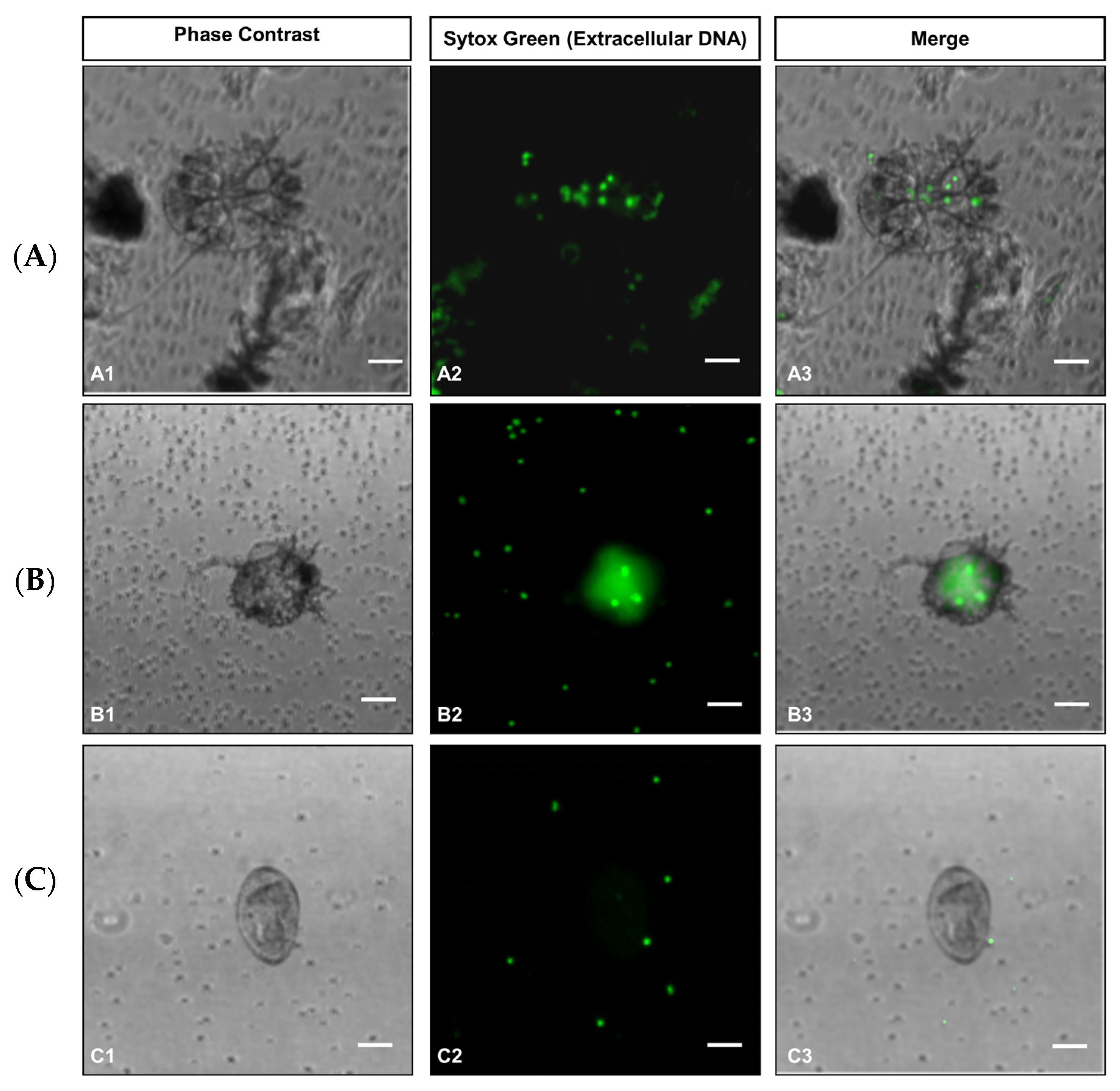
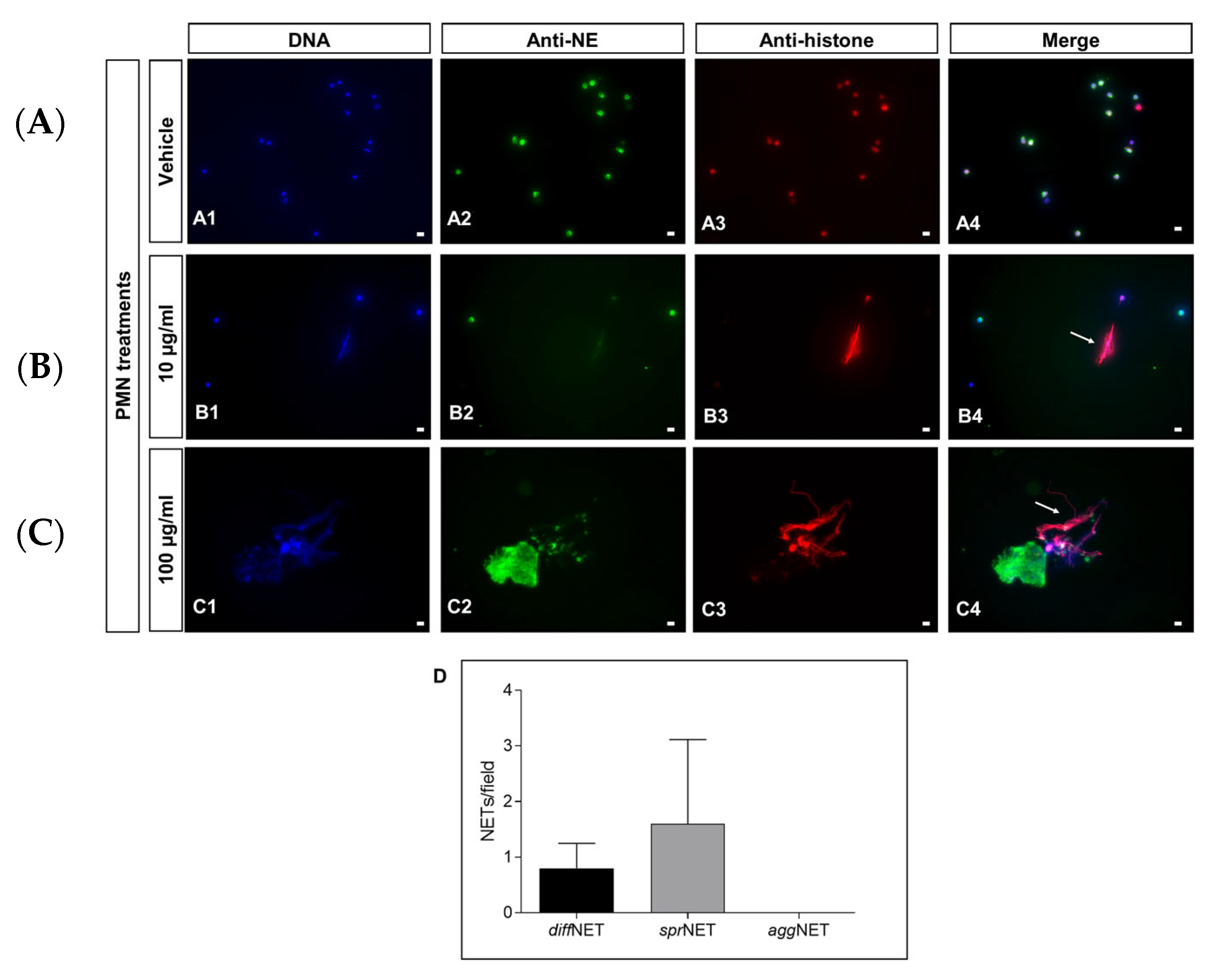
To evaluate if different stages of S. scabiei (i.e., eggs, larvae and nymphs) drive NET release in bovine PMN, DNA release was first assessed by Sytox Green-derived epifluorescence in live cells. This approach allowed for the visualization of extracellular DNA release in PMN–mite co-cultures. As shown in Figure 1, bovine PMN attached to S. scabiei nymphs (Figure 1(A3)) and larvae (Figure 1(B3)), maintaining their characteristic round morphology. However, no extracellular DNA filaments indicative of NET extrusion were clearly observed after 1 h of co-culture with nymphs (Figure 1A), larvae (Figure 1B), or eggs (Figure 1C), regardless of cellular attachment. Notably, NET structures were not evident even in PMN attached to nymphs (Figure 1(A1–A3)) or eggs (Figure 1(C1–C3)), while only a minor PMN enlargement and diffuse Sytox green-derived signal were observed on PMN attached to larvae (Figure 1(B1–B3)), suggesting that physical interaction alone does not trigger NETosis. To further assess the NET-inducing capacity of S. scabiei, ScAg was used at 10 and 100 µg/mL to stimulate bovine PMN. Immunofluorescence analysis was employed to identify classical NET components: extracellular DNA, NE, and histones. As shown in Figure 2, stimulation of PMN with 10 µg/mL ScAg induced a modest NET response after 2 h, characterized by short, thread-like NET structures (sprNETs; Figure 2(B1–B4)). Co-localization of extracellular DNA (blue, Figure 2(B1)), NE (green, Figure 2(B2)), and histones (red, Figure 2(B3)) confirmed the presence of ScAg-induced NETs (Figure 2(B4)). In contrast, vehicle-treated control cells exclusively showed an intracellular localization of these components without NET formation (Figure 2(A1–A4)). In addition, a higher ScAg concentration (100 µg/mL) triggered cytotoxic responses in stimulated PMN (Figure 2(C1–C4)), as evidenced by disorganized co-localization of NET markers and absence of intact, non-NETotic cells. Phenotypic analyses of NETs, based on previously reported classification criteria [21], revealed that sprNETs represented the predominant phenotype observed in ScAg-stimulated cells at 2 h post-treatment, followed by diffNETs. Overall, aggNETs were not detected after mite stimulation (Figure 2D).

3.2. ScAg Induces a Fast and Sustained Ca2+ Flux in Bovine PMN

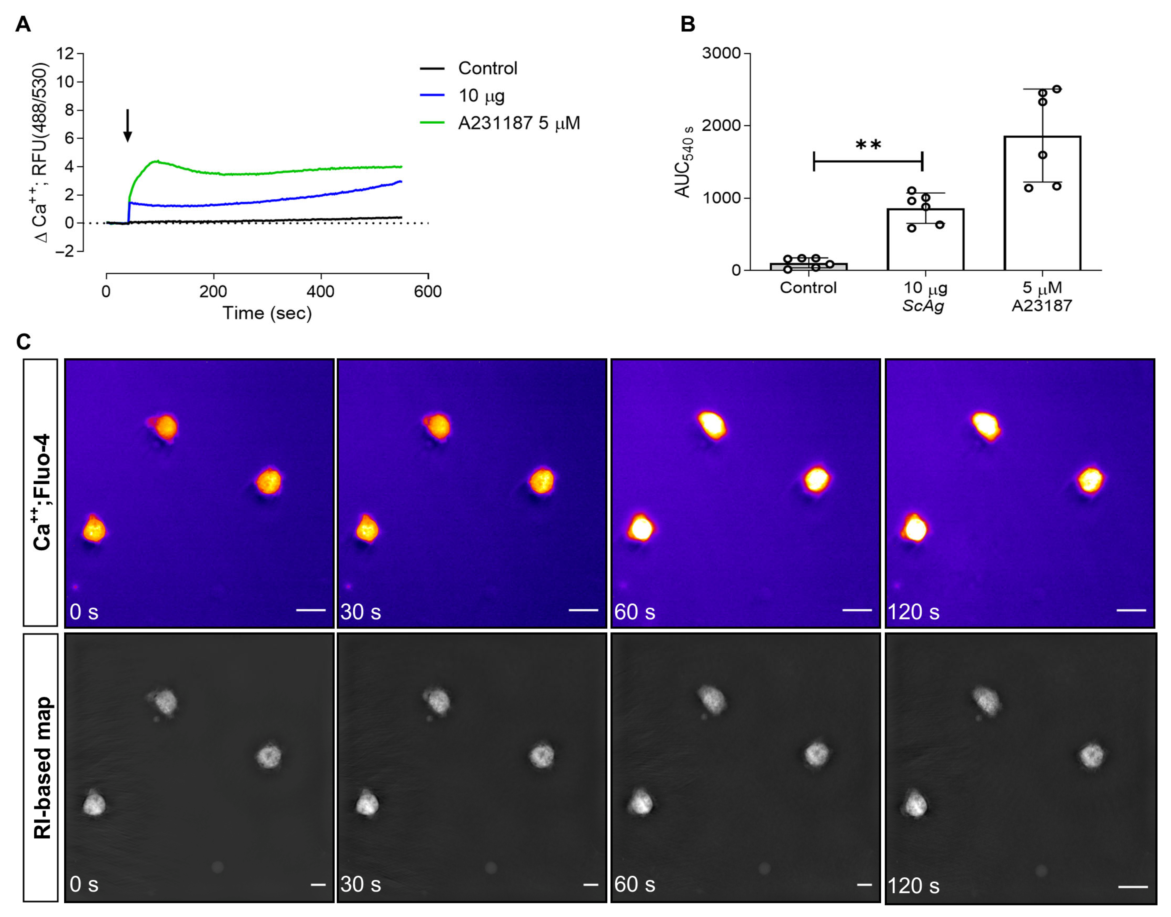
To determine whether ScAg triggers early activation of bovine PMN, intracellular Ca2+ fluxes were assessed as previously described [33]. As shown in Figure 3A, stimulation with 10 µg/mL ScAg induced a rapid and sustained increase in Ca2+-based fluorescence, even though the peak intensity was lower than that induced by the positive control A23187 (5 µM; 2.6 µg/mL). Kinetic analyses revealed that ScAg-induced Ca2+ mobilization in PMN remained consistent over time, with an increase in slope approximately 250 s after stimulation. A quantitative evaluation of the data via AUC analysis confirmed that stimulation with ScAg significantly elevated intracellular Ca2+ levels in PMN over the measurement period (p = 0.002), with the mean signal being approximately 2.2-fold lower than that generated by A23187 treatments (Figure 3B). These findings were supported by live cell 3D holotomographic fluorescence microscopy (Figure 3C), which confirmed a sustained increase in cytoplasmic Ca2+ signals following neutrophil ScAg stimulation. Notably, this activation mode occurred without phenotypic changes in PMN throughout the recording period.

3.3. ScAg Induces Oxidative Responses in Bovine PMN

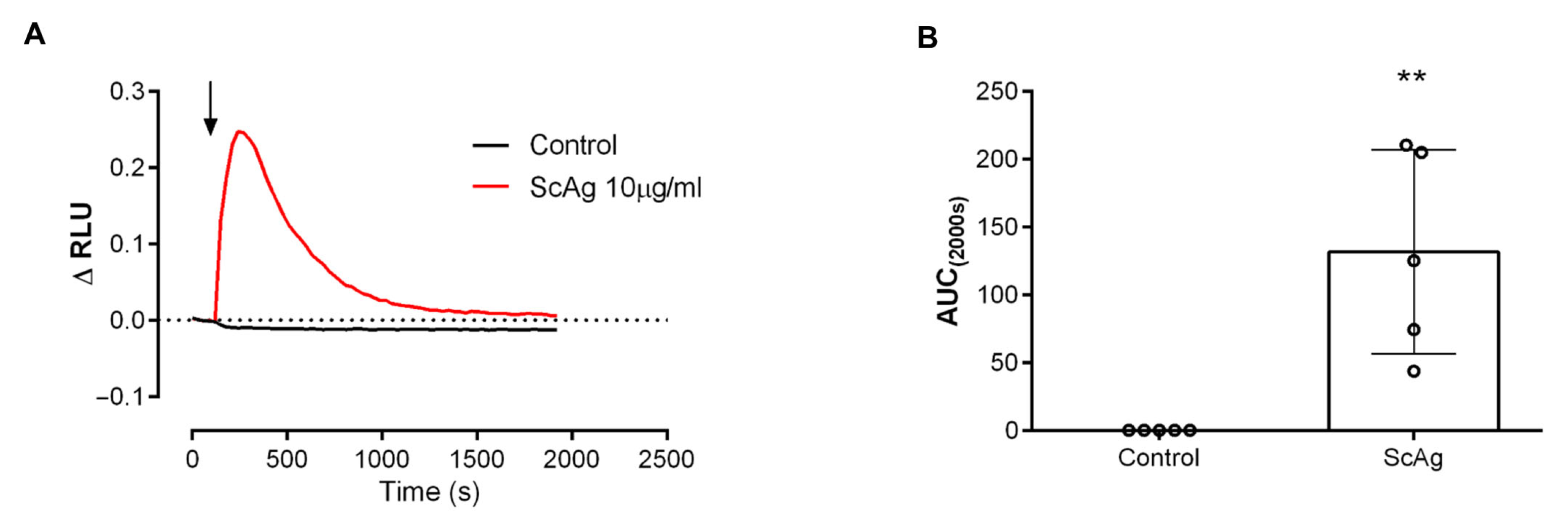
Since ScAg was shown to induce NET formation and intracellular Ca2+ fluxes in bovine PMN, we next assessed neutrophil oxidative responses driven by this mite-derived antigen using luminol-based chemiluminescence assays, as previously described [35]. Overall, stimulation with 10 μg/mL ScAg elicited a rapid and sustained increase in ROS-based luminescence, peaking approximately 240 s after exposure. This response gradually declined, returning to baseline levels around 900–1000 s post-stimulation (Figure 4A). Quantitative AUC analysis confirmed a significant elevation of ROS production over time compared to vehicle-treated controls (p = 0.0079) (Figure 4B).

3.4. ScAg Does Not Affect Bovine PMN Phagocytosis

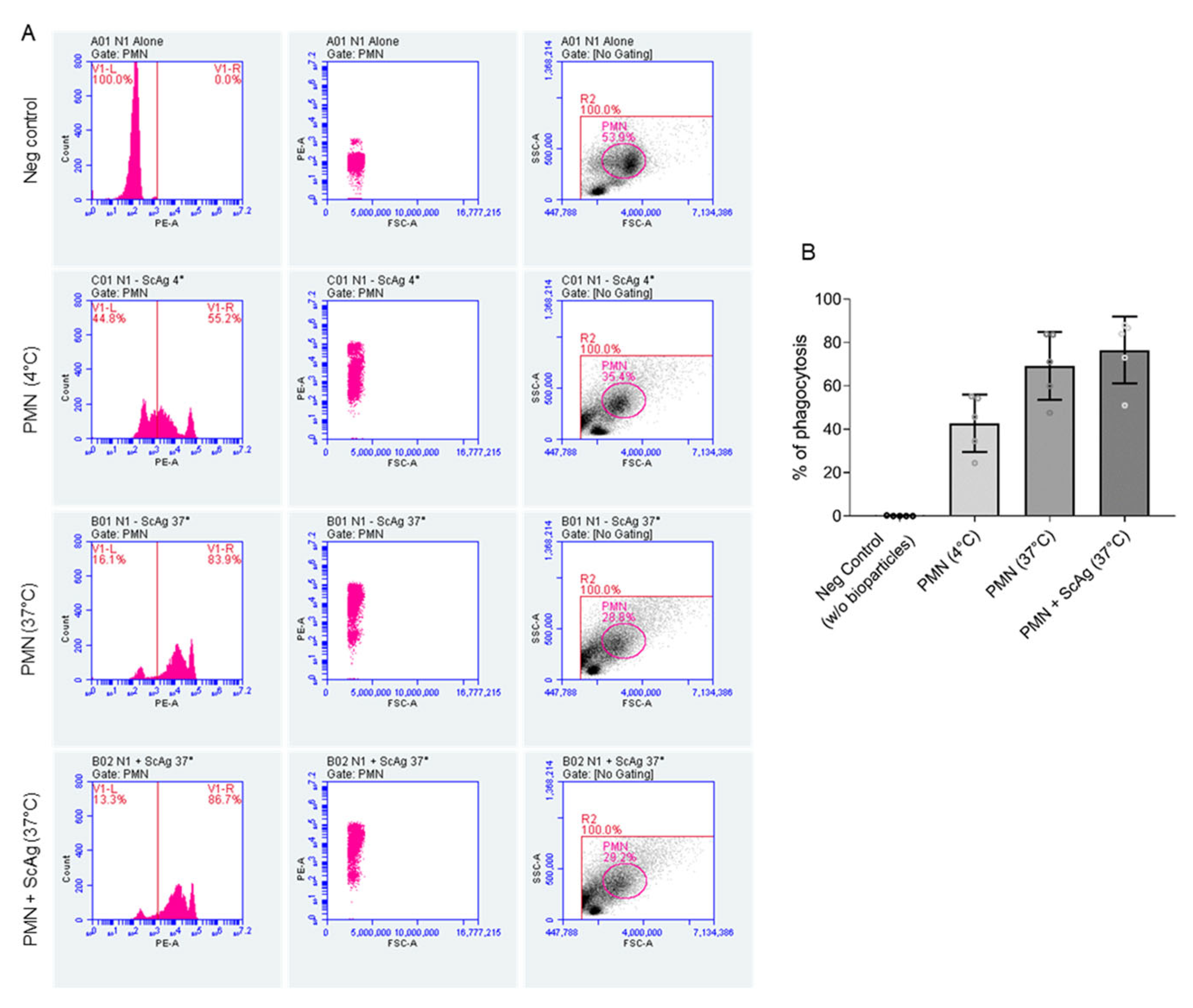
Based on previous reports indicating that S. scabiei-derived molecules can modulate PMN phagocytic activity [17], we further studied if stimulation with ScAg affects the phagocytic activity of bovine PMN. Phagocytosis was assessed by flow cytometry using pHrodo-labeled bioparticles, a reliable indicator for this defense function. As shown in Figure 5, an average of 69.2 ± 15.64% of PMN were engaged in phagocytosis under baseline conditions. Importantly, pre-treatment with ScAg did not significantly alter this percentage, with 76.52 ± 15.41% of PMN performing phagocytosis, indicating no observable effect of ScAg on neutrophil phagocytic activity.

4. Discussion

Sarcoptic mange is a globally distributed infectious skin disease of mammals caused by S. scabiei infestation. Due to the parasite’s life cycle, close interactions between its developmental stages and components of the host’s innate immune cells are very likely during infestation [1,4]. Similar to other cutaneous inflammatory responses, sarcoptic mange is associated with endothelial cell activation, increased vascular permeability, and rapid infiltration of immune cells like PMN [12,13,36,37,38,39]; however, the specific role of NET formation and other neutrophil effector mechanisms during this parasitic infestation remains largely unexplored. In this study, we examined interactions between bovine PMN and different S. scabiei stages in vitro. Co-culture of bovine PMN with S. scabiei eggs, larvae and nymphs revealed minimal PMN attachment to the parasite’s chitinous exoskeleton, suggesting that PMN are not effectively attracted by mites and fail to directly interact with the parasitic surface. The latter effect could be a consequence of the KOH-based mite isolation method, where mite surface proteins can potentially be damaged. However, this finding is consistent with previous reports showing that PMN tend to accumulate in perivascular regions of the dermis during S. scabiei infestation rather than directly interacting with mite structures [13]. The limited chemotaxis and/or attachment suggests that surface antigenic components of the mites may not represent adequate triggers of PMN engagement. Similar findings were reported in F. hepatica infections, where juvenile flukes show limited interactions with host PMN [22], supporting the idea that some metazoan parasite stages may be capable of evading initial PMN responses. The same holds true for the ruminant amphistome species C. daubneyi, where different parasitic stages and parasite-specific antigens (CdAg) merely induced discrete NET formation in exposed bovine PMN [30].
Mammalian PMN rely on several effector mechanisms to combat pathogen invasion, including phagocytosis, degranulation, oxidative burst, EV and NET release [9,40,41]. Obviously, during metazoan infections, phagocytosis proves to be ineffective due to the large size of the pathogen; thus, alternative responses like NETosis come into play [24,25,26,27,29]. In the current study, PMN co-cultures with S. scabiei stages failed to induce considerable NET formation in bovine PMN, indicating a low level of PMN activation in response to the parasite exoskeleton encounter. PMN, like other innate immune cells, rely on pattern recognition receptors (PPRs), such as TLRs and Dectin-1, to recognize pathogen-associated molecular patterns (PAMPs) and to initiate immune responses [42,43]. To test whether S. scabiei-derived molecules can activate PMN independent of physical contact, we stimulated PMN with soluble ScAg. In contrast to intact parasite stages, stimulation with 10 µg/mL of ScAg triggered moderate NET formation in bovine PMN. NET identity was confirmed by the co-localization of NE, histones, and extracellular DNA, and the predominant phenotype observed was short sprNETs. This discrete NETotic response is consistent with previous findings in other parasite stages like juvenile F. hepatica [22] and adult C. daubneyi [30]. Current results confirm that the magnitude and phenotype of NET responses may be strongly influenced by specific immunogenic properties of individual parasite stages and species, as previously proposed [22,30].
Ca2+ flux-based signaling is a central regulator of PMN effector functions, including degranulation, ROS generation and NET formation [15,44,45]. Overall, we here assessed whether stimulation with ScAg affects intracellular Ca2+ homeostasis in bovine PMN. Current data demonstrated that ScAg stimulation induces a rapid and sustained increase in neutrophil intracellular Ca2+ levels, indicating early PMN activation upon antigen exposure. In PMN, Ca2+ flux is typically induced by chemoattractants like CXCL8, PAF, or LTB4, as well as by interactions with adhesion molecules (L-selectin or CD11b) and Fc receptors [46]. These triggers of Ca2+ signaling usually precede downstream functional PMN responses. However, in the context of metazoan-derived molecules, including parasitic arthropods, the mechanisms underlying PMN activation remain less defined. For instance, excretory/secretory products of the blowfly Lucilia sericata (Calliphoridae) have been shown to elicit both oxidative and non-oxidative PMN responses via Ca2+-independent pathways [47], suggesting that helminth- or arthropod-derived molecules may activate mammalian PMN through non-canonical or PRR-independent signaling routes. In line with this, our findings indicate that S. scabiei-derived antigens can act as PMN modulators via Ca2+-dependent signaling but in a rather moderate way. Further studies should address the participation of different Ca2+-linked mechanisms, such as intra- and extra-cellular Ca2+ origin and store-operated Ca2+ entry (SOCE), upon S. scabei-induced PMN activation in bovines.
Since NET formation is classically linked to oxidative responses [9], we next evaluated the capacity of ScAg to induce neutrophil ROS production. Current results show that ScAg stimulation triggers a rapid and sustained generation of ROS in neutrophils, confirming its potential as an oxidative stimulus for bovine PMN. In PMN, ROS production is primarily mediated by NADPH oxidase (NOX) activation [9], catalyzing the reduction of molecular oxygen (O2) to superoxide anion (O2), which is subsequently converted to hydrogen peroxide (H2O2) and hypochlorous acid (HOCl) [45]. These processes create an oxidative microenvironment that mediates pathogen killing and supports NET formation. To our best knowledge, this is the first report on ROS production by PMN in response to S. scabiei-derived antigens, thus expanding the current understanding of arthropod-induced innate immune responses in bovines. This provides a novel mechanism in which PMN contribute to S. scabiei-induced inflammatory responses and tissue damage in bovines upon S. scabiei infestation.
During S. scabiei infestation in vivo, parasite-driven immunomodulation is thought to delay local host immune responses, thereby allowing a persistent (chronic) infestation [1,4]. In cases of scabiosis, secondary colonization of the epidermis by opportunistic bacteria like Staphylococcus aureus is frequently observed [4,12,14], furthermore indicating a compromised PMN-dependent defense. In this context, to assess a functional impact of ScAg on bovine PMN, we evaluated neutrophil phagocytic activities following ScAg stimulation. Current data indicate that ScAg, at concentrations sufficient to activate PMN oxidative and calcium responses, does not significantly alter the phagocytic activity of bovine PMN. This observation contrasts with previous findings reporting that recombinant SMSB4, a serpin derived from S. scabiei, can inhibit complement activity and reduce PMN phagocytosis [17]. Interestingly, the immunomodulatory function of serpins has also been documented in other parasitic arachnids, including the hard tick species Amblyomma americanum, Ixodes ricinus, and Rhipicephalus microplus, where salivary gland-derived serpins facilitate immune evasion and prolonged feeding times by dampening host immune responses [48,49,50]. In this context, it is plausible to hypothesize that either the current antigens derived from whole specimens contained amounts of serpins that were too low to induce suppressive effects or that the modulatory effects of S. scabiei-derived serpins on phagocytosis are primarily exerted through complement-dependent routes, which was not studied in the current experimental approach. However, these assumptions need further investigations for a final conclusion.

5. Conclusions

The current data indicate that S. scabiei antigens drive different events of early activation in bovine PMN, including intracellular Ca2+ flux and ROS production, and induce a modest release of NETs. However, these innate immune responses are not linked to a change in phagocytic activity. Notably, co-cultures with intact S. scabiei eggs, larvae and nymphs failed to activate PMN or to promote significant PMN chemotaxis or cell attachment, suggesting that the parasite’s exoskeleton may lack accessible antigens or immunostimulatory surface components recognizable by bovine PMN during early infestation. Despite that, since ScAg induced ROS production and Ca2+ fluxes in bovine PMN, the participation of PMN-derived responses during bovine scabiosis could enhance inflammatory responses, increasing mite-induced tissular damage or hampering tissular reparation. Nonetheless, given the exploratory design and the limited number of animals included in this study, these finding should be carefully interpreted. Further studies with larger samples sizes are required to fully exclude individual variability.

Author Contributions

Conceptualization: C.H. and I.C.; methodology: C.L. and I.C.; validation: I.C., A.T. and C.H.; formal analysis: C.L., D.G. and I.C.; investigation: C.L., D.G. and S.L.-O.; resources: A.T.; writing—original draft preparation, C.L. and D.G.; writing—review and editing, C.L., D.G., I.C., C.H. and A.T.; supervision, I.C., A.T. and C.H.; funding acquisition, A.T. All authors have read and agreed to the published version of the manuscript.

Funding

C.L. and D.G. were funded by the National Agency for Research and Development ((ANID), DOCTORADO BECAS CHILE. S.L-O. was a recipient of a PhD fellowship financed by the German Academic Exchange Service (DAAD Research Grants—One-Year Grants 2018–19). The publication fees were partially funded by the Open Access Funds of Justus Liebig University Giessen.

Institutional Review Board Statement

The study was conducted in accordance with the Declaration of Helsinki, and the Justus Liebig University Giessen (JLU) Animal Care Committee Guidelines. Protocols were approved by the Ethic Commission for Experimental Animal Studies of Federal State of Hesse (Regierungspräsidium Giessen: GI 18/10 No V2/2022; JLU-No 0002_V) and in accordance with European Animal Welfare Legislation: ART13TFEU and current applicable German Animal Protection Laws.

Informed Consent Statement

Not applicable.

Data Availability Statement

All data are included in the manuscript.

Acknowledgments

The authors would like to thank Christine Henrich, Christin Ritter and Hannah Salecker (all members of the Institute of Parasitology, JLU Giessen, Germany) for their outstanding technical support.

Conflicts of Interest

The authors declare no conflicts of interest.

References

  1. Arlian, L.G.; Morgan, M.S. A Review of Sarcoptes scabiei: Past, Present and Future. Parasites Vectors 2017, 10, 297. [Google Scholar] [CrossRef] [PubMed]
  2. Rentería-Solís, Z.; Min, A.M.; Alasaad, S.; Müller, K.; Michler, F.-U.; Schmäschke, R.; Wittstatt, U.; Rossi, L.; Wibbelt, G. Genetic Epidemiology and Pathology of Raccoon-Derived Sarcoptes Mites from Urban Areas of Germany. Med. Vet. Entomol. 2014, 28, 98–103. [Google Scholar] [CrossRef] [PubMed]
  3. Bhat, S.A.; Walton, S.F.; Ventura, T.; Liu, X.; McCarthy, J.S.; Burgess, S.T.G.; Mounsey, K.E. Early Immune Suppression Leads to Uncontrolled Mite Proliferation and Potent Host Inflammatory Responses in a Porcine Model of Crusted versus Ordinary Scabies. PLoS Negl. Trop. Dis. 2020, 14, e0008601. [Google Scholar] [CrossRef]
  4. Bhat, S.A.; Mounsey, K.E.; Liu, X.; Walton, S.F. Host Immune Responses to the Itch Mite, Sarcoptes scabiei, in Humans. Parasites Vectors 2017, 10, 385. [Google Scholar] [CrossRef]
  5. Elder, B.L.; Arlian, L.G.; Morgan, M.S. Modulation of Human Dermal Microvascular Endothelial Cells by Sarcoptes scabiei in Combination with Proinflammatory Cytokines, Histamine, and Lipid-Derived Biologic Mediators. Cytokine 2009, 47, 103–111. [Google Scholar] [CrossRef][Green Version]
  6. Morgan, M.S.; Arlian, L.G. Response of Human Skin Equivalents to Sarcoptes scabiei. J. Med. Entomol. 2010, 47, 877–883. [Google Scholar] [CrossRef]
  7. Mullins, J.S.; Arlian, L.G.; Morgan, M.S. Extracts of Sarcoptes scabiei De Geer Downmodulate Secretion of IL-8 by Skin Keratinocytes and Fibroblasts and of GM-CSF by Fibroblasts in the Presence of Proinflammatory Cytokines. J. Med. Entomol. 2009, 46, 845–851. [Google Scholar] [CrossRef][Green Version]
  8. Brinkmann, V.; Reichard, U.; Goosmann, C.; Fauler, B.; Uhlemann, Y.; Weiss, D.S.; Weinrauch, Y.; Zychlinsky, A. Neutrophil Extracellular Traps Kill Bacteria. Science 2004, 303, 1532–1535. [Google Scholar] [CrossRef]
  9. Fuchs, T.A.; Abed, U.; Goosmann, C.; Hurwitz, R.; Schulze, I.; Wahn, V.; Weinrauch, Y.; Brinkmann, V.; Zychlinsky, A. Novel Cell Death Program Leads to Neutrophil Extracellular Traps. J. Cell Biol. 2007, 176, 231–241. [Google Scholar] [CrossRef]
  10. Kolaczkowska, E.; Kubes, P. Neutrophil Recruitment and Function in Health and Inflammation. Nat. Rev. Immunol. 2013, 13, 159–175. [Google Scholar] [CrossRef] [PubMed]
  11. Papayannopoulos, V. Neutrophil Extracellular Traps in Immunity and Disease. Nat. Rev. Immunol. 2018, 18, 134–147. [Google Scholar] [CrossRef]
  12. Dagleish, M.P.; Ali, Q.; Powell, R.K.; Butz, D.; Woodford, M.H. Fatal Sarcoptes scabiei Infection of Blue Sheep (Pseudois nayaur) in Pakistan. J. Wildl. Dis. 2007, 43, 512–517. [Google Scholar] [CrossRef]
  13. Elwood, H.; Berry, R.S.; Gardner, J.M.; Shalin, S.C. Superficial Fibrin Thrombi … and Other Findings: A Review of the Histopathology of Human Scabietic Infections. J. Cutan. Pathol. 2015, 42, 346–352. [Google Scholar] [CrossRef]
  14. Swe, P.M.; Christian, L.D.; Lu, H.C.; Sriprakash, K.S.; Fischer, K. Complement Inhibition by Sarcoptes scabiei Protects Streptococcus pyogenes—An in Vitro Study to Unravel the Molecular Mechanisms behind the Poorly Understood Predilection of S. pyogenes to Infect Mite-Induced Skin Lesions. PLoS Negl. Trop. Dis. 2017, 11, e0005437. [Google Scholar] [CrossRef] [PubMed]
  15. Hann, J.; Bueb, J.-L.; Tolle, F.; Bréchard, S. Calcium Signaling and Regulation of Neutrophil Functions: Still a Long Way to Go. J. Leukoc. Biol. 2020, 107, 285–297. [Google Scholar] [CrossRef] [PubMed]
  16. Mika, A.; Reynolds, S.L.; Mohlin, F.C.; Willis, C.; Swe, P.M.; Pickering, D.A.; Halilovic, V.; Wijeyewickrema, L.C.; Pike, R.N.; Blom, A.M.; et al. Novel Scabies Mite Serpins Inhibit the Three Pathways of the Human Complement System. PLoS ONE 2012, 7, e40489. [Google Scholar] [CrossRef]
  17. Swe, P.M.; Fischer, K. A Scabies Mite Serpin Interferes with Complement-Mediated Neutrophil Functions and Promotes Staphylococcal Growth. PLoS Negl. Trop. Dis. 2014, 8, e2928. [Google Scholar] [CrossRef]
  18. Li, S.; Ying, S.; Wang, Y.; Lv, Y.; Qiao, J.; Fang, H. Neutrophil Extracellular Traps and Neutrophilic Dermatosis: An Update Review. Cell Death Discov. 2024, 10, 18. [Google Scholar] [CrossRef] [PubMed]
  19. Yamamoto, T. Role of Neutrophils in Cutaneous Lupus Erythematosus. J. Dermatol. 2024, 51, 180–184. [Google Scholar] [CrossRef]
  20. Urban, C.F.; Ermert, D.; Schmid, M.; Abu-Abed, U.; Goosmann, C.; Nacken, W.; Brinkmann, V.; Jungblut, P.R.; Zychlinsky, A. Neutrophil Extracellular Traps Contain Calprotectin, a Cytosolic Protein Complex Involved in Host Defense against Candida Albicans. PLoS Pathog. 2009, 5, e1000639. [Google Scholar] [CrossRef]
  21. Grob, D.; Conejeros, I.; Velásquez, Z.D.; Preußer, C.; Gärtner, U.; Alarcón, P.; Burgos, R.A.; Hermosilla, C.; Taubert, A. Trypanosoma brucei brucei Induces Polymorphonuclear Neutrophil Activation and Neutrophil Extracellular Traps Release. Front. Immunol. 2020, 11, 559561. [Google Scholar] [CrossRef]
  22. Peixoto, R.; Silva, L.M.R.; López-Osório, S.; Zhou, E.; Gärtner, U.; Conejeros, I.; Taubert, A.; Hermosilla, C. Fasciola hepatica Induces Weak NETosis and Low Production of Intra- and Extracellular ROS in Exposed Bovine Polymorphonuclear Neutrophils. Dev. Comp. Immunol. 2021, 114, 103787. [Google Scholar] [CrossRef]
  23. Branzk, N.; Lubojemska, A.; Hardison, S.E.; Wang, Q.; Gutierrez, M.G.; Brown, G.D.; Papayannopoulos, V. Neutrophils Sense Microbe Size and Selectively Release Neutrophil Extracellular Traps in Response to Large Pathogens. Nat. Immunol. 2014, 15, 1017–1025. [Google Scholar] [CrossRef]
  24. Muñoz-Caro, T.; Conejeros, I.; Zhou, E.; Pikhovych, A.; Gärtner, U.; Hermosilla, C.; Kulke, D.; Taubert, A. Dirofilaria immitis Microfilariae and Third-Stage Larvae Induce Canine NETosis Resulting in Different Types of Neutrophil Extracellular Traps. Front. Immunol. 2018, 9, 968. [Google Scholar] [CrossRef] [PubMed]
  25. Muñoz-Caro, T.; Rubio, R.M.C.; Silva, L.M.R.; Magdowski, G.; Gärtner, U.; McNeilly, T.N.; Taubert, A.; Hermosilla, C. Leucocyte-Derived Extracellular Trap Formation Significantly Contributes to Haemonchus contortus Larval Entrapment. Parasites Vectors 2015, 8, 607. [Google Scholar] [CrossRef]
  26. Bonne-Année, S.; Kerepesi, L.A.; Hess, J.A.; Wesolowski, J.; Paumet, F.; Lok, J.B.; Nolan, T.J.; Abraham, D. Extracellular Traps Are Associated with Human and Mouse Neutrophil and Macrophage Mediated Killing of Larval Strongyloides stercoralis. Microbes Infect. 2014, 16, 502–511. [Google Scholar] [CrossRef]
  27. Grob, D.; Conejeros, I.; López-Osorio, S.; Velásquez, Z.D.; Segeritz, L.; Gärtner, U.; Schaper, R.; Hermosilla, C.; Taubert, A. Canine Angiostrongylus vasorum-Induced Early Innate Immune Reactions Based on NETs Formation and Canine Vascular Endothelial Cell Activation In Vitro. Biology 2021, 10, 427. [Google Scholar] [CrossRef] [PubMed]
  28. McCoy, C.J.; Reaves, B.J.; Giguère, S.; Coates, R.; Rada, B.; Wolstenholme, A.J. Human Leukocytes Kill Brugia malayi Microfilariae Independently of DNA-Based Extracellular Trap Release. PLoS Negl. Trop. Dis. 2017, 11, e0005279. [Google Scholar] [CrossRef] [PubMed]
  29. Mendez, J.; Sun, D.; Tuo, W.; Xiao, Z. Bovine Neutrophils Form Extracellular Traps in Response to the Gastrointestinal Parasite Ostertagia ostertagi. Sci. Rep. 2018, 8, 17598. [Google Scholar] [CrossRef]
  30. Silva, L.M.R.; López-Osorio, S.; Peixoto, R.; Zhou, E.; Espinosa, G.; Gärtner, U.; Taubert, A.; Conejeros, I.; Hermosilla, C. Cellular Immune Responses of Bovine Polymorphonuclear Neutrophils to Calicophoron daubneyi. Front. Immunol. 2025, 16, 1515419. [Google Scholar] [CrossRef]
  31. Deplazes, P.; Eckert, J.; von Samson-Himmelstjerna, G.; Zahner, H.G. Lehrbuch der Parasitologie für die Tiermedizin; 3., überarbeitete Auflage; Enke Verlag: Stuttgart, Germany, 2013; ISBN 9783830411352. [Google Scholar]
  32. Schindelin, J.; Arganda-Carreras, I.; Frise, E.; Kaynig, V.; Longair, M.; Pietzsch, T.; Preibisch, S.; Rueden, C.; Saalfeld, S.; Schmid, B.; et al. Fiji: An Open-Source Platform for Biological-Image Analysis. Nat. Methods 2012, 9, 676–682. [Google Scholar] [CrossRef]
  33. Conejeros, I.; Jara, E.; Carretta, M.D.; Alarcón, P.; Hidalgo, M.A.; Burgos, R.A. 2-Aminoethoxydiphenyl Borate (2-APB) Reduces Respiratory Burst, MMP-9 Release and CD11b Expression, and Increases l-Selectin Shedding in Bovine Neutrophils. Res. Vet. Sci. 2012, 92, 103–110. [Google Scholar] [CrossRef] [PubMed]
  34. Larrazabal, C.; Hermosilla, C.; Taubert, A.; Conejeros, I. 3D Holotomographic Monitoring of Ca(++) Dynamics during Ionophore-Induced Neospora caninum Tachyzoite Egress from Primary Bovine Host Endothelial Cells. Parasitol. Res. 2021, 121, 1169–1177. [Google Scholar] [CrossRef]
  35. Rinaldi, M.; Moroni, P.; Paape, M.J.; Bannerman, D.D. Evaluation of Assays for the Measurement of Bovine Neutrophil Reactive Oxygen Species. Vet. Immunol. Immunopathol. 2007, 115, 107–125. [Google Scholar] [CrossRef]
  36. Lin, A.M.; Rubin, C.J.; Khandpur, R.; Wang, J.Y.; Riblett, M.; Yalavarthi, S.; Villanueva, E.C.; Shah, P.; Kaplan, M.J.; Bruce, A.T. Mast Cells and Neutrophils Release IL-17 through Extracellular Trap Formation in Psoriasis. J. Immunol. 2011, 187, 490. [Google Scholar] [CrossRef]
  37. Little, S.E.; Davidson, W.R.; Rakich, P.M.; Nixon, T.L.; Bounous, D.I.; Nettles, V.F. Responses of Red Foxes to First and Second Infection with Sarcoptes scabiei. J. Wildl. Dis. 1998, 34, 600–611. [Google Scholar] [CrossRef]
  38. Löwenstein, M.; Loupal, G.; Baumgartner, W.; Kutzer, E. Histology of the Skin and Determination of Blood and Serum Parameters during the Recovery Phase of Sarcoptic Manage in Cattle after Avermectin (Ivomec) Treatment. Appl. Parasitol. 1996, 37, 77–86. [Google Scholar]
  39. Skerratt, L.F. Cellular Response in the Dermis of Common Wombats (Vombatus ursinus) Infected with Sarcoptes scabiei Var. Wombati. J. Wildl. Dis. 2003, 39, 193–202. [Google Scholar] [CrossRef]
  40. Espinosa, G.; Salinas-Varas, C.; Rojas-Barón, L.; Preußer, C.; Pogge von Strandmann, E.; Gärtner, U.; Conejeros, I.; Hermosilla, C.; Taubert, A. Bovine PMN Responses to Extracellular Vesicles Released by Besnoitia besnoiti Tachyzoites and B. besnoiti-Infected Host Cells. Front. Immunol. 2024, 15, 1509355. [Google Scholar] [CrossRef] [PubMed]
  41. Nathan, C. Neutrophils and Immunity: Challenges and Opportunities. Nat. Rev. Immunol. 2006, 6, 173–182. [Google Scholar] [CrossRef] [PubMed]
  42. Muñoz-Caro, T.; Gibson, A.J.; Conejeros, I.; Werling, D.; Taubert, A.; Hermosilla, C. The Role of TLR2 and TLR4 in Recognition and Uptake of the Apicomplexan Parasite Eimeria bovis and Their Effects on NET Formation. Pathogens 2021, 10, 118. [Google Scholar] [CrossRef]
  43. Thomas, C.J.; Schroder, K. Pattern Recognition Receptor Function in Neutrophils. Trends Immunol. 2013, 34, 317–328. [Google Scholar] [CrossRef]
  44. Burgos, R.A.; Conejeros, I.; Hidalgo, M.A.; Werling, D.; Hermosilla, C. Calcium Influx, a New Potential Therapeutic Target in the Control of Neutrophil-Dependent Inflammatory Diseases in Bovines. Vet. Immunol. Immunopathol. 2011, 143, 1–10. [Google Scholar] [CrossRef] [PubMed]
  45. Nguyen, G.T.; Green, E.R.; Mecsas, J. Neutrophils to the ROScue: Mechanisms of NADPH Oxidase Activation and Bacterial Resistance. Front. Cell Infect. Microbiol. 2017, 7, 373. [Google Scholar] [CrossRef]
  46. Futosi, K.; Fodor, S.; Mócsai, A. Neutrophil Cell Surface Receptors and Their Intracellular Signal Transduction Pathways. Int. Immunopharmacol. 2013, 17, 638–650. [Google Scholar] [CrossRef] [PubMed]
  47. van der Plas, M.J.A.; van der Does, A.M.; Baldry, M.; Dogterom-Ballering, H.C.M.; van Gulpen, C.; van Dissel, J.T.; Nibbering, P.H.; Jukema, G.N. Maggot Excretions/Secretions Inhibit Multiple Neutrophil pro-Inflammatory Responses. Microbes Infect. 2007, 9, 507–514. [Google Scholar] [CrossRef]
  48. Chlastáková, A.; Kotál, J.; Beránková, Z.; Kaščáková, B.; Martins, L.A.; Langhansová, H.; Prudnikova, T.; Ederová, M.; Kutá Smatanová, I.; Kotsyfakis, M.; et al. Iripin-3, a New Salivary Protein Isolated From Ixodes ricinus Ticks, Displays Immunomodulatory and Anti-Hemostatic Properties In Vitro. Front. Immunol. 2021, 12, 626200. [Google Scholar] [CrossRef]
  49. Coutinho, M.L.; Bizzarro, B.; Tirloni, L.; Berger, M.; Freire Oliveira, C.J.; Sá-Nunes, A.; Silva Vaz, I.J. Rhipicephalus microplus Serpins Interfere with Host Immune Responses by Specifically Modulating Mast Cells and Lymphocytes. Ticks Tick Borne Dis. 2020, 11, 101425. [Google Scholar] [CrossRef] [PubMed]
  50. Tirloni, L.; Kim, T.K.; Berger, M.; Termignoni, C.; da Silva Vaz, I.J.; Mulenga, A. Amblyomma americanum Serpin 27 (AAS27) Is a Tick Salivary Anti-Inflammatory Protein Secreted into the Host during Feeding. PLoS Negl. Trop. Dis. 2019, 13, e0007660. [Google Scholar] [CrossRef]
Figure 1. Extracellular DNA release by bovine PMN in response to intact Sarcoptes scabiei stages. Bovine PMN were co-cultured with S. scabiei nymphs (A), larvae (B), or eggs (C) in medium containing Sytox Green to visualize extracellular DNA release. Images show phase contrast (1), fluorescence (2), and merged (3) views. Scale bar: 100 µm.
Animals 16 01628 g001
Figure 2. Immunofluorescence analysis of NET formation induced by Sarcoptes scabiei-derived antigen. Bovine PMN were incubated with vehicle control (A) or stimulated with S. scabiei antigen (ScAg) at 10 µg/mL (B) or 100 µg/mL (C). (D) Bar graph illustrating mean ± SD of different NET phenotypes per field from ScAg (10 µg/mL)-treated cells. Immunofluorescence images show NET-like structures composed of 1: DNA (blue), 2: neutrophil elastase (green) and 3: histones (red) or 4: merge. Arrow indicates NET-like structures induced by ScAg treatment. Scale bar: 5 µm.
Animals 16 01628 g002
Figure 3. Stimulation with Sarcoptes scabiei antigen induces a rapid and sustained calcium flux in bovine PMN. Bovine PMN were loaded with Fluo-4 AM in HBSS/Ca2+ buffer and stimulated with ScAg (10 µg/mL) or the calcium ionophore A23187 (5 µM). (A) Representative trace shows intracellular Ca2+ kinetics over time. Arrow indicates stimulus addition (B) Bar graph represents the AUC at 540 s. (C) Time-lapse images of Fluo-4-loaded PMN are shown as pseudocolor and refractive index (RI)-based intensity maps following stimulation. Data represents the mean ± standard deviation of six biological replicates. ** p ≤ 0.01.
Animals 16 01628 g003
Figure 4. Stimulation with Sarcoptes scabiei antigen induces ROS production in bovine PMN. ROS generation was evaluated by luminol-based chemiluminescence. (A) Representative luminometric traces over time of PMN stimulated with ScAg (10 µg/mL) or vehicle. Arrow indicates stimulus addition (B) Bar graph shows the AUC at 2000 s. Data represents the mean ± SD of six biological replicates. ** p ≤ 0.01.
Animals 16 01628 g004
Figure 5. PMN-derived phagocytosis of pHrodo–E. coli bioparticles is not affected by stimulation with Sarcoptes scabiei antigen. PMN were exposed to E. coli bioparticles conjugated with the pH-sensitive probe pHrodo. (A) Representative histograms and forward/scatter dot plots illustrating the gating strategy for untreated and ScAg-exposed PMN. (B) Bar graph representing the mean percentage of phagocytosis ± SD of five biological replicates.
Animals 16 01628 g005
Disclaimer/Publisher’s Note: The statements, opinions and data contained in all publications are solely those of the individual author(s) and contributor(s) and not of MDPI and/or the editor(s). MDPI and/or the editor(s) disclaim responsibility for any injury to people or property resulting from any ideas, methods, instructions or products referred to in the content.