Feline Infectious Peritonitis: A Challenging Diagnostic and Therapeutic Labyrinth
 , , , , ,
  and
and
Simple Summary
Abstract
1. Introduction
2. Ante-Mortem Diagnostic Approaches
2.1. Anamnesis and Physical Examination
2.2. Routinary Laboratory Analysis
2.2.1. Hematology
2.2.2. Effusion Cytology and Biochemistry
2.2.3. Rivalta’s Test
2.2.4. FNA Cytological and Fluid Analyses of CSF, and Aqueous Humor
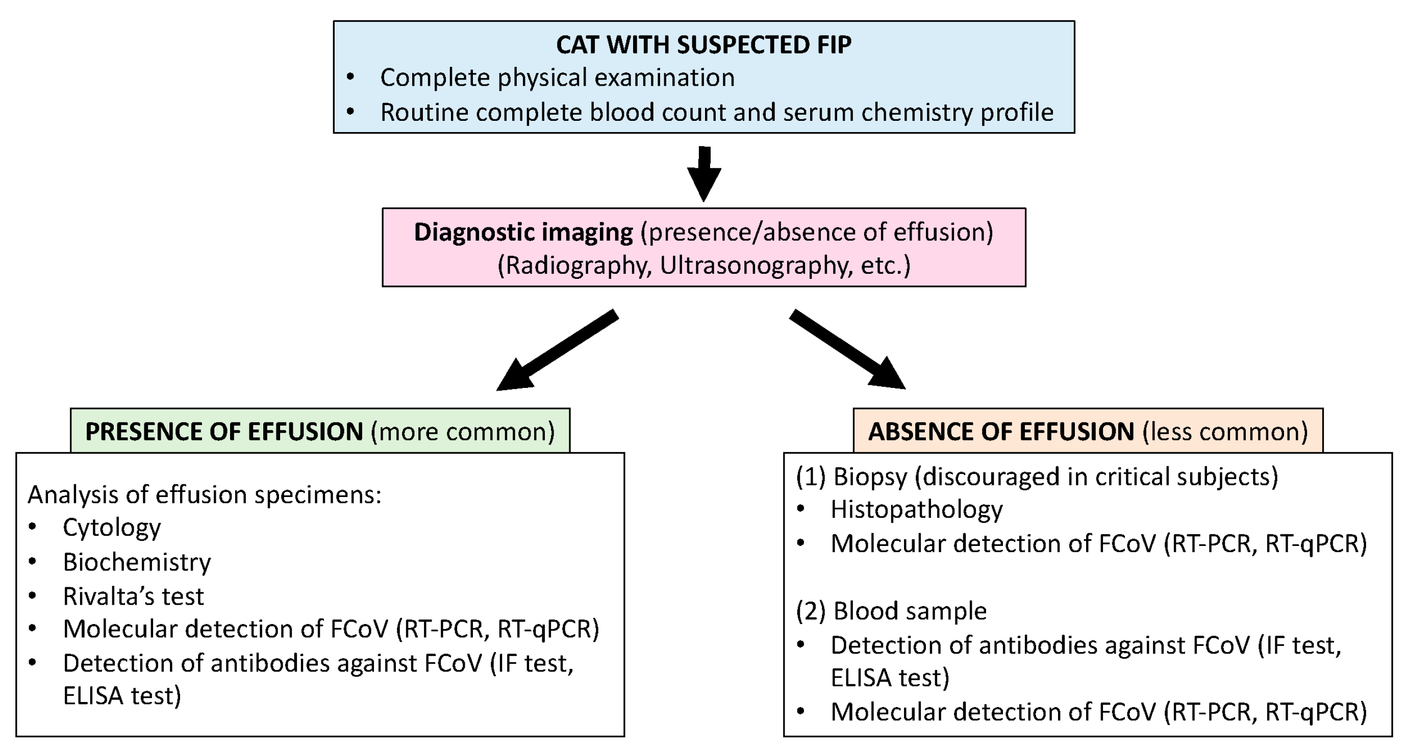
2.3. Diagnostic Imaging
2.4. FCoV Detection
2.4.1. FCoV Antigen Immunostaining
2.4.2. Molecular Detection of FCoV
2.4.3. Indirect FCoV Detection
3. General Management of Cats Suspected with FIP
4. Treatment Approaches
4.1. Nucleoside Analogs
4.2. Viral Protease Inhibitors
4.3. Interferons
4.4. Polyprenyl Immunostimulant
4.5. Other Compounds
5. Prevention
5.1. Vaccination
5.2. Measures to Reduce the Risk of FCoV Infection
6. Conclusions
Author Contributions
Funding
Institutional Review Board Statement
Informed Consent Statement
Data Availability Statement
Conflicts of Interest
References
- Wolfe, L.G.; Griesemer, R.A. Feline Infectious Peritonitis. Pathol. Vet. 1966, 3, 255–270. [Google Scholar] [CrossRef]
- Thayer, V.; Gogolski, S.; Felten, S.; Hartmann, K.; Kennedy, M.; Olah, G.A. 2022 AAFP/EveryCat Feline Infectious Peritonitis Diagnosis Guidelines. J. Feline Med. Surg. 2022, 24, 905–933. [Google Scholar] [CrossRef]
- Pearks Wilkerson, A.J.; Teeling, E.C.; Troyer, J.L.; Bar-Gal, G.K.; Roelke, M.; Marker, L.; Pecon-Slattery, J.; O’Brien, S.J. Coronavirus outbreak in cheetahs: Lessons for SARS. Curr. Biol. 2004, 14, R227–R228. [Google Scholar] [CrossRef][Green Version]
- Gaffney, P.M.; Kennedy, M.; Terio, K.; Gardner, I.; Lothamer, C.; Coleman, K.; Munson, L. Detection of feline coronavirus in Cheetah (Acinonyx jubatus) feces by reverse transcription-nested polymerase chain reaction in cheetahs with variable frequency of viral shedding. J. Zoo Wildl. Med. 2012, 43, 776–786. [Google Scholar] [CrossRef]
- Stephenson, N.; Swift, P.; Moeller, R.B.; Worth, S.J.; Foley, J. Feline Infectious Peritonitis in a Mountain Lion (Puma concolor), California, USA. J. Wildl. Dis. 2013, 49, 408–412. [Google Scholar] [CrossRef]
- Masters, P.S. The Molecular Biology of Coronaviruses. In Advances in Virus Research; Elsevier: Amsterdam, The Netherlands, 2006; pp. 193–292. Available online: https://linkinghub.elsevier.com/retrieve/pii/S0065352706660053 (accessed on 1 September 2025).
- Gao, Y.-Y.; Wang, Q.; Liang, X.-Y.; Zhang, S.; Bao, D.; Zhao, H.; Li, S.-B.; Wang, K.; Hu, G.-X.; Gao, F.-S. An updated review of feline coronavirus: Mind the two biotypes. Virus Res. 2023, 326, 199059. [Google Scholar] [CrossRef]
- Dye, C.; Siddell, S.G. Genomic RNA sequence of feline coronavirus strain FCoV C1Je. J. Feline Med. Surg. 2007, 9, 202–213. [Google Scholar] [CrossRef] [PubMed]
- Pedersen, N.C. A review of feline infectious peritonitis virus infection: 1963–2008. J. Feline Med. Surg. 2009, 11, 225–258. [Google Scholar] [CrossRef] [PubMed]
- Decaro, N.; Mari, V.; Lanave, G.; Lorusso, E.; Lucente, M.S.; Desario, C.; Colaianni, M.L.; Elia, G.; Ferringo, F.; Alfano, F.; et al. Mutation analysis of the spike protein in Italian feline infectious peritonitis virus and feline enteric coronavirus sequences. Res. Vet. Sci. 2021, 135, 15–19. [Google Scholar] [CrossRef]
- Rottier, P.J.M.; Nakamura, K.; Schellen, P.; Volders, H.; Haijema, B.J. Acquisition of Macrophage Tropism during the Pathogenesis of Feline Infectious Peritonitis Is Determined by Mutations in the Feline Coronavirus Spike Protein. J. Virol. 2005, 79, 14122–14130. [Google Scholar] [CrossRef]
- Brown, M.A.; Troyer, J.L.; Pecon-Slattery, J.; Roelke, M.E.; O’Brien, S.J. Genetics and Pathogenesis of Feline Infectious Peritonitis Virus. Emerg. Infect. Dis. 2009, 15, 1445–1452. [Google Scholar] [CrossRef]
- Chang, H.-W.; Egberink, H.F.; Halpin, R.; Spiro, D.J.; Rottier, P.J.M. Spike Protein Fusion Peptide and Feline Coronavirus Virulence. Emerg. Infect. Dis. 2012, 18, 1089–1095. [Google Scholar] [CrossRef]
- Porter, E.; Tasker, S.; Day, M.J.; Harley, R.; Kipar, A.; Siddell, S.G.; Helps, C.R. Amino acid changes in the spike protein of feline coronavirus correlate with systemic spread of virus from the intestine and not with feline infectious peritonitis. Vet. Res. 2014, 45, 49. [Google Scholar] [CrossRef]
- Pedersen, N.C. An update on feline infectious peritonitis: Diagnostics and therapeutics. Vet. J. 2014, 201, 133–141. [Google Scholar] [CrossRef] [PubMed]
- Chang, H.-W.; De Groot, R.J.; Egberink, H.F.; Rottier, P.J.M. Feline infectious peritonitis: Insights into feline coronavirus pathobiogenesis and epidemiology based on genetic analysis of the viral 3c gene. J. Gen. Virol. 2010, 91, 415–420. [Google Scholar] [CrossRef]
- Tekes, G.; Thiel, H.-J. Feline Coronaviruses. In Advances in Virus Research; Elsevier: Amsterdam, The Netherlands, 2016; pp. 193–218. Available online: https://linkinghub.elsevier.com/retrieve/pii/S0065352716300410 (accessed on 23 December 2025).
- Pedersen, N.C.; Ward, J.; Mengeling, W.L. Antigenic relationship of the feline infectious peritonitis virus to coronaviruses of other species. Arch. Virol. 1978, 58, 45–53. [Google Scholar] [CrossRef] [PubMed]
- Tekes, G.; Hofmann-Lehmann, R.; Stallkamp, I.; Thiel, V.; Thiel, H.-J. Genome Organization and Reverse Genetic Analysis of a Type I Feline Coronavirus. J. Virol. 2008, 82, 1851–1859. [Google Scholar] [CrossRef] [PubMed]
- Herrewegh, A.A.P.M.; Smeenk, I.; Horzinek, M.C.; Rottier, P.J.M.; De Groot, R.J. Feline Coronavirus Type II Strains 79-1683 and 79-1146 Originate from a Double Recombination between Feline Coronavirus Type I and Canine Coronavirus. J. Virol. 1998, 72, 4508–4514. [Google Scholar] [CrossRef]
- Terada, Y.; Matsui, N.; Noguchi, K.; Kuwata, R.; Shimoda, H.; Soma, T.; Mochizuki, M.; Maeda, K. Emergence of Pathogenic Coronaviruses in Cats by Homologous Recombination between Feline and Canine Coronaviruses. PLoS ONE 2014, 9, e106534. [Google Scholar] [CrossRef]
- Jaimes, J.A.; Millet, J.K.; Stout, A.E.; André, N.M.; Whittaker, G.R. A Tale of Two Viruses: The Distinct Spike Glycoproteins of Feline Coronaviruses. Viruses 2020, 12, 83. [Google Scholar] [CrossRef]
- Gao, Y.; Liang, X.; Wang, Q.; Zhang, S.; Zhao, H.; Wang, K.; Hu, G.; Liu, W.J.; Gao, F. Mind the feline coronavirus: Comparison with SARS-CoV-2. Gene 2022, 825, 146443. [Google Scholar] [CrossRef]
- Tresnan, D.B.; Levis, R.; Holmes, K.V. Feline aminopeptidase N serves as a receptor for feline, canine, porcine, and human coronaviruses in serogroup I. J. Virol. 1996, 70, 8669–8674. [Google Scholar] [CrossRef] [PubMed]
- Dye, C.; Temperton, N.; Siddell, S.G. Type I feline coronavirus spike glycoprotein fails to recognize aminopeptidase N as a functional receptor on feline cell lines. J. Gen. Virol. 2007, 88, 1753–1760. [Google Scholar] [CrossRef] [PubMed]
- Van Hamme, E.; Desmarets, L.; Dewerchin, H.L.; Nauwynck, H.J. Intriguing interplay between feline infectious peritonitis virus and its receptors during entry in primary feline monocytes. Virus Res. 2011, 160, 32–39. [Google Scholar] [CrossRef]
- Hartmann, K. Feline infectious peritonitis. Vet. Clin. North Am. Small Anim. Pract. 2005, 35, 39–79. [Google Scholar] [CrossRef]
- Solikhah, T.I.; Agustin, Q.A.D.; Damaratri, R.A.; Siwi, D.A.F.; Rafi’uttaqi, G.N.; Hartadi, V.A.; Solikhah, G.P. A review of feline infectious peritonitis virus infection. Vet. World 2024, 17, 2417–2432. [Google Scholar] [CrossRef]
- Tasker, S.; Addie, D.D.; Egberink, H.; Hofmann-Lehmann, R.; Hosie, M.J.; Truyen, U.; Belák, S.; Boucraut-Baralon, C.; Frymus, T.; Lloret, A.; et al. Feline Infectious Peritonitis: European Advisory Board on Cat Diseases Guidelines. Viruses 2023, 15, 1847. [Google Scholar] [CrossRef]
- Felten, S.; Hartmann, K. Diagnosis of Feline Infectious Peritonitis: A Review of the Current Literature. Viruses 2019, 11, 1068. [Google Scholar] [CrossRef] [PubMed]
- Addie, D.; Belák, S.; Boucraut-Baralon, C.; Egberink, H.; Frymus, T.; Gruffydd-Jones, T.; Hartmann, K.; Hosie, M.J.; Lloret, A.; Lutz, H.; et al. Feline Infectious Peritonitis: ABCD Guidelines on Prevention and Management. J. Feline Med. Surg. 2009, 11, 594–604. [Google Scholar] [CrossRef]
- Fischer, Y.; Sauter-Louis, C.; Hartmann, K. Diagnostic accuracy of the Rivalta test for feline infectious peritonitis. Vet. Clin. Pathol. 2012, 41, 558–567. [Google Scholar] [CrossRef]
- Felten, S.; Leutenegger, C.M.; Balzer, H.-J.; Pantchev, N.; Matiasek, K.; Wess, G.; Egberink, H.; Hartmann, K. Sensitivity and specificity of a real-time reverse transcriptase polymerase chain reaction detecting feline coronavirus mutations in effusion and serum/plasma of cats to diagnose feline infectious peritonitis. BMC Vet. Res. 2017, 13, 228. [Google Scholar] [CrossRef]
- Singh, M.; Foster, D.J.; Child, G.; Lamb, W.A. Inflammatory cerebrospinal fluid analysis in cats: Clinical diagnosis and outcome. J. Feline Med. Surg. 2005, 7, 77–93. [Google Scholar] [CrossRef] [PubMed]
- Felten, S.; Matiasek, K.; Gruendl, S.; Sangl, L.; Wess, G.; Hartmann, K. Investigation into the utility of an immunocytochemical assay in body cavity effusions for diagnosis of feline infectious peritonitis. J. Feline Med. Surg. 2017, 19, 410–418. [Google Scholar] [CrossRef]
- Stranieri, A.; Scavone, D.; Paltrinieri, S.; Giordano, A.; Bonsembiante, F.; Ferro, S.; Gelain, M.E.; Meazzi, S.; Lauzi, S. Concordance between Histology, Immunohistochemistry, and RT-PCR in the Diagnosis of Feline Infectious Peritonitis. Pathogens 2020, 9, 852. [Google Scholar] [CrossRef]
- Moyadee, W.; Sunpongsri, S.; Choowongkomon, K.; Roytrakul, S.; Rattanasrisomporn, A.; Tansakul, N.; Rattanasrisomporn, J. Feline infectious peritonitis: A comprehensive evaluation of clinical manifestations, laboratory diagnosis, and therapeutic approaches. J. Adv. Vet. Anim. Res. 2024, 11, 19–26. [Google Scholar] [CrossRef]
- Rohrbach, B.W.; Legendre, A.M.; Baldwin, C.A.; Lein, D.H.; Reed, W.M.; Wilson, R.B. Epidemiology of feline infectious peritonitis among cats examined at veterinary medical teaching hospitals. J. Am. Vet. Med. Assoc. 2001, 218, 1111–1115. [Google Scholar] [CrossRef] [PubMed]
- Healey, E.A.; Andre, N.M.; Miller, A.D.; Whittaker, G.R.; Berliner, E.A. Outbreak of feline infectious peritonitis (FIP) in shelter-housed cats: Molecular analysis of the feline coronavirus S1/S2 cleavage site consistent with a ‘circulating virulent–avirulent theory’ of FIP pathogenesis. J. Feline Med. Surg. Open Rep. 2022, 8, 20551169221074226. [Google Scholar] [CrossRef]
- Kennedy, M.A. Feline infectious peritonitis: Update on pathogenesis, diagnostics, and treatment. Vet. Clin. North Am. Small Anim. Pract. 2020, 50, 1001–1011. [Google Scholar] [CrossRef]
- Vennema, H.; Poland, A.; Foley, J.; Pedersen, N.C. Feline Infectious Peritonitis Viruses Arise by Mutation from Endemic Feline Enteric Coronaviruses. Virology 1998, 243, 150–157. [Google Scholar] [CrossRef]
- Riemer, F.; Kuehner, K.A.; Ritz, S.; Sauter-Louis, C.; Hartmann, K. Clinical and laboratory features of cats with feline infectious peritonitis—A retrospective study of 231 confirmed cases (2000–2010). J. Feline Med. Surg. 2016, 18, 348–356. [Google Scholar] [CrossRef]
- Fischer, Y.; Wess, G.; Hartmann, K. Perikarderguss bei einer Katze mit feliner infektiöser Peritonitis. Schweiz. Arch. Tierheilkd. 2012, 154, 27–31. [Google Scholar] [CrossRef]
- Ruiz, M.D.; Vessières, F.; Ragetly, G.R.; Hernandez, J.L. Characterization of and factors associated with causes of pleural effusion in cats. J. Am. Vet. Med. Assoc. 2018, 253, 181–187. [Google Scholar] [CrossRef] [PubMed]
- Repyak, K.; Atiee, G.; Cook, A.; Bryan, L.; Gremillion, C. Thoracic radiographic findings in cats with feline infectious peritonitis. J. Feline Med. Surg. 2025, 27, 1098612X241309823. [Google Scholar] [CrossRef]
- Doenges, S.J.; Weber, K.; Dorsch, R.; Fux, R.; Fischer, A.; Matiasek, L.A.; Matiasek, K.; Hartmann, K. Detection of feline coronavirus in cerebrospinal fluid for diagnosis of feline infectious peritonitis in cats with and without neurological signs. J. Feline Med. Surg. 2016, 18, 104–109. [Google Scholar] [CrossRef]
- Jinks, M.R.; English, R.V.; Gilger, B.C. Causes of endogenous uveitis in cats presented to referral clinics in North Carolina. Vet. Ophthalmol. 2016, 19, 30–37. [Google Scholar] [CrossRef]
- Yin, Y.; Li, T.; Wang, C.; Liu, X.; Ouyang, H.; Ji, W.; Liu, J.; Liao, X.; Li, J.; Hu, C. A retrospective study of clinical and laboratory features and treatment on cats highly suspected of feline infectious peritonitis in Wuhan, China. Sci. Rep. 2021, 11, 5208. [Google Scholar] [CrossRef] [PubMed]
- Norris, J.; Bosward, K.; White, J.; Baral, R.; Catt, M.; Malik, R. Clinicopathological findings associated with feline infectious peritonitis in Sydney, Australia: 42 cases (1990–2002). Aust. Vet. J. 2005, 83, 666–673. [Google Scholar] [CrossRef]
- Tsai, H.-Y.; Chueh, L.-L.; Lin, C.-N.; Su, B.-L. Clinicopathological findings and disease staging of feline infectious peritonitis: 51 cases from 2003 to 2009 in Taiwan. J. Feline Med. Surg. 2011, 13, 74–80. [Google Scholar] [CrossRef] [PubMed]
- Stranieri, A.; Giordano, A.; Paltrinieri, S.; Giudice, C.; Cannito, V.; Lauzi, S. Comparison of the performance of laboratory tests in the diagnosis of feline infectious peritonitis. J. Vet. Diagn. Investig. 2018, 30, 459–463. [Google Scholar] [CrossRef]
- Giordano, A.; Spagnolo, V.; Colombo, A.; Paltrinieri, S. Changes in some acute phase protein and immunoglobulin concentrations in cats affected by feline infectious peritonitis or exposed to feline coronavirus infection. Vet. J. 2004, 167, 38–44. [Google Scholar] [CrossRef]
- Savary, K.; Sellon, R.; Law, J. Chylous abdominal effusion in a cat with feline infectious peritonitis. J. Am. Anim. Hosp. Assoc. 2001, 37, 35–40. [Google Scholar] [CrossRef]
- Jeffery, U.; Deitz, K.; Hostetter, S. Positive predictive value of albumin: Globulin ratio for feline infectious peritonitis in a mid-western referral hospital population. J. Feline Med. Surg. 2012, 14, 903–905. [Google Scholar] [CrossRef]
- Fischer, Y.; Weber, K.; Sauter-Louis, C.; Hartmann, K. The Rivalta’s test as a diagnostic variable in feline effusions–evaluation of optimum reaction and storage conditions. Tierarztl. Prax. Ausg. K Klientiere Heimtiere 2013, 41, 297–303. [Google Scholar] [CrossRef]
- Giordano, A.; Paltrinieri, S.; Bertazzolo, W.; Milesi, E.; Parodi, M. Sensitivity of Tru-cut and fine-needle aspiration biopsies of liver and kidney for diagnosis of feline infectious peritonitis. Vet. Clin. Pathol. 2005, 34, 368–374. [Google Scholar] [CrossRef] [PubMed]
- Crawford, A.H.; Stoll, A.L.; Sanchez-Masian, D.; Shea, A.; Michaels, J.; Fraser, A.R.; Beltran, E. Clinicopathologic Features and Magnetic Resonance Imaging Findings in 24 Cats With Histopathologically Confirmed Neurologic Feline Infectious Peritonitis. J. Vet. Intern. Med. 2017, 31, 1477–1486. [Google Scholar] [CrossRef]
- Tasker, S. Diagnosis of feline infectious peritonitis: Update on evidence supporting available tests. J. Feline Med. Surg. 2018, 20, 228–243. [Google Scholar] [CrossRef] [PubMed]
- Hellemans, A.; Acar, D.D.; Stroobants, V.J.E.; Theuns, S.; Desmarets, L.M.B.; Nauwynck, H.J. A comparative study of techniques used for the diagnosis of effusive feline infectious peritonitis. Vlaams Diergeneeskd. Tijdschr. 2020, 89, 100–110. [Google Scholar] [CrossRef]
- Hartmann, K.; Binder, C.; Hirschberger, J.; Cole, D.; Reinacher, M.; Schroo, S.; Frost, J.; Egberink, H.; Lutz, H.; Hermanns, W. Comparison of Different Tests to Diagnose Feline Infectious Peritonitis. J. Vet. Intern. Med. 2003, 17, 781–790. [Google Scholar] [CrossRef]
- Kipar, A.; Meli, M.L. Feline Infectious Peritonitis: Still an Enigma? Vet. Pathol. 2014, 51, 505–526. [Google Scholar] [CrossRef]
- Barker, E.N.; Tasker, S. Advances in Molecular Diagnostics and Treatment of Feline Infectious Peritonitis. Adv. Small Anim. Care 2020, 1, 161–188. [Google Scholar] [CrossRef]
- Kipar, A.; Baptiste, K.; Barth, A.; Reinacher, M. Natural FCoV infection: Cats with FIP exhibit significantly higher viral loads than healthy infected cats. J. Feline Med. Surg. 2006, 8, 69–72. [Google Scholar] [CrossRef]
- Dunbar, D.; Kwok, W.; Graham, E.; Armitage, A.; Irvine, R.; Johnston, P.; McDonald, M.; Montgomery, D.; Nicolson, L.; Robertson, E.; et al. Diagnosis of non-effusive feline infectious peritonitis by reverse transcriptase quantitative PCR from mesenteric lymph node fine-needle aspirates. J. Feline Med. Surg. 2019, 21, 910–921. [Google Scholar] [CrossRef] [PubMed]
- Emmler, L.; Felten, S.; Matiasek, K.; Balzer, H.-J.; Pantchev, N.; Leutenegger, C.; Hartmann, K. Feline coronavirus with and without spike gene mutations detected by real-time RT-PCRs in cats with feline infectious peritonitis. J. Feline Med. Surg. 2020, 22, 791–799. [Google Scholar] [CrossRef]
- Felten, S.; Weider, K.; Doenges, S.; Gruendl, S.; Matiasek, K.; Hermanns, W.; Mueller, E.; Matiasek, L.; Fischer, A.; Weber, K.; et al. Detection of feline coronavirus spike gene mutations as a tool to diagnose feline infectious peritonitis. J. Feline Med. Surg. 2017, 19, 321–335. [Google Scholar] [CrossRef]
- Barker, E.N.; Stranieri, A.; Helps, C.R.; Porter, E.L.; Davidson, A.D.; Day, M.J.; Knowles, T.; Kipar, A.; Tasker, S. Limitations of using feline coronavirus spike protein gene mutations to diagnose feline infectious peritonitis. Vet. Res. 2017, 48, 60. [Google Scholar] [CrossRef] [PubMed]
- Addie, D.D.; Le Poder, S.; Burr, P.; Decaro, N.; Graham, E.; Hofmann-Lehmann, R.; Jarrett, O.; McDonald, M.; Meli, M.L. Utility of feline coronavirus antibody tests. J. Feline Med. Surg. 2015, 17, 152–162. [Google Scholar] [CrossRef]
- Lorusso, E.; Mari, V.; Losurdo, M.; Lanave, G.; Trotta, A.; Dowgier, G.; Colaianni, M.L.; Zatelli, A.; Elia, G.; Buonavoglia, D.; et al. Discrepancies between feline coronavirus antibody and nucleic acid detection in effusions of cats with suspected feline infectious peritonitis. Res. Vet. Sci. 2019, 125, 421–424. [Google Scholar] [CrossRef]
- Xu, L.; Ye, S.; Ding, Y.; Xiao, Y.; Yao, C.; Wang, Z.; Cai, S.; Ou, J.; Mao, J.; Hu, X.; et al. A Combined Method Based on the FIPV N Monoclonal Antibody Immunofluorescence Assay and RT-nPCR Method for the Rapid Diagnosis of FIP-Suspected Ascites. Transbound. Emerg. Dis. 2023, 2023, 8429106. [Google Scholar] [CrossRef] [PubMed]
- Attipa, C.; Warr, A.S.; Epaminondas, D.; O’Shea, M.; Hanton, A.J.; Fletcher, S.; Malbon, A.; Lyraki, M.; Hammond, R.; Hardas, A.; et al. Emergence and spread of feline infectious peritonitis due to a highly pathogenic canine/feline recombinant coronavirus. bioRxiv 2023. [Google Scholar] [CrossRef]
- Thames, J.E.; Seley-Radtke, K.L. Comparison of the Old and New—Novel Mechanisms of Action for Anti-coronavirus Nucleoside Analogues. Chimia 2022, 76, 409. [Google Scholar] [CrossRef]
- Murphy, B.G.; Perron, M.; Murakami, E.; Bauer, K.; Park, Y.; Eckstrand, C.; Liepnieks, M.; Pedersen, N.C. The nucleoside analog GS-441524 strongly inhibits feline infectious peritonitis (FIP) virus in tissue culture and experimental cat infection studies. Vet. Microbiol. 2018, 219, 226–233. [Google Scholar] [CrossRef]
- Gokalsing, E.; Ferrolho, J.; Gibson, M.S.; Vilhena, H.; Anastácio, S. Efficacy of GS-441524 for Feline Infectious Peritonitis: A Systematic Review (2018–2024). Pathogens 2025, 14, 717. [Google Scholar] [CrossRef] [PubMed]
- Pedersen, N.C.; Perron, M.; Bannasch, M.; Montgomery, E.; Murakami, E.; Liepnieks, M.; Liu, H. Efficacy and safety of the nucleoside analog GS-441524 for treatment of cats with naturally occurring feline infectious peritonitis. J. Feline Med. Surg. 2019, 21, 271–281. [Google Scholar] [CrossRef]
- Addie, D.D.; Covell-Ritchie, J.; Jarrett, O.; Fosbery, M. Rapid Resolution of Non-Effusive Feline Infectious Peritonitis Uveitis with an Oral Adenosine Nucleoside Analogue and Feline Interferon Omega. Viruses 2020, 12, 1216. [Google Scholar] [CrossRef]
- Krentz, D.; Zenger, K.; Alberer, M.; Felten, S.; Bergmann, M.; Dorsch, R.; Matiasek, K.; Kolberg, L.; Hofmann-Lehmann, R.; Meli, M.L.; et al. Curing Cats with Feline Infectious Peritonitis with an Oral Multi-Component Drug Containing GS-441524. Viruses 2021, 13, 2228. [Google Scholar] [CrossRef]
- Zuzzi-Krebitz, A.-M.; Buchta, K.; Bergmann, M.; Krentz, D.; Zwicklbauer, K.; Dorsch, R.; Wess, G.; Fischer, A.; Matiasek, K.; Hönl, A.; et al. Short Treatment of 42 Days with Oral GS-441524 Results in Equal Efficacy as the Recommended 84-Day Treatment in Cats Suffering from Feline Infectious Peritonitis with Effusion—A Prospective Randomized Controlled Study. Viruses 2024, 16, 1144. [Google Scholar] [CrossRef]
- Coggins, S.J.; Govendir, M.; Norris, J.M.; Malik, R.; Hall, E.J.; Thompson, M.F.; Kimble, B. Pharmacokinetics of GS-441524 following intravenous remdesivir in six cats and results of therapeutic drug monitoring during treatment of feline infectious peritonitis: 22 cases (2021–2024). J. Small Anim. Pract. 2025, 66, 495–506. [Google Scholar] [CrossRef]
- Furbish, A.; Allinder, M.; Austin, G.; Tynan, B.; Byrd, E.; Gomez, I.P.; Peterson, Y. First analytical confirmation of drug-induced crystal nephropathy in felines caused by GS-441524, the active metabolite of Remdesivir. J. Pharm. Biomed. Anal. 2024, 247, 116248. [Google Scholar] [CrossRef]
- Wang, J.; Reiss, K.; Shi, Y.; Lolis, E.; Lisi, G.P.; Batista, V.S. Mechanism of Inhibition of the Reproduction of SARS-CoV-2 and Ebola Viruses by Remdesivir. Biochemistry 2021, 60, 1869–1875. [Google Scholar] [CrossRef] [PubMed]
- Renner, K.A.; Cattin, R.; Kimble, B.; Munday, J.; White, A.; Coggins, S. Efficacy of oral remdesivir in treating feline infectious peritonitis: A prospective observational study of 29 cats. J. Feline Med. Surg. 2025, 27, 1098612X251335189. [Google Scholar] [CrossRef] [PubMed]
- Cook, S.; Castillo, D.; Williams, S.; Haake, C.; Murphy, B. Serotype I and II Feline Coronavirus Replication and Gene Expression Patterns of Feline Cells—Building a Better Understanding of Serotype I FIPV Biology. Viruses 2022, 14, 1356. [Google Scholar] [CrossRef]
- Coggins, S.J.; Norris, J.M.; Malik, R.; Govendir, M.; Hall, E.J.; Kimble, B.; Thompson, M.F. Outcomes of treatment of cats with feline infectious peritonitis using parenterally administered remdesivir, with or without transition to orally administered GS-441524. J. Vet. Intern. Med. 2023, 37, 1772–1783. [Google Scholar] [CrossRef] [PubMed]
- Taylor, S.S.; Coggins, S.; Barker, E.N.; Gunn-Moore, D.; Jeevaratnam, K.; Norris, J.M.; Hughes, D.; Stacey, E.; MacFarlane, L.; O’Brien, C.; et al. Retrospective study and outcome of 307 cats with feline infectious peritonitis treated with legally sourced veterinary compounded preparations of remdesivir and GS-441524 (2020–2022). J. Feline Med. Surg. 2023, 25, 1098612X231194460. [Google Scholar] [CrossRef] [PubMed]
- Roy, M.; Jacque, N.; Novicoff, W.; Li, E.; Negash, R.; Evans, S.J.M. Unlicensed Molnupiravir is an Effective Rescue Treatment Following Failure of Unlicensed GS-441524-like Therapy for Cats with Suspected Feline Infectious Peritonitis. Pathogens 2022, 11, 1209. [Google Scholar] [CrossRef]
- Sase, O. Molnupiravir treatment of 18 cats with feline infectious peritonitis: A case series. J. Vet. Intern. Med. 2023, 37, 1876–1880. [Google Scholar] [CrossRef]
- Clark, T.; Coggins, S.; Korman, R.; King, J.; Malik, R. Treatment of feline infectious peritonitis in cats with molnupiravir: Clinical observations and outcomes for 54 cases. Aust. Vet. J. 2025, 103, 339–353. [Google Scholar] [CrossRef]
- Černá, P.; Wittenburg, L.; Hawley, J.; Willis, M.; Siegenthaler, B.; Lappin, M.R. Pharmacokinetics of Molnupiravir in Cats with Naturally Occurring Feline Infectious Peritonitis. Pathogens 2025, 14, 666. [Google Scholar] [CrossRef]
- Yoshida, S.; Sugawara-Suda, M.; Sasaoka, K.; Sasaki, N.; Yokoyama, N.; Nakamura, K.; Morishita, K.; Kim, S.; Sunaga, T.; Takiguchi, M. Evaluation of the course of improvement with molnupiravir treatment for feline infectious peritonitis. Can. Vet. J. Rev. Veterinaire Can. 2025, 66, 546–554. [Google Scholar]
- Cook, S.; Wittenburg, L.; Yan, V.C.; Theil, J.H.; Castillo, D.; Reagan, K.L.; Williams, S.; Pham, C.-D.; Li, C.; Muller, F.L.; et al. An Optimized Bioassay for Screening Combined Anticoronaviral Compounds for Efficacy against Feline Infectious Peritonitis Virus with Pharmacokinetic Analyses of GS-441524, Remdesivir, and Molnupiravir in Cats. Viruses 2022, 14, 2429. [Google Scholar] [CrossRef] [PubMed]
- Delaplace, M.; Huet, H.; Gambino, A.; Le Poder, S. Feline Coronavirus Antivirals: A Review. Pathogens 2021, 10, 1150. [Google Scholar] [CrossRef]
- Kim, Y.; Mandadapu, S.R.; Groutas, W.C.; Chang, K.-O. Potent inhibition of feline coronaviruses with peptidyl compounds targeting coronavirus 3C-like protease. Antivir. Res. 2013, 97, 161–168. [Google Scholar] [CrossRef]
- Lu, J.; Chen, S.A.; Khan, M.B.; Brassard, R.; Arutyunova, E.; Lamer, T.; Vuong, W.; Fischer, C.; Young, H.S.; Vederas, J.C.; et al. Crystallization of Feline Coronavirus Mpro With GC376 Reveals Mechanism of Inhibition. Front. Chem. 2022, 10, 852210. [Google Scholar] [CrossRef]
- Pedersen, N.C.; Kim, Y.; Liu, H.; Galasiti Kankanamalage, A.C.; Eckstrand, C.; Groutas, W.C.; Bannasch, M.; Meadows, J.M.; Chang, K.-O. Efficacy of a 3C-like protease inhibitor in treating various forms of acquired feline infectious peritonitis. J. Feline Med. Surg. 2018, 20, 378–392. [Google Scholar] [CrossRef]
- Vuong, W.; Khan, M.B.; Fischer, C.; Arutyunova, E.; Lamer, T.; Shields, J.; Saffran, H.A.; McKay, R.T.; Van Belkum, M.J.; Joyce, M.A.; et al. Feline coronavirus drug inhibits the main protease of SARS-CoV-2 and blocks virus replication. Nat. Commun. 2020, 11, 4282. [Google Scholar] [CrossRef] [PubMed]
- Lv, J.; Bai, Y.; Wang, Y.; Yang, L.; Jin, Y.; Dong, J. Effect of GS-441524 in combination with the 3C-like protease inhibitor GC376 on the treatment of naturally transmitted feline infectious peritonitis. Front. Vet. Sci. 2022, 9, 1002488. [Google Scholar] [CrossRef] [PubMed]
- Mihaescu, G.; Chifiriuc, M.C.; Filip, R.; Bleotu, C.; Ditu, L.M.; Constantin, M.; Cristian, R.-E.; Grigore, R.; Bertesteanu, S.V.; Bertesteanu, G.; et al. Role of interferons in the antiviral battle: From virus-host crosstalk to prophylactic and therapeutic potential in SARS-CoV-2 infection. Front. Immunol. 2024, 14, 1273604. [Google Scholar] [CrossRef]
- Takano, T.; Satoh, K.; Doki, T.; Tanabe, T.; Hohdatsu, T. Antiviral Effects of Hydroxychloroquine and Type I Interferon on In Vitro Fatal Feline Coronavirus Infection. Viruses 2020, 12, 576. [Google Scholar] [CrossRef] [PubMed]
- Ishida, T.; Shibanai, A.; Tanaka, S.; Uchida, K.; Mochizuki, M. Use of recombinant feline interferon and glucocorticoid in the treatment of feline infectious peritonitis. J. Feline Med. Surg. 2004, 6, 107–109. [Google Scholar] [CrossRef]
- Ritz, S.; Egberink, H.; Hartmann, K. Effect of Feline Interferon-Omega on the Survival Time and Quality of Life of Cats with Feline Infectious Peritonitis. J. Vet. Intern. Med. 2007, 21, 1193–1197. [Google Scholar] [CrossRef]
- Doki, T.; Yabe, M.; Takano, T.; Hohdatsu, T. Differential induction of type I interferon by type I and type II feline coronaviruses in vitro. Res. Vet. Sci. 2018, 120, 57–62. [Google Scholar] [CrossRef]
- Legendre, A.M.; Kuritz, T.; Heidel, R.E.; Baylor, V.M. Polyprenyl Immunostimulant in Feline Rhinotracheitis: Randomized Placebo-Controlled Experimental and Field Safety Studies. Front. Vet. Sci. 2017, 4, 24. [Google Scholar] [CrossRef] [PubMed]
- Legendre, A.M.; Kuritz, T.; Galyon, G.; Baylor, V.M.; Heidel, R.E. Polyprenyl Immunostimulant Treatment of Cats with Presumptive Non-Effusive Feline Infectious Peritonitis In a Field Study. Front. Vet. Sci. 2017, 4, 7. [Google Scholar] [CrossRef]
- Legendre, A.M.; Bartges, J.W. Effect of Polyprenyl Immunostimulant on the survival times of three cats with the dry form of feline infectious peritonitis. J. Feline Med. Surg. 2009, 11, 624–626. [Google Scholar] [CrossRef]
- Černá, P.; Ayoob, A.; Baylor, C.; Champagne, E.; Hazanow, S.; Heidel, R.E.; Wirth, K.; Legendre, A.M.; Gunn-Moore, D.A. Retrospective Survival Analysis of Cats with Feline Infectious Peritonitis Treated with Polyprenyl Immunostimulant That Survived over 365 Days. Pathogens 2022, 11, 881. [Google Scholar] [CrossRef]
- Lee, I.; Desai, A.; Patil, A.; Xu, Y.; Pozza-Adams, K.; Berdis, A.J. Utilization of Flow Cytometry, Metabolomic Analyses and a Feline Infectious Peritonitis Case Study to Evaluate the Physiological Impact of Polyprenyl Immunostimulant. Cells 2025, 14, 752. [Google Scholar] [CrossRef] [PubMed]
- Triratapiban, C.; Lueangaramkul, V.; Phecharat, N.; Pantanam, A.; Lekcharoensuk, P.; Theerawatanasirikul, S. First study on in vitro antiviral and virucidal effects of flavonoids against feline infectious peritonitis virus at the early stage of infection. Vet. World 2023, 16, 618–630. [Google Scholar] [CrossRef]
- Mohseni, N.; Royster, A.; Ren, S.; Ma, Y.; Pintado, M.; Mir, M.; Mir, S. A novel compound targets the feline infectious peritonitis virus nucleocapsid protein and inhibits viral replication in cell culture. J. Biol. Chem. 2023, 299, 102976. [Google Scholar] [CrossRef]
- Camero, M.; Lanave, G.; Catella, C.; Lucente, M.S.; Sposato, A.; Mari, V.; Tempesta, M.; Martella, V.; Buonavoglia, A. ERDRP-0519 inhibits feline coronavirus in vitro. BMC Vet. Res. 2022, 18, 55. [Google Scholar] [CrossRef]
- Catella, C.; Camero, M.; Lucente, M.S.; Fracchiolla, G.; Sblano, S.; Tempesta, M.; Martella, V.; Buonavoglia, C.; Lanave, G. Virucidal and antiviral effects of Thymus vulgaris essential oil on feline coronavirus. Res. Vet. Sci. 2021, 137, 44–47. [Google Scholar] [CrossRef] [PubMed]
- Chou, A.-A.; Lin, C.-H.; Chang, Y.-C.; Chang, H.-W.; Lin, Y.-C.; Pi, C.-C.; Kan, Y.-M.; Chuang, H.-F.; Chen, H.-W. Antiviral activity of Vigna radiata extract against feline coronavirus in vitro. Vet. Q. 2024, 44, 1–13. [Google Scholar] [CrossRef]
- Jiang, Z.; Piao, L.; Ren, C.; Zhang, W.; Zhu, Y.; Kong, R. Identifying Natural Products as Feline Coronavirus Mpro Inhibitors by Structural-Based Virtual Screening and Enzyme-Based Assays. ACS Omega 2025, 10, 2092–2101. [Google Scholar] [CrossRef]
- Lueangaramkul, V.; Termthongthot, P.; Mana, N.; Panichayupakaranant, P.; Semkum, P.; Lekcharoensuk, P.; Theerawatanasirikul, S. Antiviral and Immunomodulatory Effects of α-Mangostin Against Feline Infectious Peritonitis Virus: In Vitro Assay. Animals 2025, 15, 2417. [Google Scholar] [CrossRef]
- Tizard, I.R. Vaccination against coronaviruses in domestic animals. Vaccine 2020, 38, 5123–5130. [Google Scholar] [CrossRef] [PubMed]
- Scott, F.W. Evaluation of Risks and Benefits Associated with Vaccination against Coronavirus Infections in Cats. In Advances in Veterinary Medicine; Elsevier: Amsterdam, The Netherlands, 1999; pp. 347–358. Available online: https://linkinghub.elsevier.com/retrieve/pii/S0065351999800263 (accessed on 7 November 2025).
- Gerber, J.D. Overview of the development of a modified live temperature-sensitive FIP virus vaccine. Feline Pract. 1995, 23, 62–66. [Google Scholar]
- Si, F.; Yu, R.; Dong, S.; Chen, B.; Li, C.; Song, S. Towards a Safer Future: Enhancing Vaccine Development to Combat Animal Coronaviruses. Vaccines 2024, 12, 330. [Google Scholar] [CrossRef]
- Gerber, J.D.; Pfeiffer, N.E.; Ingersoll, J.D.; Christianson, K.K.; Landon, R.M.; Selzer, N.L.; Beckenhauer, W.H. Characterization of an Attenuated Temperature Sensitive Feline Infectious Peritonitis Vaccine Virus. In Coronaviruses and Their Diseases; Cavanagh, D., Brown, T.D.K., Eds.; Advances in Experimental Medicine and Biology; Springer: Boston, MA, USA, 1990; Volume 276, pp. 481–489. Available online: http://link.springer.com/10.1007/978-1-4684-5823-7_67 (accessed on 6 November 2025).
- Chawla, M.; Cuspoca, A.F.; Akthar, N.; Magdaleno, J.S.L.; Rattanabunyong, S.; Suwattanasophon, C.; Jongkon, N.; Choowongkomon, K.; Shaikh, A.R.; Malik, T.; et al. Immunoinformatics-aided rational design of a multi-epitope vaccine targeting feline infectious peritonitis virus. Front. Vet. Sci. 2023, 10, 1280273. [Google Scholar] [CrossRef]
- Karthik, K.; Senthilkumar, T.M.A.; Udhayavel, S.; Raj, G.D. Role of antibody-dependent enhancement (ADE) in the virulence of SARS-CoV-2 and its mitigation strategies for the development of vaccines and immunotherapies to counter COVID-19. Hum. Vaccines Immunother. 2020, 16, 3055–3060. [Google Scholar] [CrossRef]
- Takano, T.; Morioka, H.; Gomi, K.; Tomizawa, K.; Doki, T.; Hohdatsu, T. Screening and identification of T helper 1 and linear immunodominant antibody-binding epitopes in spike 1 domain and membrane protein of feline infectious peritonitis virus. Vaccine 2014, 32, 1834–1840. [Google Scholar] [CrossRef] [PubMed]
- Chen, C.; Li, Y.-L.; Lv, F.-L.; Xu, L.-D.; Huang, Y.-W. Surface Display of Peptides Corresponding to the Heptad Repeat 2 Domain of the Feline Enteric Coronavirus Spike Protein on Bacillus subtilis Spores Elicits Protective Immune Responses Against Homologous Infection in a Feline Aminopeptidase-N-Transduced Mouse Model. Front. Immunol. 2022, 13, 925922. [Google Scholar] [CrossRef]
- Glansbeek, H.L.; Haagmans, B.L.; Te Lintelo, E.G.; Egberink, H.F.; Duquesne, V.; Aubert, A.; Horzinek, M.C.; Rottier, P.J.M. Adverse effects of feline IL-12 during DNA vaccination against feline infectious peritonitis virus. J. Gen. Virol. 2002, 83, 1–10. [Google Scholar] [CrossRef]
- Wasmoen, T.L.; Kadakia, N.P.; Unfer, R.C.; Fickbohm, B.L.; Cook, C.P.; Chu, H.-J.; Acree, W.M. Protection of Cats from Infectious Peritonitis by Vaccination with a Recombinant Raccoon Poxvirus Ex-pressing the Nucleocapsid Gene of Feline Infectious Peritonitis Virus. In Corona- and Related Viruses; Talbot, P.J., Levy, G.A., Eds.; Advances in Experimental Medicine and Biology; Springer: Boston, MA, USA, 1995; Volume 380, pp. 221–228. Available online: http://link.springer.com/10.1007/978-1-4615-1899-0_36 (accessed on 6 November 2025).
- Hohdatsu, T.; Yamato, H.; Ohkawa, T.; Kaneko, M.; Motokawa, K.; Kusuhara, H.; Kaneshima, T.; Arai, S.; Koyama, H. Vaccine efficacy of a cell lysate with recombinant baculovirus-expressed feline infectious peritonitis (FIP) virus nucleocapsid protein against progression of FIP. Vet. Microbiol. 2003, 97, 31–44. [Google Scholar] [CrossRef] [PubMed]
- Wang, Y.; Liu, Y.; Wang, J.; Zhang, M.; Deng, X.; Song, J.; Zhu, J.; Yu, L.; Li, G.; Liu, G. An adenovirus-vectored vaccine based on the N protein of feline coronavirus elicit robust protective immune responses. Antivir. Res. 2024, 223, 105825. [Google Scholar] [CrossRef] [PubMed]
- Brostoff, T.; Savage, H.P.; Jackson, K.A.; Dutra, J.C.; Fontaine, J.H.; Hartigan-O’Connor, D.J.; Carney, R.P.; Pesavento, P.A. Feline Infectious Peritonitis mRNA Vaccine Elicits Both Humoral and Cellular Immune Responses in Mice. Vaccines 2024, 12, 705. [Google Scholar] [CrossRef] [PubMed]
- Mir, S.; Peters, M.; Penny, G.; Agsaoa, A.; Mir, M. From Challenge to Cure: A Look at Feline Infectious Peritonitis and Emerging Treatment Strategies and Breakthroughs. Vet. Sci. 2025, 12, 650. [Google Scholar] [CrossRef]
- Addie, D.D.; Jarrett, O. Use of a reverse-transcriptase polymerase chain reaction for monitoring the shedding of feline coronavirus by healthy cats. Vet. Rec. 2001, 148, 649–653. [Google Scholar] [CrossRef]
- Addie, D.D.; Paltrinieri, S.; Pedersen, N.C. Recommendations from workshops of the second international feline coronavirus/feline infectious peritonitis symposium. J. Feline Med. Surg. 2004, 6, 125–130. [Google Scholar] [CrossRef]
- Italian Ministry of Health. Official Circular on the Use of Remdesivir (Veklury®) and GS-441524 for the Treatment of Feline Infectious Peritonitis (FIP); Italian Ministry of Health: Rome, Italy, 2025. [Google Scholar]


| Sample Type | Diagnostic Method | Clinical Utility | Reference |
|---|---|---|---|
| Blood | Hematology Serum biochemistry | Supportive but non-specific findings. Low–moderate sensitivity and low specificity. | [29] |
| Molecular detection (RT-PCR, RT-qPCR) | FCoV can also be present in cats with systemic FECV infection. Very high titers may raise FIP suspicion. Moderate sensitivity and low specificity. | [30] | |
| Antibody detection (ELISA, IF) | Anti-FCoV antibodies can be detected in cats without FIP. If negative, FIP is unlikely. High sensitivity and very low specificity. | [31] | |
| Effusion | Cytology | Typically, pyogranulomatous inflammatory reaction with macrophages. Moderate to high sensitivity and moderate specificity. | [2] |
| Rivalta’s test | Excellent NPV. Useful to exclude FIP. High sensitivity and moderate specificity. | [32] | |
| Molecular detection (RT-PCR, RT-qPCR) | Strong supportive evidence in case of effusive FIP. High sensitivity and moderate to high specificity. | [33] | |
| FNA CSF Aqueous humor | Cytology | May demonstrate pyogranulomatous inflammation with macrophages. Moderate sensitivity and specificity. | [34] |
| Immunostaining (ICC, IF) | Strong confirmatory evidence of FIP. Moderate to high sensitivity and very high specificity. | [35] | |
| Molecular detection (RT-PCR, RT-qPCR) | Useful in combination with cytology and antigen detection. High sensitivity and moderate specificity. | [31] | |
| Tissue | Histopathology | Moderate specificity but low sensitivity depending on the lesion. | [36] |
| Immunostaining (IHC) | Diagnostic gold standard for FIP. High sensitivity and very high specificity. | ||
| Molecular detection (RT-PCR, RT-qPCR) | High titers are reliable for FIP confirmation in combination with other tests. High sensitivity and moderate specificity. |
| Therapeutic Class | Agent | Comments/Notes |
|---|---|---|
| Nucleoside Analogs | GS-441524 | Promising clinical results; reported success rates ~83% as monotherapy and up to ~90% in combination therapy; not licensed and available only as costly extemporaneous preparations |
| Remdesivir (GS-5734) | Prodrug of GS-441524; promising results as monotherapy or in combination protocols; not approved for veterinary use; available only as extemporaneous formulations | |
| Molnupiravir | Promising results; generally, more affordable than GS-441524-based treatments; approved for human use and potentially accessible under the prescribing cascade | |
| Viral Protease Inhibitors | GC376/GC373 | Good antiviral activity; particularly promising in combination therapy; not commercially available |
| Interferons | rfINF-ω | Suggested as adjunct or maintenance therapy rather than primary treatment; commercially available for veterinary use |
| Immunomodulators | PI | May help reduce treatment duration when used in combination therapy; avoid co-administration with glucocorticoids |
| Non-nucleoside Inhibitors | ERDRP-0516 | Preliminary antiviral activity reported in vitro only |
| Natural Compounds | Flavonoids | Antiviral activity reported in vitro only |
| K31 | Preliminary antiviral activity reported in vitro only | |
| Curcuminoids | Immunomodulatory and antiviral effects reported in vitro only | |
| Thymus vulgaris essential oil | Antiviral activity reported in vitro only | |
| Vigna Radiata extract | Preliminary antiviral activity reported in vitro only | |
| α-mangostin | Antiviral activity reported in vitro only |
Disclaimer/Publisher’s Note: The statements, opinions and data contained in all publications are solely those of the individual author(s) and contributor(s) and not of MDPI and/or the editor(s). MDPI and/or the editor(s) disclaim responsibility for any injury to people or property resulting from any ideas, methods, instructions or products referred to in the content. |
© 2026 by the authors. Licensee MDPI, Basel, Switzerland. This article is an open access article distributed under the terms and conditions of the Creative Commons Attribution (CC BY) license.
Share and Cite
Vasinioti, V.I.; Lucente, M.S.; Catella, C.; Buonavoglia, C.; Decaro, N.; Pratelli, A.; Capozza, P. Feline Infectious Peritonitis: A Challenging Diagnostic and Therapeutic Labyrinth. Animals 2026, 16, 128. https://doi.org/10.3390/ani16010128
Vasinioti VI, Lucente MS, Catella C, Buonavoglia C, Decaro N, Pratelli A, Capozza P. Feline Infectious Peritonitis: A Challenging Diagnostic and Therapeutic Labyrinth. Animals. 2026; 16(1):128. https://doi.org/10.3390/ani16010128
Chicago/Turabian StyleVasinioti, Violetta Iris, Maria Stella Lucente, Cristiana Catella, Canio Buonavoglia, Nicola Decaro, Annamaria Pratelli, and Paolo Capozza. 2026. "Feline Infectious Peritonitis: A Challenging Diagnostic and Therapeutic Labyrinth" Animals 16, no. 1: 128. https://doi.org/10.3390/ani16010128
APA StyleVasinioti, V. I., Lucente, M. S., Catella, C., Buonavoglia, C., Decaro, N., Pratelli, A., & Capozza, P. (2026). Feline Infectious Peritonitis: A Challenging Diagnostic and Therapeutic Labyrinth. Animals, 16(1), 128. https://doi.org/10.3390/ani16010128