Article

Emergence of Novel Anaplasma Species in the Mediterranean Area

1
Istituto Zooprofilattico Sperimentale “G. Pegreffi” della Sardegna, Via Duca degli Abruzzi 8, 07100 Sassari, Italy
2
Dipartimento di Medicina Veterinaria, Università degli Studi di Sassari, 07100 Sassari, Italy
*
Authors to whom correspondence should be addressed.
These authors contributed equally to this work.
Animals 2025, 15(7), 1029; https://doi.org/10.3390/ani15071029
Submission received: 8 January 2025 / Revised: 24 March 2025 / Accepted: 1 April 2025 / Published: 2 April 2025
(This article belongs to the Topic Advances in Infectious and Parasitic Diseases of Animals)

Simple Summary

Bacterial tick-borne diseases are a major concern for both human and animal health in subtropical, tropical, and Mediterranean regions. The island of Sardinia has been identified as a hotspot for tick-borne infections, particularly those caused by bacteria from the Anaplasma genus. The distribution and species composition of these bacteria are expanding globally, driven by factors such as climate change, human activity, animal movements, and the shifting distribution of tick vectors. This paper provides evidence for the emergence of novel Anaplasma candidate species and strains in the Mediterranean area and suggests a possible role for hosts as reservoirs of these infections. Given the overlap in clinical symptoms caused by different tick-borne pathogens, and the fact that only Anaplasma phagocytophilum is currently included in human and animal diagnostic tests, it is crucial to include these new Anaplasma species in the diagnostic routine for tick-borne infections. This will help develop effective control strategies based on a comprehensive One Health approach.

Abstract

The emergence of new Anaplasma strains in the Mediterranean region poses a challenge for both veterinary medicine and public health, as it can lead to more complex diagnostic and treatment strategies. Species related to Anaplasma platys and A. phagocytophilum, two important tick-borne pathogens, have been reported in several Mediterranean countries. However, the data on their presence in Sardinia remain limited. This study aimed to identify the Anaplasma species genetically related to zoonotic agents, such as A. phagocytophilum or A. platys, in Sardinian horses, cattle, and swine. Using various molecular approaches targeting the groEL and gltA genes, the results confirm the presence of Candidatus (Ca.) Anaplasma turritanum and reveal the emergence of Candidatus (Ca.) Anaplasma cinensis and an A. phagocytophilum-like strain, designated as A. phagocytophilum-like 2, in Sardinian horses and cattle. These findings underscore the importance of ongoing surveillance in Sardinia and similar Mediterranean regions, as well as the urgent need for enhanced diagnostic tools and preventive strategies. Given the zoonotic potential of these emerging strains, these findings emphasize the need for greater vigilance in both veterinary and human health sectors to mitigate the risks associated with tick-borne Anaplasma species transmission. Ongoing research and proactive measures are essential to reduce the public health burden and prevent the spread of these pathogens across animal and human populations.

1. Introduction

Anaplasmosis is a tick-borne disease caused by the bacterial organisms of the genus Anaplasma, which affect a wide range of wild and domestic mammalian hosts, including humans [1]. Although Anaplasma species are primarily of veterinary importance, human cases have also been reported worldwide since 1994 [2,3,4,5,6]. Thanks to the molecular characterization of multiple target genes, Anaplasma species have been classified into six distinct species that infect various mammals and specific host cell types, such as monocytes (Anaplasma bovis), neutrophils (Anaplasma phagocytophilum), erythrocytes (Anaplasma ovis, Anaplasma marginale and Anaplasma centrale), and platelets (Anaplasma platys) [1,7,8]. In addition to these well-recognized Anaplasma species, several new strains with potential diagnostic and therapeutic relevance have recently been identified in a variety of hosts [7]. For example, Anaplasma odocoilei sp. nov. was isolated from the white-tailed deer (Odocoileus virginianus) in the United States [9], Candidatus Anaplasma boleense was initially identified in Hyalomma asiaticum ticks in China [10,11], and Anaplasma capra was initially isolated from asymptomatic goats in China and subsequently recognized as a novel tick-transmitted zoonotic pathogen [4,12,13,14]. Candidatus Anaplasma cinensis is a newly described species within the Anaplasma genus, closely related to A. platys, an established pathogen of both veterinary and public health concern [10,15].
Ixodid ticks are the primary vectors and reservoirs of Anaplasma species in nature, with species from the genera Ixodes, Dermacentor, Rhipicephalus and Amblyomma frequently reported as vectors of Anaplasma in various regions worldwide [16]. Ixodidae ticks can transmit Anaplasma species through transstadial transmission, although, with a few exceptions, they do not do so transovarially. Furthermore, Anaplasma species have also been discovered in various stages of mosquito development, including eggs, larvae, pupae, and adults [10]. Nevertheless, the role of mosquitoes in the transmission of Anaplasma species needs further investigation.
The clinical symptoms of the Anaplasma infection range from subclinical to severe, with signs depending on the species involved, bacterial load, and host factors, such as susceptibility, immunity and age [16,17,18]. In general, Anaplasma infection in mammals occurs as a subclinical infection or co-infection, and the clinical symptoms may include fever, anemia, ataxia, anorexia, jaundice, or lymph node enlargement [19].
The clinical diagnosis of Anaplasma infections is challenging due to non-specific clinical signs. During the acute phase, diagnosis is typically based on the microscopic examination of blood smears stained with Giemsa to detect Anaplasma organisms [20]. Although this method is inexpensive and fast, it lacks sensitivity and relies heavily on the skill of the examiner.
To detect pathogen DNA, molecular techniques, such as loop-mediated isothermal amplification (LAMP), polymerase chain reaction (PCR), and real-time quantitative PCR (qPCR), are commonly used. However, the sensitivity of these techniques can be limited, especially in persistently infected animals with low-level bacteremia [21,22]. On the other hand, serological tests, such as the indirect fluorescent antibody technique (IFAT) and the enzyme-linked immunosorbent assay (ELISA), offer advantages for detecting antibodies in infected animals at all stages of the Anaplasma infection and for identifying previous exposure to pathogens and carrier animals [1,23,24,25].
Despite the increasing number of studies on Anaplasma species in Sardinian ticks [26,27,28,29], knowledge gaps remain regarding the spectrum of these bacterial species among domestic hosts, the potential role of livestock as sources of human infection, and the overall epidemiology of Anaplasma in the region. Sardinia is a hotspot for tick-borne infections, characterized by different landscapes, climatic patterns, and vegetation coverage that influence the occurrence of tick-borne diseases. The objective of this study was to investigate the presence and diversity of Anaplasma species, including the novel Anaplasma species potentially emerging in bovines, horses, and swine in Sardinia, Italy.

2. Materials and Methods

2.1. Ethical Statement

This study adhered to ethical guidelines and regulations and did not involve any experimental procedures on animals. Sample collection was conducted during routine clinical visits to farms as part of standard veterinary care or disease monitoring.

2.2. Study Area and Design

From January 2019 to December 2020, a study was conducted to assess the prevalence of Anaplasma species in domestic mammals across various farms on the island of Sardinia, Italy. As part of this study, EDTA blood samples were collected by qualified veterinarians from a total of 158 asymptomatic domestic mammals, including 80 horses, 72 cattle, and 6 pigs. These animals were selected from 49 farming locations across the island, representing a broad cross-section of Sardinia’s agricultural landscape.
All procedures adhered to European Union regulations on animal health and welfare, ensuring that the sample collection did not disrupt animal care or management practices. In compliance with ethical standards, informed consent was obtained from the animal owners, who were thoroughly briefed on the study’s objectives, the sampling process, and any potential risks. The consent process was conducted transparently, and the owners were assured that their animals’ health and well-being would remain a priority throughout the study.
Sardinia’s Mediterranean climate plays a crucial role in the local ecology of ticks and tick-borne diseases. With mild, rainy winters and hot, dry summers, coupled with an average annual temperature of approximately 25 °C, the island provides an ideal environment for tick survival and proliferation. The average annual rainfall of around 200 mm further supports the necessary humidity for tick populations. These climatic conditions create ideal circumstances for the spread of tick-borne pathogens [30]. Consequently, Sardinia has a high incidence of tick-borne diseases, making it a key location for studies of vector-borne pathogens [31]. All the animals sampled in this study were treated for ticks at the beginning of the tick activity period.

2.3. DNA Extraction and PCR Strategies

Genomic DNA was extracted from the collected blood samples using the commercially available DNeasy® Blood & Tissue kit (Qiagen, Hilden, Germany), following the manufacturer’s instructions. In our study, all 158 animals were initially screened for Anaplasma spp. by PCR amplification using the primers HGE-521F and HGE-790R, which amplify a fragment of the 16S ribosomal RNA gene conserved across all Anaplasma species. The PCRs were performed as previously described [32]. Following the initial screening, only the samples that tested positive for Anaplasma spp. underwent a second round of testing to further identify and differentiate the Anaplasma species using more specific genetic markers. The presence of the species related to A. platys and A. phagocytophilum was assessed through various PCR assays targeting the gltA and groEL genes (Table 1). Each PCR included both positive and negative controls. By focusing on the animals that initially tested positive, we ensured that the follow-up PCR assays were performed on individuals with confirmed Anaplasma infections, thereby increasing the likelihood of obtaining clear and specific species-level identifications.
The PCR products were examined using 1% agarose gels containing SYBR Safe DNA Gel Stain (Invitrogen, Carlsbad, CA, USA) and visualized under UV light. The gels were then stored at −20 °C for further analysis.

2.4. Purification, Sequencing and Phylogenetic Analyses

The PCR products were purified using the QIAquick Purification Kit (Qiagen, Hilden, Germany), following the manufacturer’s instructions. The purified amplicons were then sequenced, assembled, and analyzed using the ChromasPro software (version 1.7.7, Technelysium Pty. Ltd., Tewantin, Australia). The resulting sequences were assigned to unique sequence types, which were named after the host and identified by progressive numbers. These sequence types were compared with entries in the GenBank database using the standard nucleotide Basic Local Alignment Search Tool (BLASTn version 2.16.0; https://blast.ncbi.nlm.nih.gov, accessed on 1 September 2022). All the sequences obtained in this study were deposited in the GenBank database (accession numbers are shown in Table 2).
For the phylogenetic analyses, the four gltA sequence types (C3–C6) were aligned with a set of nine sequences representing the gltA variability of the Ca. A. cinensis (MH716424, LC645225), Candidatus Anaplasma africanum (LC592667, OP654651), Candidatus Anaplasma turritanum (MW296131, MW296123, MW296140, OP573281), and A. platys (AY077620). The Rickettsia rickettsii (MT958042) sequence was used as an outgroup.
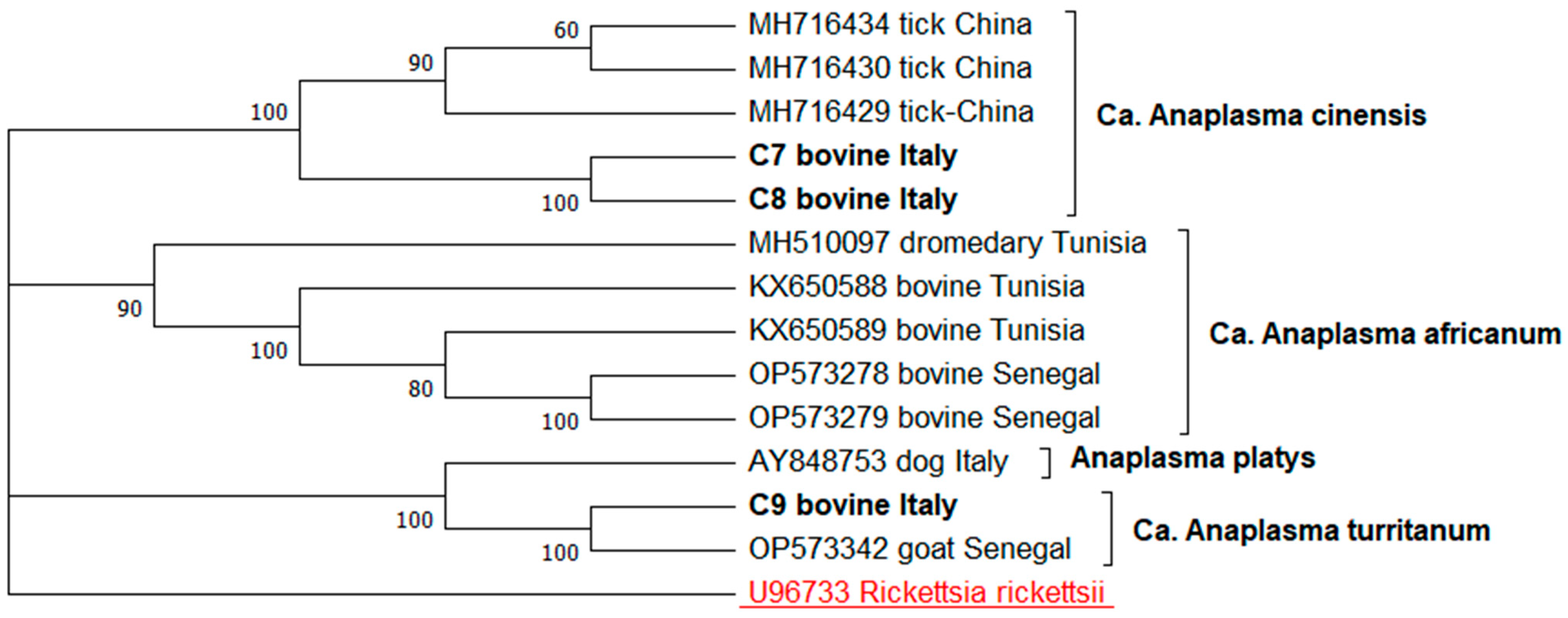
The three groEL sequence types (C7–C9) related to the A. platys-like strains were aligned with a set of ten sequences representing the groEL gene variability of the Ca. A. cinensis (MH716434, MH716430, MH716429), Ca. Anaplasma africanum (MH610097, KX650588, KX650589, OP573278, OP573279), Ca. A. turritanum (OP573342), and A. platys (AY848753). The R. rickettsii (U96733) sequence was used as an outgroup.
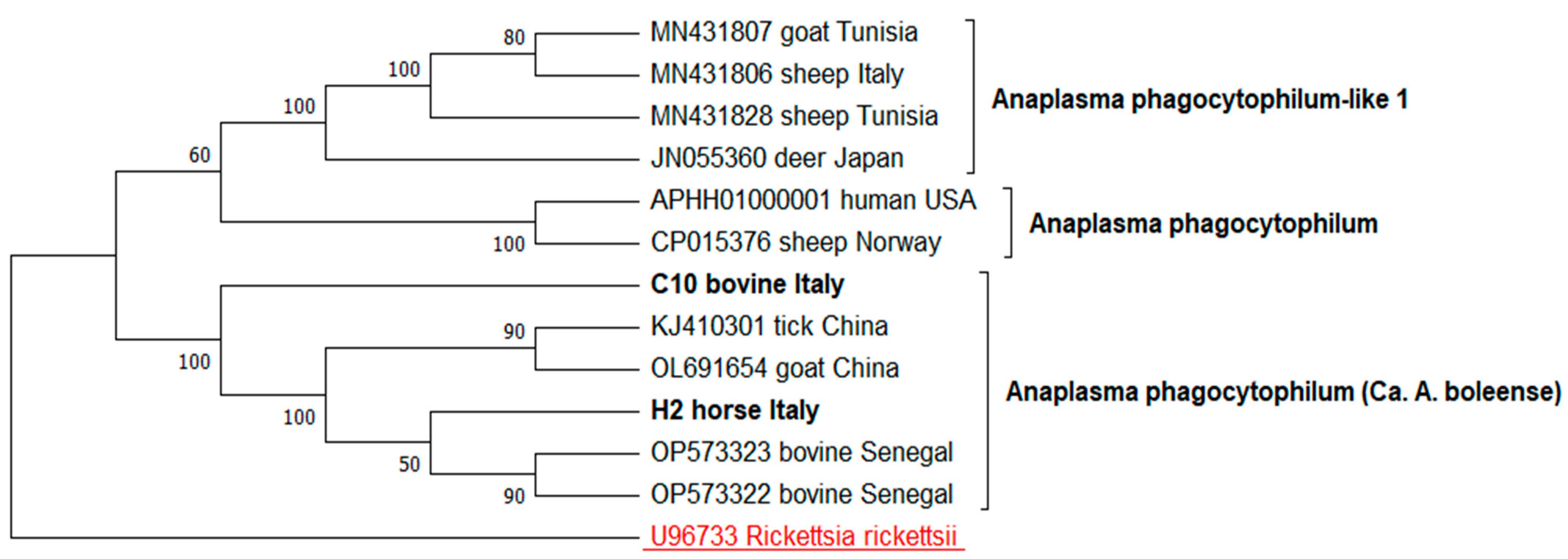
The two groEL sequence types (C10, H2) related to A. phagocytophilum were aligned with a set of ten sequences representing groEL gene variability of the A. phagocytophilum like-1 (MN431807, MN431806, MN431828, JN055360), A. phagocytophilum (APHH01000001, CP015376), Ca. A. boleense (OP573322, OP573323, KJ410301, OL691654). The R. rickettsii (U96733) sequence was used as an outgroup.
The evolutionary history for all the phylogenetic trees was inferred by using the maximum likelihood method. The Kimura 2-parameter model was applied to the gltA tree and the A. phagocytophilum groEL tree, while the Tamura–Nei model was used for the A. platys-like groEL tree. The trees with the highest log likelihood (−2331.22, −1767.97, −2710.66, respectively) are shown. The percentage of trees in which the associated taxa clustered together is shown next to the branches. The initial trees for the heuristic search were obtained automatically by applying the neighbor-joining and BioNJ algorithms to a matrix of pairwise distances estimated using the maximum composite likelihood (MCL) approach, and the Tamura–Nei model, respectively. The topology with the superior log likelihood value was then selected. A discrete Gamma distribution was used to model the evolutionary rate differences among the sites (five categories (+G, parameter = 1.1311; +G, parameter = 0.2919; +G, parameter = 0.5544, respectively)). These analyses involved 14, 14 and 13 nucleotide sequences, respectively. There was a total of 486, 468, and 719 positions in the final dataset. The evolutionary analyses were conducted in MEGA12 [35].

3. Results

Anaplasma DNA was detected in 56 out of 158 mammals (35.4%) in this study, including 13/80 horses (16.3%), 39/72 cattle (54.1%), and 4/6 swine (66.7%), using standard PCR targeting the 16S rRNA gene.
The sequencing of the 16S rRNA PCR products generated clear electropherograms in 23 of the 56 positive samples. The sequence alignment revealed four distinct sequence types (Table 2).
The GenBank BLAST analysis showed that ten sequences (HCS1; PQ579998–PQ580007) from two horses, five cattle, and three swine were 100% identical to the A. capra/A. ovis/A. centrale/A. marginale group isolated in mammals and ticks worldwide. Although the 16S rRNA gene is highly conserved across the Anaplasma species, it was unable to differentiate between these closely related species. Additionally, two sequences from two cattle (C1; PQ580008–PQ580009) were 100% identical to A. platys and the related strains. Similarities in the A. phagocytophilum/A. platys/Ca. A. cinensis group varied from 98.78% in the H1 (PQ580014) sequence isolated from one horse to 100% in the C2 sequence type from four cattle (PQ580010–PQ580013) (Table 2). To achieve greater species-level resolution, these positive samples were subjected to a second round of testing using more specific genetic markers.
PCR testing of the gltA gene, which targets the Anaplasma sp. related to A. platys and A. capra strains, combined with the sequencing and BLAST analysis (Table 2), identified three sequence types in four cattle, showing 99.47% to 99.56% similarity to Ca. A. cinensis (C3, PQ609681–PQ609682; C4, PQ609683; and C5, PQ609684). Additionally, one sequence type was detected in a single cattle, exhibiting 99.47% similarity to Ca. A. turritanum (C6, PQ609685).
The sequencing and BLAST analysis of the three cattle that tested positive for the A. platys-like groEL gene revealed two sequence types (C7 and C8, PQ588703–PQ588704) with 99.22% to 99.32% similarity to Ca. A. cinensis and one sequence type (C9, PQ588706) with 92.48% similarity to the A. platys gene.
Finally, one cattle and one horse positive for the groEL gene specific to the A. phagocytophilum-like 2 (Table 2) showed 99.47% (C10, PQ588707) and 97.74% (H2, PQ588708) similarity to the Anaplasma sp. BL099-6 found in ticks in China.
The phylogenetic analysis, based on the alignment of the four gltA gene sequence types obtained in this study with the nine reference Anaplasma sequences, revealed four main clades strongly supported by a bootstrap analysis: (a) one clade, including C3, C4 and C5 sequence strains, which clustered with the Ca. A. cinensis reference sequences; (b) one clade containing the C6 sequence strains and reference sequences of Ca. A. turritanum; (c) one clade containing A. platys; (d) one clade containing Ca. A. africanum. The C3–C5 sequence types formed a well-supported monophyletic group with Ca. A. cinensis reference sequences, which were also identified in ticks from China and bovines from Bolivia. The C6 sequence type grouped with the Ca. A. turritanum reference strains, which represent the strains previously reported in ruminants from Italy, Tunisia, and Senegal (Figure 1).
The phylogenetic tree based on the alignment of the three sequence types named C7, C8 and C9, with 11 reference Anaplasma sequence strains, was consistent with the BLASTN results. The sequence types C7 and C8 formed a statistically supported monophyletic clade with the Ca. A. cinensis groEL sequences isolated from ticks in China. Meanwhile, the C9 sequence type clustered in a statistically supported clade with the Ca. A. turritanum groEL gene sequences isolated from dogs in Italy (Figure 2).
Finally, the phylogenetic tree constructed from the alignment of the C10 and H2 sequence types with 11 reference Anaplasma sequence strains, was consistent with the BLASTN results. Both sequence types were resolved into a monophyletic clade, strongly supported by a bootstrap value of 100%, and grouped with Chinese A. phagocytophilum-like 2 sequences (Figure 3).

4. Discussion

To date, several Anaplasma species have been detected in the Mediterranean area, including the zoonotic A. phagocytophilum (found in humans, ticks, dogs, sheep, and horses), A. platys (in dogs and ticks), and A. ovis (in ticks, sheep, and goats). Additionally, A. marginale (in calves and goats), A. bovis (in sheep), and Ca. A. turritanum have been reported in the same area, including Sardinia [15,26,36,37,38,39].
The molecular approach applied in this study enabled the identification of novel sequence types linked to A. phagocytophilum and A. platys, while also confirming the presence of Ca. A. turritanum, a species commonly found in Mediterranean countries [28]. This species is primarily transmitted through the animal trade and the passive transport of ticks by migratory birds, but it has recently emerged in sub-Saharan Africa, possibly due to the founder effect [40].
The identification and characterization of the Ca. A. turritanum strains using the gltA and groEL gene markers underscores the value of these genes in the genetic studies of the Anaplasma species related to A. phagocytophilum and A. platys [41]. Detecting genetic variations is crucial for understanding the epidemiology of the emerging Anaplasma species, their transmission dynamics, and their potential zoonotic role, which could help develop more targeted, region-specific control strategies, thereby reducing the risk of outbreaks in both livestock and human populations.
These results contribute to understanding the geographic spread of Ca. A. turritanum and emphasize the need for continuous surveillance. Further research should focus on tracking its movement between regions, identifying primary and alternative reservoirs and vectors, and evaluating its role in zoonotic transmission. Also, Ca. A. turritanum should be included in the diagnostic routine of tick-borne infections in humans to assess its possible impact as a zoonotic agent.
The detection of Ca. A. cinensis in Sardinian cattle provides new insights into the geographic distribution of this species. Three distinct sequence types of Ca. A. cinensis were identified in cattle, a species genetically related to A. platys [40], confirming its presence in Sardinia for the first time in Europe. Initially isolated in 2019 from Rhipicephalus microplus ticks in Ankang, Northwest China [33], the presence of Ca. A. cinensis in Sardinia constitutes a novel record for Europe. This finding not only expands the understanding of the species’ geographic range but also highlights the potential epidemiological trends common to other Mediterranean countries where similar arthropod vectors are prevalent.
Another significant discovery is the first detection in Sardinia of A. phagocytophilum-like 2 (the Chinese variant of A. phagocytophilum) in one horse and one cattle. This strain, previously described in China in mosquitoes (Aedus albopictus, Anopheles sinensis, and Armigeres subalbatus) at various life stages, had been classified as Ca. A. boleense [10]. This finding underscores the need for further research to understand its transmission pathways and the potential impact on animal and human health.
Currently, Ca. A. turritanum, Ca. A. cinensis, and Ca. A. boleense are not included in standard diagnostic panels for tick-borne infections of human and veterinary significance, despite evidence of their presence in domestic mammals across various Mediterranean regions. Furthermore, none of the animals analyzed in this study exhibited the clinical signs of illness, which supports the hypothesis that ruminants and equines may act as reservoirs for these Anaplasma species. The choice of asymptomatic animals was intentional. By selecting animals that were apparently healthy at the time of sampling, this study aimed to assess the presence of latent or subclinical infections, which are common in tick-borne diseases but often go undetected without targeted testing. This approach is crucial for understanding the true prevalence of infections in livestock, as many tick-borne pathogens can persist in animals without causing overt symptoms. In this way, the study aimed to provide a more accurate picture of the circulation of tick-borne pathogens in the region, beyond the clinical cases that might be detected only during outbreaks.
The impact of Ca. A. turritanum, Ca. A. cinensis, and Ca. A. boleense on human health requires further investigation, as these species may represent emerging zoonotic threats. The naming of variants as Candidatus is crucial for understanding the scientific, diagnostic, pathogenic, and epidemiological implications. Taxonomically, Candidatus indicates species that have been identified but not fully described, creating diagnostic challenges due to genetic variations that can lead to cross-reactions in tests. These genetic differences can influence the pathogenicity, immune response, and treatment effectiveness. Epidemiologically, the classification highlights the need for further research on the distribution and spread of these species, as even slight genetic variations can affect the transmission and impact on populations, with implications for public health strategies and zoonotic disease monitoring.
As the geographic range of Anaplasma species continues to expand, there is an increasing need for enhanced surveillance systems to able to detect and to monitor these pathogens across regions. Additionally, improving diagnostic strategies for both humans and animals to enable the early detection of these pathogens is key to preventing severe disease outcomes. Molecular-based diagnostics, including PCR and sequencing technologies, have proven invaluable in accurately identifying Anaplasma species and their genetic variants, facilitating timely interventions and treatment.

5. Conclusions

The identification of Ca. A. turritanum, Ca. A. cinensis, and Ca. A. boleense in domestic mammals reveals that these species, closely related to the zoonotic A. platys and A. phagocytophilum, are emerging. Climate-related changes may be facilitating their spread by altering tick population dynamics and expanding the distribution of vector species. This highlights the urgent need to include these emerging Anaplasma species in routine diagnostic practices for both humans and animals, as they could represent a significant threat to public and veterinary health. Effective surveillance and control measures are crucial to mitigate the potential spread of these pathogens and to reduce the risk of the zoonotic transmission of Anaplasma species. Further research is needed to fully understand the epidemiology of these species and refine strategies for their detection, prevention, and control.

Author Contributions

Conceptualization, V.C., R.Z., G.M. and A.A.; methodology, all authors; software, V.C., R.Z. and A.A.; validation V.C. and R.Z.; formal analysis, V.C., R.Z. and A.A.; investigation, V.C., R.Z. and A.A.; resources, V.C. and G.M.; data curation, V.C. and R.Z.; writing—original draft preparation, V.C., R.Z. and A.A.; writing—review and editing, V.C., R.Z. and A.A.; visualization, V.C., R.Z., G.M., G.C., L.G., P.B., C.C., E.B. and A.A.; supervision, V.C., R.Z., G.M., G.C., L.G., P.B., C.C., E.B. and A.A.; project administration, V.C. and R.Z. All authors have read and agreed to the published version of the manuscript.

Funding

This research received no external funding.

Institutional Review Board Statement

Not applicable.

Informed Consent Statement

Not applicable.

Data Availability Statement

All data generated and analyzed during this study are included in this published article.

Conflicts of Interest

The authors declare no conflicts of interest.

References

  1. Atif, F.A. Alpha proteobacteria of genus Anaplasma (Rickettsiales: Anaplasmataceae): Epidemiology and characteristics of Anaplasma species related to veterinary and public health importance. Parasitology 2016, 143, 659–685. [Google Scholar] [CrossRef] [PubMed]
  2. Karpathy, S.E.; Kingry, L.; Pritt, B.S.; Berry, J.C.; Chilton, N.B.; Dergousoff, S.J.; Cortinas, R.; Sheldon, S.W.; Oatman, S.; Anacker, M.; et al. Anaplasma bovis—Like infections in humans, United States, 2015–2017. Emerg. Infect. Dis. 2023, 29, 1904–1907. [Google Scholar] [CrossRef] [PubMed]
  3. Lu, M.; Li, F.; Liao, Y.; Shen, J.-J.; Xu, J.-M.; Chen, Y.-Z.; Li, J.-H.; Holmes, E.C.; Zhang, Y.-Z. Epidemiology and Diversity of Rickettsiales Bacteria in Humans and Animals in Jiangsu and Jiangxi Provinces, China. Sci. Rep. 2019, 9, 13176. [Google Scholar] [CrossRef]
  4. Li, H.; Zheng, Y.-C.; Ma, L.; Jia, N.; Jiang, B.-G.; Jiang, R.-R.; Huo, Q.-B.; Wang, Y.-W.; Liu, H.-B.; Chu, Y.-L.; et al. Human infection with a novel tick-borne Anaplasma species in China: A surveillance study. Lancet Infect. Dis. 2015, 15, 663–670. [Google Scholar] [CrossRef]
  5. Bakken, J.S.; Dumler, J.S.; Chen, S.M.; Eckman, M.R.; Van Etta, L.L.; Walker, D.H. Human granulocytic ehrlichiosis in the upper midwest United States. A New Species Emerging? JAMA 1994, 272, 212–218. [Google Scholar] [CrossRef]
  6. Chen, S.M.; Dumler, J.S.; Bakken, J.S.; Walker, D.H. Identification of a granulocytotropic Ehrlichia species as the etiologic agent of human disease. J. Clin. Microbiol. 1994, 32, 589–595. [Google Scholar] [CrossRef]
  7. Rar, V.; Tkachev, S.; Tikunova, N. Genetic diversity of Anaplasma bacteria: Twenty years later. Infect. Genet. Evol. 2021, 91, 104833. [Google Scholar] [CrossRef] [PubMed]
  8. Dumler, J.S.; Barbet, A.F.; Bekker, C.P.; Dasch, G.A.; Palmer, G.H.; Ray, S.C.; Rikihisa, Y.; Rurangirwa, F.R. Reorganization of genera in the families Rickettsiaceae and Anaplasmataceae in the order Rickettsiales: Unification of some species of Ehrlichia with Anaplasma, Cowdria with Ehrlichia and Ehrlichia with Neorickettsia, descriptions of six new species combinations and designation of Ehrlichia equi and ‘HGE agent’ as subjective synonyms of Ehrlichia phagocytophila. Int. J. Syst. Evol. Microbiol. 2001, 51, 2145–2165. [Google Scholar]
  9. Tate, C.M.; Howerth, E.W.; Mead, D.G.; Dugan, V.G.; Luttrell, M.P.; Sahora, A.I.; Munderloh, U.G.; Davidson, W.R.; Yabsley, M.J. Anaplasma odocoilei sp. nov. (family Anaplasmataceae) from white-tailed deer (Odocoileus virginianus). Ticks Tick-Borne Dis. 2013, 4, 110–119. [Google Scholar] [CrossRef]
  10. Guo, W.-P.; Tian, J.-H.; Lin, X.-D.; Ni, X.-B.; Chen, X.-P.; Liao, Y.; Yang, S.-Y.; Dumler, J.S.; Holmes, E.C.; Zhang, Y.-Z. Extensive genetic diversity of Rickettsiales bacteria in multiple mosquito species. Sci. Rep. 2016, 6, 38770. [Google Scholar] [CrossRef]
  11. Kang, Y.-J.; Diao, X.-N.; Zhao, G.-Y.; Chen, M.-H.; Xiong, Y.; Shi, M.; Fu, W.-M.; Guo, Y.-J.; Pan, B.; Chen, X.-P.; et al. Extensive diversity of Rickettsiales bacteria in two species of ticks from China and the evolution of the Rickettsiales. BMC Evol. Biol. 2014, 14, 167. [Google Scholar] [CrossRef] [PubMed]
  12. Shi, Y.; Yang, J.; Guan, G.; Liu, Z.; Luo, J.; Song, M. Molecular investigation of Anaplasma species in sheep from Heilongjiang province, Northeast China identified four Anaplasma species and a novel genotype of Anaplasma capra. Parasitol. Int. 2020, 76, 102072. [Google Scholar]
  13. Liu, Z.; Ma, M.; Wang, Z.; Wang, J.; Peng, Y.; Li, Y.; Guan, G.; Luo, J.; Yin, H. Molecular survey and genetic identification of Anaplasma species in goats from central and southern China. Appl. Environ. Microbiol. 2012, 78, 464–470. [Google Scholar] [PubMed]
  14. Altay, K.; Erol, U.; Sahin, O.F. Anaplasma capra: A new emerging tick-borne zoonotic pathogen. Vet. Res. Commun. 2024, 48, 1329–1340. [Google Scholar]
  15. Zobba, R.; Schianchi, E.; Ben Said, M.; Belkahia, H.; Messadi, L.; Piredda, R.; Pittau, M.; Alberti, A. GltA typing of Anaplasma strains related to A. platys: Taxonomical and one health implications. Ticks Tick-Borne Dis. 2022, 13, 101850. [Google Scholar]
  16. Rymaszewska, A.; Grenda, S. Bacteria of the genus Anaplasma—Characteristics of Anaplasma and their vectors: A review. Veterinární Medicína 2008, 53, 573–584. [Google Scholar]
  17. Aubry, P.; Geale, D.W. A review of bovine anaplasmosis. Transbound. Emerg. Dis. 2011, 58, 1–30. [Google Scholar]
  18. Potgieter, F. Bovine anaplasmosis. In Infectious Diseases of Livestock, 2nd ed.; Coetzer, J.A.W., Tustin, R.C., Eds.; Oxford University Press: Cape Town, South Africa, 2004; pp. 594–616. [Google Scholar]
  19. Battilani, M.; De Arcangeli, S.; Balboni, A.; Dondi, F. Genetic diversity and molecular epidemiology of Anaplasma. Infect. Genet. Evol. 2017, 49, 195–211. [Google Scholar]
  20. Shabana, I.I.; Alhadlag, N.M.; Zaraket, H. Diagnostic tools of caprine and ovine anaplasmosis: A direct comparative study. BMC Vet. Res. 2018, 14, 165. [Google Scholar]
  21. Silaghi, C.; Santos, A.S.; Gomes, J.; Christova, I.; Matei, I.A.; Walder, G.; Domingos, A.; Bell-Sakyi, L.; Sprong, H.; Von Loewenich, F.D.; et al. Guidelines for the direct detection of Anaplasma spp. in diagnosis and epidemiological studies. Vector-Borne Zoonotic Dis. 2017, 17, 12–22. [Google Scholar]
  22. Pan, L.; Zhang, L.; Wang, G.; Liu, Q.; Yu, Y.; Wang, S.; Yu, H.; He, J. Rapid, simple, and sensitive detection of Anaplasma phagocytophilum by loop-mediated isothermal amplification of the msp2 gene. J. Clin. Microbiol. 2011, 49, 4117–4120. [Google Scholar] [PubMed]
  23. McGuire, T.C.; Davis, W.C.; Brassfield, A.L.; McElwain, T.F.; Palmer, G.H. Identification of Anaplasma marginale long-term carrier cattle by detection of serum antibody to isolated MSP-3. J. Clin. Microbiol. 1991, 29, 788–793. [Google Scholar]
  24. Von Fricken, M.E.; Lkhagvatseren, S.; Boldbaatar, B.; Nymadawa, P.; Weppelmann, T.A.; Baigalmaa, B.-O.; Anderson, B.D.; Reller, M.E.; Lantos, P.M.; Gray, G.C. Estimated seroprevalence of Anaplasma spp. and spotted fever group Rickettsia exposure among herders and livestock in Mongolia. Acta Trop. 2018, 177, 179–185. [Google Scholar]
  25. Yoo, J.; Chung, J.-H.; Kim, C.-M.; Yun, N.R.; Kim, D.-M. Asymptomatic-anaplasmosis confirmation using genetic and serological tests and possible coinfection with spotted fever group Rickettsia: A case report. BMC Infect. Dis. 2020, 20, 458. [Google Scholar]
  26. Alberti, A.; Zobba, R.; Chessa, B.; Addis, M.F.; Sparagano, O.; Pinna Parpaglia, M.L.; Cubeddu, T.; Pintori, G.; Pittau, M. Equine and canine Anaplasma phagocytophilum strains isolated on the island of Sardinia (Italy) are phylogenetically related to pathogenic strains from the United States. Appl. Environ. Microbiol. 2005, 71, 6418–6422. [Google Scholar]
  27. Zobba, R.; Anfossi, A.G.; Pinna Parpaglia, M.L.; Dore, G.M.; Chessa, B.; Spezzigu, A.; Rocca, S.; Visco, S.; Pittau, M.; Alberti, A. Molecular investigation and phylogeny of Anaplasma spp. in mediterranean ruminants reveal the presence of neutrophil-tropic strains closely related to Anaplasma platys. Appl. Environ. Microbiol. 2014, 80, 271–280. [Google Scholar]
  28. Zobba, R.; Ben Said, M.; Belkahia, H.; Pittau, M.; Cacciotto, C.; Pinna Parpaglia, M.L.; Messadi, L.; Alberti, A. Molecular epidemiology of Anaplasma spp. related to A. phagocytophilum in mediterranean small ruminants. Acta Trop. 2020, 202, 105286. [Google Scholar]
  29. Chisu, V.; Zobba, R.; Lecis, R.; Sotgiu, F.; Masala, G.; Foxi, C.; Pisu, D.; Alberti, A. GroEL Typing and phylogeny of Anaplasma species in ticks from domestic and wild vertebrates. Ticks Tick-Borne Dis. 2018, 9, 31–36. [Google Scholar]
  30. Chisu, V.; Loi, F.; Foxi, C.; Chessa, G.; Masu, G.; Rolesu, S.; Masala, G. Coexistence of tick-borne pathogens in ticks collected from their hosts in Sardinia: An update. Acta Parasitol. 2020, 65, 999–1004. [Google Scholar]
  31. Guccione, C.; Colomba, C.; Tolomeo, M.; Trizzino, M.; Iaria, C.; Cascio, A. Rickettsiales in Italy. Pathogens 2021, 10, 181. [Google Scholar] [CrossRef]
  32. Kolbert, C. Detection of the agent of human granulocytic ehrlichiosis by PCR. In PCR Protocols for Emerging Infectious Diseases; Persing, D.H., Ed.; ASM Press: Washington, DC, USA, 1996; pp. 106–111. [Google Scholar]
  33. Guo, W.P.; Zhang, B.; Wang, Y.H.; Xu, G.; Wang, X.; Ni, X.; Zhou, E.M. Molecular identification and characterization of Anaplasma capra and Anaplasma platys-like in Rhipicephalus microplus in Ankang, Northwest China. BMC Infect. Dis. 2019, 19, 434. [Google Scholar]
  34. Ybañez, A.P.; Matsumoto, K.; Kishimoto, T.; Inokuma, H. Molecular analyses of a potentially novel Anaplasma species closely related to Anaplasma phagocytophilum detected in sika deer (Cervus nippon yesoensis) in Japan. Vet. Microbiol. 2012, 157, 232–236. [Google Scholar] [PubMed]
  35. Kumar, S.; Stecher, G.; Suleski, M.; Sanderford, M.; Sharma, S.; Tamura, K. MEGA12: Molecular Evolutionary Genetic Analysis version 12 for adaptive and green computing. Mol. Biol. Evol. 2024, 41, 263. [Google Scholar]
  36. Torina, A.; Alongi, A.; Naranjo, V.; Estrada-Peña, A.; Vicente, J.; Scimeca, S.; Marino, A.M.F.; Salina, F.; Caracappa, S.; De La Fuente, J. Prevalence and genotypes of Anaplasma species and habitat suitability for ticks in a mediterranean ecosystem. Appl. Environ. Microbiol. 2008, 74, 7578–7584. [Google Scholar]
  37. Ben Said, M.; Belkahia, H.; Karaoud, M.; Bousrih, M.; Yahiaoui, M.; Daaloul-Jedidi, M.; Messadi, L. First molecular survey of Anaplasma bovis in small ruminants from Tunisia. Vet. Microbiol. 2015, 179, 322–326. [Google Scholar]
  38. Chisu, V.; Dei Giudici, S.; Foxi, C.; Chessa, G.; Peralta, F.; Sini, V.; Masala, G. Anaplasma Species in ticks infesting mammals of Sardinia, Italy. Animals 2023, 13, 1332. [Google Scholar] [CrossRef]
  39. Saratsis, A.; Ligda, P.; Aal, F.; Jelicic, M.; Polgar, J.; De Vries, M.; Mastranestasis, I.; Musella, V.; Rinaldi, L.; Jongejan, F.; et al. The scenario of ticks and tick-borne pathogens of sheep on a mediterranean island. Microorganisms 2022, 10, 1551. [Google Scholar] [CrossRef]
  40. Zobba, R.; Murgia, C.; Dahmani, M.; Mediannikov, O.; Davoust, B.; Piredda, R.; Schianchi, E.; Scagliarini, A.; Pittau, M.; Alberti, A. Emergence of Anaplasma species related to A. phagocytophilum and A. platys in Senegal. Int. J. Mol. Sci. 2022, 24, 35. [Google Scholar] [CrossRef]
  41. Kolo, A. Anaplasma species in Africa—A century of discovery: A review on molecular epidemiology, genetic diversity, and control. Pathogens 2023, 12, 702. [Google Scholar] [CrossRef]
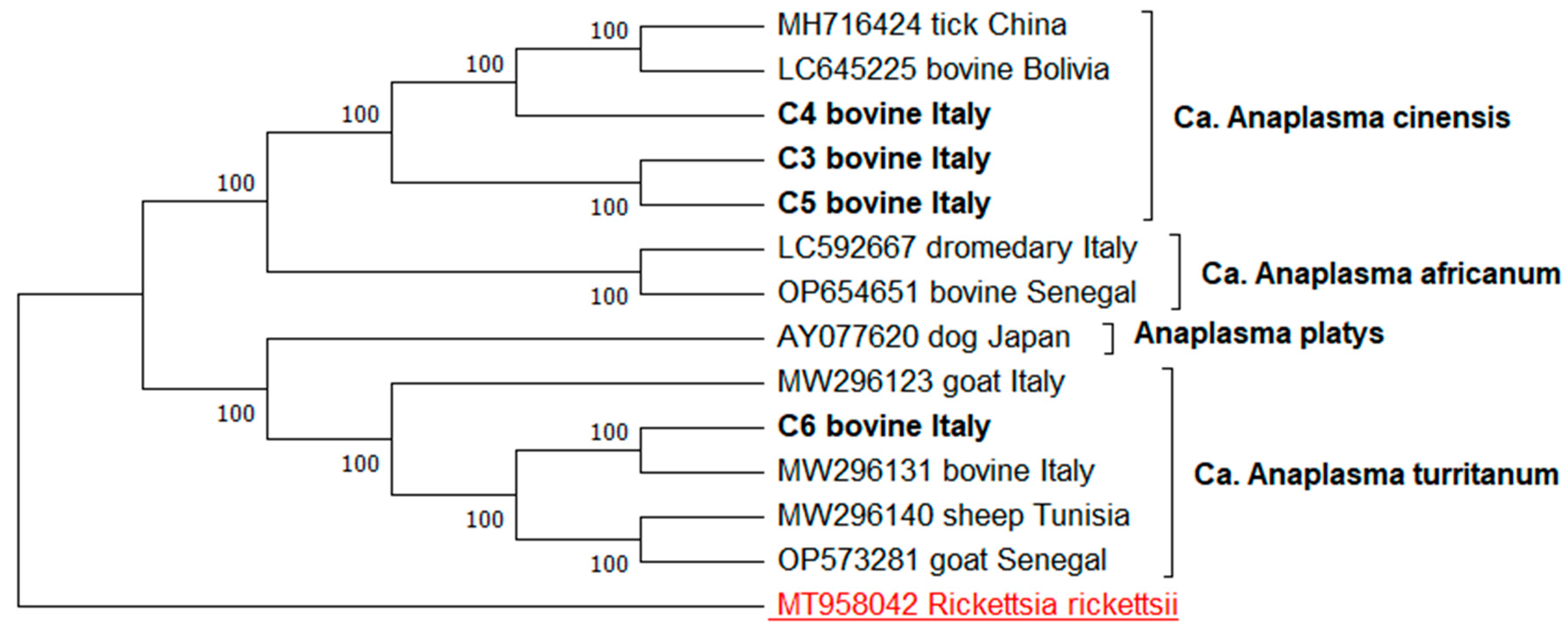
Figure 1. Phylogenetic tree of the Anaplasma spp. strains based on gltA sequencing. Evolutionary history was inferred using the maximum likelihood method and Tamura–Nei model.
Animals 15 01029 g001
Figure 2. Phylogenetic analysis of the Anaplasma spp. strains based on groEL sequencing. The evolutionary history was inferred using the maximum likelihood method with the Tamura–Nei model.
Animals 15 01029 g002
Figure 3. Phylogeny of A. phagocytophilum strains based on groEL sequencing. The evolutionary history was inferred using the maximum likelihood method, applying the Tamura–Nei model.
Animals 15 01029 g003
Table 1. Primer sequences used in this study for the detection of Anaplasma species from the blood samples of the mammal hosts analyzed.
PathogensGene Target PCR AssayGene PrimersPrimer Sequence
(5′ to 3′)
Amplicon Size (bp)References
Anaplasma spp.16S rRNAPCRHGE–521F
HGE–790R
TGTAGGCGGTTCGGTAAGTTAAAG293 bp[32]
CTTAACGCGTTAGCTACAACACAG
A. platysgltANested PCRPglt-FATGAWAGAAAAWGCTGTTTT660 bp[33]
Pglt-R1TCATGRTCTGCATGCATKATG
Pglt-R2CATGCATKATGAARATMGCAT
Pglt-L-F1ATGAWAGAAAAWGCTGTTTT610 bp
Pglt-L-F2TCATGRTCTGCATGCATKATG
Pglt-L-RCATGCATKATGAARATMGCAT
GroELNested PCRPgro_F_F GATGCWCATCCYATSGCMATG1195 bp
Pgro_F_R1CGTGMTSGCTATAGCGMAART
Pgro_F_R2TCAYACCATTGDGAYRCCCAT
A. phagocytophilumGroELNested PCREphplGroEL(569)F ATGGTATGCAGTTTGATCGC573 bp[26]
EphplGroEL(1193)R TCTACTCTGTCTTTGCGTTC
EphGroEL(1142)R TTGAGTACAGCAACACCACCGGAA
A. phagocytophilum-like 1 GroELNested PCRAPGRSP41F3 GAATCTAGCTATGTTGCATGATAATGT1446 bp[34]
APSPGR1697R1CAGCATAAACACGCACTACGAA
APSPGR234F2CGTAGTAGGACTTTCCGGTTTTTG
APSPGR1680R2TGCACAGCATAAACACGCACTA
A. phagocytophilum-like 2GroELNested PCRAPHAGOVAR2GROEL_FTACTCTAGAAGACGCGGTAG793 bp[28]
APHAGOVAR2GROEL_R1ACGAACATTCTTAGCAGTCC
APHAGOVAR2GROEL_R2CTTCTATCACCAAATCCTGG
Table 2. Definition of 16S rRNA, gltA and groEL gene sequence types, host sources, BLAST analysis and GenBank accession numbers.
Target GeneSequence Type *Host SourcesLocalityBlastNGenBank Accession Number (s)
16S rRNAHCS1Horse, Cattle, SwineAglientu, Arzachena, Baunei, Birori, Chiaramonti, Fordongianus, Laerru, Orgosolo, Santu LussurgiuSedini, Villanova Monteleone100% A. capra/A. ovis/A. centrale/A. marginale groupPQ579998–PQ580007
C1CattleAnela, Chiaramonti, Milis, Oristano, Monti100% A. phagocytophilum/A. platys/Ca. A. cinensis groupPQ580008–PQ580009
C2CattleOrgosolo, Santa Giusta, Villacidro, Laerru, Chiaramonti100% A. phagocytophilum/A. platys/Ca. A. cinensis groupPQ580010–PQ580013
H1HorseSanta Giusta98.78% A. phagocytophilum/A. platys/Ca. A. cinensis groupPQ580014
gltAC3CattleOrgosolo99.56% Ca. A. cinensisPQ609681–PQ609682
C4CattleOrgosolo99.47% Ca. A. cinensisPQ609683
C5CattleOrgosolo99.47% Ca. A. cinensisPQ609684
C6CattleIllorai99.47% Ca. A. turritanumPQ609685
GroELC7CattleOrgosolo99.32% Ca A. cinensisPQ588703–PQ588704
C8CattleOrgosolo99.22% Ca. A. cinensisPQ588705
C9CattleMilis99–100% Ca. A. turritanumPQ588706
GroELC10CattleBortigiadas97.74% Anaplasma spPQ588707
H2HorseSanta Giusta99.47% Anaplasma sp.PQ588708
* Hosts are numbered according to the order in which they were collected and processed in the laboratory.
Disclaimer/Publisher’s Note: The statements, opinions and data contained in all publications are solely those of the individual author(s) and contributor(s) and not of MDPI and/or the editor(s). MDPI and/or the editor(s) disclaim responsibility for any injury to people or property resulting from any ideas, methods, instructions or products referred to in the content.