Article

Comprehensive Phenotypic Characterization and Genomic Analysis Unveil the Probiotic Potential of Bacillus velezensis K12

1
State Key Laboratory of Animal Nutrition and Feeding, College of Animal Science and Technology, China Agricultural University, Beijing 100193, China
2
College of Animal Science and Technology & College of Veterinary Medicine, Zhejiang A&F University, Hangzhou 311300, China
3
Ningxia Eppen Biotech Co., Ltd., Yinchuan 750100, China
4
Department of Basic Veterinary Medicine, College of Veterinary Medicine, China Agricultural University, Beijing 100193, China
*
Authors to whom correspondence should be addressed.
Animals 2025, 15(6), 798; https://doi.org/10.3390/ani15060798
Submission received: 25 January 2025 / Revised: 1 March 2025 / Accepted: 10 March 2025 / Published: 11 March 2025
(This article belongs to the Section Animal Genetics and Genomics)

Simple Summary

The increasing utilization of antibiotics has sparked widespread public concern, and the search for antibiotic alternatives has become a research hotspot. Bacillus spp. have emerged as pivotal sources of probiotic preparations, garnering considerable attention in recent years owing to their strong bacteriostatic activity and antimicrobial resistance. In this study, a strain of Bacillus velezensis K12 was screened from broiler intestine and assumed to be probiotic. The results of an in vitro assay and whole-genome sequencing showed that K12 possesses potent bacteriostatic properties and exhibits safety in vitro, positioning it as a promising candidate for further probiotic development.

Abstract

Bacillus spp. have emerged as pivotal sources of probiotic preparations, garnering considerable attention in recent years owing to their vigorous bacteriostatic activity and antimicrobial resistance. This study aimed to investigate these probiotic characteristics in depth and verify the safety of Bacillus velezensis K12, a strain isolated from broiler intestine. The K12 strain was identified as Bacillus velezensis based on its morphology and 16S rDNA sequence homology analysis. Subsequently, B. velezensis K12 was evaluated for acid resistance, bile salt resistance, gastrointestinal tolerance, drug sensitivity, and antimicrobial activity. Additionally, whole-genome sequencing technology was employed to dissect its genomic components further, aiming to explore its potential applications as a probiotic strain. B. velezensis K12 was sensitive to six antibiotics and had acid tolerance. Furthermore, it showed potent antimicrobial activity against a wide range of pathogenic bacteria, including Escherichia coli (E. coli), Staphylococcus aureus, Salmonella, Clostridium perfringens, Bacillus cereus, and Vibrio parahaemolyticus. The complete genome sequencing of B. velezensis K12 revealed a genomic length of 3,973,105 base pairs containing 4123 coding genes, among which 3973 genes were functionally annotated. The genomic analysis identified genes associated with acid and bile tolerance, adhesion, antioxidants, and secondary metabolite production, whereas no functional genes related to enterotoxins or transferable antibiotic resistance were detected, thereby confirming the probiotic properties of B. velezensis K12. B. velezensis K12 exhibits broad-spectrum bacteriostatic activity and in vitro safety, positioning it as a potential candidate strain for developing probiotic Bacillus preparations.

1. Introduction

The increasing utilization of antibiotics has sparked widespread public concern since 1990. Several alternatives to antibiotics have been proposed to control the problems associated with their overuse, such as bacteriocins [1], oligosaccharides [2], enzymes [3], and probiotics [4]. The International Scientific Association for Probiotics and Prebiotics (ISAPP) define probiotics as “live microorganisms that, when administered in adequate amounts, confer a health benefit on the host” [5]. The main bacterial probiotics commonly used are Lactobacillus, Bifidobacterium, and Bacillus [6]. Bacillus is a Gram-positive bacterium that produces spores. Compared to Lactobacillus and Bifidobacterium, Bacillus has higher acid resistance and better stability during heat treatment and cold storage [7]. Bacillus species can secrete numerous metabolites and peptides with antimicrobial activity, which exert various effects, including antibacterial, antifungal, and growth-promoting impact [8].
Bacillus velezensis was initially isolated from the Vélez River in Torredelmar, Málaga, southern Spain [9]. It can be readily isolated and cultured and is widely found in nature [10]. The Bacillus velezensis strain exhibits a notable capacity to produce secondary metabolites with potent antibacterial characteristics, including antibiotic lipopeptides, polyketides, and peptides [11]. Given its extensive natural distribution and the enrichment of its metabolites, Bacillus velezensis has gained increasing recognition in recent years as a probiotic [12]. Bacillus velezensis has been widely studied as a biocontrol agent due to its antagonistic potential against various phytopathogenic fungi [13,14,15]. Moreover, numerous studies have described the probiotic properties of different Bacillus velezensis strains in the aquaculture and poultry industries [16,17,18,19]. Several studies have also supported the safety and promise of Bacillus velezensis strains for application as human oral probiotics [20,21].
Whole-genome sequencing provides tremendous insight into the potential properties of microbes as probiotics, as well as valuable information regarding their use [22]. The genomes of Bacillus velezensis from diverse sources, including potato tubers [23], termite gut [24], and soil [25], have been thoroughly described, thereby elucidating the underlying mechanisms responsible for its probiotic attributes.
In the current study, B. velezensis K12 isolated from broiler intestine was investigated for its probiotic potential, focusing on its growth characteristics, resistance to acid and bile salts, antibiotic susceptibility, and in vitro bacteriostatic properties. We also performed a complete genome analysis of B. velezensis K12 to explore its genetic determinants concerning its probiotic properties. This study provides valuable insights into the genome and phenotypic features of B. velezensis K12, thereby identifying it as a potent antimicrobial agent in future research and animal husbandry.

2. Materials and Methods

2.1. Isolation, Purification, and Characterization of Strains

Strain K12 was isolated from broiler intestine. The indicator strains for the in vitro studies were E. coli K88, E. coli CVCC25922, Staphylococcus aureus CVCC1822, Salmonella CVCC519, Clostridium perfringens CVCC66, Bacillus cereus CICC21290, and Vibrio parahaemolyticus CICC23924, which were all provided by the Animal Nutrition Process Development Department of the R&D Center of Ningxia Yipin Biotechnology Co., Ltd. (Yinchuan, China).
Strain K12 was inoculated onto Luria–Bertani (LB) solid medium using the line dilution method and incubated inverted at 37 °C for 14 h to observe the morphology of the colonies. Single colonies on LB medium were picked for Gram staining, and the morphological characteristics of the bacteria were examined under an upright microscope (Olympus CX23, Yijingtong Optics Technology Co., Shanghai, China). Subsequently, single colonies on LB medium were picked and inoculated into LB liquid medium (Beijing YiKeRan Biotechnology Co., Beijing, China). They were then incubated in a constant temperature shaker (Shanghai Jinghong Laboratory Equipment Co., Shanghai, China) at 37 °C and 220 rpm for 14 h. Centrifugation was performed to collect the bacterial bodies, which were then forwarded to Shanghai Parsonage Bio-technology Co. (Shanghai, China). for 16S rDNA gene sequencing. PCR amplification was conducted with 27F: 5′-AGAGTTTGATCCTGGCTCAG-3′ and 1492R: 5′-GGTTACCTTGTTACGACTT-3′ primers [26]. The PCR reaction mixture (50 μL) included 5 μL of 10× Buffer, 1 μL of Taq polymerase, 1 μL of dNTPs, 1.5 μL of each primer (upstream and downstream), and 1 μL of template. The PCR reaction program was as follows: pre-denaturation at 95 °C for 5 min, denaturation at 95 °C for 30 s, annealing at 58 °C for 30 s, extension at 72 °C for 1 min and 30 s, repeated for 35 cycles, followed by a final extension at 72 °C for 7 min, and storage at 4 °C. The amplified products were analyzed via 1.0% agarose gel (Beijing YiKeRan Biotechnology Co., Beijing, China) electrophoresis and sequenced. The obtained sequences were submitted to the NCBI database for BLAST comparison (BLAST v2.13.0) [27]. Sequences from 11 closely related species were retrieved, and a 16S evolutionary tree was constructed using MEGA11, using the neighbor-joining method, with a bootstrap value of 1000 [28], based on multiple sequence alignments performed with MAFFT software [29].

2.2. Determination of Strain Biology

2.2.1. Determination of the Growth Curve of Strain K12 Versus the pH Change Curve

Referring to Renschler et al. [30] with minor modifications, the K12 strain was activated and cultivated in LB liquid medium for 14 h. The cultured bacterial suspension was transferred to fresh LB liquid medium at a 1% inoculum ratio and incubated in a shaker at 37 °C and 220 r/min for 36 h. Samples were collected every 2 h, and the absorbance at 600 nm and the pH of the bacterial culture were measured.

2.2.2. Hemolysis Analysis

The activated K12 strain was cultured on sheep blood agar plates (Beijing YiKeRan Biotechnology Co., Beijing, China), and the results were observed after 14 h of incubation at 37 °C [31]. The formation of a green circle around the colonies indicated α-hemolytic activity, while the formation of a transparent circle indicated β-hemolytic activity. In contrast, the absence of a transparent circle indicated γ-hemolytic activity.

2.2.3. Acid Resistance Analysis

To study the acid resistance of the strains, the method of Soni et al. [31] was referred to and slightly modified. The pH of the LB liquid medium was adjusted to 1.5, 2.5, 3.5, 4.5, 5.5, and 6.5 using 1 mol/L HCl. The activated bacterial broth was inoculated into LB liquid medium with different pH values at an inoculum rate of 1%, with pH 7.1 inoculated and non-inoculated LB liquid medium as the control. The samples were incubated at 37 °C and 220 r/min for 24 h. After incubation, samples were taken at 0, 2, 4, 8, 12, and 24 h, and the absorbance was measured at a wavelength of 600 nm using a UV spectrophotometer (TU-1950, Beijing Puxi General Instrument Co., Beijing, China).

2.2.4. Bile Resistance Analysis

The bile resistance of the strains was determined by the method of Reyes-Cortes et al. [32] with slight modifications. The corresponding masses of bile salts were weighed and dissolved into sterile LB liquid medium, and 0.00%, 0.02%, 0.04%, 0.06%, 0.08%, 0.10%, 0.20%, and 0.30% bile salts were prepared. The bile salts were filtered to remove bacteria. The LB liquid medium without bacteria was used as a control. The activated bacterial solution was added to the LB liquid medium with varying bile salt concentrations at an inoculum rate of 1% and then incubated at 37 °C and 220 r/min for 24 h. After incubation, samples were taken at 0, 2, 4, 8, 12, and 24 h, and the absorbance was measured at a wavelength of 600 nm using a UV spectrophotometer.

2.2.5. Determination of Tolerance to Simulated Gastrointestinal Fluids

To determine the tolerance of strain K12 to simulated gastrointestinal fluids, the method of Tanvi et al. [33] was used with minor modifications. The activated bacterial solution was centrifuged at 3000 r/min for 10 min, and the bacterial bodies were collected and washed twice with sterile PBS buffer (pH 7.3–7.5) to remove the residual medium. The bacteria were resuspended in 5.0 mL of PBS, and 1.0 mL of each bacterial suspension was separately mixed with 9.0 mL of sterile simulated artificial gastric fluid (containing 1% pepsin, pH 2.0) and 9.0 mL of sterile simulated artificial intestinal fluid (containing 0.2% trypsin and 1.2% bovine bile salts, pH 8.0). The simulated gastrointestinal fluids were purchased from Beijing Yikeran Biotechnology Co (Beijing, China). The mixtures were cultured at 37 °C. After 3 h of both initial and co-culturing, the viable bacteria were counted on agar plates, and the relative content was calculated.

2.2.6. Antibiotic Susceptibility Testing

The disk diffusion method was used for the drug sensitivity test, with results interpreted according to the American Society for Clinical Laboratory Standardization (CLSI) 2021 standards [34]. A fresh bacterial suspension with a concentration of 1.0 × 10⁸ CFU/mL (100 μL) was inoculated onto an LB agar plate. Drug-sensitive tablets containing cefotaxime, florfenicol, ciprofloxacin, cotrimoxazole, doxycycline, and gentamicin were placed onto plates coated with the bacterial solution. The tablets were gently pressed to ensure they adhered tightly to the plate, and then inverted and placed on the LB plate, and the inhibition zone diameters were measured after incubation at 37 °C for 24 h.

2.2.7. Determination of Bacteriostatic Capacity

The bacteriostatic capacity of strain K12 was determined using a modified version of the Oxford cup double plate method [35]. E. coli K88, E. coli CVCC25922, Staphylococcus aureus CVCC1822, Salmonella CVCC519, Bacillus cereus CICC21290, Clostridium perfringens CVCC66, and Vibrio parahaemolyticus CICC23924 were used as the indicator bacteria, which were inoculated into LB liquid medium, RCM liquid medium, and high-salt LB liquid medium containing 3.5% NaCl, respectively, to make suspensions containing 1 × 10⁸ CFU/mL indicator bacteria. Strain K12 was inoculated into LB liquid medium and incubated at 37 °C and 220 r/min for 14 h, and then diluted 10, 100, 1000, 4000, 8000, and 10,000 times via gradient dilution with sterile water. Pure agar plates (containing 1.8% agar) were prepared by placing 8 mm diameter Oxford cups (Beijing YiKeRan Biotechnology Co., Beijing, China) evenly on the solidified agar plates. To LB solid medium/anaerobic broth solid medium/high-salt LB solid medium (containing 1.8% agar) at around 50 °C, add a 1% (v/v) suspension of indicator bacteria; mix the two and pour onto an agar plate (containing 1.8% agar). After the medium solidified, the Oxford cup was pulled out using sterile forceps and 200 μL of the above-treated strain K12 dilution was injected into each well. After static diffusion for 2 h, the sample was positioned in a 37 °C constant-temperature incubator for 12–20 h. The presence of an inhibition zone was observed, and the size of the inhibition zone was measured using a vernier caliper. Each experimental group was repeated three times.
Strain K12 was cultured in LB liquid medium at 37 °C with agitation at 220 r/min for 14 h. The culture was then centrifuged at 3000 rpm for 10 min to separate the supernatant from the sediment. After that, the supernatant was filtered through a 0.22 μm microfiltration membrane to remove bacteria, and the sediment was washed with sterile PBS buffer (Beijing YiKeRan Biotechnology Co., Beijing, China) 3 times to remove residual medium and supernatant and then resuspended in PBS. The indicator bacteria were subjected to the same as the above, and the inhibition effect of strain K12 whole culture, supernatant, and sediment was determined by the Oxford cup double plate method. Each set of experiments was repeated three times.
Strain K12 was cultured in LB liquid medium, incubated at 37 °C and 220 r/min on a shaker (SPH-110X24, Shanghai Shiping Experimental Equipment Co., Shanghai, China) for 14 h, and then boiled to inactivate it. The bacteriostatic ability of strain K12 after inactivation was assessed using the Oxford cup double plate method, with E. coli K88 serving as an indicator bacterium. Each group of experiments was repeated three times.

2.2.8. Whole-Genome Sequencing and Annotation

The genomic DNA of strain K12 was extracted utilizing the TruSeqTM DNA Sample Prep Kit (Tiangen, Beijing, China). Then, 1% agarose gel electrophoresis and a NanoDrop 2000 spectrophotometer (Thermo Fisher Scientific, Waltham, MA, USA) were used to assess the quality and concentration of the extracted DNA [36]. The complete genome sequencing of strain B. velezensis K12 was performed in two stages: second-generation sequencing using the Illumina NovaSeq 6000 platform (Illumina, San Diego, CA, USA) and third-generation sequencing using the Oxford Nanopore platform (Oxford Nanopore, Oxford, UK) [37]. To generate second-generation data, libraries were prepared from the samples and sequenced. Raw data quality was assessed and controlled using fastp software (v0.23.1), including splice contamination removal, length filtering, reads quality filtering, and fuzzy base N filtering [38]. After data filtering, the number of high-quality reads of strain K12 was 7,011,760, and the percentage of high-quality reads in the downstream reads was 99.53%. The number of high-quality reads was 1,058,327,828 bp, and the percentage of high-quality reads in the downstream bases was 99.49%. SOAPec [39] was used to quality-correct all reads according to the K-mer frequency, and the K-mer used for quality correction was 19. Third-generation sequencing data were assembled using Unicycler [40], followed by correction of the assembly results using Pilon software (v1.24) based on high-quality second-generation data [41]. Finally, the spliced complete sequence was obtained.
Average Nucleotide Identity (ANI) is an important parameter based on the whole-genome sequences of species, and is used to determine the genetic relatedness between species by analyzing and comparing homologous gene sequences [42]. It provides an intuitive representation of the phylogenetic distance between species. Using the complete sequences from the assembly results, fastANI software (v1.33) (https://github.com/ParBLiSS/FastANI/releases, accessed on 19 September 2024) was employed to search the Up-to-Date Bacterial Core Gene (UBCG) database for 20 closely related species [43]. ANI analysis was performed to obtain ANI values, and UBCG software (v1) (https://help.ezbiocloud.net/ubcg-users-manual/, accessed on 19 September 2024) was used to construct a phylogenetic tree based on the core genes of the sample.
The prediction of genome components included identifying protein-coding genes, non-coding RNAs, repetitive sequences, prophages, genomic islands, and clustered regularly interspaced short palindromic repeat (CRISPR) sequences.
Protein-coding genes in the bacterial genome were predicted using GeneMarkS software (v4.32) [44]. tRNA genes were found utilizing tRNAscan-SE [45], while rRNA genes were identified employing Barrnap (https://github.com/tseemann/barrnap, accessed on 17 September 2024). Additional non-coding RNAs were primarily predicted through comparison with the Rfam database (v12.2) [46]. RepeatMasker software (v4.0.7) was employed to predict scattered repeat sequences [47], and TRF software was used for predicting tandem repeat sequences [48]. CRISPR sequences were identified across the genome using CRISPRCasFinder (v4.2.20) [49]. Prophage presence was predicted with PhiSpy (v4.2.21) [50], and gene islands were identified using IslandViewer 4 (v0.2) [51].
To predict gene functions, four distinct databases were utilized: Kyoto Encyclopedia of Genes and Genomes (KEGG) (v111.1) [52], Clusters of Orthologous Groups (COGs) v2.1.12 [53], Gene Ontology (GO) (v2024.9.8) [54], and the Carbohydrate-Active Enzymes database (CAZy) (v12) [55]. The whole genome of strain K12 was searched by BLAST against the above functional databases (https://blast.ncbi.nlm.nih.gov/Blast.cgi, accessed on 19 September 2024). Meanwhile, the predicted genomic sequences were aligned with the Comprehensive Antibiotic Research Database (CARD) (v2023.12) to analyze resistance genes and with the Virulence Factors of Pathogenic Bacteria Database (VFDB) (v2024.03) to annotate virulence genes [56,57]. Additionally, gene clusters for secondary metabolites were identified with the Antibiotics and Secondary Metabolite Analysis Shell (antiSMASH) [58]. Probiotic-related genes were identified through manual extraction from genome annotations and validated via BLASTp analysis against NCBI’s nonredundant protein database (https://blast.ncbi.nlm.nih.gov/Blast.cgi, accessed on 19 September 2024) [59].

2.3. Statistical Analysis

The in vitro bacterial inhibitory activity was analyzed with one-way ANOVA, and the in vitro bacterial inhibitory activity data were analyzed with linear regression using SPSS 27.0 software [60]. The results are presented as the mean ± standard deviation, with significance determined at p < 0.05. The phylogenetic tree of B. velezensis K12 was plotted using MEGA11 software, and the graphs depicting acid and bile salt resistance were prepared using GraphPad Prism 9.5 software.

3. Results

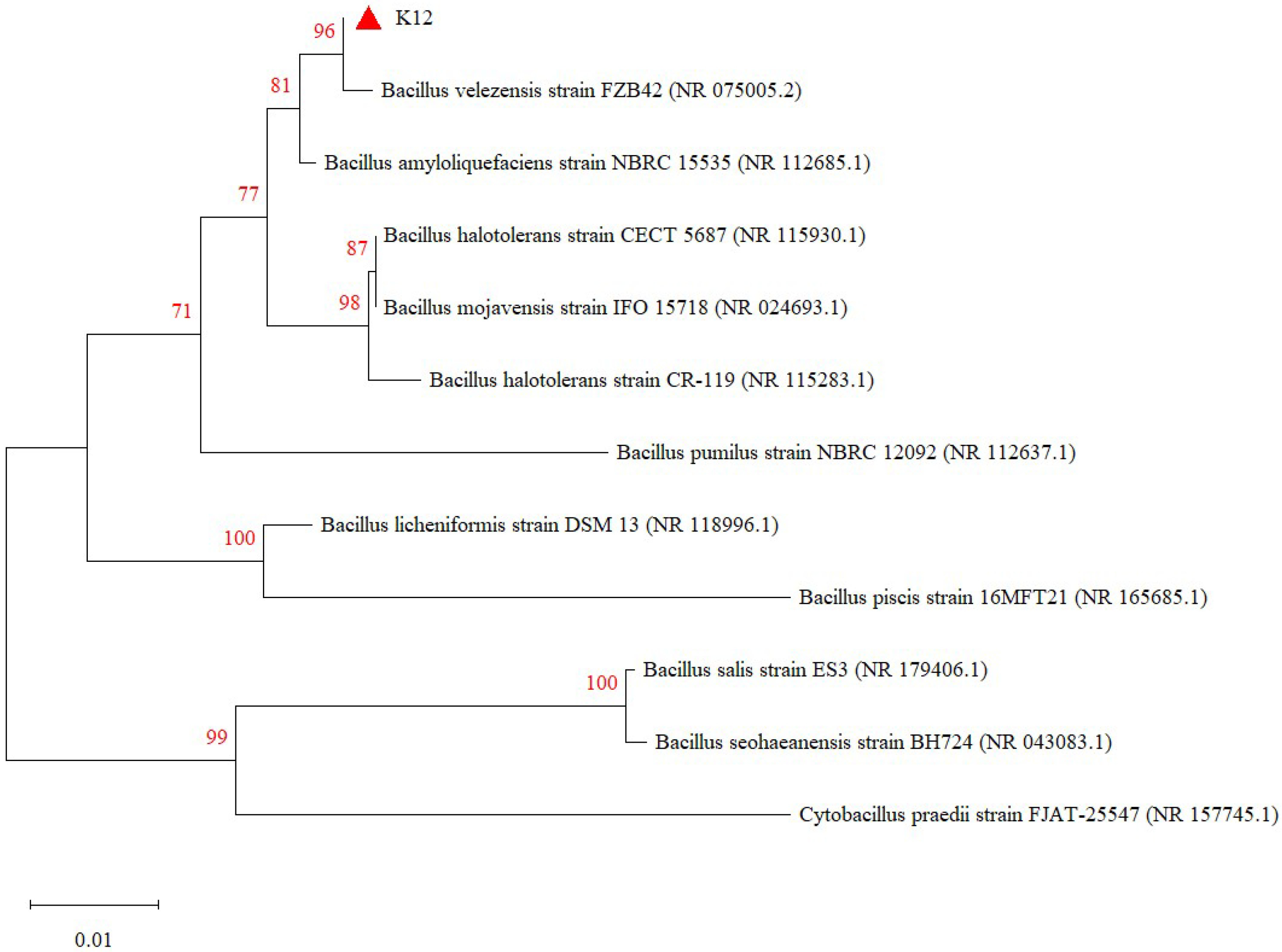
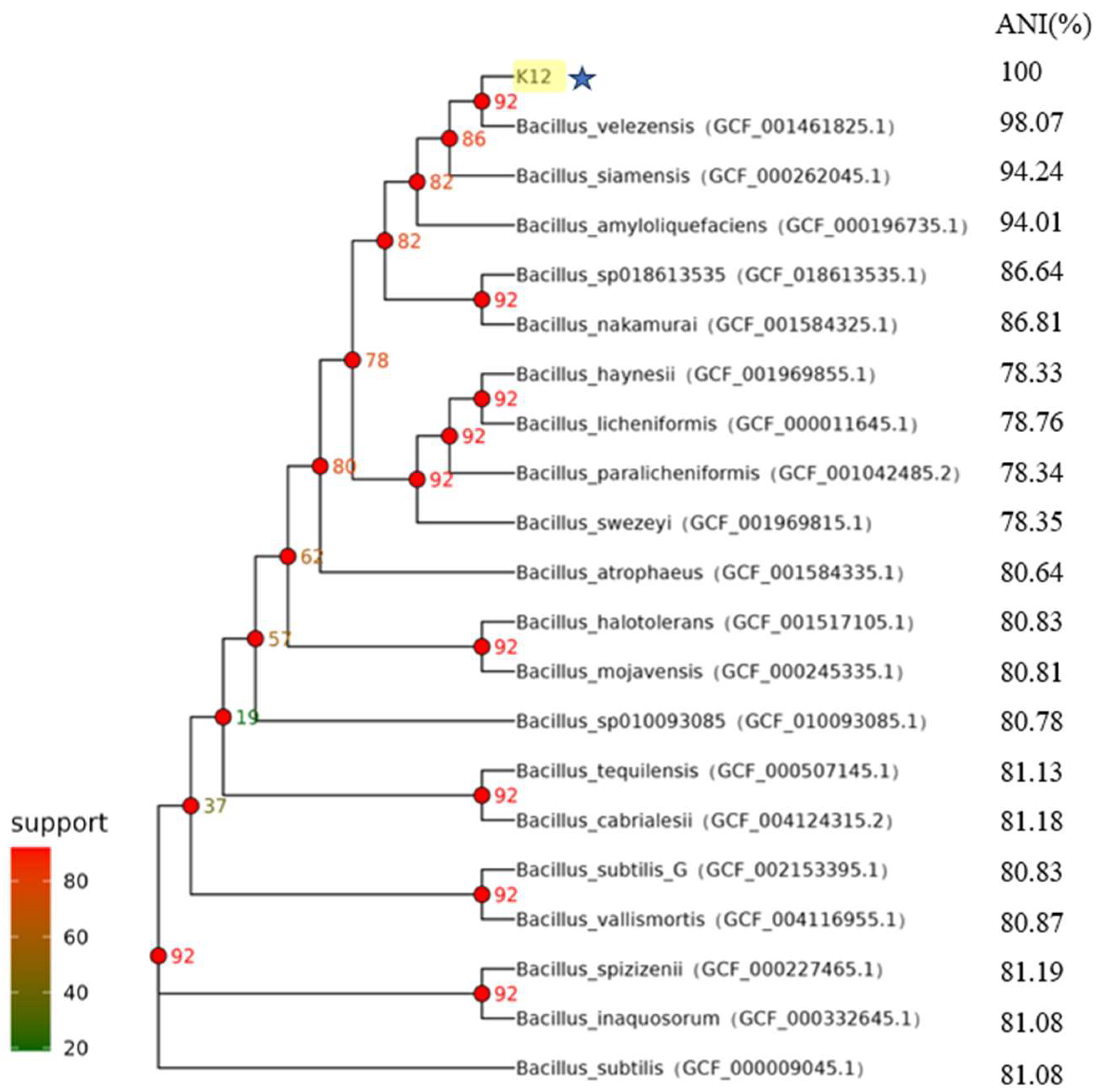
3.1. Identification of Strain K12

The test strains were isolated on LB solid medium using the line dilution method, and the inoculated medium was incubated in a constant temperature incubator at 37 °C for 14 h. The morphology of the colonies formed on the medium’s surface was observed. Strain K12 grew well on the LB solid medium, with milky-white colonies and slightly irregular edges (Figure 1A). Strain K12 was Gram-stained and observed under the light microscope as short purple rods, indicating Gram-positive bacteria (Figure 1B). The 16S rDNA sequence was amplified and sequenced, followed by a gene sequence comparison with the NCBI database. Phylogenetic tree analysis indicated that the target strain was the most closely related to B. velezensis FZB42 (Figure 2). ANI calculates the percentage of nucleotide identity between two strains by comparing the similarity of their whole-genome sequences. Typically, an ANI value ≥95% indicates that the two strains belong to the same species [42]. The ANI homology indices were calculated between strain K12 and 20 closely related species (Figure 3), and the results showed that strain K12 had the highest homology with the species Bacillus velezensis (ANI = 98.07%). Based on the morphological features and molecular analysis, strain K12 was confirmed as Bacillus velezensis.

3.2. Characterization of the Growth of B. velezensis K12

B. velezensis K12 was inoculated into LB liquid medium to monitor its growth dynamics and pH fluctuations over 36 h. Samples were collected at regular intervals to chart the growth curve (Figure 4A) and the pH variation curve (Figure 4B) of strain B. velezensis K12 from the moment of inoculation until 36 h later. During the initial phase, from 0 to 2 h, B. velezensis K12 exhibited slow growth. Subsequently, after 2 h, the strain entered the logarithmic growth phase, during which its concentration increased rapidly. This rapid growth continued until 8 h of incubation, when the strain reached the stabilization phase, marking a point where its population no longer increased significantly. Concurrently, the pH value of the medium was measured at various time points, revealing a range of 7 to 8.5 throughout the experiment.
B. velezensis K12 exhibited minimal growth within the pH range of 1.5 to 2.5. However, a gradual increase in growth was observed as the pH surpassed 3.5, as illustrated in Figure 4C. Furthermore, as the concentration of bile salts increased, the development of B. velezensis K12 was progressively inhibited. Specifically, growth was virtually absent after adding 0.06% bile salts, as shown in Figure 4D. After incubation, no hemolytic ring appeared around a single colony of B. velezensis K12 on sheep blood agar, indicating that it was not hemolytic (Supplementary Figure S1). When subjected to an artificial gastric solution for 3 h, B. velezensis K12 demonstrated a survival rate of merely 0.01%, indicating almost no growth. Conversely, after being exposed to an artificial intestinal solution for 3 h, the strain exhibited a survival rate of 40.70% (Table 1).
B. velezensis K12 was extremely sensitive to cefotaxime, florfenicol, ciprofloxacin, cotrimoxazole, doxycycline, and gentamicin (inhibitory circle diameter > 20.00 mm) (Table 2). Images of antibiotic susceptibility analysis of B. velezensis K12 are shown in the Supplementary Materials (Figure S2).

3.3. In Vitro Bacteriostatic Activity of B. Velezensis K12

The fermentation broth of B. velezensis K12 exhibited varying inhibitory activity against seven indicator bacteria (p < 0.05). As the broth was diluted, its inhibitory potential decreased. Notably, the fermentation broth was highly sensitive to E. coli CVCC25922, E. coli K88, Salmonella CVCC519, Staphylococcus aureus CVCC1882, and Vibrio parahaemolyticus CICC23924, with a minimum inhibitory dilution of 1:10,000. The minimum inhibitory dilution for Bacillus cereus CICC21290 was 1:4000, and the minimum inhibitory dilution for Clostridium perfringens CVCC66 was 1:1000 (Table 3). The diameter of the B. velezensis K12 inhibition circle decreased linearly with the increasing dilution gradient of the indicator bacteria, except for Staphylococcus aureus CVCC1822 and Bacillus cereus CICC21290 (line p-value < 0.001). Images of the circle-of-inhibition diameter of the B. velezensis K12 bacterial solution against seven indicator bacteria are shown in the Supplementary Materials (Figure S3).
After isolating the organism and supernatant of B. velezensis K12, it was found that the organism exhibited bacteriostatic activity against all indicator bacteria (p < 0.05). In addition, the sterile supernatant demonstrated bacteriostatic activity against E. coli K88, Bacillus cereus CICC21290, Clostridium perfringens CVCC66, and Vibrio parahaemolyticus CICC23924 (p < 0.05). Notably, the inhibitory effect of whole bacteria was more potent than that of sediment, and the inhibitory effect of sediment was more substantial than that of the sterile supernatant (p < 0.05) (Table 4). Images of the circle-of-inhibition diameters of different components of B. velezensis K12 against seven indicator bacteria are shown in the Supplementary Materials (Figure S4). Furthermore, after inactivating B. velezensis K12, no inhibitory effect on the indicator bacterium E. coli K88 was observed (p < 0.05) (Table 5).

3.4. Whole-Genome Sequencing Results of B. velezensis K12

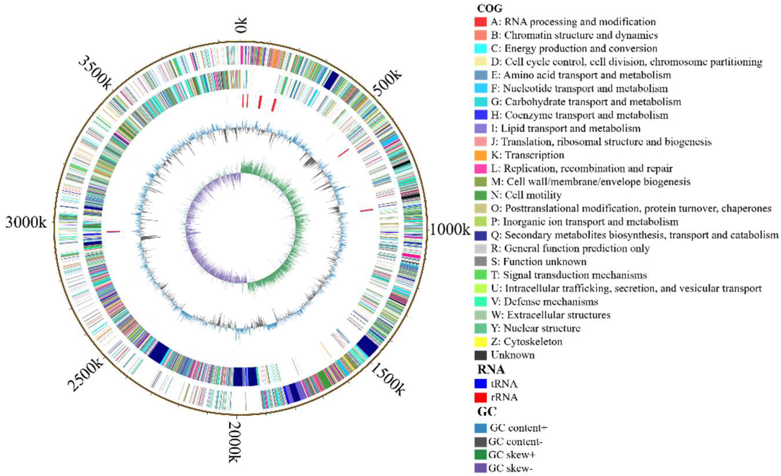
3.4.1. Genome Composition of B. velezensis K12

The genome of B. velezensis K12 comprises a circular sequence measuring 3,973,105 bp long, with an average GC content of 46.69% (Table 6). It includes 3913 protein-coding genes, which constitute 88.68% of the genome, and 97 ncRNAs, 86 tRNAs, and 27 rRNAs. The genome of B. velezensis K12 included 121 long terminal repeats, 35 long interspersed nuclear elements, 17 short interspersed elements, 19 prophages, and 21 genomics islands. The distribution of genes predicted by B. velezensis K12 gene islands is mapped in the Supplementary Materials (Figure S5). No CRISPR elements were detected in the genome. The circular genome diagram illustrates various features. Starting from the innermost ring and moving outward, the following features are noticed: GC skew in the first ring; GC content in the second ring; tRNA and rRNA positions in the third and fourth rings, respectively [61]; CDSs on the positive and negative strands of the fifth and sixth rings, with colors indicating different COG classifications; and the chromosomal karyotype in the outermost ring (Figure 5). The complete genome sequence of strain B. velezensis K12 has been submitted to GenBank (NCBI) with accession number CP171691.

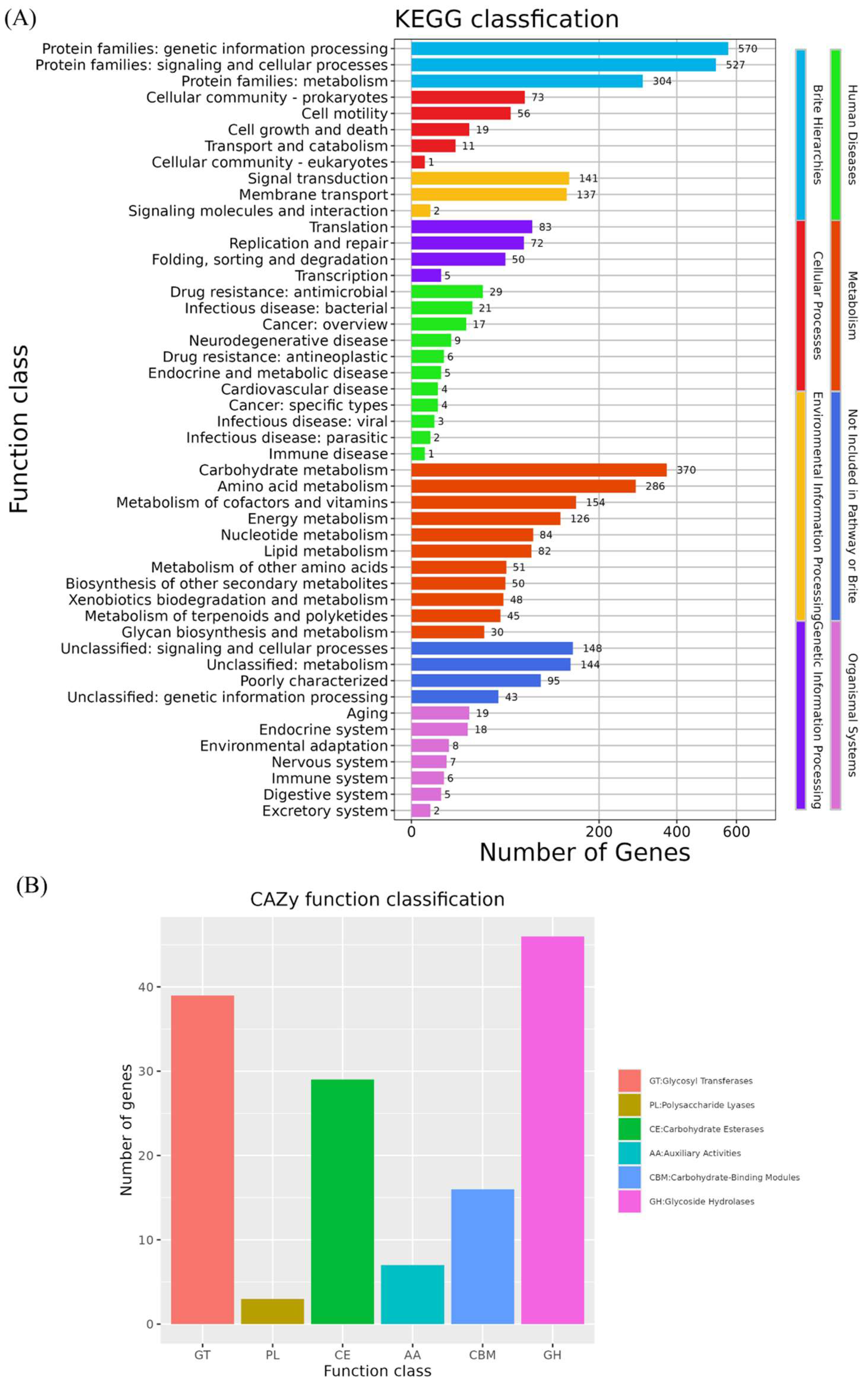
3.4.2. Functional Annotation of B. velezensis K12 Genome

By comparing the amino acid sequences of the B. velezensis K12 genome to the GO database, we identified 2199 functional genes. Among these, 736 genes were related to molecular functions, 812 to biological processes, and 651 to cellular components (Figure 6A). Specifically, in terms of molecular functions, 88 genes were classified as having oxidoreductase activity and 74 as having ion-binding functions. Within biological processes, 361 genes were classified as being involved in cellular nitrogen compound metabolism and 331 in biosynthesis. For cellular components, 600 genes were classified as cellular and 372 as intracellular.
A total of 3706 protein-coding genes in the B. velezensis K12 genome were annotated in the COG database (Figure 6B). Among these, the largest group consisted of 893 genes with unknown functions. The functional categories with high numbers of annotated genes were as follows: amino acid transport and metabolism (E) with 332 genes, transcription (K) with 310 genes, and carbohydrate transport and metabolism (G) with 256 genes.
The genome of B. velezensis K12 contains 3973 genes annotated through the KEGG database (Figure 7A). Among them, 570 genes were related to genetic information processing, making this the most represented category. This category was followed by annotations related to environmental information processing, genetic information processing, cellular processes, human diseases, and organic systems. Other frequently annotated functions included signaling and cellular processes (527 genes), carbohydrate metabolism (370 genes), protein family metabolism (307 genes), and amino acid metabolism (286 genes).
The genome of B. velezensis K12 has 140 genes annotated in the CAZy database (Figure 7B). The data indicated the presence of 46 genes associated with glycoside hydrolases (GHs) and 39 genes associated with glycosyltransferases (GTs). In addition, 29 genes were associated with carbohydrate esterases (CEs), and 16 were linked to carbohydrate-binding modules (CBMs). There are seven genes associated with auxiliary activities (AAs) and three genes associated with polysaccharide lyases (PLs).
CARD is the most widely used and comprehensive database of bacterial drug-resistant genes. Through the annotation of the CARD database, with IDENTITY ≥ 80% as the screening condition, the genome of B. velezensis K12 had six drug-resistant genes, among which lmrB, clbA, ykkD, and rpoB genes had multi-drug resistance, and it was hypothesized that B. velezensis K12 might have specific drug resistance (Table 7).
The genome of B. velezensis K12 had four virulence factors, including the hlyIII virulence gene related to exotoxin, the dhbE virulence gene related to nutrient metabolism, and dep/capD and capB related to immune regulation, as annotated by the VFDB database with IDENTITY ≥ 80% as the screening condition (Table 8).
Thirteen secondary metabolite gene clusters were identified in the genome of strain B. velezensis K12 by AntiSMASH 7.0 software, of which 10 were associated with synthesizing known repressor secondary metabolites (Table 9). Of these, four gene clusters were predicted to have 95%, 100%, 100%, and 100% synthetic similarity to fengycin, surfactin, bacillothiazol, and bacillibactin in non-ribosomal peptides (NRPs), respectively. Three clusters were predicted to have 100% synthetic similarity to the polyketide synthases (PKSs) in the synthesis of macrolactin H, bacillaene, and difficidin; two clusters of genes had 100% and 91% similarity to lanthipeptide and plantazolicin in ribosomal synthesis and post-translationally modified peptides (RiPPs), respectively; and one cluster of genes had 100% similarity to bacillinolysin. In addition, there are three unknown gene clusters in the B. velezensis K12 genome, including two terpenes and one polyketide (T3PKS), suggesting that the B. velezensis K12 gene cluster may synthesize new bacteriostatic substances.
Whole-genome sequence analysis showed that the B. velezensis K12 genome contains several putative probiotic genes for acid tolerance, bile salt tolerance, adhesion aggregation, and antioxidant resistance that may help B. velezensis K12 to survive in the gastrointestinal environment (Table 10).

4. Discussion

The dire resistance to traditional feed antibiotics has accelerated the pursuit of antibiotic alternatives. Recently, probiotic products have garnered increasing attention in the livestock and poultry industries. In 2019, the European Food Safety Authority (EFSA) granted B. velezensis Qualified Presumption of Safety (QPS) status, proving that it does not have toxicological potential and has aminoglycoside production capacity [62]. Nonetheless, B. velezensis has not received substantial recognition or endorsement as a safe microbial feed additive, likely due to the paucity of data regarding its biosafety and probiotic properties at phenotypic and genetic levels [63]. This study assessed the probiotic potential and safety profile of B. velezensis K12 through in vitro experiments complemented by genomic analyses.
Antimicrobial properties against pathogenic bacteria are the main selection criteria for probiotics in animal feed and nutrition [64]. In this study, the strain K12, with a strong inhibitory effect against bacteria, was screened and identified as Bacillus velezensis. Escherichia coli, Salmonella spp., Staphylococcus aureus, and Clostridium perfringens are common pathogens in poultry [65]. Vibrio parahaemolyticus is the most critical aquatic pathogen in the genus Vibrio and poses a significant hazard to aquaculture [66]. Bacillus cereus is one of the essential human enteric pathogens [67]. B. velezensis K12 showed varying degrees of inhibitory effects on the seven pathogenic bacteria mentioned above, and its whole bacteria exhibited greater inhibitory potential than sediments and supernatants, with the inhibitory effect ceasing after inactivation. These findings hint that the bacteriostatic effect of B. velezensis K12 may come from the secretion of heat-intolerant bacteriostatic substances by live bacteria. These inhibitory substances may be NRPs, PKS, bacilysin, and RiPPs (Table 9). Among them, fengycin, difficidin, bacilysin, macrolactin, and bacillibactin have been reported to have broad-spectrum antibacterial effects [68,69,70,71]. These antibacterial compounds have been reported to operate in three mechanisms: cell wall disruption, the control of critical cell machinery DNA and RNA, and the inhibition of enzyme activity [11].
Probiotics must endure the oxidative challenges present in the gastrointestinal tract to extend benefits to the host [72]. The whole-genome sequencing revealed genes associated with acid and bile tolerance in the B. velezensis K12 genome (Table 10). Based on the annotation results, B. velezensis K12 utilizes eight ATP synthase atp genes, organized in an operon to sustain H+ homeostasis in acidic conditions by hydrolyzing ATP to extrude protons from the cytoplasm [63]. The Na+/H+ antiporter and Na+ (K+, Li+, and/or alkali)/H+ antiporter genes play an essential role in Na+ resistance, pH homeostasis, and osmoregulation, enabling bacterial survival in acidic environments [73]. The enzyme bile salt hydrolase was encoded by bsh genes (bshA, bshB1, bshC), which play an essential role in the cholesterol-lowering effect by hydrolyzing conjugated bile salts into amino acids and deconjugated bile salts [74]. In addition, the genes encoding dnaK, dnaJ, pryG, eno, and clp protease chaperones are up-regulated in acid shock, facilitating the strain’s tolerance to thermal and osmotic stress while aiding in the repair of damaged proteins [75,76]. By measuring the growth of the strain under different conditions, it was found that B. velezensis K12 was not adapted to the artificial gastric fluid environment compared to the artificial intestinal fluid, which is consistent with the results of B. velezensis K12 intolerance in environments with pH below 3.5. In the future, B. velezensis K12 could be delivered to the intestine with the help of embedding technology or enteric formulations.
Mucus adherence and penetration potential are crucial probiotic characteristics facilitating the transitory colonization of the host intestinal epithelial surface and the competitive exclusion of pathogens [77]. The B. velezensis K12 genome encodes numerous reported surface proteins implicated in adhesion and aggregation (Table 10), including mucus adhesion domain protein (lspA, glnH, tuf), fibronectin-binding protein (fbp), Bacillus subtilis matrix protein TasA (tasA 1), teichoic-acid (tagO), and exopoly saccharides (eps, lip, galE) [77,78,79,80,81]. Multiple investigations have demonstrated that Bacillus velezensis has probiotic properties that enhance the antioxidant capacity of the host [82,83]. In this study, we identified numerous genes within B. velezensis K12 that encode glutathione peroxidase (bshA), alkyl hydroperoxide reductase (ahp), catalases (kat), and superoxide dismutase (sodF), which also confirmed the antioxidant properties of Bacillus velezensis.
Probiotic candidates must be analyzed for safety, including assessments of hemolysis, resistance, and virulence. The surfactin and hlyIII genes related to hemolysis were annotated in the B. velezensis K12 genome. As an antimicrobial peptide, surfactin exerts hemolytic activity by modifying the integrity of cell membranes [84]. It produces surface-active effects that are not pathogenic. The hlyIII gene encoding hemolysin alone did not determine bacterial pathogenicity and was not found on transferable plasmids. It has been shown that B. velezensis FCW2 MCC4686, which carries the hlyIII gene, did not cause mortality or disease symptoms in the zebrafish model, suggesting that the strain is non-pathogenic [59]. In addition, in vitro hemolysis tests showed that B. velezensis K12 did not induce the hemolysis of hemocytes on sheep blood agar and was not hemolytic. Antibiotic resistance genes associated with antibiotics, such as aminoglycoside (rpsE) and tetracycline (rpoB) in the B. velezensis K12 genome, were identified by CARD analysis. However, in vitro antibiotic susceptibility testing showed that B. velezensis K12 was susceptible to six antibiotics, including gentamicin and tetracycline. This suggests that the B. velezensis K12 resistance genes may lead to an inherent resistance mechanism during microbial formation, but this is not generally transferred between organisms [85]. Although the genome of B. velezensis K12 has four virulence factors annotated by the VFDB, they are not considered genuinely harmful. B. velezensis TS5, which carries virulence genes similar to B. velezensis K12, related to immunomodulatory and nutritional/metabolic factors, is safe in mice [86]. In addition, common virulence genes of Bacillus enterotoxins, including hemolysin A (hlyA), hemolysin II (hlyII), hemolysin BL (hbl), non-hemolytic enterotoxin (Nhe), enterotoxin T (bceT), and cytotoxin K (CytK), were not found in the B. velezensis K12 genome [87,88]. The above safety analysis shows that B. velezensis K12 has a favorable in vitro safety profile. However, to fully understand its safety and effects in vivo, additional animal studies are necessary in the future.

5. Conclusions

These in vitro and genomic studies provide valuable insights into the essential properties of B. velezensis K12. This strain possesses potent bacteriostatic properties and has exhibited safety in vitro. However, the specific mechanism of bacterial inhibition needs to be further investigated. B. velezensis K12 was sensitive to six antibiotics and had acid tolerance. Genome mining revealed that B. velezensis K12 contains genes encoding host intestinal adhesion and antioxidants. The results of this study prove that B. velezensis K12 is a promising candidate for further probiotic development.

Supplementary Materials

The following supporting information can be downloaded at: https://www.mdpi.com/article/10.3390/ani15060798/s1, Figure S1: Hemolysis analysis of B. velezensis K12. Figure S2: Antibiotic susceptibility analysis of B. velezensis K12. Figure S3: Circle-of-inhibition diameter of B. velezensis K12 bacterial solution against seven indicator bacteria. Figure S4: Circle-of-inhibition diameters of different components of B. velezensis K12 against seven indicator bacteria. Figure S5: Map of gene distribution on genetic islands of B. velezensis K12.

Author Contributions

Conceptualization, J.Y.; methodology, Y.H., Y.T. and L.W.; investigation, Y.T., T.L., Y.H., L.W. and X.L.; formal analysis: Y.T.; Visualization: Y.T., T.L. and Y.H.; validation: Y.T. and T.L.; writing—original draft preparation, Y.T.; writing—review and editing, J.Y. and R.Y. All authors have read and agreed to the published version of the manuscript.

Funding

This work was supported by the Scientific Research startup Funding (College of Veterinary Medicine, China Agricultural University, No. 10050523).

Institutional Review Board Statement

Not applicable.

Informed Consent Statement

Not applicable.

Data Availability Statement

All data are contained within the article.

Conflicts of Interest

Authors Liangliang Wu and Xiaobo Liu were employed by the company Ningxia Eppen Biotech Co., Ltd. The remaining author declares that the research was conducted in the absence of any commercial or financial relationships that could be construed as a potential conflict of interest. The funders had no role in the design of the study; in the collection, analyses, or interpretation of data; in the writing of the manuscript; or in the decision to publish the results.

Abbreviations

The following abbreviations are used in this manuscript:
ISAPPInternational Scientific Association for Probiotics and Prebiotics
E. coliEscherichia coli
LBLuria–Bertani
CLSIAmerican Society for Clinical Laboratory Standardization
ANIAverage Nucleotide Identity
UBCGUp-to-Date Bacterial Core Gene
KEGGKyoto Encyclopedia of Genes and Genomes
COGsClusters of Orthologous Groups
GOGene Ontology
CAZyCarbohydrate-Active Enzymes database
CARDComprehensive Antibiotic Research Database
VFDBVirulence Factors of Pathogenic Bacteria Database
antiSMASHAntibiotics and Secondary Metabolite Analysis Shell
ANOVAOne-way analysis of variance
NCBINational Center for Biotechnology Information
NRPsNon-ribosomal peptides
PKSPolyketide synthases
RiPPsRibosomal synthesis and post-translationally modified peptides
EFSAEuropean Food Safety Authority
QPSQualified Presumption of Safety

References

  1. Cotter, P.D.; Ross, R.P.; Hill, C. Bacteriocins—A viable alternative to antibiotics? Nat. Rev. Microbiol. 2013, 11, 95–105. [Google Scholar] [CrossRef] [PubMed]
  2. Corrigan, A.; de Leeuw, M.; Penaud-Frézet, S.; Dimova, D.; Murphy, R.A. Phylogenetic and functional alterations in bacterial community compositions in broiler ceca as a result of mannan oligosaccharide supplementation. Appl. Environ. Microbiol. 2015, 81, 3460–3470. [Google Scholar] [CrossRef] [PubMed]
  3. de Vries, S.; Pustjens, A.M.; Kabel, M.A.; Salazar-Villanea, S.; Hendriks, W.H.; Gerrits, W.J. Processing technologies and cell wall degrading enzymes to improve nutritional value of dried distillers grain with solubles for animal feed: An in vitro digestion study. J. Agric. Food Chem. 2013, 61, 8821–8828. [Google Scholar] [CrossRef]
  4. Millet, S.; Maertens, L. The European ban on antibiotic growth promoters in animal feed: From challenges to opportunities. Vet. J. 2011, 187, 143–144. [Google Scholar] [CrossRef]
  5. Hill, C.; Guarner, F.; Reid, G.; Gibson, G.R.; Merenstein, D.J.; Pot, B.; Morelli, L.; Canani, R.B.; Flint, H.J.; Salminen, S.; et al. Expert consensus document. The International Scientific Association for Probiotics and Prebiotics consensus statement on the scope and appropriate use of the term probiotic. Nat. Rev. Gastroenterol. Hepatol. 2014, 11, 506–514. [Google Scholar] [CrossRef] [PubMed]
  6. Yousaf, S.; Nouman, H.M.; Ahmed, I.; Husain, S.; Waseem, M.; Nadeem, S.; Tariq, M.; Sizmaz, O.; Chudhry, M.F.Z. A Review of Probiotic Applications in Poultry: Improving Immunity and Having Beneficial Effects on Production and Health. Adv. Microbiol. 2022, 61, 115–123. [Google Scholar] [CrossRef]
  7. Bilal, M.; Si, W.; Barbe, F.; Chevaux, E.; Sienkiewicz, O.; Zhao, X. Effects of novel probiotic strains of Bacillus pumilus and Bacillus subtilis on production, gut health, and immunity of broiler chickens raised under suboptimal conditions. Poult. Sci. 2021, 100, 100871. [Google Scholar] [CrossRef]
  8. Tran, C.; Cock, I.E.; Chen, X.; Feng, Y. Antimicrobial Bacillus: Metabolites and Their Mode of Action. Antibiotics 2022, 11, 88. [Google Scholar] [CrossRef]
  9. Ruiz-García, C.; Béjar, V.; Martínez-Checa, F.; Llamas, I.; Quesada, E. Bacillus velezensis sp. nov., a surfactant-producing bacterium isolated from the river Vélez in Málaga, southern Spain. Int. J. Syst. Evol. Microbiol. 2005, 55, 191–195. [Google Scholar] [CrossRef]
  10. Ye, M.; Tang, X.; Yang, R.; Zhang, H.; Li, F.; Tao, F.; Li, F.; Wang, Z. Characteristics and Application of a Novel Species of Bacillus: Bacillus velezensis. ACS Chem. Biol. 2018, 13, 500–505. [Google Scholar] [CrossRef]
  11. Khalid, F.; Khalid, A.; Fu, Y.; Hu, Q.; Zheng, Y.; Khan, S.; Wang, Z. Potential of Bacillus velezensis as a probiotic in animal feed: A review. J. Microbiol. 2021, 59, 627–633. [Google Scholar] [CrossRef] [PubMed]
  12. Johny, L.C.; Suresh, P.V. Complete genome sequencing and strain characterization of a novel marine Bacillus velezensis FTL7 with a potential broad inhibitory spectrum against foodborne pathogens. World J. Microbiol. Biotechnol. 2022, 38, 164. [Google Scholar] [CrossRef] [PubMed]
  13. Zhang, L.; Liu, Z.; Pu, Y.; Zhang, B.; Wang, B.; Xing, L.; Li, Y.; Zhang, Y.; Gu, R.; Jia, F.; et al. Antagonistic Strain Bacillus velezensis JZ Mediates the Biocontrol of Bacillus altitudinis m-1, a Cause of Leaf Spot Disease in Strawberry. Int. J. Mol. Sci. 2024, 25, 8872. [Google Scholar] [CrossRef] [PubMed]
  14. Dutilloy, E.; Arias, A.A.; Richet, N.; Guise, J.F.; Duban, M.; Leclere, V.; Selim, S.; Jacques, P.; Jacquard, C.; Clément, C.; et al. Bacillus velezensis BE2 controls wheat and barley diseases by direct antagonism and induced systemic resistance. Appl. Microbiol. Biotechnol. 2024, 108, 64. [Google Scholar] [CrossRef]
  15. Ye, Q.; Zhong, Z.; Chao, S.; Liu, L.; Chen, M.; Feng, X.; Wu, H. Antifungal Effect of Bacillus velezensis ZN-S10 against Plant Pathogen Colletotrichum changpingense and Its Inhibition Mechanism. Int. J. Mol. Sci. 2023, 24, 16694. [Google Scholar] [CrossRef]
  16. Yang, H.; Du, D.; Zhang, Q.; Teame, T.; Wang, A.; Hao, Q.; Liu, S.; Ding, Q.; Yao, Y.; Yang, Y.; et al. Dietary Bacillus velezensis T23 fermented products supplementation improves growth, hepatopancreas and intestine health of Litopenaeus vannamei. Fish Shellfish. Immunol. 2024, 149, 109595. [Google Scholar] [CrossRef]
  17. Chang, X.; Yun, L.; Liu, Z.; Shen, Y.; Feng, S.; Yang, G.; Meng, X. Antagonistic Effects and the Underlying Mechanisms of Bacillus velezensis and its Antibacterial Peptide LCI Against Aeromonas hydrophila Infection in Largemouth Bass. Probiotics Antimicrob. Proteins 2024, 1–18. [Google Scholar] [CrossRef]
  18. Li, C.; Li, S.; Dang, G.; Jia, R.; Chen, S.; Deng, X.; Liu, G.; Beckers, Y.; Cai, H. Screening and characterization of Bacillus velezensis LB-Y-1 toward selection as a potential probiotic for poultry with multi-enzyme production property. Front. Microbiol. 2023, 14, 1143265. [Google Scholar] [CrossRef]
  19. Dong, H.; Gao, R.; Dong, Y.; Yao, Q.; Zhu, H. Bacillus velezensis RC116 Inhibits the Pathogens of Bacterial Wilt and Fusarium Wilt in Tomato with Multiple Biocontrol Traits. Int. J. Mol. Sci. 2023, 24, 8527. [Google Scholar] [CrossRef]
  20. Brutscher, L.M.; Gebrechristos, S.; Garvey, S.M.; Spears, J.L. Genetic and Phenotypic Characterization of Bacillus velezensis Strain BV379 for Human Probiotic Applications. Microorganisms 2024, 12, 436. [Google Scholar] [CrossRef]
  21. Larsen, I.S.; Chenaux, M.; Collins, F.W.J.; Mandic, A.; Hansen, L.B.S.; Lauridsen, C.A.S.; Haller, R.F.; Elvig-Jørgensen, S.; Horwell, E.; Christiansen, J.; et al. Bacillus velezensis DSM 33864 reduces Clostridioides difficile colonization without disturbing commensal gut microbiota composition. Sci. Rep. 2023, 13, 14941. [Google Scholar] [CrossRef] [PubMed]
  22. Liu, G.; Kong, Y.; Fan, Y.; Geng, C.; Peng, D.; Sun, M. Whole-genome sequencing of Bacillus velezensis LS69, a strain with a broad inhibitory spectrum against pathogenic bacteria. J. Biotechnol. 2017, 249, 20–24. [Google Scholar] [CrossRef] [PubMed]
  23. Yu, C.; Chen, H.; Zhu, L.; Song, Y.; Jiang, Q.; Zhang, Y.; Ali, Q.; Gu, Q.; Gao, X.; Borriss, R.; et al. Profiling of Antimicrobial Metabolites Synthesized by the Endophytic and Genetically Amenable Biocontrol Strain Bacillus velezensis DMW1. Microbiol. Spectr. 2023, 11, e0003823. [Google Scholar] [CrossRef] [PubMed]
  24. Zhang, X.; He, X.; Chen, J.; Li, J.; Wu, Y.; Chen, Y.; Yang, Y. Whole-Genome Analysis of Termite-Derived Bacillus velezensis BV-10 and Its Application in King Grass Silage. Microorganisms 2023, 11, 2697. [Google Scholar] [CrossRef]
  25. Ablimit, N.; Zheng, F.; Wang, Y.; Wen, J.; Wang, H.; Deng, K.; Cao, Y.; Wang, Z.; Jiang, W. Bacillus velezensis strain NA16 shows high poultry feather-degrading efficiency, protease and amino acid production. Ecotoxicol. Environ. Saf. 2024, 278, 116353. [Google Scholar] [CrossRef] [PubMed]
  26. Klindworth, A.; Pruesse, E.; Schweer, T.; Peplies, J.; Quast, C.; Horn, M.; Glöckner, F.O. Evaluation of general 16S ribosomal RNA gene PCR primers for classical and next-generation sequencing-based diversity studies. Nucleic Acids Res. 2012, 41, e1. [Google Scholar] [CrossRef]
  27. Sayers, E.W.; Bolton, E.E.; Brister, J.R.; Canese, K.; Chan, J.; Comeau, D.C.; Connor, R.; Funk, K.; Kelly, C.; Kim, S.; et al. Database resources of the national center for biotechnology information. Nucleic Acids Res. 2022, 50, D20–D26. [Google Scholar] [CrossRef]
  28. Tamura, K.; Stecher, G.; Kumar, S. MEGA11: Molecular Evolutionary Genetics Analysis Version 11. Mol. Biol. Evol. 2021, 38, 3022–3027. [Google Scholar] [CrossRef]
  29. Katoh, K.; Standley, D.M. MAFFT multiple sequence alignment software version 7: Improvements in performance and usability. Mol. Biol. Evol. 2013, 30, 772–780. [Google Scholar] [CrossRef]
  30. Renschler, M.A.; Wyatt, A.; Anene, N.; Robinson-Hill, R.; Pickerill, E.S.; Fox, N.E.; Griffith, J.A.; McKillip, J.L. Using nitrous acid-modified de Man, Rogosa, and Sharpe medium to selectively isolate and culture lactic acid bacteria from dairy foods. J. Dairy Sci. 2020, 103, 1215–1222. [Google Scholar] [CrossRef]
  31. Soni, R.; Keharia, H.; Shah, K.; Jain, N. Phenotypic characterization and genome analysis reveal the probiotic potential of a banyan endophyte Bacillus velezensis K1. J. Appl. Microbiol. 2023, 134, lxac057. [Google Scholar] [CrossRef] [PubMed]
  32. Reyes-Cortes, J.L.; Azaola-Espinosa, A.; Lozano-Aguirre, L.; Ponce-Alquicira, E. Physiological and Genomic Analysis of Bacillus pumilus UAMX Isolated from the Gastrointestinal Tract of Overweight Individuals. Microorganisms 2021, 9, 1076. [Google Scholar] [CrossRef]
  33. Tanvi, S.; Ravichandra, V.; Madhur, D.S.; Agampodi Promoda, P.; Stephen, T.; Roger, S.; Rajaraman, E. Probiotic Bacillus coagulans MTCC 5856 spores exhibit excellent in-vitro functional efficacy in simulated gastric survival, mucosal adhesion and immunomodulation. J. Funct. Foods 2019, 52, 100–108. [Google Scholar] [CrossRef]
  34. Humphries, R.; Bobenchik, A.M.; Hindler, J.A.; Schuetz, A.N. Overview of Changes to the Clinical and Laboratory Standards Institute Performance Standards for Antimicrobial Susceptibility Testing, M100, 31st Edition. J. Clin. Microbiol. 2021, 59, e0021321. [Google Scholar] [CrossRef] [PubMed]
  35. Yi-Zhou, X.; Xin-Yue, L.; Hai-Long, Z.; Jun-Yuan, C.; Lian-Bing, L.; Qi-Lin, Z. Purification and antibacterial properties of a novel bacteriocin against Escherichia coli from Bacillus subtilis isolated from blueberry ferments. LWT 2021, 146, 111456. [Google Scholar] [CrossRef]
  36. Mark, D.; Tairo, F.; Ndunguru, J.; Kweka, E.; Saggaf, M.; Bachwenkizi, H.; Chiunga, E.; Lusana, J.L.; Sikazwe, G.; Maghembe, R. Assessing the effect of sample storage time on viral detection using a rapid and cost-effective CTAB-based extraction method. Plant Methods 2024, 20, 64. [Google Scholar] [CrossRef]
  37. Safar, H.A.; Alatar, F.; Nasser, K.; Al-Ajmi, R.; Alfouzan, W.; Mustafa, A.S. The impact of applying various de novo assembly and correction tools on the identification of genome characterization, drug resistance, and virulence factors of clinical isolates using ONT sequencing. BMC Biotechnol. 2023, 23, 26. [Google Scholar] [CrossRef]
  38. Chen, S. Ultrafast one-pass FASTQ data preprocessing, quality control, and deduplication using fastp. iMeta 2023, 2, e107. [Google Scholar] [CrossRef]
  39. Luo, R.; Liu, B.; Xie, Y.; Li, Z.; Huang, W.; Yuan, J.; He, G.; Chen, Y.; Pan, Q.; Liu, Y.; et al. SOAPdenovo2: An empirically improved memory-efficient short-read de novo assembler. GigaScience 2012, 1, 18. [Google Scholar] [CrossRef]
  40. Wick, R.R.; Judd, L.M.; Gorrie, C.L.; Holt, K.E. Unicycler: Resolving bacterial genome assemblies from short and long sequencing reads. PLoS Comput. Biol. 2017, 13, e1005595. [Google Scholar] [CrossRef]
  41. Walker, B.J.; Abeel, T.; Shea, T.; Priest, M.; Abouelliel, A.; Sakthikumar, S.; Cuomo, C.A.; Zeng, Q.; Wortman, J.; Young, S.K.; et al. Pilon: An integrated tool for comprehensive microbial variant detection and genome assembly improvement. PLoS ONE 2014, 9, e112963. [Google Scholar] [CrossRef] [PubMed]
  42. Jain, C.; Rodriguez, R.L.; Phillippy, A.M.; Konstantinidis, K.T.; Aluru, S. High throughput ANI analysis of 90K prokaryotic genomes reveals clear species boundaries. Nat. Commun. 2018, 9, 5114. [Google Scholar] [CrossRef]
  43. Na, S.I.; Kim, Y.O.; Yoon, S.H.; Ha, S.M.; Baek, I.; Chun, J. UBCG: Up-to-date bacterial core gene set and pipeline for phylogenomic tree reconstruction. J. Microbiol. 2018, 56, 280–285. [Google Scholar] [CrossRef] [PubMed]
  44. Besemer, J.; Lomsadze, A.; Borodovsky, M. GeneMarkS: A self-training method for prediction of gene starts in microbial genomes. Implications for finding sequence motifs in regulatory regions. Nucleic Acids Res. 2001, 29, 2607–2618. [Google Scholar] [CrossRef]
  45. Chan, P.P.; Lin, B.Y.; Mak, A.J.; Lowe, T.M. tRNAscan-SE 2.0: Improved detection and functional classification of transfer RNA genes. Nucleic Acids Res. 2021, 49, 9077–9096. [Google Scholar] [CrossRef]
  46. Nawrocki, E.P.; Burge, S.W.; Bateman, A.; Daub, J.; Eberhardt, R.Y.; Eddy, S.R.; Floden, E.W.; Gardner, P.P.; Jones, T.A.; Tate, J.; et al. Rfam 12.0: Updates to the RNA families database. Nucleic Acids Res. 2015, 43, D130–D137. [Google Scholar] [CrossRef] [PubMed]
  47. Tarailo-Graovac, M.; Chen, N. Using RepeatMasker to identify repetitive elements in genomic sequences. Curr. Protoc. Bioinform. 2009, 5, 4.10.11–14.10.14. [Google Scholar] [CrossRef]
  48. Benson, G. Tandem repeats finder: A program to analyze DNA sequences. Nucleic Acids Res. 1999, 27, 573–580. [Google Scholar] [CrossRef]
  49. Couvin, D.; Bernheim, A.; Toffano-Nioche, C.; Touchon, M.; Michalik, J.; Néron, B.; Rocha, E.P.C.; Vergnaud, G.; Gautheret, D.; Pourcel, C. CRISPRCasFinder, an update of CRISRFinder, includes a portable version, enhanced performance and integrates search for Cas proteins. Nucleic Acids Res. 2018, 46, W246–W251. [Google Scholar] [CrossRef]
  50. Akhter, S.; Aziz, R.K.; Edwards, R.A. PhiSpy: A novel algorithm for finding prophages in bacterial genomes that combines similarity- and composition-based strategies. Nucleic Acids Res. 2012, 40, e126. [Google Scholar] [CrossRef]
  51. Bertelli, C.; Laird, M.R.; Williams, K.P.; Lau, B.Y.; Hoad, G.; Winsor, G.L.; Brinkman, F.S.L. IslandViewer 4: Expanded prediction of genomic islands for larger-scale datasets. Nucleic Acids Res. 2017, 45, W30–W35. [Google Scholar] [CrossRef] [PubMed]
  52. Kanehisa, M.; Furumichi, M.; Sato, Y.; Kawashima, M.; Ishiguro-Watanabe, M. KEGG for taxonomy-based analysis of pathways and genomes. Nucleic Acids Res. 2023, 51, D587–D592. [Google Scholar] [CrossRef] [PubMed]
  53. Cantalapiedra, C.P.; Hernández-Plaza, A.; Letunic, I.; Bork, P.; Huerta-Cepas, J. eggNOG-mapper v2: Functional Annotation, Orthology Assignments, and Domain Prediction at the Metagenomic Scale. Mol. Biol. Evol. 2021, 38, 5825–5829. [Google Scholar] [CrossRef]
  54. Consortium, T.G.O. The Gene Ontology Resource: 20 years and still GOing strong. Nucleic Acids Res. 2019, 47, D330–D338. [Google Scholar] [CrossRef]
  55. Ausland, C.; Zheng, J.; Yi, H.; Yang, B.; Li, T.; Feng, X.; Zheng, B.; Yin, Y. dbCAN-PUL: A database of experimentally characterized CAZyme gene clusters and their substrates. Nucleic Acids Res. 2021, 49, D523–D528. [Google Scholar] [CrossRef] [PubMed]
  56. Alcock, B.P.; Huynh, W.; Chalil, R.; Smith, K.W.; Raphenya, A.R.; Wlodarski, M.A.; Edalatmand, A.; Petkau, A.; Syed, S.A.; Tsang, K.K.; et al. CARD 2023: Expanded curation, support for machine learning, and resistome prediction at the Comprehensive Antibiotic Resistance Database. Nucleic Acids Res. 2023, 51, D690–D699. [Google Scholar] [CrossRef]
  57. Liu, B.; Zheng, D.; Zhou, S.; Chen, L.; Yang, J. VFDB 2022: A general classification scheme for bacterial virulence factors. Nucleic Acids Res. 2022, 50, D912–D917. [Google Scholar] [CrossRef]
  58. Blin, K.; Shaw, S.; Augustijn, H.E.; Reitz, Z.L.; Biermann, F.; Alanjary, M.; Fetter, A.; Terlouw, B.R.; Metcalf, W.W.; Helfrich, E.J.N.; et al. antiSMASH 7.0: New and improved predictions for detection, regulation, chemical structures and visualisation. Nucleic Acids Res. 2023, 51, W46–W50. [Google Scholar] [CrossRef]
  59. Dhanya Raj, C.T.; Suryavanshi, M.V.; Kandaswamy, S.; Ramasamy, K.P.; James, R.A. Whole genome sequence analysis and in-vitro probiotic characterization of Bacillus velezensis FCW2 MCC4686 from spontaneously fermented coconut water. Genomics 2023, 115, 110637. [Google Scholar] [CrossRef]
  60. McHugh, M.L. Multiple comparison analysis testing in ANOVA. Biochem. Medica 2011, 21, 203–209. [Google Scholar] [CrossRef]
  61. Stothard, P.; Wishart, D.S. Circular genome visualization and exploration using CGView. Bioinformatics 2005, 21, 537–539. [Google Scholar] [CrossRef]
  62. Koutsoumanis, K.; Allende, A.; Alvarez-Ordóñez, A.; Bolton, D.; Bover-Cid, S.; Chemaly, M.; Davies, R.; De Cesare, A.; Hilbert, F.; Lindqvist, R.; et al. Update of the list of QPS-recommended biological agents intentionally added to food or feed as notified to EFSA 11: Suitability of taxonomic units notified to EFSA until September 2019. EFSA J. Eur. Food Saf. Auth. 2020, 18, e05965. [Google Scholar] [CrossRef]
  63. Quach, N.T.; Vu, T.H.N.; Nguyen, N.A.; Nguyen, V.T.; Bui, T.L.; Ky, S.C.; Le, T.L.; Hoang, H.; Ngo, C.C.; Le, T.T.M.; et al. Phenotypic features and analysis of genes supporting probiotic action unravel underlying perspectives of Bacillus velezensis VTX9 as a potential feed additive for swine. Ann. Microbiol. 2021, 71, 36. [Google Scholar] [CrossRef]
  64. Twinkle, B.; Bhargab, G.; Ankita, K.; Madhurjya, G.; Aparoop, D.; Debajit, B. Probiotic characterization of indigenous Bacillus velezensis strain DU14 isolated from Apong, a traditionally fermented rice beer of Assam. Biocatal. Agric. Biotechnol. 2019, 18, 101008. [Google Scholar] [CrossRef]
  65. Żbikowska, K.; Michalczuk, M.; Dolka, B. The Use of Bacteriophages in the Poultry Industry. Animals 2020, 10, 872. [Google Scholar] [CrossRef]
  66. Liu, Z.; Zhou, Y.; Wang, H.; Liu, C.; Wang, L. Recent advances in understanding the fitness and survival mechanisms of Vibrio parahaemolyticus. Int. J. Food Microbiol. 2024, 417, 110691. [Google Scholar] [CrossRef] [PubMed]
  67. Yang, S.; Wang, Y.; Liu, Y.; Jia, K.; Zhang, Z.; Dong, Q. Cereulide and Emetic Bacillus cereus: Characterizations, Impacts and Public Precautions. Foods 2023, 12, 833. [Google Scholar] [CrossRef]
  68. Wu, L.; Wu, H.; Chen, L.; Yu, X.; Borriss, R.; Gao, X. Difficidin and bacilysin from Bacillus amyloliquefaciens FZB42 have antibacterial activity against Xanthomonas oryzae rice pathogens. Sci. Rep. 2015, 5, 12975. [Google Scholar] [CrossRef]
  69. Li, Y.; Gu, Y.; Li, J.; Xu, M.; Wei, Q.; Wang, Y. Biocontrol agent Bacillus amyloliquefaciens LJ02 induces systemic resistance against cucurbits powdery mildew. Front. Microbiol. 2015, 6, 883. [Google Scholar] [CrossRef]
  70. Sabaté, D.C.; Audisio, M.C. Inhibitory activity of surfactin, produced by different Bacillus subtilis subsp. subtilis strains, against Listeria monocytogenes sensitive and bacteriocin-resistant strains. Microbiol. Res. 2013, 168, 125–129. [Google Scholar] [CrossRef]
  71. Romero-Tabarez, M.; Jansen, R.; Sylla, M.; Lünsdorf, H.; Häussler, S.; Santosa, D.A.; Timmis, K.N.; Molinari, G. 7-O-malonyl macrolactin A, a new macrolactin antibiotic from Bacillus subtilis active against methicillin-resistant Staphylococcus aureus, vancomycin-resistant enterococci, and a small-colony variant of Burkholderia cepacia. Antimicrob. Agents Chemother. 2006, 50, 1701–1709. [Google Scholar] [CrossRef] [PubMed]
  72. Zhang, Y.; Overbeck, T.J.; Skebba, V.L.P.; Gandhi, N.N. Genomic and Phenotypic Safety Assessment of Probiotic Bacillus coagulans Strain JBI-YZ6.3. Probiotics Antimicrob. Proteins 2024, 1–11. [Google Scholar] [CrossRef]
  73. Fujisawa, M.; Kusumoto, A.; Wada, Y.; Tsuchiya, T.; Ito, M. NhaK, a novel monovalent cation/H+ antiporter of Bacillus subtilis. Arch. Microbiol. 2005, 183, 411–420. [Google Scholar] [CrossRef]
  74. Xiong, Z.Q.; Wang, Q.H.; Kong, L.H.; Song, X.; Wang, G.Q.; Xia, Y.J.; Zhang, H.; Sun, Y.; Ai, L.Z. Short communication: Improving the activity of bile salt hydrolases in Lactobacillus casei based on in silico molecular docking and heterologous expression. J. Dairy Sci. 2017, 100, 975–980. [Google Scholar] [CrossRef] [PubMed]
  75. Mazhar, S.; Khokhlova, E.; Colom, J.; Simon, A.; Deaton, J.; Rea, K. In vitro and in silico assessment of probiotic and functional properties of Bacillus subtilis DE111®. Front. Microbiol. 2022, 13, 1101144. [Google Scholar] [CrossRef]
  76. Prasad, J.; McJarrow, P.; Gopal, P. Heat and osmotic stress responses of probiotic Lactobacillus rhamnosus HN001 (DR20) in relation to viability after drying. Appl. Environ. Microbiol. 2003, 69, 917–925. [Google Scholar] [CrossRef]
  77. Lipke, P.N.; Ragonis-Bachar, P. Sticking to the Subject: Multifunctionality in Microbial Adhesins. J. Fungi 2023, 9, 419. [Google Scholar] [CrossRef]
  78. Sam-On, M.F.S.; Mustafa, S.; Mohd Hashim, A.; Yusof, M.T.; Zulkifly, S.; Malek, A.Z.A.; Roslan, M.A.H.; Mohd Asrore, M.S. Mining the genome of Bacillus velezensis FS26 for probiotic markers and secondary metabolites with antimicrobial properties against aquaculture pathogens. Microb. Pathog. 2023, 181, 106161. [Google Scholar] [CrossRef] [PubMed]
  79. da Costa, T.M.; Viljoen, A.; Towell, A.M.; Dufrêne, Y.F.; Geoghegan, J.A. Fibronectin binding protein B binds to loricrin and promotes corneocyte adhesion by Staphylococcus aureus. Nat. Commun. 2022, 13, 2517. [Google Scholar] [CrossRef]
  80. Butorac, K.; Novak, J.; Bellich, B.; Terán, L.C.; Banić, M.; Leboš Pavunc, A.; Zjalić, S.; Cescutti, P.; Šušković, J.; Kos, B. Lyophilized alginate-based microspheres containing Lactobacillus fermentum D12, an exopolysaccharides producer, contribute to the strain’s functionality in vitro. Microb. Cell Factories 2021, 20, 85. [Google Scholar] [CrossRef]
  81. Lunderberg, J.M.; Liszewski Zilla, M.; Missiakas, D.; Schneewind, O. Bacillus anthracis tagO Is Required for Vegetative Growth and Secondary Cell Wall Polysaccharide Synthesis. J. Bacteriol. 2015, 197, 3511–3520. [Google Scholar] [CrossRef] [PubMed]
  82. Yun, L.; Kang, M.; Shen, Y.; Feng, J.; Yang, G.; Zhang, J.; Meng, X.; Chang, X. Dietary Bacillus velezensis R-71003 and sodium gluconate improve antioxidant capacity, immune response and resistance against Aeromonas hydrophila in common carp. Fish Shellfish Immunol. 2023, 139, 108921. [Google Scholar] [CrossRef] [PubMed]
  83. Jiang, Z.; Su, W.; Li, W.; Wen, C.; Du, S.; He, H.; Zhang, Y.; Gong, T.; Wang, X.; Wang, Y.; et al. Bacillus amyloliquefaciens 40 regulates piglet performance, antioxidant capacity, immune status and gut microbiota. Anim. Nutr. (Zhongguo Xu Mu Shou Yi Xue Hui) 2023, 12, 116–127. [Google Scholar] [CrossRef]
  84. Wei-Chuan, C.; Ruey-Shin, J.; Yu-Hong, W. Applications of a lipopeptide biosurfactant, surfactin, produced by microorganisms. Biochem. Eng. J. 2015, 103, 158–169. [Google Scholar] [CrossRef]
  85. Holzapfel, W.; Arini, A.; Aeschbacher, M.; Coppolecchia, R.; Pot, B. Enterococcus faecium SF68 as a model for efficacy and safety evaluation of pharmaceutical probiotics. Benef. Microbes 2018, 9, 375–388. [Google Scholar] [CrossRef]
  86. Chen, B.; Zhou, Y.; Duan, L.; Gong, X.; Liu, X.; Pan, K.; Zeng, D.; Ni, X.; Zeng, Y. Complete genome analysis of Bacillus velezensis TS5 and its potential as a probiotic strain in mice. Front. Microbiol. 2023, 14, 1322910. [Google Scholar] [CrossRef] [PubMed]
  87. Dietrich, R.; Jessberger, N.; Ehling-Schulz, M.; Märtlbauer, E.; Granum, P.E. The Food Poisoning Toxins of Bacillus cereus. Toxins 2021, 13, 98. [Google Scholar] [CrossRef]
  88. Fu, R.; Chen, D.; Tian, G.; Zheng, P.; Mao, X.; Yu, J.; He, J.; Huang, Z.; Luo, Y.; Yu, B. Effect of dietary supplementation of Bacillus coagulans or yeast hydrolysates on growth performance, antioxidant activity, cytokines and intestinal microflora of growing-finishing pigs. Anim. Nutr. (Zhongguo Xu Mu Shou Yi Xue Hui) 2019, 5, 366–372. [Google Scholar] [CrossRef]
Figure 1. Morphological characteristics of B. velezensis K12. (A) Colony morphology in LB solid medium. (B) Gram-stained bacteria observed under a 400× light microscope.
Animals 15 00798 g001
Figure 2. Phylogenetic tree of B. velezensis K12 derived from 16S rDNA. The values on the tree reflect the confidence level that the corresponding branch is correct.
Animals 15 00798 g002
Figure 3. Core genetic evolutionary tree of B. velezensis K12 derived from ANI. Values corresponding to the branch node are the number of genes supporting the branch. B. velezensis K12 is labelled in yellow and star symbols in the figure.
Animals 15 00798 g003
Figure 4. Growth characterization analysis of B. velezensis K12. (A) Growth curve. (B) pH variation curve. (C) Acid resistance. (D) Bile salt resistance.
Animals 15 00798 g004
Figure 5. The genomic map of B. velezensis K12.
Animals 15 00798 g005
Figure 6. Functional annotation of GO and COG database. (A) GO annotation of B. velezensis K12. (B) COG annotation of B. velezensis K12.
Animals 15 00798 g006
Figure 7. Functional annotation of KEGG and CAZy database. (A) KEGG annotation of B. velezensis K12. (B) CAZy annotation of B. velezensis K12.
Animals 15 00798 g007
Table 1. Tolerance analysis of B. velezensis K12 to simulated gastrointestinal fluids.
Simulated Gastrointestinal FluidsTime/hSurvival Rate/%
03
Bacterial concentration
(CFU/mL)
Simulated gastric fluid1.1 × 1081.1 × 1040.01%
Simulated intestinal fluid8.6 × 1073.5 × 10740.70%
Table 2. Antibiotic susceptibility analysis of B. velezensis K12.
AntibioticsInhibitory Circle Diameter/mm
Florfenicol29.08 ± 1.17
Ciprofloxacin27.69 ± 0.11
Cotrimoxazole22.94 ± 0.04
Gentamycin23.54 ± 0.11
Doxycycline21.59 ± 1.04
Cefotaxime22.33 ± 5.43
Table 3. Circle-of-inhibition diameter of B. velezensis K12 bacterial solution against seven indicator bacteria (mm).
Dilution FactorE. coli
CVCC25922
E. coli
K88
Staphylococcus aureus CVCC1822Salmonella CVCC519Bacillus
cereus CICC21290
Clostridium perfringens CVCC66Vibrio parahaemolyticus CICC23924
Control8.00 ± 0.00 e8.00 ± 0.00 d8.00 ± 0.00 g8.00 ± 0.00 f8.00 ± 0.00 e8.00 ± 0.00 d8.00 ± 0.00 g
1022.06 ± 2.24 a18.11 ± 2.84 a20.89 ± 0.19 a19.50 ± 0.44 a17.00 ± 0.00 a13.11 ± 0.19 a21.02 ± 0.03 a
10018.89 ± 1.92 b16.66 ± 2.32 ab18.36 ± 0.70 b17.06 ± 0.42 b14.11 ± 0.19 b11.56 ± 0.51 b19.50 ± 0.50 b
100016.33 ± 2.08 c15.23 ± 2.17 abc14.33 ± 0.44 c15.45 ± 0.39 c12.67 ± 0.34 c10.89 ± 0.35 c17.83 ± 0.58 c
400014.17 ± 0.73 cd14.19 ± 2.01 abc13.07 ± 0.09 d13.50 ± 0.44 d10.94 ± 0.10 d8.00 ± 0.00 d16.83 ± 0.29 d
800013.28 ± 1.11 d12.72 ± 2.33 bc12.06 ± 0.05 e13.06 ± 0.10 d8.00 ± 0.00 e8.00 ± 0.00 d15.67 ± 0.58 e
10,00012.44 ± 0.96 d11.33 ± 3.51 cd11.00 ± 0.00 f11.78 ± 0.39 e8.00 ± 0.00 e8.00 ± 0.00 d14.50 ± 0.87 f
Line
p-value
<0.001<0.0010.057<0.0010.256<0.001<0.001
a–g Superscripts without common letters in the same column denote significant differences (p < 0.05).
Table 4. Circle-of-inhibition diameters of different components of B. velezensis K12 against seven indicator bacteria (mm).
SampleE. coli
CVCC25922
E. coli
K88
Staphylococcus aureus
CVCC1822
Salmonella CVCC519Bacillus
cereus CICC21290
Clostridium perfringens CVCC66Vibrio parahaemolyticus CICC23924
Sterile water8.00 ± 0.00 b8.00 ± 0.00 c8.00 ± 0.00 b8.00 ± 0.00 c8.00 ± 0.00 d8.00 ± 0.00 c8.00 ± 0.00 d
Whole bacteria16.83 ± 1.61 a16.20 ± 0.26 a16.83 ± 1.61 a18.67 ± 1.15 a18.78 ± 0.84 a13.00 ± 1.00 a17.83 ± 0.29 a
Supernatant8.00 ± 0.00 b15.03 ± 0.50 b8.00 ± 0.00 b8.00 ± 0.00 c12.11 ± 0.19 c11.00 ± 1.00 b14.87 ± 0.32 c
Sediment15.50 ± 1.50 a15.07 ± 1.01 b15.50 ± 1.50 a17.17 ± 1.04 b15.56 ± 0.51 b11.33 ± 1.15 b17.37 ± 0.23 b
LB8.00 ± 0.00 b8.00 ± 0.00 c8.00 ± 0.00 b8.00 ± 0.00 c8.00 ± 0.00 d8.00 ± 0.00 c8.00 ± 0.00 d
PBS8.00 ± 0.00 b8.00 ± 0.00 c8.00 ± 0.00 b8.00 ± 0.00 c8.00 ± 0.00 d8.00 ± 0.00 c8.00 ± 0.00 d
a–d Superscripts without common letters in the same column denote significant differences (p < 0.05).
Table 5. Circle-of-inhibition diameters of B. velezensis K12 inactivated supernatant (mm).
SamplesE. coli K88
Whole bacteria16.56 ± 0.51 a
Boiled8.00 ± 0.00 b
High-pressure treatment8.00 ± 0.00 b
LB8.00 ± 0.00 b
a,b Superscripts without common letters in the same column denote significant differences (p < 0.05).
Table 6. Genome composition of B. velezensis K12.
GenomeValue
Seq length (bp)3,973,405
Seq typeCircular
G + C content (%)46.69
N20 length (bp)28,206
N50 length (bp)9802
N90 length (bp)1825
Max length (bp)174,061
Min length (bp)1
Total number of genes4123
Number of coding sequences (CDS)3913
Average length (bp)900.47
Gene/genome (%)88.68
Prophages19
Genomic islands21
Number of RNA genes210
Table 7. Results of CARD annotation of B. velezensis K12.
Gene NumberResistant GeneAntibiotic ResistanceResistance Mechanism
chr_263lmrBlincosamide antibiotic; nucleoside antibioticAntibiotic efflux
chr_519clbAlincosamide antibiotic; oxazolidinone antibiotic; phenicol antibiotic; pleuromutilin antibiotic; streptogramin A antibiotic; streptogramin antibioticAntibiotic target alteration
chr_1328ykkDaminoglycoside antibiotic; phenicol antibiotic; tetracycline antibioticAntibiotic efflux
chr_124EF-Tuelfamycin antibioticAntibiotic target alteration
chr_118rpoBpeptide antibiotic; rifamycin antibioticAntibiotic target alteration; antibiotic target replacement
chr_142rpsEaminoglycoside antibioticAntibiotic target alteration
Table 8. Results of VFDB annotation of B. velezensis K12.
Gene NumberVF Id
(gb Number)
Virulence GeneVF FunctionIdentity/%
chr 1907VFG016303
(gb|NP 389723)
dep/capDImmune modulation84.615
chr 2046VFG016235
(gb|WP 003182805)
hlyIIIExotoxin80.189
chr 2967VFG050021
(gb|NP 391078)
dhbENutritional/Metabolic factor81.051
chr 3389VFG016299
(gb|NP 391471)
capBImmune modulation93.13
Table 9. The predicted gene clusters for secondary metabolites of B. velezensis K12.
TypeStartEndMost Similar Known ClusterSimilarity
lanthipeptide-class-iii193,784216,399andalusicin A; andalusicin B RiPP:Lanthipeptide100%
NRPS314,996380,403surfactin NRP:Lipopeptide95%
RRE-containing, LAP708,772731,949plantazolicin RiPP:LAP91%
terpene1,097,9171,118,657--
transAT-PKS1,421,0881,509,330macrolactin H Polyketide100%
transAT-PKS, T3PKS, NRPS1,731,5931,841,698bacillaene Polyketide + NRP100%
NRPS, transAT-PKS, betalactone1,898,7332,036,597fengycin NRP100%
terpene2,061,8822,083,765--
T3PKS2,143,2242,184,324--
transAT-PKS2,300,2332,406,424difficidin Polyketide100%
NRPS2,892,0562,942,291bacillothiazol100%
NRP-metallophore, NRPS, RiPP-like3,039,1453,090,934bacillibactin NRP100%
Other3,624,7303,666,148bacilysin other100%
Table 10. Putative probiotic genes in the genome of B. velezensis K12.
Functional CategoryGeneDescription/FunctionGene Number
Acid stressatpATP synthasechr 3493, chr 3495, chr 3487, chr 3488, chr 3489, chr 3490, chr 3492, chr 3494
atpHF(1)F(0)-ATP synthasechr 3491
nhaCNa+/H+ antiporterchr 2382
nhaC 1Na+/H+ antiporterchr 530
mleNNa+/H+ antiporterchr 2225
yuiFNa+/H+ antiporterchr 2973
ycgANa+/H+ antiporterchr 291
-Na+/H+ antiporterchr 904
mrpNa+ (K+, Li+ and/or alkali)/H+ antiporterchr 164, chr 2931, chr 2932, chr 2933, chr 2934, chr 2935, chr 2936, chr 2937
Bile tolerancebshCGlucosaminyl-malate cysteine ligasechr 1545
bshAN-acetyl-alpha-D-glucosaminyl L-malate synthasechr 2114
bshB1Acetylglucosamine-malate deacetylasechr 2115
pyrGCTP synthasechr 3523
dnaKChaperone proteinchr 2440
dnaJChaperone proteinchr 2439
enoEnolase C/Enolase Nchr 3202
clpCLP proteasechr 97, chr 1386, chr 2596, chr 3264
oppAABC transporter substrate-binding proteinchr 1159
nagBGlucosamine-6-phosphate deaminasechr 3301
-Sodium bile acid symporterchr 526
AdhesionfbpFirmicute fructose-1,6-bisphosphatasechr 3790, chr 3797
epsGlycosyltransferaseschr 3232, chr 3233, chr 3234, chr 3235, chr 3236, chr 3237, chr 3238, chr 3239, chr 3240, chr 3241, chr 3242, chr 3243
lipLipasechr 266
galENAD(P)-dependent epimerase dehydratasechr 1228
lspAPeptidasechr 1575
glnHBacterial solute-binding proteinchr 2519
tufGTP EFTUchr 124
tasA 1Cell division proteinchr 2356
tagOUDP-N-acetylglucosamine-1-phosphate transferasechr 3351
OxidativekatCatalasechr 914, chr 3685, chr 3712
ahpAlkyl hydroperoxide reductasechr 3786, chr 3787
bsaAglutathione peroxidasechr 2056
sodFSuperoxide dismutasechr 1962
Disclaimer/Publisher’s Note: The statements, opinions and data contained in all publications are solely those of the individual author(s) and contributor(s) and not of MDPI and/or the editor(s). MDPI and/or the editor(s) disclaim responsibility for any injury to people or property resulting from any ideas, methods, instructions or products referred to in the content.

