Article

Morphological and Molecular Identification of Obligatory Myiasis-Causing Species in Wild Cervids in Croatia

1
Department of Bacteriology and Parasitology, Croatian Veterinary Institute, 10000 Zagreb, Croatia
2
Department of Wildlife Management and Nature Conservation, Karlovac University of Applied Sciences, 47000 Karlovac, Croatia
3
Centre for Microbiology and Environmental Systems Science (CMESS), Department of Microbiology and Ecosystem Science, University of Vienna, 1010 Vienna, Austria
4
Department of Pathology, Croatian Veterinary Institute, 10000 Zagreb, Croatia
*
Author to whom correspondence should be addressed.
Animals 2025, 15(2), 208; https://doi.org/10.3390/ani15020208
Submission received: 11 December 2024 / Revised: 3 January 2025 / Accepted: 9 January 2025 / Published: 14 January 2025

Simple Summary

The larvae of the Oestridae family parasitize the tissues, body cavities and gastrointestinal tract of mammalian hosts and thereby cause obligatory subcutaneous, nasopharyngeal and gastrointestinal myiasis, affecting wild and domestic mammals worldwide. The study aimed to present initial morphological and molecular findings regarding nasopharyngeal and subcutaneous myiasis in red deer and roe deer populations in Croatia. Larval specimens were obtained from 45 wild cervids during the timeframe spanning from 2015 to 2024. Subcutaneous larvae in both cervid species were identified as Hypoderma diana, while nasopharyngeal larvae were identified as Cephenemyia simulator in roe deer and Pharyngomyia picta in red deer. Our results provide a reference for similar investigations elsewhere in southeastern Europe, which are important for obtaining a complete understanding of botfly distribution throughout Europe as climate conditions become increasingly favorable to their growth.

Abstract

The infestation of ruminants by fly larvae of the Oestridae family can lead to nasopharyngeal or subcutaneous myiasis, which can harm animal welfare and productivity on sheep and cattle farms. The infestation of wild ruminants allows them to serve as reservoirs for subsequent transfer to domestic animals. The fly species most commonly responsible for myiasis have been well studied in many parts of the world, but less so in southeastern Europe. Here, we identified three botfly species among 197 subcutaneous larvae and 138 nasopharyngeal larvae from 36 wild roe deer (Capreolus capreolus) and 9 wild red deer (Cervus elaphus) at 30 locations across Croatia. Instars were stored in 75% ethanol and their species and developmental stage were identified based on morphological and morphometric keys. Subcutaneous larvae in both cervid species were identified as Hypoderma diana, while nasopharyngeal larvae were identified as Cephenemyia simulator in roe deer and Pharyngomyia picta in red deer. Partial sequencing of the gene-encoding mitochondrial cytochrome oxidase subunit I confirmed the taxonomic classification of C. stimulator and P. picta but was unable to confirm that of H. diana for lack of previously published sequences. This study appears to be the first to combine the morphological and molecular characterization of myiasis-causing botflies in wild cervids in southeastern Europe.

1. Introduction

While adult flies of the Oestridae family live freely, their larvae require a vertebrate host to complete their life cycle [1]. The larvae parasitize the tissues, body cavities and gastrointestinal tract of mammalian hosts and thereby cause obligatory subcutaneous, nasopharyngeal and gastrointestinal myiasis, affecting wild and domestic herds of ruminants and equids worldwide [2]. The infestation of domestic animals can immunosuppress them, increasing risk of secondary infections that reduce animal well-being, hide quality and milk production [3,4]. The infestation of wild cervids has been linked to weight loss and the severe inflammation of the nasal mucosa [5,6].
The diverse species of the Oestridae family have adapted to a wide range of ruminant hosts. In western and southeastern Europe, species of the genus Hypoderma (subfamily Hypodermatinae) are the most frequent cases of myiasis, including H. bovis (Linnaeus, 1758) and H. lineatum (Villers, 1789) in cattle (Bos taurus, Linnaeus, 1758); H. diana (Brauer, 1858) and H. actaeon (Brauer, 1858) in roe deer (Capreolus capreolus, Linnaeus, 1758) and red deer (Cervus elaphus, Linnaeus, 1758); and H. tarandi (Linnaeus, 1758) in reindeer (Rangifer tarandus, Linnaeus, 1758) [7]. Nasopharyngeal botflies of the subfamily Oestrinae also show different host preferences depending on fly species [8], with Cephenemyia auribarbis (Meigen, 1824) infecting primarily red deer; Cephenemyia stimulator (Clark, 1815) infecting roe deer; Cephenemyia ulrichii (Brauer, 1862) infecting moose (Alces alces, Linnaeus, 1758); Oestrus ovis (Linnaeus, 1758) infecting domestic sheep (Ovis aries, Linnaeus, 1758), goats (Capra hircus, Linnaeus, 1758), European mouflon (Ovis gmelini musimon, Pallas, 1811) and alpine ibex (Capra ibex, Linnaeus, 1758); and Pharyngomyia picta (Meigen, 1824) infecting red deer, sika deer (Cervus nippon, Linnaeus, 1758), fallow deer (Dama dama, Linnaeus, 1758), Iberian red deer (Cervus elaphus hispanicus, Hilzheimer, 1909) and roe deer [9,10]. In addition to mammals, certain species of oestrid flies, including O. ovis, H. tarandi, and C. ulrichii, have the potential to infect humans [11,12].
Much less is known about the botfly species responsible for myiasis among domestic and wild ruminants in southeastern Europe. Croatia is a good area to examine this question because it contains continental and Mediterranean regions suitable for various oestrid fly species and their wild cervid hosts, particularly roe deer and red deer, which are the most common wild cervid hosts in other parts of Europe [13,14,15]. Studies based solely on morphology have reported Cephenemyia stimulator, C. ulrichii, and P. picta in roe deer and red deer in Croatia [16,17], and one such study reported a 12% prevalence of C. stimulator in roe deer in one part of the country [18]. Those studies did not cover most of the country, nor did they validate their species identification using molecular techniques. The present study therefore undertook the morphological and molecular characterization of botflies linked to subcutaneous and nasopharyngeal myiasis in roe deer and red deer across Croatia.

2. Materials and Methods

Larvae were sampled from wild cervids that had been killed within legally sanctioned game management plans or that had been found dead and sent to the Pathology Laboratory at the Croatian Veterinary Institute (Zagreb, Croatia). A total of 36 roe deer and 9 red deer were sampled between 2015 and 2024.
Larvae were analyzed for developmental stage, and third-instar larvae were analyzed for species based on morphological criteria that included the shape and arrangement of the antennal lobes, as well as the presence or absence of the cephalic spines at the anterior part; the shape of the peritremes at the posterior ends; and the location, orientation, distribution pattern and shape of cuticular spines [1,7,8,19]. Morphology was examined under a Stereo Discovery 20 microscope (Zeiss, Jena, Germany) and an Imager M.2 microscope (Zeiss) equipped with Axiovision and ZEN2 Pro software (blue edition, version number 3.5.093.00010 (Zeiss).
After preservation in absolute ethanol at −21 °C, larvae were analyzed molecularly to determine the species. From each animal, 1–8 larvae in the second- or third-instar stages were analyzed. A piece of tissue weighing ~20 mg was cut from each larva and placed into a 2 mL microcentrifuge tube, and then DNA was extracted using the King Fisher and Mag Max Core extraction kit (Thermo Fisher Scientific, Waltham, MA, USA) following the manufacturer’s instructions. The DNA was eluted in 100 μL of elution buffer, and a region of the gene-encoding mitochondrial cytochrome oxidase subunit I was amplified using primers LCO1490 and HCO2198 [20] in 20 μL PCR reactions containing 1 μL of DNA template, 0.4 μL of each primer (10 pmol/μL), 10 μL of G2 GOTaq Mastermix (Promega, Madison, WI, USA) and 8.2 μL of DNase/RNase-free distilled water (Promega). During all amplifications, positive and negative controls were run in parallel. Amplified DNA was analyzed using the QIAxcel system of capillary electrophoresis and DNA Fast Analysis (Qiagen, Hilden, Germany), and amplicon size was estimated using DNA QX Alignment Markers 15 bp/3 kb and QX DNA Size Markers 50–3000 bp (Qiagen).
The amplicon of the expected size was purified using the EXOSAP-it® kit (Thermo Fisher Scientific, Waltham, MA, USA) according to the manufacturer’s instructions, then sequenced in both directions using the same primer pair by the firm Macrogen (Amsterdam, The Netherlands). Sequences were assembled using SeqMan Pro 17 and edited using SeqBuilder Pro 17 with the Lasergene Molecular Biology software 17.6 suite (DNASTAR, Madison, WI, USA), then input into the Basic Local Alignment Search Tool (https://blast.ncbi.nlm.nih.gov) to identify relevant sequences in GenBank® (accessed on 18 December 2024).
The sequences from the present study were aligned to sequences in the database using the ClustalW algorithm with default parameters, as implemented in BioEdit 7.2.5 [20]. Aligned sequences were manually trimmed to cover 591 nucleotides.
Phylogenies were reconstructed using maximum likelihood analysis (as implemented in MEGA 7.0 [21]. Based on the corrected Akaike information criterion, the optimal nucleotide substitution model during reconstruction was found to be the general time-reversible model with gamma-distributed rate variation and a proportion of invariant sites (GTR + G + I). Phylogenetic trees were generated using the heuristic nearest-neighbor-interchange approach and 1000 bootstrap replicates to assess node support.

3. Results

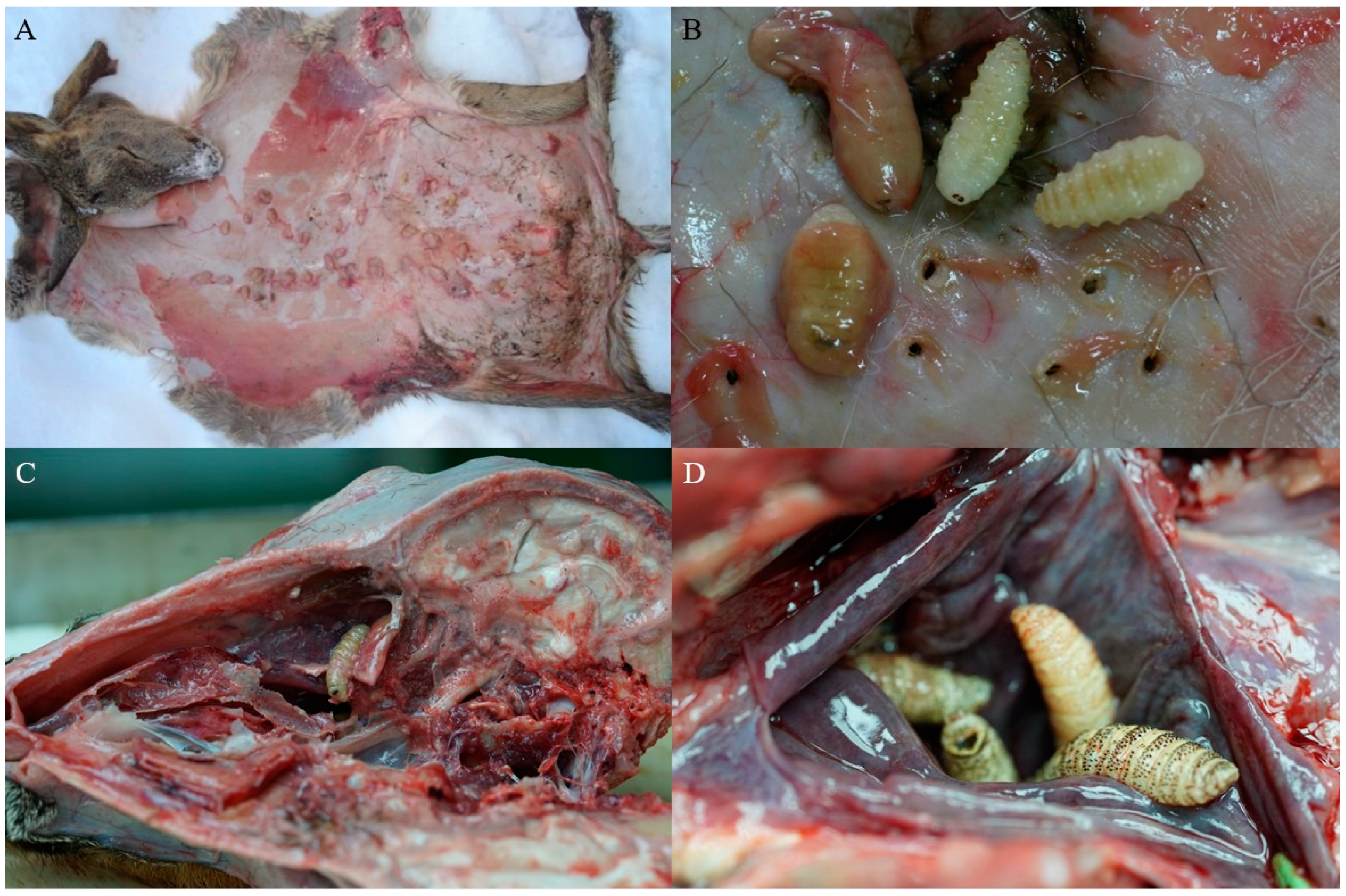
Fly larvae were sampled from wild cervids throughout the country that were brought to the Pathology Laboratory at the Croatian Veterinary Institute. One roe deer came from the northern coastal region of Croatia, while the others came from 19 continental locations and 10 mountainous locations (Figure 1). Larvae were found in the nasopharynx of 27 roe deer (125 larvae) and one red deer (13 larvae), while subcutaneous larvae were retrieved from nine roe deer (138 larvae) and eight red deer (59 larvae) (Figure 2). Details about the larvae and their hosts are provided in Table S1 in the Supplementary Materials.
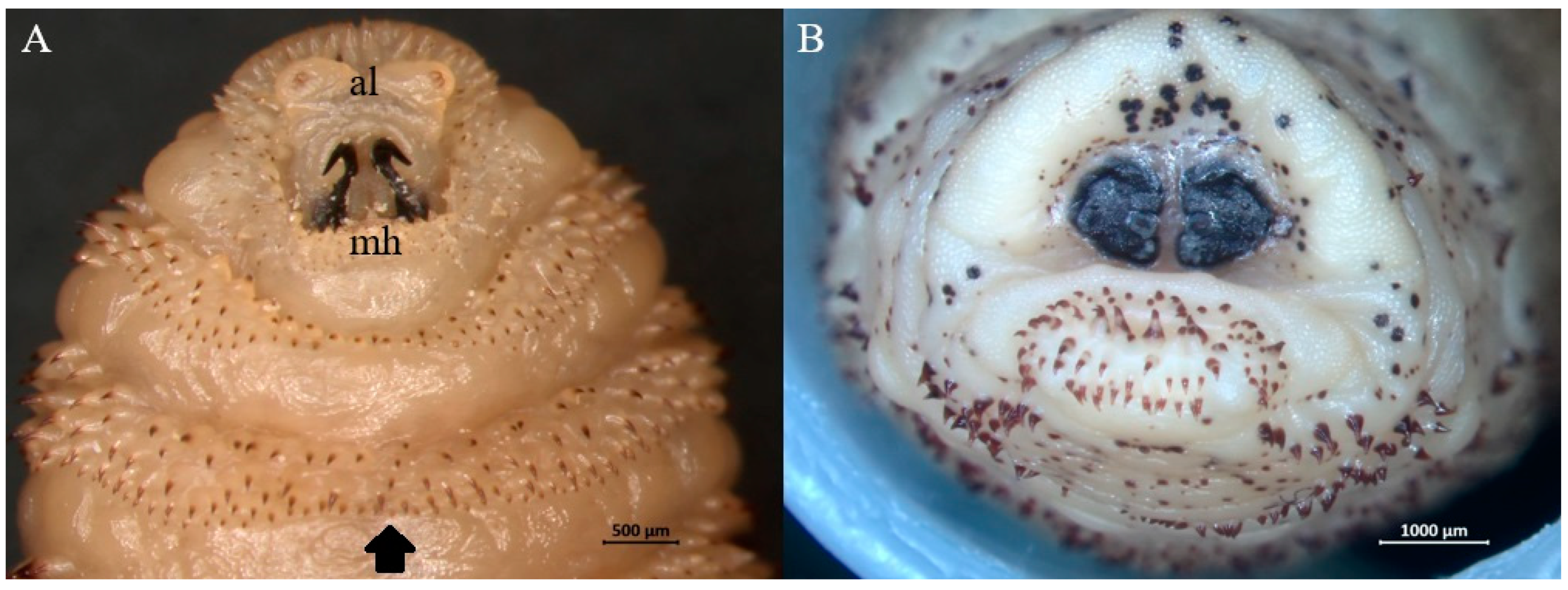
Morphological examination of third-instar larvae suggested that all belonged to one of the following three species: H. diana (Figure 3), C. stimulator (Figure 4), and P. picta (Figure 5). All subcutaneous larvae were identified as H. diana, while nasopharyngeal larvae were identified as C. stimulator in 27 roe deer, or P. picta in the one red deer.
Taxonomic classification was further explored through the partial sequencing of the gene-encoding cytochrome oxidase subunit I. A total of 23 second-instar and 40 third-instar larvae that had been assigned to H. diana based on morphology were sequenced. All 63 amplicons shared the same sequence, which was >90% similar to the corresponding sequences for the following species: H. lineatum (GenBank accession NC013932), 90.44% similar; H. bovis (NC080982), 90.41% similar; H. sinense (NC071819), 90.29% similar; and H. sinense (AF295558), 90.36% similar. Similarity to previously published H. diana sequences could not be determined because the deposited sequences from that species came from regions of the cytochrome oxidase subunit I gene that did not overlap with the sequence that we amplified. A total of 19 second-instar and 47 third-instar larvae that had been assigned to C. stimulator based on morphology were sequenced. All 66 sequences were identical and 99.85% similar to the corresponding sequence reported for C. stimulator collected from roe deer in Spain (NC_059850). Two second-instar and two third-instar larvae of putative P. picta were sequenced. All sequences were the same and 99.70% similar to P. picta obtained from red deer in Austria (KX146940).
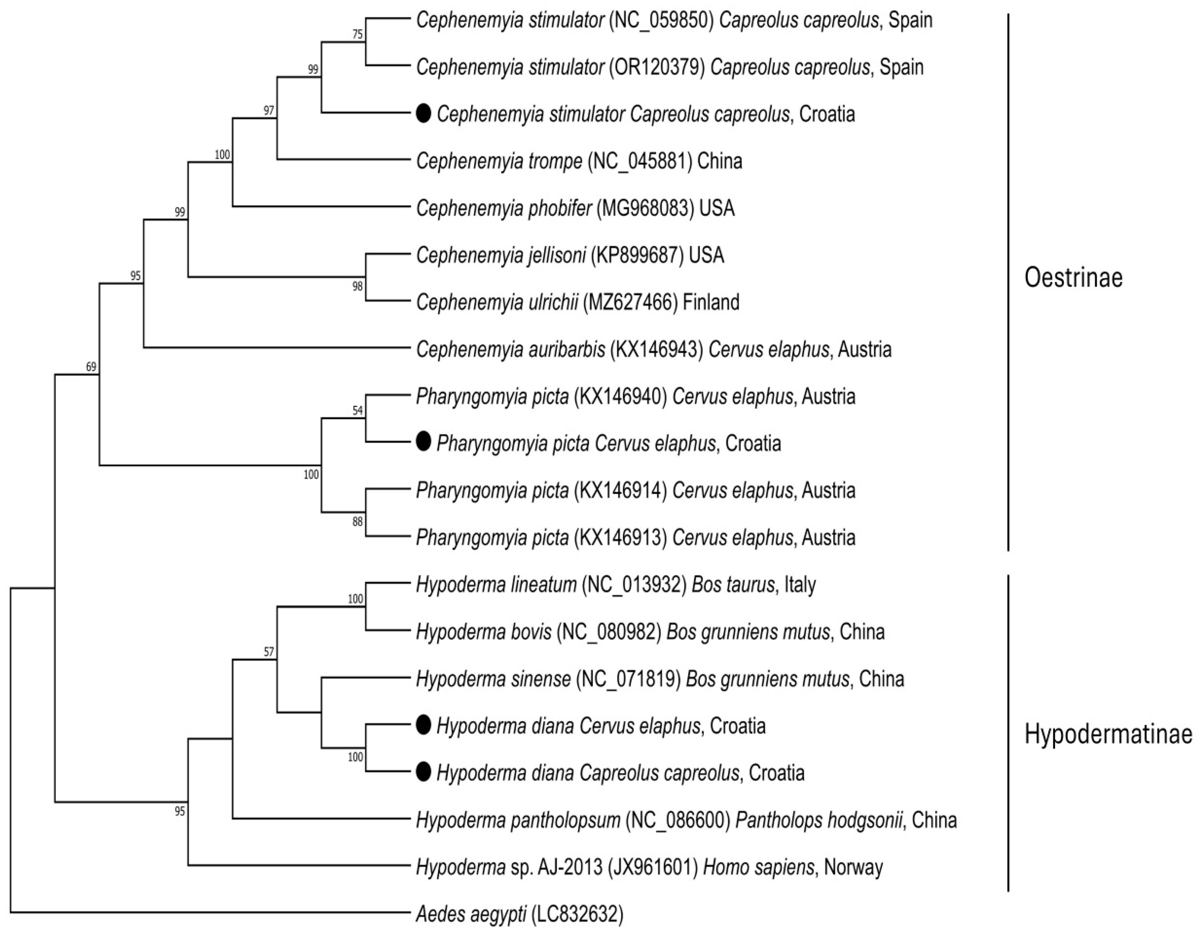
Phylogenetics were clearly differentiated among the three botfly species (Figure 6), validating our species assignments based on the region of the cytochrome oxidase subunit I gene that we amplified.

4. Discussion

The obligatory myiasis of wild cervids is found in diverse regions across the globe, including southeastern Europe [1]. Despite their growing significance and expansion due to environmental changes, their distribution and prevalence in southeastern Europe is virtually unknown [8]. Our analysis of wild deer from across Croatia indicates that at least three species of myiasis-causing flies are prevalent: H. diana, C. stimulator and P. picta. Our results are the first attempt at detecting botflies based on the combination of morphology and genetic sequencing, which is important in light of the morphological similarities among dipteran species [22]. Our phylogenetic analysis supports the morphology-based taxonomic classification of C. stimulator and P. picta in our samples, but it did not confirm our assignment of H. diana because of the lack of reference sequences in GenBank.
In 1959, C. stimulator and H. bovis were reported in the nasopharynx and subcutaneous tissue of roe deer in Croatia, although those taxonomic assignments were based purely on morphology [23]. We recently detected O. ovis in a human and confirmed the taxonomy through sequencing [24]. The present study substantially extends our understanding of botfly species in the country. It also provides data for analyzing the prevalence of these species in other parts of southeastern Europe.
H. diana in our study was detected in roe deer and red deer, and all 63 samples from 17 animals collected from around the country during a 10-year period showed the same sequence in the region of the cytochrome oxidase subunit I gene that we amplified. These observations suggest that H. diana has not diverged into strains preferring one host or another. Such a lack of host specificity is consistent with previous studies of its prevalence in red deer (Spain) and roe deer (Romania) [25,26] or its detection in horses and alpacas in Germany [27,28]. It seems likely that H. diana may be present in other wild ruminants in Croatia and elsewhere in southeastern Europe. We also suggest that the H. bovis identified in Croatian roe deer in 1959 [23] may actually have been H. diana, given that no cases of deer infection with H. bovis have been reported since then. In the present study, we did not identify H. actaeon in subcutaneous tissue, despite its documented presence in northern regions in Hungary [29] that are geographically distant from the Croatian border. It is noteworthy that the Hungarian study did not provide molecular confirmation of H. actaeon. It has predominantly been reported in wild cervids in Spain [30] and Portugal [31,32], making its absence in our findings somewhat expected.
Our detection of C. stimulator is consistent with the high prevalence of this nasal botfly in roe deer across Europe, especially central Europe [8,33]. It was first reported in Croatia in 1959 [23], and more recent work detected it in up to 12% of roe deer in a mountainous region of the country as well as in the flat central region near the capital [17,18]. Batinjanin (2022) [17] also detected nasopharyngeal C. ulrichii in two roe deer, at least based on the morphology of three third-instar larvae from Zagreb County, which we failed to detect in 125 nasopharyngeal larvae from 27 animals nearly entirely from continental and mountain regions. It seems likely that future molecular studies in Croatia and southeastern Europe will identify C. ulrichii and other botfly species beyond the three we detected here.
P. picta was recently detected based on morphology in Croatian red deer [34], and the present study confirms its presence across the country using molecular methods. This extends the list of European countries where the botfly has been definitively detected, which already includes Poland [35], Spain [36], Hungary [37], Austria [38], and Portugal [39]. We speculate that the species is also present in other parts of southeastern Europe.

5. Conclusions

The current study is, to our knowledge, the first analysis of myiasis-causing botflies in wild cervids in Croatia that combines morphological and molecular analyses. Our work establishes H. diana, C. stimulator and P. picta as prevalent in many parts of the country. We believe that C. ulrichii and likely additional botfly species are present as well, which should be examined in future work. Such work may also uncover multiple haplotypes of these species. Our results provide a reference for similar investigations elsewhere in southeastern Europe, which are important for obtaining a complete understanding of botfly distribution throughout Europe as climate conditions become increasingly favorable to their growth.

Supplementary Materials

The following supporting information can be downloaded at: https://www.mdpi.com/article/10.3390/ani15020208/s1, Table S1: Data summary from the research.

Author Contributions

Conceptualization, R.B., K.P., A.H. and E.G.; methodology, E.G., D.J.Ž., K.P., A.H. and Š.N.; writing—original draft, E.G. and R.B.; formal analysis, E.G., D.J.Ž., A.H., Š.N. and R.B.; investigation, D.J.Ž., K.P. and Š.N.; data curation, E.G. and K.P.; writing—review & editing, A.H. and R.B.; resources and funding acquisition, R.B. All authors have read and agreed to the published version of the manuscript.

Funding

This study was funded by the European Union (NextGenerationEU) and supported by the Ministry of Science and Education of the Republic of Croatia through project NPOO 5 at the Croatian Veterinary Institute (“Molecular identification of nematodes, tapeworms, flukes and obligate myiasis of domestic and wild animals”, PARAKOD). The work of AH is supported by the Austrian Science Fund through project P 36130.

Institutional Review Board Statement

The animals from which samples were taken had been killed during regular hunting events carried out in accordance with regional and national regulations. The present study was approved by the Ethics Committee of the Croatian Veterinary Institute (Z-VI-4-4185-6/23, approval date: 17 October 2023).

Informed Consent Statement

Not applicable.

Data Availability Statement

The datasets from the current study are available upon request to the corresponding author.

Acknowledgments

We are grateful to Kristina Skrbin, Ada Vilić, Petra Cikoš, Mario Maršić and Mario Vinkšel for their invaluable help with sample processing.

Conflicts of Interest

The authors declare no conflicts of interest.

References

  1. Colwell, D.D. Bot flies and warble flies (Order Diptera: Family Oestridae). In Parasitic Diseases of Wild Mammals, 2nd ed.; Samuel, W.M., Pybus, M.J., Kocan, A.A., Eds.; Iowa State University Press: Ames, IA, USA, 2001; Volume 2, pp. 46–71. [Google Scholar]
  2. Scholl, P.J.; Colwell, D.D.; Cepeda-Palacios, R. Myiasis (Muscoidea, Oestroidea). In Medical and Veterinary Entomology, 3rd ed.; Mullen, G.R., Durden, L.A., Eds.; Elsevier: Amsterdam, The Netherlands, 2018; pp. 383–419. [Google Scholar]
  3. Hassan, M.-u.; Khan, M.N.; Abubakar, M.; Waheed, H.M.; Iqbal, Z.; Hussain, M. Bovine hypodermosis-a global aspect. Trop. Anim. Health Prod. 2010, 42, 1615–1625. [Google Scholar] [CrossRef]
  4. El-Tahawy, A.S. The prevalence of selected diseases and syndromes affecting Barki sheep with special emphasis on their economic impact. Small Ruminant Res. 2010, 90, 83–87. [Google Scholar] [CrossRef]
  5. Vicente, J.; Fierro, Y.; Martínez, M.; Gortázar, C. Long-term epidemiology, effect on body condition and interspecific interactions of concomitant infection by nasopharyngeal bot fly larvae (Cephenemyia auribarbis and Pharyngomyia picta, Oestridae) in a population of Iberian red deer (Cervus elaphus hispanicus). Parasitology 2004, 129, 349–361. [Google Scholar] [CrossRef]
  6. Ortiz-Leal, I.; Torres, M.V.; López-Beceiro, A.; Sanchez-Quinteiro, P.; Fidalgo, L. Dissecting the Effects of Cephenemyia stimulator on the Olfactory Turbinates and Nasopharynx of Roe Deers (Capreolus capreolus). Animals 2024, 14, 1297. [Google Scholar] [CrossRef] [PubMed]
  7. Otranto, D.; Colwell, D.D.; Traversa, D.; Stevens, J.R. Species identification of Hypoderma affecting domestic and wild ruminants by morphological and molecular characterization. Med. Vet. Entomol. 2003, 17, 316–325. [Google Scholar] [CrossRef] [PubMed]
  8. Morrondo, P.; Pajares, G.; Arias, M.S.; Martínez-Calabuig, N.; Remesar, S.; García-Dios, D.; Díaz, P.; López, C.M.; Panadero, R.; Díez-Baños, P. An Update on Cephenemyiosis in the European Roe Deer: Emergent Myiasis in Spain. Animals 2021, 11, 3382. [Google Scholar] [CrossRef]
  9. Granados, J.E.; Forte-Gil, D.; Ramos, B.; Cano-Manuel, F.J.; Soriguer, R.C.; Fandos, P.; Pérez, J.M. First record of Pharyngomyia picta (Diptera: Oestridae) parasitizing Cervus elaphus in Sierra Nevada National Park. Parasitol. Res. 2021, 120, 3895–3898. [Google Scholar] [CrossRef] [PubMed]
  10. Martínez-Calabuig, N.; Vieira-Pinto, M.; López, C.M.; Remesar, S.; Panadero, R. Cephenemyia stimulator (Diptera: Oestridae) myiasis in a roe deer (Capreolus capreolus) from Portugal. Vet. Parasitol. Reg. Stud. Rep. 2023, 41, 100883. [Google Scholar] [CrossRef]
  11. Singh, A.; Singh, Z. Incidence of myiasis among humans-a review. Parasitol. Res. 2015, 114, 3183–3199. [Google Scholar] [CrossRef] [PubMed]
  12. Bernhardt, V.; Finkelmeier, F.; Verhoff, M.A. Jens Amendt. Myiasis in humans-a global case report evaluation and literature analysis. Parasitol. Res. 2019, 118, 389–397. [Google Scholar] [CrossRef]
  13. Otranto, D.; Stevens, J.R.; Brianti, E.; Dorchies, P. Human and livestock migrations: A history of bot fly biodiversity in the Mediterranean region. Trends Parasitol. 2006, 22, 209–213. [Google Scholar] [CrossRef] [PubMed]
  14. Kusak, J.; Krapinec, K. Ungulates and their management in Croatia. In European Ungulates and Their Management in the 21st Century, 1st ed.; Apollonio, M., Andersen, R., Putman, R., Eds.; Cambridge University Press: Cambridge, UK, 2010; Volume 1, pp. 527–539. [Google Scholar]
  15. Linnell, J.D.C.; Zachos, F.E. Status and distribution patterns of European ungulates: Genetics, population history and conservation. In Ungulate Management in Europe: Problems and Practices, 1st ed.; Putman, R., Apollonio, M., Andersen, R., Eds.; Cambridge University Press: Cambridge, UK, 2011; Volume 1, pp. 12–53. [Google Scholar]
  16. Konjević, D.; Janicki, Z.; Slavica, A.; Severin, K. Nosna štrkljivost u srna (Capreolus capreolus L.). Vet Stanica 2006, 37, 153–159. [Google Scholar]
  17. Batinjan, M. Morphological Characterization of Deer Botfly from the Airways of Roe Deer. Rector’s Award, FVMUZ, Heinzelova 55, 10000 Zagreb, 4.10.2022. Available online: https://apps.unizg.hr/rektorova-nagrada/javno/radovi/1545/preuzmi (accessed on 4 December 2024).
  18. Kusak, J.; Špičić, S.; Slijepčević, V.; Bosnić, S.; Rajković Janje, R.; Duvnjak, S.; Sindičić, M.; Majnarić, D.; Cvetnić, Ž.; Huber, Đ. Health status of red deer and roe deer in Gorski kotar, Croatia. Vet. Arhiv. 2012, 82, 59–73. Available online: https://hrcak.srce.hr/77399 (accessed on 4 December 2024).
  19. Radhakrishnan, S.; Ajithkumar, K.G.; Ravindran, R.; Rajagopal, K. First record in South Asia of deer throat bot fly larvae Pharyngomyia picta (Meigen, 1824) (Diptera: Oesteridae) from Sambar deer (Rusa unicolor), a new host record. Trop. Biomed. 2012, 29, 265–269. [Google Scholar]
  20. Hall, T.A. BioEdit: A user-friendly biological sequence alignment editor and analysis program for Windows 95/98/NT. Nucleic Acids Symp. 1999, 41, 95–98. [Google Scholar]
  21. Kumar, S.; Stecher, G.; Tamura, K. MEGA7: Molecular Evolutionary Genetics Analysis Version 7.0 for Bigger Datasets. Mol. Biol. Evol. 2016, 33, 1870–1874. [Google Scholar] [CrossRef] [PubMed]
  22. Otranto, D.; Stevens, J.R. Molecular approaches to the study of myiasis-causing larvae. Int. J. Parasitol. 2002, 32, 1345–1360. [Google Scholar] [CrossRef] [PubMed]
  23. Richter, S. Parasitska fauna srne (Capreolus capreolus L.) u NR Hrvatskoj. Vet. Arh. Zagreb. 1959, 29, 34–45. [Google Scholar]
  24. Pupić-Bakrač, A.; Pupić-Bakrač, J.; Škara Kolega, M.; Beck, R. Human ophthalmomyiasis caused by Oestrus ovis—First report from Croatia and review on cases from Mediterranean countries. Parasitol. Res. 2020, 119, 783–793. [Google Scholar] [CrossRef]
  25. Martínez-Gómez, F.; Hernández-Rodríguez, S.; Ruiz-Sánchez, P.; Molina-Rodero, R.; Martínez-Moreno, A. Hypodermosis in the red deer Cervus elaphus in Cordoba, Spain. Med. Vet. Entomol. 1990, 4, 311–314. [Google Scholar] [CrossRef] [PubMed]
  26. Ilie, M.S.; Imre, M.I.; Hotea, I.O.; Imre, K.; Sorescu, I.D.; Andrei, S.; Onita, P.; Oprescu, I.; Morariu, S.; Mihali, C.; et al. Prevalence of Hypoderma infestation in deer in Western Romania. Luc. Sti. Med. 2012, 19, 3. [Google Scholar]
  27. Borges, F.; Sybrecht, G.W.; von Samson-Himmelstjerna, G. First reported case of Hypoderma diana Brauer, 1985 (Diptera: Oestridae)-associated myiasis in a horse in Germany. Equine Vet. Educ. 2019, 31, 122–125. [Google Scholar] [CrossRef]
  28. Venjakob, P.L.; Vogel, C.; Clausen, P.H.; Nijhof, A.M. First report of a Hypoderma diana infestation in alpaca (Vicugna pacos) in Germany. Parasitol. Res. 2019, 118, 1963–1966. [Google Scholar] [CrossRef]
  29. Husvéth, B.; Egri, B. Retrospective study on the occurrence of warble fly infestation (Hypodermosis) of the red deer and roe deer in North-West Hungary (Szigetkoz, District of Ravazd and Tarjan). Int. J. Zool. Anim. Biol. 2021, 4, 1–7. [Google Scholar] [CrossRef]
  30. González, S.; del Rio, M.L.; Diez, M.N.; Hidalgo, M.R.; Martínez, A. Identification of Hypoderma actaeon (Diptera: Oestridae) in red deer (Cervus elaphus) from northern Spain: Microscopy study and molecular analysis. Microsc. Res. Tech. 2023, 86, 3–11. [Google Scholar] [CrossRef]
  31. Ahmed, H.; Ramalho Sousa, S.; Simsek, S.; Anastácio, S.; Gunyakti Kilind, S. First molecular characterization of Hypoderma actaeon in cattle and red deer (Cervus elaphus) in Portugal. Korean J. Parasitol. 2017, 55, 653–658. [Google Scholar] [CrossRef] [PubMed]
  32. Panadero, R.; Varas, G.; Pajares, G.; Markina, F.; López, C.; Díaz, P.; Pérez-Creo, A.; Prieto, A.; Díez-Baños, P.; Morrondo, P. Hypoderma actaeon: An emerging myiasis in roe deer (Capreolus capreolus). Med. Vet. Entomol. 2017, 31, 94–96. [Google Scholar] [CrossRef] [PubMed]
  33. Király, I.; Egri, B. Epidemiological characteristics of Cephenemyia stimulator (Clark, 1815) larval infestation in European roe deer (Capreolus capreolus) in Hungary. Acta Zool. Acad. Sci. Hung. 2007, 53, 271–279. [Google Scholar]
  34. Martinković, F.; Štimac, I.; Bujanić, M.; Konjević, D. First identification of Pharyngomyia picta in red deer in Republic of Croatia. In Proceedings of the 10th International Deer Biology Congress, Osijek, Hrvatska, 4–9 September 2022; p. 93. [Google Scholar]
  35. Draber-Mońko, A.; Bystrowski, C. Several new data from Poland on the occurrence of imagines of the hypodermatid and oestrid flies (Diptera: Hypodermatidae and Oestridae). Fragm. Faun. 2016, 59, 105–113. [Google Scholar] [CrossRef]
  36. Ruiz, I.; Soriguer, R.C.; Perez, J.M. Pharyngeal bot flies (Oestridae) from sympatric wild cervids in Southern Spain. J. Parasitol. 1993, 79, 623–626. [Google Scholar] [CrossRef] [PubMed]
  37. Sugár, L. The occurrence of nasal throat bot flies (Oestridae) in wild ruminants in Hungary. Parasitol. Hung. 1974, 7, 181–189. [Google Scholar]
  38. Leitner, N.; Schwarzmann, L.; Zittra, C.; Palmieri, N.; Eigner, B.; Otranto, D.; Glawischnig, W.; Fuehrer, H.P. Morphological and molecular identification of nasopharyngeal bot fly larvae infesting red deer (Cervus elaphus) in Austria. Parasitol. Res. 2016, 115, 4417–4422. [Google Scholar] [CrossRef] [PubMed]
  39. Miranda, R.; Serejo, J.; Pérez, J.M.; Aranha, J.; Venâncio, C.; Vieira-Pinto, M. First Study of Pharingomyia picta and Cephanemyia auribarbis in Wild Populations of Red Deer (Cervus elaphus) in Portugal. Animals 2022, 12, 1896. [Google Scholar] [CrossRef] [PubMed]
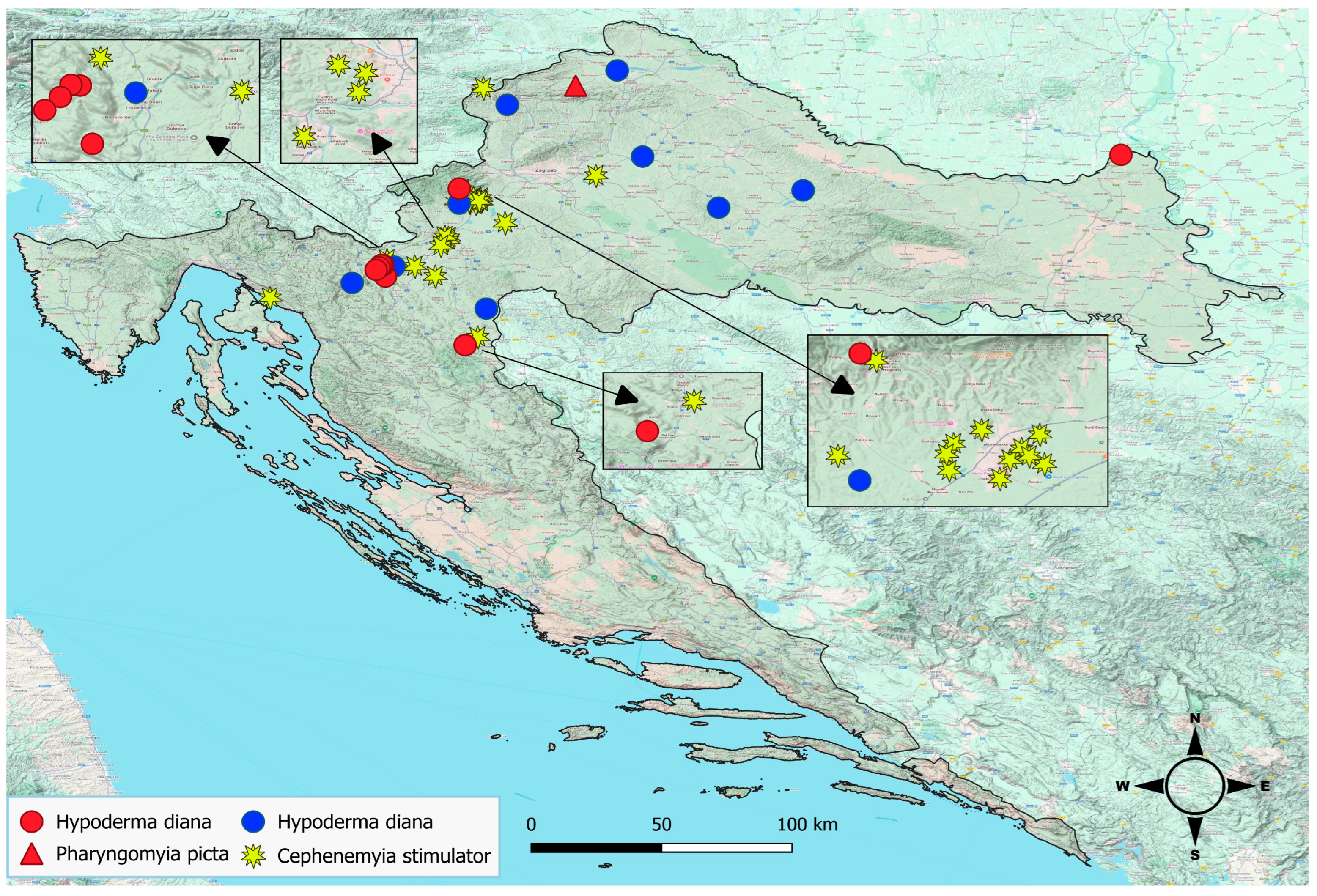
Figure 1. Locations of sampled roe deer (blue and yellow symbols) and red deer (red symbols) in Croatia, annotated according to the botfly species subsequently identified. Insets show zoomed regions of the map.
Animals 15 00208 g001
Figure 2. Examples of macroscopic findings in a roe deer. (A) Subcutaneous warbles. (B) Larvae of Hypoderma sp. extracted from subcutaneous warbles. (C,D) Larvae of Cephenemyia sp. in the pharyngeal pouch.
Animals 15 00208 g002
Figure 3. Representative third-instar larva of Hypoderma diana. (A) Anterior end showing the mouth (m), opercular suture (os) and band of spines between mouth and opercular suture (black arrow) on the cephalic segment. (B) Ventral spines on the first thoracic segment showing posteriorly directed spines with narrow bases and rounded tips (red arrow) and anteriorly directed spines with narrow bases and sharp tips (blue arrow). (C) Flat, C-shaped spiracular plates incompletely surrounding an ecdysal scar (es).
Animals 15 00208 g003
Figure 4. Representative third-instar larva of Cephenemyia stimulator. (A) Anterior end with V shaped antennal lobes (al), mouth hooks (mh) and arrays of cuticular spines (arrow). (B) Posterior end with reniform spiracular plates.
Animals 15 00208 g004
Figure 5. Representative third-instar larva of Pharyngomyia picta. (A) Anterior end with mouth hooks (mh) and widely separated antennal lobes (al). (B) Posterior end with crescent-shaped spiracular plates.
Animals 15 00208 g005
Figure 6. Maximum-likelihood phylogenetic tree based on cox1 nucleotide sequences of species in subfamilies Osterinae and Hypodermatinae. The strains identified in the present study are indicated with a solid circle. Previously reported strains of H. diana, H. tarandi and H. actaeon were not included because the corresponding sequences in the database come from different regions of the cox1 gene than the region sequenced in the present study.
Animals 15 00208 g006
Disclaimer/Publisher’s Note: The statements, opinions and data contained in all publications are solely those of the individual author(s) and contributor(s) and not of MDPI and/or the editor(s). MDPI and/or the editor(s) disclaim responsibility for any injury to people or property resulting from any ideas, methods, instructions or products referred to in the content.