Article

Occurrence and Multi-Locus Genotyping of Giardia duodenalis in Black Goats from Fujian Province, China

1
Key Laboratory of Fujian-Taiwan Animal Pathogen Biology, College of Animal Sciences, Fujian Agriculture and Forestry University, Fuzhou 350002, China
2
Fujian Zhuyian Agriculture Development Co., Ltd., Fuzhou 350000, China
*
Authors to whom correspondence should be addressed.
These authors contributed equally to this work.
Animals 2025, 15(2), 199; https://doi.org/10.3390/ani15020199
Submission received: 30 November 2024 / Revised: 29 December 2024 / Accepted: 8 January 2025 / Published: 13 January 2025

Simple Summary

Giardia duodenalis is a parasite that infects the intestines of both animals and humans, leading to health concerns and economic losses in livestock farming. In black goats, infections can cause severe diarrhea or long-term asymptomatic carriers, which impacts animal husbandry. This study aimed to determine the prevalence of G. duodenalis in black goats from nine districts of Fujian Province, China. A total of 539 goat fecal samples were collected and analyzed using nested PCR targeting the SSU rRNA gene. Further analyses focused on the beta-giardin, glutamate dehydrogenase, and triosephosphate isomerase genes. Results showed that 115 samples (21.34%) tested positive for G. duodenalis, with assemblages A and E detected. Statistical analysis revealed significant differences in infection rates between regions but no significant variation by age or gender. These findings contribute valuable data for understanding G. duodenalis in black goats and for improving prevention strategies in livestock.

Abstract

Giardia duodenalis is a zoonotic parasite that causes gastrointestinal diseases in both humans and animals. To evaluate the prevalence and genetic diversity of G. duodenalis in black goats, we collected 539 fecal samples from nine districts in Fujian Province, China. The presence of G. duodenalis was confirmed through nested PCR targeting the SSU rRNA gene, and genotyping was performed at the beta-giardin, glutamate dehydrogenase, and triosephosphate isomerase loci. Among the samples, 115 tested positive, yielding an overall infection rate of 21.34%. Assemblages A and E were identified, with assemblage E being predominant. Statistical analysis revealed significant regional differences in infection rates (p < 0.01), with Zhangzhou exhibiting the highest infection rate (39%) and Fuzhou the lowest (3.13%). No significant differences in infection rates were observed based on age: 24.56% (56/228) for goats <1 year, 14.92% (27/181) for goats 1–2 years, 26.8% (26/97) for goats 2–3 years, and 18.18% (6/33) for goats ≥ 3 years. Similarly, no significant differences were found between sexes: 24.84% (40/161) for males and 19.84% (75/378) for females. Notably, assemblage A, a zoonotic genotype, was detected, indicating a potential risk of cross-species transmission. This study contributes to a deeper understanding of G. duodenalis in black goats and provides critical data for the development of targeted control strategies in Fujian Province.

1. Introduction

Giardia duodenalis is a flagellated protozoan parasite first identified in human feces in 1681. Mammals become infected by ingesting cysts of G. duodenalis through contaminated food or water via the fecal-oral route [1,2]. This parasite is one of the most common causes of diarrhea globally, affecting over 300 million people annually, particularly in developing and least-developed countries. Infected hosts often suffer from gastrointestinal distress, malnutrition, and stunted growth over extended periods. Children and individuals with weakened immune systems are especially vulnerable to G. duodenalis infections [3]. The lack of commercial vaccines and specialized treatments for G. duodenalis leads to significant economic losses in livestock production [4,5]. Additionally, adult animals that are asymptomatic carriers play a crucial role in the epidemiology of G. duodenalis, as they can spread the parasite to other susceptible animals without showing any symptoms [4].
The genus Giardia includes six species characterized by G. duodenalis morphological features: G. duodenalis, Giardia muris, Giardia minor, Giardia psittaci, and Giardia agilis. Among these, G. duodenalis has eight genetic assemblages (A-H) [6]. Assemblages A and B can infect both humans and animals, and assemblages C through H primarily infect animals.
Assemblages C and D, for example, have been found in domestic dogs and other canines. Assemblage F predominantly infects cats. Assemblage H primarily affects wild animals [7,8,9,10,11,12]. Notably, assemblage E is the most frequently detected in goats worldwide, followed by assemblages A and B [13].
In 1979, the World Health Organization (WHO) listed giardiasis as one of the top ten parasitic diseases threatening human health. The prevalence of giardiasis ranges from 2% to 7% in developed countries but is significantly higher (20–30%) in developing and developing countries [14]. The reported prevalence rates in developed countries include 0.11% in the United States, 4.6% in Canada, 2% in the United Kingdom, and 0.63% in Italy [15,16,17,18]. The reported prevalence rates in developing countries include 23.8% in Brazil, 12.7% in Bangladesh, 23.7% in Malaysia, and 20.3% in Thailand [19,20,21,22].
Epidemiological surveys in China indicate an average infection rate in humans of 0.85%, with a peak infection rate of 9.5% reported in Shanghai [23]. G. duodenalis infections have also been widely documented in Chinese cattle, with the first case recorded in Guangdong Province in 2006 [23]. The average infection rate in cattle was 5.43%, with a peak of 18.87% in Shanxi Province [23]. In sheep and goats, the average infection rate is 6.07%. In Henan Province, 24.31% of goats were infected, and the highest infection rate of 27.78% was reported in Chongqing municipality [23]. However, the infection rate in animals from Fujian Province remains unknown.
In order to deepen the understanding of the epidemiology of G. duodenalis in Fujian Province, this study utilized molecular detection and genotype identification of G. duodenalis in sheep feces from nine administrative regions. There are several methods available for detecting G. duodenalis, with fecal microscopy being the most common. In order to provide more accurate detection results, nested PCR was used to analyze the samples and amplify the genes of G. duodenalis for genotype identification. These findings offer foundational data for the prevention and control of G. duodenalis in black goats in Fujian, and they deepen our comprehension of the genetic makeup and zoonotic potential associated with this parasite.

2. Materials and Methods

2.1. Study Areas and Sample Collection

Between September and November 2023, a total of 539 fecal samples were randomly collected from black goats on farms in nine administrative districts of Fujian Province (for collection details, refer to Table 1 and Figure 1). The sample set included 161 samples from male goats and 378 from female goats. Age categorization consisted of 228 samples from goats aged ≤1 years, 181 from goats aged 1–2 years, and 97 from goats aged 2–3 years. Fecal samples were obtained directly from the rectum of each goat, individually sealed to prevent cross-contamination, and labeled with information regarding region, gender, and age. The samples were subsequently refrigerated and transported to the laboratory, where they were stored at −80 °C until further analysis.

2.2. Genomic DNA Extraction and PCR Amplifications

Total genomic DNA was extracted from approximately 200 mg of each fecal specimen using the Genomic DNA Extraction Kit (TianGen Biotech, Beijing, China). All DNA samples were stored at −20 °C until required for subsequent experiments. The 292bp fragment for the small subunit (SSU) rRNA gene by nested PCR amplification was to screen G. duodenalis-positive samples. G. duodenalis-positive samples were analyzed by MLGs using the three loci in beta-giardin (bg), glutamate dehydrogenase (gdh), and triosephosphate isomerase (tpi). The sizes of the fragments for bg, gdh, and tpi are 511 bp, 530 bp and 530 bp, respectively. PCR reactions were carried out using a thermocycler, with 2 µL of genomic DNA (for primary PCR) or 2 µL of PCR amplification product (for secondary PCR), 2.5 µL of 10 × PCR Buffer, 2 µL of dNTPs, 0.2 µL of Ex Taq polymerase, 0.25 µL of each primer, 1.5 µL of MgCl2 (all form Takara, Dalian, China), and ddH2O added to a final volume of 25 µL.

2.3. Genotype Identification

The identification of G. duodenalis was performed using nested PCR based on the amplification of the SSU rRNA gene in black goat fecal samples. These amplification products were stored at 4 °C for short-term use or at −40 °C for long-term storage.
Positive G. duodenalis samples were amplified using the bg, gdh, and tpi loci to determine their genotypes. The primers and amplification used in this study have been previously described [3,24]. Amplification products were stored at 4 °C for short-term use or at −40 °C for long-term storage.

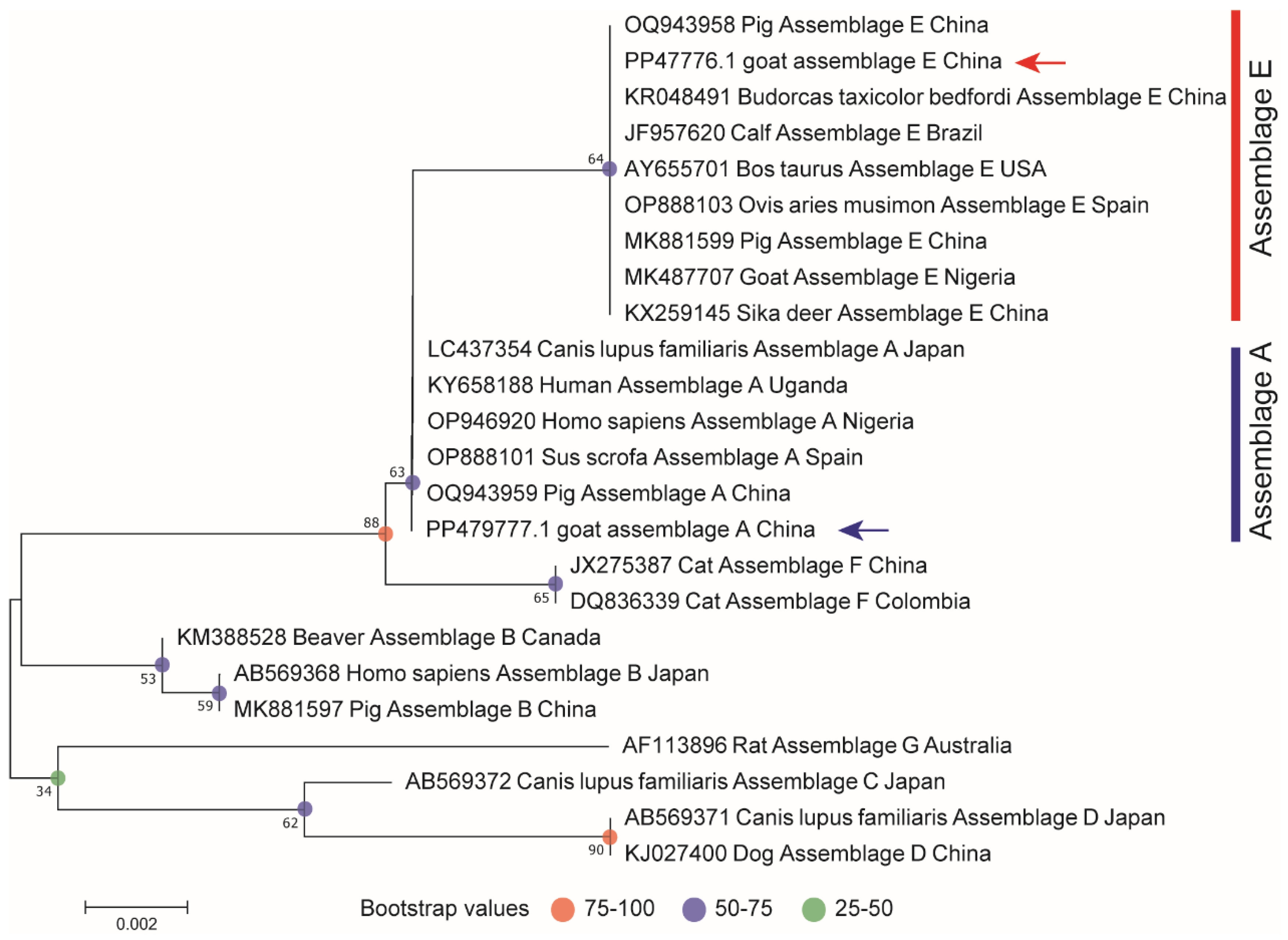
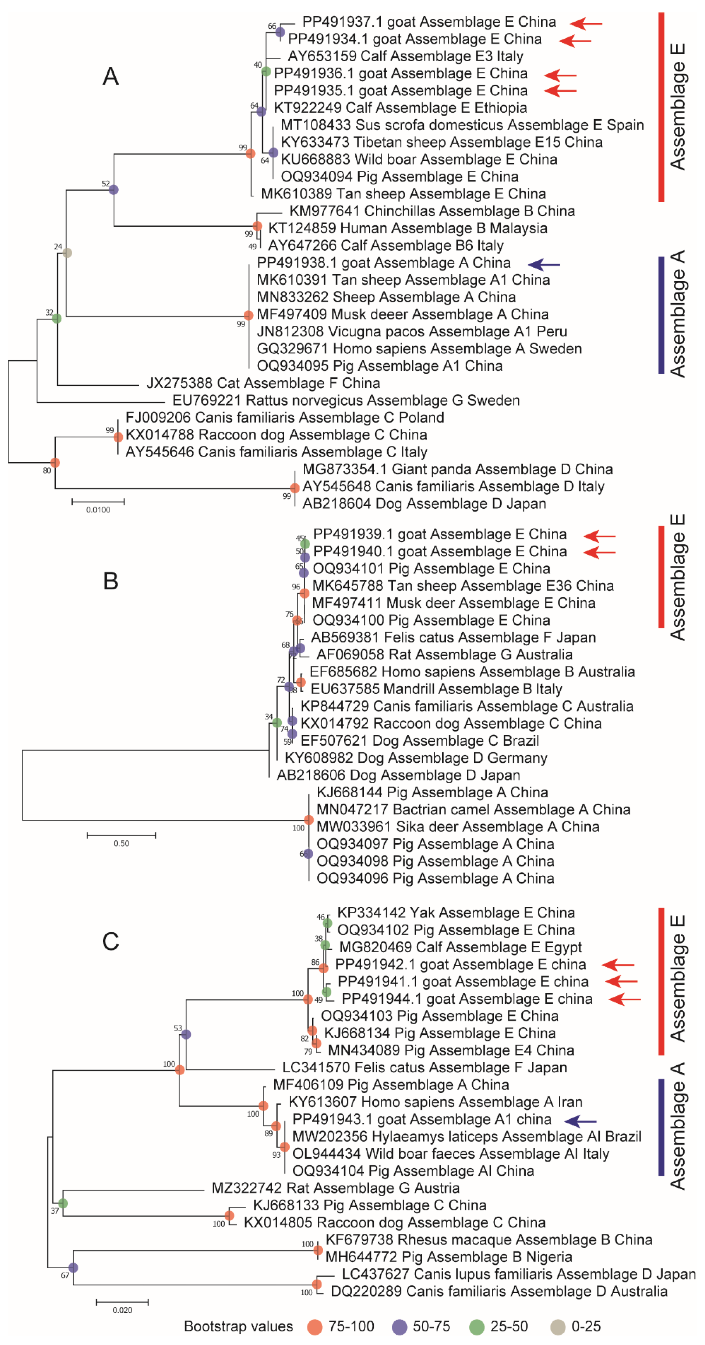
2.4. Sequencing and Phylogenetic Analysis

Following amplification, the products were analyzed by gel electrophoresis. A 1.0% agarose gel containing nucleic acid dye was prepared. Each sample (5 µL of second-round PCR product mixed with 1 µL of 6× loading buffer) was loaded into the wells, with the Takara DL2000 Marker (Takara, Dalian, China) used as a molecular weight reference. Electrophoresis was performed at 125 V for 30 min in a TAE buffer (Shanghai Toscience Biotechnology Co., Ltd., Shanghai, China). The gel was visualized using a UV gel imaging system, and the results were photographed and saved. Positive PCR products were stored at 4 °C for short-term storage or at −40 °C for long-term storage. All positive secondary PCR products were sent to Sangon Bioengineering Ltd., (Shanghai Sangon Biotechnology Co., Ltd, Shanghai, China) for bidirectional sequencing. Sequence editing and alignment construction were performed using DNAstar 7.1 software, and the final sequences were submitted to the NCBI database for comparative analysis to identify G. duodenalis genotypes and polymorphisms. Phylogenetic trees were constructed using the Neighbor-Joining (NJ) method with 1000 bootstrap replicates in Mega11. Separate phylogenetic trees were generated based on identified genotypes or subtypes.

2.5. Statistical Analysis

Statistical analysis of the infection rate of G. duodenalis in black goats across different districts, age groups, and sex categories was conducted using the χ2 test in SPSS Statistics 26.0. The analysis also incorporates a t-test and Bonferroni correction for multiple comparisons. Differences between groups were considered statistically significant when p < 0.05.

3. Results

3.1. The Positivity Rate of G. duodenalis

The SSU rRNA gene was employed for screening positivity to assess the prevalence of G. duodenalis in black goats. A total of 539 fecal samples from black goats, collected across nine administrative districts of Fujian Province, were analyzed using nested PCR. The results indicated that 115 samples tested positive, with a positivity rate of 21.34%. Partial results of the amplified products are displayed in the gel electrophoresis.

3.2. Prevalence and Genotypic Diversity of G. duodenalis in Black Goats

Infection rates varied significantly across regions, ranging from 3.13% to 39%. The highest prevalence was observed in Zhangzhou City (39%, 32/89), followed by Putian City (37.1%, 23/62) and Longyan City (31.58%, 24/76) (Table 2). Sequence analysis of the gdh, tpi, and bg loci, in conjunction with phylogenetic analysis of the tpi locus, revealed that the G. duodenalis-positive samples belonged to assemblages E (114) and A (1). Statistical analysis further revealed significant differences in G. duodenalis detection rates among the nine districts, emphasizing the importance of considering regional variation when developing control strategies.

3.3. Age-Elated Infection Rates of G. duodenalis

The infection rate of G. duodenalis in black goats of varying ages across nine administrative districts in Fujian Province is a key factor in understanding the epidemiology of this parasite. The overall infection rates ranged from 0% to 26.8%. Specifically, the detection rates were 24.56% (56/228) for goats aged ≤1 year, 14.92% (27/181) for those aged 1–2 years, 26.8% (26/97) for those aged 2–3 years, and 18.18% (6/33) for goats aged ≥3 years (Table 3). Notably, both assemblages E and A were detected in goats aged 1 year or younger, while assemblage E was found exclusively in goats aged 1–3 years and those older than 3 years. Statistical analysis revealed no significant differences in the detection rates of G. duodenalis among the different age groups (p > 0.05), suggesting that age may not be a critical factor influencing infection rates in this population.

3.4. Prevalence of G. duodenalis Infection in Male and Female Black Goats

The infection rate of G. duodenalis in male and female black goats across nine administrative districts in Fujian Province serves as a key indicator of the parasite’s epidemiological patterns. The overall infection rates ranged from 19.84% to 24.84%. Specifically, the infection rate was 24.84% (40/161) for males and 19.84% (75/378) for females (Table 3). This gender distinction is important for understanding the transmission dynamics. Statistical analysis revealed no significant difference in the detection rates of G. duodenalis between genders (p > 0.05), suggesting that both sexes exhibit similar susceptibility to the infection.

3.5. Genotyping of G. duodenalis

Two different sequences corresponding to the SSU rRNA were obtained from the 115 positive samples and subsequently submitted to the GenBank database (IDs: PP479776-PP479777). The evolution of the SSU rRNA was analyzed using MEGA 11 software, employing Clustal W for alignment and the maximum likelihood (ML) method for phylogenetic analysis (Figure 2).

3.6. G. duodenalis MLST Typing

A total of 115 positive samples were identified based on the SSU rRNA locus. Among these, 35 samples tested positive for the bg locus, yielding an amplified fragment of 511 bp, which corresponds to an infection rate of 30.4% (35/115). Likewise, 24 samples were positive for the gdh locus, with an amplified fragment length of 530 bp, representing an infection rate of 20.8% (24/115). Additionally, 27 samples tested positive for the triosephosphate isomerase (tpi) locus, with a target fragment of 530 bp, resulting in an infection rate of 23.4% (27/115). bg, gdh, and tpi partial results of the amplified products are displayed in the gel electrophoresis.
Genotyping of the 115 positive samples revealed three distinct genotypes: assemblage A, A1 and E. Specifically, one sample was identified as assemblage A, one as assemblage A1, and 84 as assemblage E. At the bg locus, one sample of assemblage A and 34 samples of assemblage E were detected, while at the gdh locus, 24 samples of assemblage E were identified. At the tpi locus, one sample of assemblage A1 and 26 samples of assemblage E were found (Table 4 and Table 5). The sequencing of three loci from 17 samples has been successfully completed. One sample showed mixed infection with combinations A, A1, and E, while the other 16 samples were combinations E. In total, two novel multi-locus genotypes (MLGs) were identified and designated as MLG I and MLG II (Table 6).
DNA sequences from the bg, gdh, and tpi loci were subsequently obtained and submitted to the GenBank database. Specifically, five sequences representing the bg locus were obtained from the 35 bg-positive samples (IDs: PP491934-PP491938), two sequences from the 24 gdh-positive samples (IDs: PP491939-PP491940), and four sequences from the 27 tpi-positive samples (IDs: PP491941-PP491944). Phylogenetic analysis was performed using Clustal W alignment and the maximum likelihood (ML) method to construct evolutionary trees for the bg, gdh, and tpi loci (Figure 3). The results indicate that the detected assemblage E consistently clusters with the reported assemblage E, showing a close relationship with assemblage E found in both cattle and goats. Notably, the detection of assemblage A in the bg genotype and assemblage A1 in the tpi genotype aligns with prior studies that suggest assemblage A1 is predominantly found in animals.

4. Discussion

G. duodenalis infection is relatively common in black sheep, though its prevalence differs greatly, ranging from Anhui (1.62%), Qinghai (2.88%), and Henan (3.4%), Inner Mongolia (28.93%) and Guangdong (24.78%) [25,26,27]. In comparison to other provinces, the notably higher infection rate of G. duodenalis in black goats in Fujian Province underscores its widespread prevalence in the region, thereby necessitating further investigation into its epidemiology and molecular characteristics within livestock populations. The detection of two genotypes, assemblages E and A, in the black goats of Fujian Province represents a significant finding. Assemblage E primarily infects livestock, which was identified in 98.26% of the positive samples, particularly ruminants, such as goats and cattle. This genotype’s dominance in livestock is consistent with findings from previous studies across various regions [25,28,29,30]. In contrast, the detection of assemblage A, a common zoonotic genotype, in black goats is noteworthy, as assemblage A infections have been found in a wide range of hosts, including humans, cats, dogs, and wildlife [26]. The presence of this genotype in black goats suggests a potential zoonotic risk to farmers and others in close contact with these animals, highlighting the critical need for preventive measures. Interestingly, this study found no significant differences in infection rates across different age groups of black goats, which contrasts with previous research showing higher susceptibility in younger animals [31,32]. The lack of significant differences in infection rates based on gender and age among black goats may be attributed to various factors, including differences in feeding practices, sample sizes, and dietary components. In black goat breeding facilities in Fujian, the widespread use of fecal leakage boards facilitates the rapid removal of waste. Proactive feces management strategies are critical for controlling the proliferation and survival of parasites. No significant differences in infection rates were observed among different ages, but regional differences in infection rates were observed, with Putian City showing the highest prevalence compared with other regions. These differences may be attributed to various factors, including environmental conditions, local agricultural practices, and the immune status of the animals. The subtropical climate of Fujian, the high humidity, moderate temperatures, and frequent rainfall may further facilitate the persistence and transmission of G. duodenalis in this region for extended periods [33]. These environmental factors, rather than host age, are likely to play a significant role in the transmission dynamics of the parasite in this region.
The study also identified two MLG I and MLG II of the bg, gdh, and tpi genes. MLG analysis provides higher resolution than single-locus genotyping, revealing more detailed genetic diversity of G. duodenalis strains. This study confirms previous findings that assemblage E is the predominant strain in livestock, with mixed infections of assemblages A and E being rare but present [25,28,29,30]. Assemblage A1 was detected in only one case at the tpi locus. While MLG analysis is more sensitive than single-copy markers, it still showed variability in its efficiency, with some loci, such as bg, yielding more positive samples than others. This variability highlights the need for comprehensive multi-locus typing in molecular epidemiological studies.
The zoonotic potential of G. duodenalis is particularly concerning. While assemblage E is primarily associated with livestock, recent studies have reported an increasing incidence of human infections [34,35,36,37]. The detection of assemblage A, a known zoonotic genotype, in black goats indicates that farmers and other individuals in close contact with these animals may be at risk of infection. The high prevalence of G. duodenalis in black goats in this region further emphasizes the need for targeted public health measures to prevent zoonotic transmission.
This study enhances our understanding of the epidemiology and genetic diversity of G. duodenalis in black goats in Fujian Province. The identification of both assemblage E and zoonotic assemblage A highlights the need for continued surveillance of this pathogen in livestock populations. Future research should focus on the zoonotic potential of G. duodenalis in both humans and animals across China to more accurately assess the risks and develop more effective control strategies.

5. Conclusions

This study provides the first molecular evidence of G. duodenalis infection in black goats from Fujian Province, China, with an overall infection rate of 21.34% based on the analysis of 539 fecal samples across nine districts. The infection rates varied significantly across regions, with the highest prevalence of 39% recorded in Zhangzhou City. Genotyping revealed that assemblage E, commonly associated with livestock, was the dominant genotype, while assemblage A was detected at a lower frequency, highlighting potential zoonotic risks. The findings showed no significant differences in infection rates based on gender or age, suggesting these factors may not significantly influence susceptibility to G. duodenalis infection. These research results enrich our understanding of the epidemiology and genetic diversity of G. duodenalis in livestock and emphasize the need for region-specific control measures to prevent the spread of this parasite. Given the zoonotic potential, especially with assemblage A, there is an urgent need for effective strategies to mitigate cross-infections between animals and humans, thereby enhancing overall disease prevention and public health protection.

Author Contributions

D.-H.Z. conceived and designed this study. S.-X.H., K.H. and P.-F.F. performed the experiments. S.-X.H. drafted the manuscript. S.-A.L., Y.L. and K.H. contributed reagents/materials/analysis tools. D.-H.Z. and Z.N. critically revised the manuscript. All authors have read and agreed to the published version of the manuscript.

Funding

This work was funded by the Natural Science Foundation of Fujian Province of China (2022J01131), Industry-university-research Cooperation Project in Fujian Province University and enterprise (Grant No. 2022N5005) and Special Fund for Science and Technology Innovation of Fujian Agriculture and Forestry University (KFb22059XA).

Institutional Review Board Statement

Not applicable.

Informed Consent Statement

Not applicable.

Data Availability Statement

The original contributions presented in this study are included in the article.

Conflicts of Interest

Author Kai Hu was employed by the company Fujian Zhuyian Agriculture Development Co., Ltd. The remaining authors declare that the research was conducted in the absence of any commercial or financial relationships that could be construed as a potential conflict of interest.

References

  1. Thompson, R.C.; Monis, P.T. Variation in Giardia: Implications for taxonomy and epidemiology. Adv. Parasitol. 2004, 58, 69–137. [Google Scholar] [PubMed]
  2. Appelbee, A.J.; Thompson, R.C.; Olson, M.E. Giardia and Cryptosporidium in mammalian wildlife–current status and future needs. Trends Parasitol. 2005, 21, 370–376. [Google Scholar] [CrossRef] [PubMed]
  3. Feng, Y.; Xiao, L. Zoonotic potential and molecular epidemiology of Giardia species and giardiasis. Clin. Microbiol. Rev. 2011, 24, 110–140. [Google Scholar] [CrossRef] [PubMed]
  4. Lux, L.; Ulrich, R.G.; Santos-Silva, S.; Queirós, J.; Imholt, C.; Klotz, C.; Paupério, J.; Pita, R.; Vale-Gonçalves, H.; Alves, P.C.; et al. Detection and Molecular Characterization of Giardia and Cryptosporidium spp. Circulating in Wild Small Mammals from Portugal. Animals 2023, 13, 515. [Google Scholar]
  5. Fan, Y.; Wang, X.; Yang, R.; Zhao, W.; Li, N.; Guo, Y.; Xiao, L.; Feng, Y. Molecular characterization of the waterborne pathogens Cryptosporidium spp., Giardia duodenalis, Enterocytozoon bieneusi, Cyclospora cayetanensis and Eimeria spp. in wastewater and sewage in Guangzhou, China. Parasites Vectors 2021, 14, 66. [Google Scholar] [CrossRef] [PubMed]
  6. Prabakaran, M.; Weible, L.J.; Champlain, J.D.; Jiang, R.Y.; Biondi, K.; Weil, A.A.; Van Voorhis, W.C.; Ojo, K.K. The Gut-Wrenching Effects of Cryptosporidiosis and Giardiasis in Children. Microorganisms 2023, 11, 2323. [Google Scholar] [CrossRef] [PubMed]
  7. Sprong, H.; Cacciò, S.M.; van der Giessen, J.W. Identification of zoonotic genotypes of Giardia duodenalis. PLoS Neglected Trop. Dis. 2009, 3, e558. [Google Scholar] [CrossRef]
  8. Thompson, R.C.; Monis, P. Giardia—From genome to proteome. Adv. Parasitol. 2012, 78, 57–95. [Google Scholar]
  9. Cacciò, S.M.; Thompson, R.C.; McLauchlin, J.; Smith, H.V. Unravelling Cryptosporidium and Giardia epidemiology. Trends Parasitol. 2005, 21, 430–437. [Google Scholar] [CrossRef] [PubMed]
  10. Liu, H.; Wang, B.; Yin, J.; Yuan, Z.; Jiang, Y.; Zhang, J.; Cao, J.; Shen, Y.; Liu, H. Investigation of giardiasis in captive animals in zoological gardens with strain typing of assemblages in China. Parasitology 2021, 148, 1360–1365. [Google Scholar] [CrossRef]
  11. Cacciò, S.M.; Lalle, M.; Svärd, S.G. Host specificity in the Giardia duodenalis species complex, Infection, genetics and evolution. J. Mol. Epidemiol. Evol. Genet. Infect. Dis. 2018, 66, 335–345. [Google Scholar] [CrossRef]
  12. Heyworth, M.F. Giardia duodenalis genetic assemblages and hosts. Parasite 2016, 23, 13. [Google Scholar] [CrossRef] [PubMed]
  13. Kanski, S.; Weber, K.; Busch, K. Feline and canine giardiosis: An Update. Tierarztl. Prax. Ausg. K Kleintiere/Heimtiere 2023, 51, 411–421. [Google Scholar] [PubMed]
  14. Roxström-Lindquist, K.; Palm, D.; Reiner, D.; Ringqvist, E.; Svärd, S.G. Giardia immunity–an update. Trends Parasitol. 2006, 22, 26–31. [Google Scholar] [CrossRef]
  15. Amar, C.F.; East, C.L.; Gray, J.; Iturriza-Gomara, M.; Maclure, E.A.; McLauchlin, J. Detection by PCR of eight groups of enteric pathogens in 4,627 faecal samples: Re-examination of the English case-control Infectious Intestinal Disease Study (1993–1996). Eur. J. Clin. Microbiol. Infect. Dis. Off. Publ. Eur. Soc. Clin. Microbiol. 2007, 26, 311–323. [Google Scholar] [CrossRef]
  16. Zylberberg, H.M.; Green, P.H.; Turner, K.O.; Genta, R.M.; Lebwohl, B. Prevalence and Predictors of Giardia in the United States. Dig. Dis. Sci. 2017, 62, 432–440. [Google Scholar] [CrossRef]
  17. Bartolini, A.; Zorzi, G.; Besutti, V. Prevalence of intestinal parasitoses detected in Padua teaching hospital, Italy, March 2011–February 2013. Infez. Med. 2017, 25, 133–141. [Google Scholar]
  18. Iqbal, A.; Goldfarb, D.M.; Slinger, R.; Dixon, B.R. Prevalence and molecular characterization of Cryptosporidium spp. and Giardia duodenalis in diarrhoeic patients in the Qikiqtani Region, Nunavut, Canada. Int. J. Circumpolar Health 2015, 74, 27713. [Google Scholar] [CrossRef] [PubMed]
  19. Haque, R.; Roy, S.; Kabir, M.; Stroup, S.E.; Mondal, D.; Houpt, E.R. Giardia assemblage A infection and diarrhea in Bangladesh. J. Infect. Dis. 2005, 192, 2171–2173. [Google Scholar] [CrossRef] [PubMed]
  20. Volotão, A.C.; Costa-Macedo, L.M.; Haddad, F.S.; Brandão, A.; Peralta, J.M.; Fernandes, O. Genotyping of Giardia duodenalis from human and animal samples from Brazil using beta-giardin gene: A phylogenetic analysis. Acta Trop. 2007, 102, 10–19. [Google Scholar] [CrossRef] [PubMed]
  21. Mohammed Mahdy, A.K.; Surin, J.; Wan, K.L.; Mohd-Adnan, A.; Al-Mekhlafi, M.S.; Lim, Y.A. Giardia intestinalis genotypes: Risk factors and correlation with clinical symptoms. Acta Trop. 2009, 112, 67–70. [Google Scholar] [CrossRef]
  22. Traub, R.J.; Inpankaew, T.; Reid, S.A.; Sutthikornchai, C.; Sukthana, Y.; Robertson, I.D.; Thompson, R.C. Transmission cycles of Giardia duodenalis in dogs and humans in Temple communities in Bangkok—A critical evaluation of its prevalence using three diagnostic tests in the field in the absence of a gold standard. Acta Trop. 2009, 111, 125–132. [Google Scholar] [CrossRef] [PubMed]
  23. Li, J.; Wang, H.; Wang, R.; Zhang, L. Giardia duodenalis Infections in Humans and Other Animals in China. Front. Microbiol. 2017, 8, 2004. [Google Scholar] [CrossRef]
  24. Geurden, T.; Vandenhoute, E.; Pohle, H.; Casaert, S.; De Wilde, N.; Vercruysse, J.; Claerebout, E. The effect of a fenbendazole treatment on cyst excretion and weight gain in calves experimentally infected with Giardia duodenalis. Vet. Parasitol. 2010, 169, 18–23. [Google Scholar] [CrossRef] [PubMed]
  25. Geng, H.L.; Yan, W.L.; Wang, J.M.; Meng, J.X.; Zhang, M.; Zhao, J.X.; Shang, K.M.; Liu, J.; Liu, W.H. Meta-analysis of the prevalence of Giardia duodenalis in sheep and goats in China. Microb. Pathog. 2023, 179, 106097. [Google Scholar] [CrossRef]
  26. Xiao, H.D.; Su, N.; Zhang, Z.D.; Dai, L.L.; Luo, J.L.; Zhu, X.Q.; Xie, S.C.; Gao, W.W. Prevalence and Genetic Characterization of Giardia duodenalis and Blastocystis spp. in Black Goats in Shanxi Province, North China: From a Public Health Perspective. Animals 2024, 14, 1808. [Google Scholar] [CrossRef] [PubMed]
  27. Yu, X.; Wang, H.; Li, Y.; Mu, X.; Yuan, K.; Wu, A.; Guo, J.; Hong, Y.; Zhang, H. Occurrence and Genotypic Identification of Blastocystis spp., Enterocytozoon bieneusi, and Giardia duodenalis in Leizhou Black Goats in Zhanjiang City, Guangdong Province, China. Animals 2023, 13, 2777. [Google Scholar] [CrossRef] [PubMed]
  28. Zhang, W.; Zhang, X.; Wang, R.; Liu, A.; Shen, Y.; Ling, H.; Cao, J.; Yang, F.; Zhang, X.; Zhang, L. Genetic characterizations of Giardia duodenalis in sheep and goats in Heilongjiang Province, China and possibility of zoonotic transmission. PLoS Neglected Trop. Dis. 2012, 6, e1826. [Google Scholar] [CrossRef]
  29. Jafari, H.; Jalali, M.H.; Shapouri, M.S.; Hajikolaii, M.R. Determination of Giardia duodenalis genotypes in sheep and goat from Iran. J. Parasit. Dis. Off. Organ Indian Soc. Parasitol. 2014, 38, 81–84. [Google Scholar] [CrossRef]
  30. Yin, Y.L.; Zhang, H.J.; Yuan, Y.J.; Tang, H.; Chen, D.; Jing, S.; Wu, H.X.; Wang, S.S.; Zhao, G.H. Prevalence and multi-locus genotyping of Giardia duodenalis from goats in Shaanxi province, northwestern China. Acta Trop. 2018, 182, 202–206. [Google Scholar] [CrossRef] [PubMed]
  31. Cui, Z.; Wang, L.; Cao, L.; Sun, M.; Liang, N.; Wang, H.; Chang, Y.; Lin, X.; Yu, L.; Wang, R.; et al. Genetic characteristics and geographic segregation of Giardia duodenalis in dairy cattle from Guangdong Province, southern China, Infection, genetics and evolution. J. Mol. Epidemiol. Evol. Genet. Infect. Dis. 2018, 66, 95–100. [Google Scholar] [CrossRef] [PubMed]
  32. Santín, M.; Trout, J.M.; Fayer, R. A longitudinal study of Giardia duodenalis genotypes in dairy cows from birth to 2 years of age. Vet. Parasitol. 2009, 162, 40–45. [Google Scholar] [CrossRef]
  33. Erickson, M.C.; Ortega, Y.R. Inactivation of protozoan parasites in food, water, and environmental systems. J. Food Prot. 2006, 69, 2786–2808. [Google Scholar] [CrossRef] [PubMed]
  34. Helmy, Y.A.; Klotz, C.; Wilking, H.; Krücken, J.; Nöckler, K.; Von Samson-Himmelstjerna, G.; Zessin, K.H.; Aebischer, T. Epidemiology of Giardia duodenalis infection in ruminant livestock and children in the Ismailia province of Egypt: Insights by genetic characterization. Parasites Vectors 2014, 7, 321. [Google Scholar] [CrossRef] [PubMed]
  35. Zou, Y.; Yuan, X.D.; Zhang, S.Y.; Zhang, H.Y.; Chen, X.Q. Molecular Detection and Characterization of Giardia duodenalis in Farmed Pigs in Three Provinces of Southern China. Pathogens 2021, 10, 1481. [Google Scholar] [CrossRef]
  36. Fantinatti, M.; Bello, A.R.; Fernandes, O.; Da-Cruz, A.M. Identification of Giardia lamblia Assemblage E in Humans Points to a New Anthropozoonotic Cycle. J. Infect. Dis. 2016, 214, 1256–1259. [Google Scholar] [CrossRef] [PubMed]
  37. Zahedi, A.; Field, D.; Ryan, U. Molecular typing of Giardia duodenalis in humans in Queensland-first report of Assemblage E. Parasitology 2017, 144, 1154–1161. [Google Scholar] [CrossRef]
Figure 1. Distribution of sampling sites in Fujian.
Animals 15 00199 g001
Figure 2. The phylogenetic tree illustrating the evolutionary relationships of G. duodenalis from sheep was constructed based on SSU rRNA gene sequences using maximum likelihood analysis. Sequences representative of each sequence type identified in this study were included in the phylogenetic analysis. The arrow indicates the genotypes identified in this experiment.
Animals 15 00199 g002
Figure 3. Phylogenetic evolutionary tree diagram of the bg (A), gdh (B), and tpi (C) loci. The arrow indicates the genotypes identified in this experiment.
Animals 15 00199 g003
Table 1. Sampling information of Fujian region.
RegionGenderAgeSummationTotal
1 Years2 Years3 Years4 Years5 Years
PutianMale9072 62539
Female1078163
SanmingMale6102 73
Female173341
QuanzhouMale511 39
Female106115
Zhang zhouMale1481 82
Female1332131
LongyanMale2172 76
Female191611
XiamenMale73 16
Female16
NanpingMale12111 70
Female181891
NingdeMale172 57
Female2791 1
FuzhouMale10 64
Female12152331
Table 2. Genotypes of G. duodenalis in different areas of Fujian Province.
DistrictSample NumberInfection RateAssemblages
Sanming7310 (13.7%)E (10)
Putian6223 (37.1%)E (23)
Quanzhou393 (7.69%)E (3)
Xiamen162 (12.5%)A (1), E (1)
Zhang zhou8232 (39%)E (32)
Fuzhou642 (3.13%)E (2)
longyan7624 (31.58%)E (24)
Ningde575 (8.78%)E (5)
Nanping7014 (20%)E (14)
Total539115 (21.34%)A (1), E (114)
Table 3. Prevalence and assemblage distribution of Giardia duodenalis by age and gender of black goats.
Type No. ExaminedNo. Positive (%)Genotypes
AE
GenderMale16140 (24.84)139
Female37875 (19.84)-75
Age≤1 year22856 (24.56)155
1–2 year18127 (14.92)-27
2–3 year9726 (26.8)-26
≥3 year336 (188)-6
Total539115 (21.34)1114
Table 4. Genotypes of three loci in different regions of Fujian Province.
DistrictSample NumberGenotype
bggdhtpi
Sanming73E (8)E (4)E (5)
Putian62E (7)E (3)E (5)
Quanzhou39E (2)E (2)E (2)
Xiamen16A (1), E (1)E (2)A1(1), E (1)
Zhang zhou82E (2)E (1)E (3)
Fuzhou64
Longyan76E (9)E (7)E (6)
Ningde57E (3)E (3)E (2)
Nanping70E (2)E (2)E (2)
Total539A (1), E (34)E (24)A1(1), E (26)
Table 5. Genotypic analysis of three genetic loci and assemblage distribution of Giardia duodenalis by age and gender of black goats.
Type No. ExaminedGenetic Loci
bggdhtpi
AEAEA1E
GenderMale161116-12112
Female378-18-12-14
Age≤1 year228124-17119
1–2 year181-6-6-5
2–3 year97-4-1-2
≥3 year33------
Total539134-24126
Table 6. MLST typing of G. duodenalis of Black sheep.
RegionsSample NumberGenotypeMLGs
bggdhtpi
LongyanLy (1, 32, 35, 36, 38, 52)EEEI
NanpingNp (46, 52, 51, 57)EEEI
PutianPt (28)EEEI
QuanzhouQz (34, 36)EEEI
SanmingSm (44)EEEI
ZhangzhouZz (72)EEEI
XiamenXm (14)EEEI
Xm (4)AEA1II
Disclaimer/Publisher’s Note: The statements, opinions and data contained in all publications are solely those of the individual author(s) and contributor(s) and not of MDPI and/or the editor(s). MDPI and/or the editor(s) disclaim responsibility for any injury to people or property resulting from any ideas, methods, instructions or products referred to in the content.

