Article

Current Enzooticity of Dirofilaria immitis and Angiostrongylus vasorum in Central and Southern Italy

1
Department of Veterinary Medicine, University of Teramo, 64100 Teramo, Italy
2
Department of Agricultural Sciences, University of Foggia, 71122 Foggia, Italy
3
Department of Veterinary Medicine, University of Perugia, 06126 Perugia, Italy
4
Freelance Veterinary Practitioner, 00189 Rome, Italy
5
Clinica Veterinaria Città di Bari, 70125 Bari, Italy
6
Boehringer Ingelheim Animal Health Italia, 20139 Milan, Italy
*
Author to whom correspondence should be addressed.
Animals 2025, 15(2), 172; https://doi.org/10.3390/ani15020172
Submission received: 26 November 2024 / Revised: 6 January 2025 / Accepted: 8 January 2025 / Published: 10 January 2025
(This article belongs to the Section Companion Animals)

Simple Summary

Dirofilaria immitis and Angiostrongylus vasorum are major parasitic nematodes affecting the pulmonary arteries of dogs. In Italy, these two parasites have recently changed their distribution patterns and have been detected in areas where their presence was previously unexpected. The present study has updated available data on the distribution and prevalence of D. immitis and A. vasorum in canine populations living in selected regions of Central and Southern Italy. The results indicate that both parasites occur stably in the investigated territories and that increased awareness and surveillance methods are of utmost importance.

Abstract

Dirofilaria immitis and Angiostrongylus vasorum are major parasitic nematodes of dogs. Many environmental and phenological changes have recently modified their geographic patterns in many countries; thus, this study has updated the distribution of D. immitis and A. vasorum in dog populations of selected regions of Central and Southern Italy. Also, collateral data on other endoparasites affecting the study population have been collected. Blood and fecal samples collected from 2000 dogs were tested using Knott’s test and copromicroscopy (i.e., Baermann’s and fecal flotation tests), respectively. Binomial logistic regression was performed to evaluate statistically significant associations between positivity for D. immitis and/or A. vasorum and potential risk factors. Overall, 35 (1.7%) and 62 (3.1%) dogs were positive for microfilariae of D. immitis and first stage larvae (L1) of A. vasorum, respectively, while 3 (0.1%) were co-infected by both nematodes. Microfilariae of Dirofilaria repens were found in 148 (7.4%) dogs, while at the flotation, eggs of Ancylostomatidae, Trichuris vulpis, and ascarids were found in the feces of 323 (16.5%), 249 (12.4%), and 172 (8.6%), dogs, respectively. Overall, 217 (10.8%) and 44 (2.2%) dogs were positive for eggs of Capillaria aerophila and Capillaria boehmi. The presence of cardiorespiratory clinical signs or non-specific signs, history of travel, and an age of >4 years old were significantly associated with positivity for D. immitis, while A. vasorum was significantly recorded in dogs with cardiorespiratory signs, or with a history of mollusk ingestion or permanent outdoor housing. These results confirm that D. immitis is enzootic in the investigated regions of Central and Southern Italy, even where it was rare/undetected until recently. Indeed, although some dogs positive for D. immitis had a history of travel in enzootic areas, the majority of them were never moved, indicating that they acquired the parasite in the region where they live. Additionally, A. vasorum is stably enzootic in the study areas, as also are other extraintestinal nematodes (i.e., D. repens and C. aerophila) that are more frequently detected today than in the past. A high level of vigilance and routine parasitological screening are necessary, considering the high prevalence of intestinal parasites in owned dogs that are also co-infected by respiratory parasites. The implementation of chemoprevention against D. immitis in dogs living in the examined area should be encouraged.

1. Introduction

Dirofilaria immitis and Angiostrongylus vasorum are major cardiorespiratory parasites of dogs transmitted by intermediate hosts, i.e., mosquitoes and terrestrial gastropods, respectively; paratenic hosts (e.g., frogs) may also be a source of infection with A. vasorum [1,2]. The adults of both nematodes reside in the pulmonary arteries of definitive hosts and cause different clinical signs. Dirofilaria immitis is responsible for canine heartworm disease, which is a chronic cardiorespiratory illness that can be fatal if not treated [3]. Humans may be accidentally infected by D. immitis and suffer from pulmonary infarctions radiologically detectable as “coin lesions” [4]. Dog angiostrongylosis is an unpredictable disease and may present with different clinical presentations, including different combinations of non-specific, cardiorespiratory, neurological, gastrointestinal, and coagulopathy-related clinical signs [5,6,7].
The geographical distribution of D. immitis and A. vasorum in Europe is influenced by various environmental factors including climate change, the introduction of new invasive vector species (e.g., Aedes albopictus, Aedes koreicus for D. immitis, and Arion vulgaris for A. vasorum), urbanization, and increased dog movements and relocation [8]. In recent years, a changing distribution and the spread of both nematodes in previously non-enzootic areas have been documented across Europe. In particular, D. immitis is establishing itself in central and northeastern regions of Europe where only imported or isolated cases were previously known, and at the same time, A. vasorum is constantly reported in European enzootic and non-enzootic territories [1,8].
In Italy, a progressive southward expansion of D. immitis has been observed in the last decade, and autochthonous foci in previously non-enzootic areas have been documented [9,10]. Similarly, A. vasorum has been recently detected in areas where its presence was previously unexpected [11], with a simultaneous rise in cases in enzootic regions, even in co-infections with D. immitis [12,13]. Therefore, the present study aimed to update the current knowledge on the infections caused by D. immitis and A. vasorum in selected regions of Central and Southern Italy where novel information on their occurrence is warranted. The presence of other extraintestinal and intestinal parasites affecting dogs has been also investigated.

2. Materials and Methods

2.1. Study Design

From October 2023 to June 2024, a total of 2000 dogs were selected in regions of Central and Southern Italy, i.e., Umbria (Site A—n. 400), Marche (Site B—n. 400), Abruzzo (Site C—n. 400), Molise and northern Apulia (Site D—n. 366), and Latium (Site E—n. 434) (Figure 1). Dogs were referred to their veterinarians for routine check-up or because they presented clinical signs.
Only dogs fulfilling the following inclusion criteria were included in this study: (i) older than 1 year, (ii) not treated in the previous six months with antiparasitic drugs used for the prevention of dirofilariosis and/or control of angiostrongylosis, and (iii) regular or permanent outdoor access.
For each dog, information on the signalment, history, and presence of clinical signs was recorded. The occurrence of cardiorespiratory, neurological, ocular, non-specific signs and signs possibly related to coagulation disorders was investigated. Individual whole blood and fecal samples were collected for each dog. A signed informed consent was obtained from the owner of/the individual responsible for each dog, and this study was approved by the Ethical Committee of the Department of Veterinary Medicine of the University of Teramo (Prot. No. 34556, 18 October 2023). Detailed demographic data are reported in Table 1.

2.2. Laboratory Techniques

Blood samples were examined using Knott’s test for the detection of circulating microfilariae [14]. Fecal samples were subjected to copromicroscopy via Baermann’s test for nematode larvae and to a standard flotation with a 1.350-specific-gravity zinc sulfate solution for the detection of helminth eggs and (oo-)cysts of protozoa [14]. All blood samples that tested positive for microfilariae were subjected to a confirmatory rapid test for the detection of circulating D. immitis antigen (Snap4DX—IDEXX Laboratories, Inc., Westbrook, ME, USA).

2.3. Statistical Analysis

Statistically significant associations between positivity to D. immitis/A. vasorum and possible risk factors, i.e., presence of clinical signs, sex, age, gastropod ingestion, history of travel, hunting, cohabitation with other dogs, and housing, were evaluated with binomial logistic regression using the software GraphPad Prism® 10.1.1. Associations were considered significant when the p-value (p) was ≤0.05. The odds ratio (OR) and the relative 95% confidence interval (95% CI) were calculated. The median age of the dog population studied was 4 years; thus, the dogs were divided into two groups for the statistical analysis on age, i.e., >4 years old and ≤4 years old.

3. Results

Out of the 2000 examined dogs, 35 (1.7%) were positive for D. immitis microfilariae via Knott’s test. For all of them, the infection was confirmed by the SNAP 4DX test. Upon Baermann’s test, larvae of A. vasorum were found in 62 (3.1%) fecal samples [Table 2]. Three dogs in Site A were co-infected with D. immitis and A. vasorum, of which one was also infected with Capillaria aerophila (syn. Eucoleus aerophilus) and Ancylostomatidae and another one with ascarids, Ancylostomatidae, and Trichuris vulpis. Overall, 22 dogs were co-infected with D. immitis and at least one other nematode (11, 6, 0, 2, and 3 in sites A-E, respectively), while 41 dogs were co-infected with A. vasorum and at least one other nematode (i.e., 28, 8, 2, 2, and 1 in sites A–E, respectively).
Microfilariae of Dirofilaria repens and Acanthocheilonema reconditum were found in 148 (7.4%) and 6 dogs, respectively. The most common parasites found via flotation were Ancylostomatidae (16.5%), followed by T. vulpis (12.4%) and C. aerophila (10.8%) [Table 3].
Infection rates for the individual parasites detected in each study site are shown in Table 2 and Table 3.
Out of the 35 dogs positive for D. immitis, 14 (40%) displayed at least one clinical sign, and of them, 9 (25.7%) showed more than one category of clinical signs. Among dogs positive for A. vasorum, 26/62 (41.9%) had at least one clinical sign, with 10 (16.1%) suffering from more than one category of clinical signs.
Details on the clinical signs shown by dogs infected with D. immitis and/or A. vasorum are presented in Table 4.

Statistical Analysis

Statistically significant associations were found among the presence of cardiorespiratory signs (p < 0.001; OR = 4.93; 95% CI = 2.00–12.20), the presence of non-specific signs (p = 0.044; OR = 2.41; 95% CI = 1.03–5.67), history of travel to other regions of Italy (p = 0.003; OR = 3.47; 95% CI = 1.55–7.79), age of >4 years old (p = 0.041; OR = 2.15; 95% CI = 1.03–4.47), and positivity to D. immitis [Table 5].
Also, A. vasorum infection was significantly associated with the presence of cardiorespiratory signs (p < 0.001; OR = 3.51; 95% CI = 1.79–6.90), mollusk ingestion (p < 0.001; OR = 4.74; 95% CI = 2.72–8.26), and permanent outdoor housing (p = 0.029; OR = 0.50; 95% CI = 0.27–0.93) [Table 5].

4. Discussions

The results of the present study show that D. immitis is enzootic in the examined areas of Central and Southern Italy, even in regions where its presence was unexpected or undetected in the last decades; at the same time, the stable occurrence of A. vasorum in the same areas is confirmed [15,16].
In a recent large-scale survey involving most of the areas examined here (i.e., Sites B–E), D. immitis was not found using Knott’s test [16]. However, the parasite was detected in other recent sero-epidemiological investigations carried out in various territories, including Sites A–C and E [16,17,18], and a recent retrospective analysis of serological tests performed by reference diagnostic laboratories showed a steady increase in the prevalence of D. immitis in dogs from Central Italy [9]. Thus, there is a factual increased distribution of D. immitis in the areas examined here, with regional infection rates higher than in the past in certain regions, e.g., Sites A and E [16,17].
These changes are likely influenced by different factors; among these, dog movements and relocation play a key role. Dogs infected with D. immitis may spread the parasite to non-enzootic regions when travelling with their owners, and dogs that are moved from non-enzootic to enzootic areas may become infected and introduce the parasite in territories previously free of it upon their return [19,20]. This is corroborated by the statistically significant correlation between the history of travel and positivity to D. immitis obtained here, underlining the impact on the epidemiology of heartworm disease. In this study, 10 dogs that had a history of travel and were positive to D. immitis had previously travelled with their owners to endemic regions of Northern Italy for hunting or holidays. However, the remaining dogs positive for D. immitis were never moved/relocated, indicating that they acquired the parasite in the region where they live.
Global warming is among the primary drivers impacting the epidemiology of D. immitis, as it enhances the development rate of this nematode in their vectors, favoring its spread and expanding distribution in new territories [21,22]. Accordingly, it appears clear that, in Italy, D. immitis has expanded its distribution southwards, and it is now enzootic in dog populations throughout the peninsula. This confirms that heartworm disease should no longer be considered limited to Northern Italy, as traditionally thought [2,10,23].
Angiostrongylus vasorum is herein confirmed to occur stably in the examined regions, with positivity rates in line with those obtained in the recent past [16]. On the other hand, it should be considered that the positivity for A. vasorum could have been underestimated here because a single fecal sample was examined. A higher infection rate would have probably been recorded here if the three consecutive defecations usually required were examined. However, this was unfeasible due to the high number of study dogs.
In the last two decades, A. vasorum has spread in the canine populations of Italy and elsewhere, mainly for bridging infections from wild reservoirs to domestic dogs. In particular, conurbation enhances the possibility of contact and/or habitat sharing between foxes, which are the reservoir hosts of A. vasorum [24]. Other drivers that have promoted a stable enzooticity of canine angiostrongylosis in several areas are, again, climatic changes, the invasion of exotic snail species, and dog movements [1]. Therefore, A. vasorum is now enzootic in canine populations living in central and southern regions of Italy, where it is now a primary pathogen of dogs. This scenario suggests the need to implement awareness and efficacious control strategies against this disease. As canine angiostrongylosis may be unpredictable, severe, and life-threatening, effective control plans should be implemented in enzootic regions.
Co-infections by D. immitis and A. vasorum have rarely been documented [12,13]. However, the recent establishment of D. immitis in Central and Southern Italy, where A. vasorum is highly enzootic, can likely cause an increase in mixed infections, as suggested by the records presented here. This also accounts for northern territories, where the two nematodes occur simultaneously [11]. The occurrence of mixed infections may have an important impact in canine practice, and they should be cautiously approached by clinical practitioners due to the unpredictable clinical consequences and serious difficulties in the diagnostic approaches [12].
Several dogs in the present study were diagnosed with D. immitis and/or A. vasorum during routine examinations and not for the presence of clinical signs. This underlines the crucial role of frequent parasitological analyses in enzootic regions. In fact, both infections may be subtle, with angiostrongylosis being initially subclinical/mild before the onset of severe and sometimes life-threatening clinical signs [25]. Conversely, heartworm disease is typically chronic with gradual appearance of clinical signs, though acute hazardous events such as caval syndrome, thromboembolism, and pneumothorax may occur suddenly, with possible fatal consequences. Thus, the later the diagnosis, the greater the damage (and their eventual irreversibility) [2]. Thus, routine checks for parasites are even more relevant considering the frequent occurrence of co-infections with other extraintestinal and/or intestinal parasites (see Section 3).
Despite the possible occurrence of subclinical or mild infections, any dog presenting with compatible cardiorespiratory signs should be tested for both diseases, as corroborated by the results of the statistical analysis performed here. Among dogs infected with D. immitis or A. vasorum that showed cardiorespiratory signs, only few (i.e., one and six, respectively) were co-infected by another respiratory parasite, i.e., C. aerophila; thus, this statistical result can be considered as not biased by parasitic co-infections. On the whole, the statistical analyses indicated that, in enzootic areas, heartworm disease should be always suspected even if dogs suffer only from non-specific signs, e.g., fatigue/exercise intolerance and weight loss. Regarding the other possible risk factors, the fact that dogs aging >4 years were more likely than younger animals to be infected by D. immitis is with all likelihood due to longer exposure to vectors during life; the correlation between the habit of mollusk ingestion and positivity to A. vasorum suggests that dogs primarily become infected by eating intermediate rather than paratenic hosts or via other suggested transmission routes [26,27,28]. The positive statistical association between permanent outdoor housing and positivity to A. vasorum is due to the increased chances of mollusk ingestion when dogs are not under owner supervision.
Dirofilaria repens has been detected here with higher positivity rates than those obtained previously in both the same and other Italian areas [10,16]. Italy is traditionally one of the European countries with the highest prevalence of canine D. repens infection [4,21]. Given that subcutaneous dirofilariosis is considered an emerging parasitic zoonosis in Europe, such increased prevalence is of relevance under both veterinary and public health perspectives [29].
Infections by respiratory Capillaria spp. were here found with a higher percentage compared to older studies in the same regions [15,16]. This epidemiological scenario suggests, at the same time, the merit of investigating more in depth the clinical occurrence and impact of respiratory capillarioses in the canine populations of Italy and the need to implement control strategies for these erroneously overlooked parasitosis.
According to previous data gathered in Italy, Ancylostomatidae and T. vulpis were here the most frequently detected intestinal parasites [16], followed by ascarids; the high rates of infection with intestinal parasites in owned dogs suggest that they should receive more attention in daily veterinary practice. These results indicate that, other than D. immitis and A. vasorum, common intestinal parasitic infections may go unnoticed during routine veterinary check-ups. This may be due to the often-subclinical nature of intestinal parasitoses mainly in adult dogs [30]. Nevertheless, overlooked infections may (i) produce chronic intestinal diseases in dogs and (ii) increase the chances of environmental contamination with infective stages, enhancing the risk of infection for other dogs and for humans in the case of zoonotic species [31]. Importantly, the high positivity rate of co-infections with respiratory parasites indicates that dogs living in the study regions are at high risk of being infected simultaneously by both extraintestinal and intestinal nematodes, calling for a focused implementation of control methods [4,30].

5. Conclusions

In conclusion, the current epidemiological scenario of cardiopulmonary nematodes affecting dogs in Central and Southern Italy (present data; [10]) calls for a higher level of surveillance and control strategies, as canine heartworm disease should be no longer considered rare or occasional in such regions and angiostrongylosis is stably enzootic.
Increased attention should be given to dog relocation, rehoming, and/or transportation, which can easily determine the establishment of these nematodes, especially D. immitis, in new areas. This is of paramount importance also considering that, very recently, a macrocyclic lactone (ML)-resistant D. immitis strain was reported for the first time, in Europe, in a dog living in Italy but arriving from a US region where ML-resistant heartworm strains are present [30]. To date, autochthonous records of resistant strains have never been described in Europe, where ML are still fully efficacious in the chemoprevention of canine dirofilariosis. Nonetheless, a future introduction and expansion of ML-resistant D. immitis is not unlikely due to increased travelling of dogs with their owners and dog relocation [19,32]. Heartworm disease control strategies must include diagnostic testing of any travelling dog and appropriate pharmacological prevention. There is merit to consider chemoprophylaxis against D. immitis as a standard practice in the areas examined here and no longer considering it as optional. It is worth underlining that MLs contained in formulations labeled for the prevention of D. immitis infection are also often effective against the establishment of A. vasorum and D. repens infections [30]. Such broad-spectrum formulations available on the market are also effective for the treatment of other extraintestinal and intestinal parasites that could be found in co-infections with D. immitis and A. vasorum. Accordingly, the awareness of both veterinarians and owners and the implementation of specific and epidemiologically based control plans for Dirofilaria spp. and A. vasorum and other parasites living in sympatry are crucial to safeguard the health of both animals and humans.

Author Contributions

Conceptualization, D.T., F.V., A.P. (Alessia Pede) and M.D.; methodology, S.M., A.D.C. and C.A.; software, S.M.; validation, D.T., F.V., A.P. (Alessia Pede) and M.D.; formal analysis, D.T., S.M. and A.D.C.; investigation, D.T., S.M., C.A., A.D.C., F.V., B.P., R.I., R.M., A.P. (Alessandra Passarelli), M.C., A.B. and L.R.; data curation, D.T., S.M., C.A. and M.D.; writing—original draft preparation, D.T., S.M. and A.D.C.; writing—review and editing, D.T., S.M. and A.D.C.; visualization, D.T., A.P. (Alessia Pede) and M.D.; supervision, D.T. and M.D.; project administration, D.T. and M.D.; funding acquisition, D.T. All authors have read and agreed to the published version of the manuscript.

Funding

This study was funded by Boehringer Ingelheim Animal Health Italia.

Institutional Review Board Statement

This study was approved by the Ethical Committee of the Department of Veterinary Medicine of the University of Teramo (Prot. No. 34556 of 18 October 2023).

Informed Consent Statement

Informed consent was obtained from all dog owners involved in this study.

Data Availability Statement

All the data obtained are included in this manuscript.

Acknowledgments

The authors are grateful to Folco Scappaticci, Andrea Mauti, Sara Scaramucci, Antonello Falcone, Antonella Di Tella, Valentina Colaberardino, Pasquale Orlando, and Giacomo Ruffini for contributing to the sample collection and to Roberto Bartolini for his support in the laboratory activities.

Conflicts of Interest

Alessia Pede is an employee of Boehringer Ingelheim Animal Health Italia. The authors declare that this study was carried out in the absence of any potential conflict of interests.

References

  1. Morgan, E.R.; Modry, D.; Paredes-Esquivel, C.; Foronda, P.; Traversa, D. Angiostrongylosis in Animals and Humans I Europe. Pathogens 2021, 10, 1236. [Google Scholar] [CrossRef]
  2. Noack, S.; Harrington, J.; Carithers, D.S.; Kaminsky, R.; Selzer, P.M. Heartworm Disease—Overview, Intervention, and Industry Perspective. Int. J. Parasitol. Drugs Drug Resist. 2021, 16, 65–89. [Google Scholar] [CrossRef] [PubMed]
  3. Ames, M.K.; Atkins, C.E. Treatment of Dogs with Severe Heartworm Disease. Vet. Parasitol. 2020, 283, 109131. [Google Scholar] [CrossRef] [PubMed]
  4. Simón, F.; Siles-Lucas, M.; Morchón, R.; González-Miguel, J.; Mellado, I.; Carretón, E.; Montoya-Alonso, J.A. Human and Animal Dirofilariasis: The Emergence of a Zoonotic Mosaic. Clin. Microbiol. Rev. 2012, 25, 507–544. [Google Scholar] [CrossRef] [PubMed]
  5. Morgan, E.; Shaw, S. Angiostrongylus vasorum Infection in Dogs: Continuing Spread and Developments in Diagnosis and Treatment. J. Small. Anim. Pract. 2010, 51, 616–621. [Google Scholar] [CrossRef]
  6. Paradies, P.; Schnyder, M.; Capogna, A.; Lia, R.P.; Sasanelli, M. Canine angiostrongylosis in naturally infected dogs: Clinical approach and monitoring of infection after treatment. Sci. World J. 2013, 2013, 702056. [Google Scholar] [CrossRef] [PubMed]
  7. Thomsen, A.S.; Petersen, M.P.; Willesen, J.L.; Bach, M.B.T.; Kieler, I.N.; Kristensen, A.T.; Koch, J.; Nielsen, L.N. Clinical Bleeding Diathesis, Laboratory Haemostatic Aberrations and Survival in Dogs Infected with Angiostrongylus vasorum: 180 cases (2005–2019). J. Small. Anim. Pract. 2024, 65, 234–242. [Google Scholar] [CrossRef] [PubMed]
  8. Fuehrer, H.P.; Morelli, S.; Unterköfler, M.S.; Bajer, A.; Bakran-Lebl, K.; Dwużnik-Szarek, D.; Farkas, R.; Grandi, G.; Heddergott, M.; Jokelainen, P.; et al. Dirofilaria spp. and Angiostrongylus vasorum: Current Risk of Spreading in Central and Northern Europe. Pathogens 2021, 10, 1268. [Google Scholar] [CrossRef]
  9. Mendoza-Roldan, J.; Benelli, G.; Panarese, R.; Iatta, R.; Furlanello, T.; Beugnet, F.; Zatelli, A.; Otranto, D. Leishmania infantum and Dirofilaria immitis Infections in Italy, 2009-2019: Changing Distribution Patterns. Parasites Vectors 2020, 13, 193. [Google Scholar] [CrossRef] [PubMed]
  10. Napoli, E.; De Benedetto, G.; Ciuca, L.; Bosco, A.; Lia, R.P.; Veneziano, V.; Bezerra Santos, M.A.; Otranto, D.; Rinaldi, L.; Brianti, E. New Distribution Patterns of Dirofilaria immitis in Italy. Front. Vet. Sci. 2023, 10, 1162403. [Google Scholar] [CrossRef] [PubMed]
  11. Olivieri, E.; Zanzani, S.A.; Gazzonis, A.L.; Giudice, C.; Brambilla, P.; Alberti, I.; Romussi, S.; Lombardo, R.; Mortellaro, C.M.; Banco, B.; et al. Angiostrongylus vasorum Infection in Dogs from a Cardiopulmonary Dirofilariosis Endemic Area of Northwestern Italy: A Case Study and a Retrospective Data Analysis. BMC Vet. Res. 2017, 13, 165. [Google Scholar] [CrossRef]
  12. Di Cesare, A.; Traversa, D.; Manzocchi, S.; Meloni, S.; Grillotti, E.; Auriemma, E.; Pampurini, F.; Garofani, C.; Ibba, F.; Venco, L. Elusive Angiostrongylus vasorum Infections. Parasites Vectors 2015, 8, 438. [Google Scholar] [CrossRef] [PubMed]
  13. Del Prete, L.; Maurelli, M.P.; Pennacchio, S.; Bosco, A.; Musella, V.; Ciuca, L.; Cringoli, G.; Rinaldi, L. Dirofilaria immitis and Angiostrongylus vasorum: The Contemporaneous Detection in Kennels. BMC Vet. Res. 2015, 11, 305. [Google Scholar] [CrossRef]
  14. Sloss, M.W.; Kemp, R.L.; Zajac, A.M. Veterinary Clinical Parasitology, 6th ed.; Iowa State University Press: Ames, IA, USA, 1994; ISBN 0813817331. [Google Scholar]
  15. De Liberato, C.; Berrilli, F.; Odorizi, L.; Scarcella, R.; Barni, M.; Amoruso, C.; Scarito, A.; Filippo, M.M.D.; Carvelli, A.; Iacoponi, F.; et al. Parasites in Stray Dogs from Italy: Prevalence, Risk Factors and Management Concerns. Acta. Parasitol. 2018, 63, 27–32. [Google Scholar] [CrossRef] [PubMed]
  16. Traversa, D.; Morelli, S.; Cassini, R.; Crisi, P.E.; Russi, I.; Grillotti, E.; Manzocchi, S.; Simonato, G.; Beraldo, P.; Viglietti, A.; et al. Occurrence of Canine and Feline Extra-intestinal Nematodes in Key Endemic Regions of Italy. Acta. Trop. 2019, 193, 227–235. [Google Scholar] [CrossRef] [PubMed]
  17. Colombo, M.; Morelli, S.; Simonato, G.; Di Cesare, A.; Veronesi, F.; Frangipane di Regalbono, A.; Grassi, L.; Russi, I.; Tiscar, P.G.; Morganti, G.; et al. Exposure to Major Vector-Borne Diseases in Dogs Subjected to Different Preventative Regimens in Endemic Areas of Italy. Pathogens 2021, 10, 507. [Google Scholar] [CrossRef] [PubMed]
  18. Morelli, S.; Gori, F.; Colombo, M.; Traversa, D.; Sarrocco, G.; Simonato, G.; Nespeca, C.; Di Cesare, A.; Frangipane di Regalbono, A.; Veronesi, F.; et al. Simultaneous Exposure to Angiostrongylus vasorum and Vector-Borne Pathogens in Dogs from Italy. Pathogens 2021, 10, 1200. [Google Scholar] [CrossRef] [PubMed]
  19. Drake, J. Dog Relocation and Rapidly Changing Parasite Threats. Biol. Life Sci. Forum 2021, 5, 4. [Google Scholar] [CrossRef]
  20. Available online: https://www.heartwormsociety.org/images/A-News/SKO_Transport_Guidelines_for_Web_G.pdf (accessed on 23 November 2024).
  21. Genchi, C.; Kramer, L.H. The Prevalence of Dirofilaria immitis and Dirofilaria repens in the Old World. Vet. Parasitol. 2020, 280, 108995. [Google Scholar] [CrossRef] [PubMed]
  22. Széll, Z.; Bacsadi, Á.; Szeredi, L.; Nemes, C.; Fézer, B.; Bakcsa, E.; Kalla, H.; Tolnai, Z.; Sréter, T. Rapid Spread and Emergence of Heartworm Resulting from Climate and Climate-driven Ecological Changes in Hungary. Vet. Parasitol. 2020, 280, 109067. [Google Scholar] [CrossRef]
  23. Gizzarelli, M.; Foglia Manzillo, V.; Ciuca, L.; Morgoglione, M.E.; El Houda Ben Fayala, N.; Cringoli, G.; Oliva, G.; Rinaldi, L.; Maurelli, M.P. Simultaneous Detection of Parasitic Vector Borne Diseases: A Robust Cross-Sectional Survey in Hunting, Stray and Sheep Dogs in a Mediterranean Area. Front. Vet. Sci. 2019, 6, 288. [Google Scholar] [CrossRef] [PubMed]
  24. Gillis-Germitsch, N.; Tritten, L.; Hegglin, D.; Deplazes, P.; Schnyder, M. Conquering Switzerland: The emergence of Angiostrongylus vasorum in Foxes over Three Decades and its Rapid Regional Increase in Prevalence Contrast with the Stable Occurrence of Lungworms. Parasitology 2020, 147, 1071–1079. [Google Scholar] [CrossRef] [PubMed]
  25. Chapman, P.S.; Boag, A.K.; Guitian, J.; Boswood, A. Angiostrongylus vasorum Infection in 23 Dogs (1999–2002). J. Small. Anim. Pract. 2004, 45, 435–440. [Google Scholar] [CrossRef] [PubMed]
  26. Bolt, G.; Monrad, J.; Frandsen, F.; Henriksen, P.; Dietz, H.H. The Common Frog (Rana temporaria) as a Potential Paratenic and Intermediate Host for Angiostrongylus vasorum. Parasitol. Res. 1993, 79, 428–430. [Google Scholar] [CrossRef]
  27. Mozzer, L.R.; Lima, W.S. Gallus gallus domesticus: Paratenic Host of Angiostrongylus vasorum. Vet. Parasitol. 2015, 207, 81–84. [Google Scholar] [CrossRef]
  28. Robbins, W.; Conboy, G.; Greenwood, S.; Schaper, R. Infectivity of Gastropod-shed Third-stage Larvae of Angiostrongylus vasorum and Crenosoma vulpis to Dogs. Parasites Vectors 2021, 14, 307. [Google Scholar] [CrossRef] [PubMed]
  29. Capelli, G.; Genchi, C.; Baneth, G.; Bourdeau, P.; Brianti, E.; Cardoso, L.; Danesi, P.; Fuehrer, H.P.; Giannelli, A.; Ionică, A.M.; et al. Recent Advances on Dirofilaria repens in Dogs and Humans in Europe. Parasites Vectors 2018, 11, 663. [Google Scholar] [CrossRef]
  30. ESCCAP, European Scientific Counsel Companion Animal Parasites. Available online: https://www.esccap.org/guidelines/ (accessed on 23 November 2024).
  31. Lorenzo-Rebenaque, L.; López-Fernández, S.; Marco-Jiménez, F.; Montoro-Dasi, L.; Marin, C.; Vega, S.; Martínez-Manzanares, E.; Fariñas, F. Zoonotic Parasites in Playgrounds in Southern Spain: A One Health Approach. Microorganisms 2023, 11, 721. [Google Scholar] [CrossRef]
  32. Traversa, D.; Diakou, A.; Colombo, M.; Kumar, S.; Long, T.; Chaintoutis, S.C.; Venco, L.; Betti Miller, G.; Prichard, R. First Case of Macrocyclic Lactone-resistant Dirofilaria immitis in Europe—Cause for Concern. Int. J. Parasitol. Drugs Drug Resist. 2024, 25, 100549. [Google Scholar] [CrossRef] [PubMed]
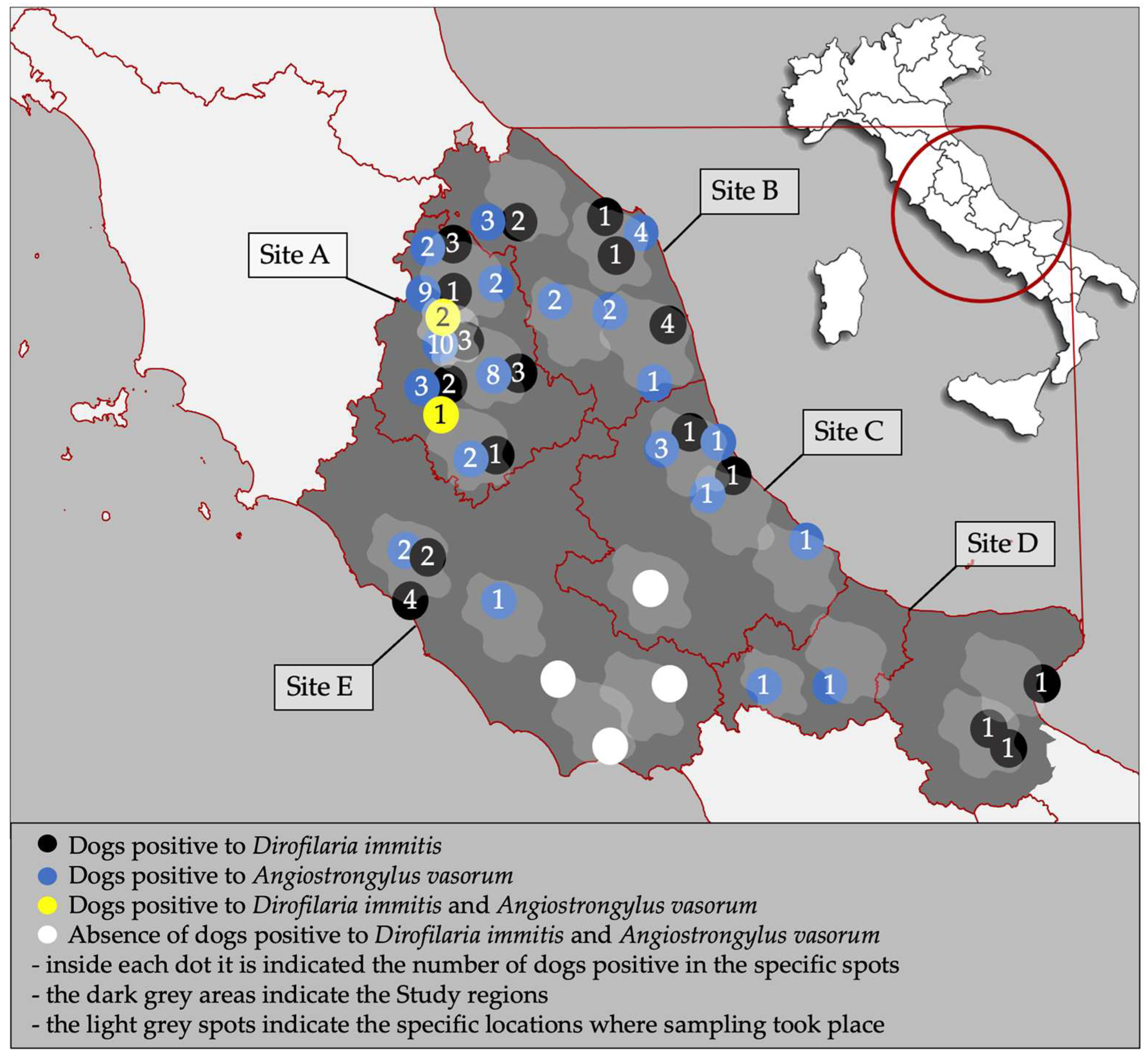
Figure 1. Study sites and distribution of dogs positive for Dirofilaria immitis and Angiostrongylus vasorum in the present study. Light-grey spots indicate the specific areas of sampling within the regions.
Animals 15 00172 g001
Table 1. Demographic data of the dogs enrolled in the present study. Site A: Umbria; Site B: Marche; Site C: Abruzzo; Site D: Molise/northern Apulia; Site E: Latium.
Site ASite BSite CSite DSite ETot
Owned3874003833133741857
Kenneled130175360143
Males2312281961802341069
Females169172204186173931
>4 years old150137185194187853
≤4 years old2502632151722471147
Gastropod ingestion10958272664284
History of travel5096624413265
Hunting13629521244197883
Contact/Cohabitation with other dogs2513313262663911565
Permanent outdoor housing1492022521513031057
Table 2. Number (n) and percentage (%) of dogs testing positive via Knott’s and Baermann’s tests in the different sites examined in this study. Site A: Umbria; Site B: Marche; Site C: Abruzzo; Site D: Molise/northern Apulia; Site E: Latium.
TestParasiteSite A
n/400 (%)
Site B
n/400 (%)
Site C
n/400 (%)
Site D
n/366 (%)
Site E
n/434 (%)
Tot
n/2000 (%)
KnottDirofilaria immitis16 (4)8 (2)2 (0.6)3 (0.8)6 (1.7)35 (1.7)
Dirofilaria repens69 (17.2)52 (13)10 (2.5)3 (0.8)14 (3.4)148 (7.4)
Acanthocheilonema reconditum-3 (0.8)--3 (0.7)6 (0.3)
BaermannAngiostrongylus vasorum39 (10.2)12 (3)6 (1.5)2 (0.5)3 (0.7)62 (3.1)
Strongyloides stercoralis13 (3.2)11 (2.7)---24 (1.2)
Table 3. Number (n) and percentage (%) of dogs testing positive via the flotation method in the different sites. Site A: Umbria; Site B: Marche; Site C: Abruzzo; Site D: Molise/northern Apulia; Site E: Latium.
ParasiteSite A
n/400 (%)
Site B
n/400 (%)
Site C
n/400 (%)
Site D
n/366 (%)
Site E
n/434 (%)
Tot
n/2000 (%)
Ascarids23 (5.7)64 (16)28 (7)12 (3.3)45 (10.4)172 (8.6)
Ancylostomatidae92 (23)90 (22.5)59 (14.7)17 (4.6)65 (15)323 (16.5)
Trichuris vulpis99 (24.7)83 (20.7)20 (5)14 (3.8)33 (7.6)249 (12.4)
Taeniidae2 (0.5)1 (0.2)4 (1)--7 (0.3)
Capillaria aerophila62 (15.5)63 (15.7)20 (5)7 (1.9)65 (15)217 (10.8)
Capillaria boehmi21 (5.2)4 (1)6 (1.5)-13 (3.2)44 (2.2)
Dipylidium caninum-2 (0.5)---2 (0.1)
Cystoisospora spp.-5 (1.2)5 (1.2)2 (0.5)7 (1.6)19 (0.9)
Giardia spp.20 (5)14 (3.5)2 (0.5)-2 (0.5)38 (1.9)
Strongyloides stercoralis-1 (0.25)---1 (0.05)
Table 4. Clinical signs displayed by dogs infected by Dirofilaria immitis (n. 35 dogs) and by Angiostrongylus vasorum (n. 62 dogs).
Dirofilaria immitis
Category of Clinical Signsn (%)Clinical Signn (%)
Cardiorespiratory signs 10 (28.6)Pale mucous membranes10 (28.6)
Dyspnea2 (5.7)
Cough 2 (5.7)
Ocular signs1 (2.85)Uveitis1 (2.85)
Non-specific signs 10 (28.6)Fatigue5 (14.3)
Weight loss5 (14.3)
Vomiting1 (2.9)
Diarrhea1 (2.9)
No clinical signs21 (60)--
Angiostrongylus vasorum
Category of Clinical Signsn (%)Clinical Signn (%)
Neurological2 (3.2)Seizures2 (3.2)
Skin lesions4 (6.4)Dermatitis3 (4.8)
Nodules1 (1.6)
Cardiorespiratory signs18 (29)Dyspnea5 (8.1)
Pale mucous membranes8 (12.9)
Cough12 (19.3)
Signs possibly related to coagulation disorders1 (4.3)Hemoptysis1 (4.3)
Melena1 (4.3)
Hematuria1 (4.3)
Non-specific signs14 (22.6)Weight loss7 (11.3)
Fatigue2 (3.2)
Diarrhea5 (8.1)
No clinical signs36 (58.1)-
Table 5. Results of the binomial logistic regression evaluating statistical associations between potential risk factors and positivity to Dirofilaria immitis and to Angiostrongylus vasorum in the dogs of the present study.
Dirofilaria immitisAngiostrongylus vasorum
FactorpOR95% CIpOR95% CI
Neurological signs0.9942.870–infinity0.1473.670.63–21.30
Skin lesions0.0581.951.32–10.150.5241.440.47–4.39
Cardiorespiratory signs<0.001 *4.932.00–12.20<0.001 *3.511.79–6.90
Ocular signs0.9892.020–infinity0.9845.790–infinity
Signs possibly related to coagulation disorders0.9938.430–infinity0.7991.350.13–13.90
Non-specific signs0.044 *2.411.03–5.670.1451.680.84–3.39
Male sex0.0572.101.00–4.530.3951.270.74–2.18
Gastropod ingestion0.4511.420.57–3.57<0.001 *4.742.72–8.26
Animal movements (within Italy)0.003 *3.471.55–7.790.0920.290.07–1.22
Hunting0.9751.010.45–2.300.3311.370.73–2.56
Contact/cohabitation with other dogs0.8771.070.43–2.670.4390.770.39–1.50
Permanently outdoor housing0.5270.770.35–1.720.029 *0.500.27–0.93
>4 years old0.041 *2.151.03–4.470.9941.000.58–1.73
* significant result; p = p-value; OR = odds ratio; CI = confidence interval.
Disclaimer/Publisher’s Note: The statements, opinions and data contained in all publications are solely those of the individual author(s) and contributor(s) and not of MDPI and/or the editor(s). MDPI and/or the editor(s) disclaim responsibility for any injury to people or property resulting from any ideas, methods, instructions or products referred to in the content.