Article

Effects of Vitamin D3 and 25(OH)D3 Supplementation on Growth Performance, Bone Parameters and Gut Microbiota of Broiler Chickens

by
Rakchanok Phutthaphol
,
Chaiyapoom Bunchasak
,
Wiriya Loongyai
and
Choawit Rakangthong
*
Department of Animal Science, Faculty of Agriculture, Kasetsart University, Bangkok 10900, Thailand
*
Author to whom correspondence should be addressed.
Animals 2025, 15(19), 2900; https://doi.org/10.3390/ani15192900
Submission received: 8 September 2025 / Revised: 29 September 2025 / Accepted: 30 September 2025 / Published: 4 October 2025
(This article belongs to the Section Poultry)

Abstract

Simple Summary

Broiler chickens grow quickly but are usually raised indoors without access to sunlight, which limits their ability to produce vitamin D naturally. Vitamin D is essential for bone strength, growth, and overall health. A special form of vitamin D, called 25-hydroxycholecalciferol [25(OH)D3], is more easily used by the body than normal vitamin D3. In this study, we tested whether adding 25(OH)D3 to diets could benefit broilers. A total of 952 birds were fed diets with no vitamin D3, standard vitamin D3, or vitamin D3 plus two levels of 25(OH)D3. Birds fed diets with either vitamin D3 or 25(OH)D3 grew faster and used feed more efficiently than birds without vitamin D3, while growth was similar between the vitamin D3 and 25(OH)D3 groups. Bone strength did not differ significantly among treatments, although birds given the higher level of 25(OH)D3 showed a slight numerical trend toward stronger bones. We also studied gut bacteria and found that 25(OH)D3 changed the balance of microbes: it reduced excess Lactobacillus and increased bacteria linked with fiber digestion and nutrient absorption. These results show that 25(OH)D3 is more effective than vitamin D3 alone, helping broilers grow better, use feed more efficiently, and maintain a healthier gut.

Abstract

Broiler chickens are commonly reared in closed housing systems with limited exposure to sunlight, thereby relying entirely on dietary sources of vitamin D. The hydroxylated metabolite 25-hydroxycholecalciferol [25(OH)D3] has been proposed as a more potent form than native vitamin D3 (cholecalciferol). This study evaluated the effects of dietary supplementation with vitamin D3 alone or in combination with 25(OH)D3 on growth performance, bone characteristics, and cecal microbiota in Ross 308 broilers. A total of 952 one-day-old male chicks were allocated to four treatments: a negative control (no vitamin D3), a positive control (vitamin D3 according to Ross 308 specifications), and a positive control supplemented with 25(OH)D3 at 1394 or 2788 IU/kg, in a randomized design with 17 replicates per treatment and 14 birds per replicate. Over a 40-day feeding trial, diets containing vitamin D3 (positive control) or supplemented with 25(OH)D3 significantly improved final body weight, weight gain, average daily gain, and feed conversion ratio compared with the negative control (p < 0.01), with no significant differences among the positive control and 25(OH)D3-supplemented groups, with a clear linear dose-dependent response. Although tibia ash and bone-breaking strength were not significantly affected, linear responses indicated a slight numerical trend toward improved skeletal mineralization with increasing 25(OH)D3. Microbiota analysis indicated that 25(OH)D3 affected cecal microbial ecology: low-dose inclusion showed reduced species richness and evenness, whereas high-dose inclusion restored richness to levels comparable to the positive control and enriched taxa associated with fiber fermentation and bile acid metabolism while reducing Lactobacillus dominance. In conclusion, supplementation with 25(OH)D3 in addition to vitamin D3 enhanced growth performance and selectively shaped the cecal microbiota of broilers, with suggestive benefits for bone mineralization. These findings highlight 25(OH)D3 as a more potent source of vitamin D than cholecalciferol alone and support its practical use in modern broiler nutrition to improve efficiency, skeletal health, and microbial balance.

1. Introduction

Modern broiler production has achieved remarkable improvements in growth rate and feed efficiency through intensive genetic selection. However, the rapid increase in body weight often surpasses skeletal development, predisposing birds to leg disorders and locomotion problems. These conditions compromise animal welfare, restrict feed intake, reduce growth, and ultimately lead to economic losses during processing [1,2].
Vitamin D plays an essential role in calcium and phosphorus absorption, which are critical for bone mineralization [3]. Under natural conditions, vitamin D3 (cholecalciferol) is synthesized in the skin upon exposure to ultraviolet B (UV-B) sunlight. Broilers reared in closed housing systems with artificial lighting rarely receive sunlight; thus, their diets must supply adequate vitamin D3 [4]. Once ingested, vitamin D3 is hydroxylated in the liver to form 25-hydroxycholecalciferol (25(OH)D3), the primary circulating metabolite, and subsequently converted in the kidneys into the biologically active 1,25-dihydroxycholecalciferol (1,25(OH)2D3), which regulates calcium and phosphorus homeostasis and bone health [5,6].
In recent years, 25(OH)D3 has become commercially available as a direct dietary supplement in poultry nutrition. Because it bypasses the hepatic conversion step, it may be more efficiently utilized by the body [7,8]. Several studies have reported that 25(OH)D3 exhibits higher bioavailability than vitamin D3 and can improve growth, bone strength, and reduce leg abnormalities in broilers [9,10].
Beyond skeletal functions, vitamin D also contributes to intestinal health by strengthening the gut barrier, modulating inflammation, and including effects on beneficial taxa such as Lactobacillus and suppression of pathogens such as Salmonella [11,12]. Although the positive effects of vitamin D3 or 25(OH)D3 supplementation have been widely documented, limited information is available regarding the potential benefits of combining these two sources at different dietary inclusion levels. Clarifying how such combinations affect growth performance, skeletal development, and gut microbiota could help optimize vitamin D supplementation strategies for modern broiler production. Therefore, the objective of this study was to evaluate the effects of dietary vitamin D3 alone and in combination with two graded levels of 25(OH)D3 on growth performance, tibial bone characteristics, and cecal microbiota composition in broiler chickens.

2. Materials and Methods

2.1. Ethical Approval and Animal Care

All experimental procedures were reviewed and approved by the Institutional Animal Care and Use Committee of Kasetsart University, Bangkok, Thailand (ACKU68-AGR-025). Birds were managed in accordance with the guidelines for the care and use of agricultural animals in research and teaching [13].

2.2. Animals, Experimental Design, and Husbandry

The trial was conducted at Sun Broiler Farm, Nakhon Nayok Province, Thailand. A total of 952 one-day-old male Ross 308 broiler chicks (initial body weight: 41.5 ± 0.5 g) were used in a 40-day feeding trial. Upon arrival, birds were randomly assigned to four dietary treatments using a completely randomized design (CRD), with 14 replicate pens per treatment and 17 birds per pen. Birds were housed in an environmentally controlled facility equipped with an evaporative cooling system. Each of the 56 pens (1 × 2 m; 2 m2 floor area) was bedded with 5 cm-deep rice husk litter. Feed and water were provided ad libitum throughout the experiment. Lighting was maintained at 50 lux according to a standard broiler lighting program (18 h light: 6 h dark), in compliance with the Thai Agricultural Standard TAS 6901-2017 [14]. All birds were vaccinated according to commercial practices: Newcastle disease (day 1), infectious bursal disease (days 1 and 20), and infectious bronchitis (days 1 and 17).

2.3. Experimental Diets

Experimental diets were formulated with maize, wheat, and soybean meal, and nutrient composition was calculated based on Ross 308 nutritional recommendations [15]. Four dietary treatments were tested:
-
Negative control (NC): Negative control (NC): Diet without vitamin D3 supplementation in the premix, used as a biological baseline rather than a commercial model, with welfare carefully maintained.
-
Positive control (PC): Diet supplemented with vitamin D3 according to Ross 308 recommendations.
-
PC + 25(OH)D3_1X: Diet supplemented with vitamin D3 and 25(OH)D3 (commercial product: Bio D®, supported by Huvepharma, Bangkok, Thailand) was supplemented at levels of 1394 IU/kg diet.
-
PC + 25(OH)D3_2X: Diet supplemented with vitamin D3 and 25(OH)D3 from the same source at 2788 IU/kg.
All diets were provided in pellet form and were isoenergetic and isonitrogenous. The detailed ingredient composition and nutrient values are presented in Table 1, and the treatment allocations are summarized in Table 2.

2.4. Growth Performance

Growth performance was evaluated across three growth phases: days 1–11 (starter), days 12–30 (grower), and days 31–40 (finisher). Body weight and feed intake of each replicate pen were recorded at the end of each period. From these data, body weight gain (BWG), average daily gain (ADG), feed conversion ratio (FCR), and mortality rate were calculated. For mortality adjustment, the body weight of dead birds and the amount of residual feed in the corresponding pen were recorded immediately to correct performance calculations. Flock uniformity was assessed in each phase by calculating the coefficient of variation (CV, %) based on the standard deviation of body weight within each pen.

2.5. Bone Parameters

At 40 days of age, sixty birds (15 per treatment) with body weights closest to the pen average were selected for bone sampling. The left tibia was collected for ash content analysis, and the right tibia was collected for mechanical strength measurement. Soft tissues were removed, and bones were sealed in labeled plastic bags and stored at −20 °C until analysis.

2.5.1. Tibia Ash Analysis

Left tibiae were thawed, dried, and finely ground. Ash content was determined following AOAC procedures [16]. Approximately 5–10 g of bone powder was weighed into pre-weighed porcelain crucibles. Samples were charred on a hot plate under a fume hood until smoke emission ceased and the residue turned black. Crucibles were then placed in a muffle furnace at 550 °C for 3 h. If necessary, further incineration was performed until consistent white or gray ash was obtained. After cooling in a desiccator for 30 min, crucibles were reweighed, and tibia ash content was expressed on a dry matter basis.

2.5.2. Tibia Breaking Strength

Right tibiae were used for mechanical testing. After thawing, bones were positioned horizontally on a three-point bending device, with the midpoint subjected to vertical compression using a texture analyzer. Compression was applied at a rate corresponding to 3–6% of the expected ultimate load, with a force increase of 0.9–1.2 MPa/min (9–12 kgf/cm2/min), until fracture occurred. The maximum force at the breaking point was recorded as tibia breaking strength (N).

2.6. Gut Microbiota Analysis

2.6.1. Sample Collection

At 40 days of age, a total of 28 birds (n = 7 replicates per group) with body weights closest to the treatment mean were selected for cecal sampling. Birds were euthanized by carbon dioxide (CO2) stunning. Cecal were excised, ligated with sterile string, and immediately placed into sterile test tubes. Samples were sealed, stored at −20 °C, and subsequently used for nucleic acid extraction to preserve microbial community composition.

2.6.2. DNA Extraction and 16S rRNA Gene Amplicon Sequencing

Microbial DNA was extracted from 300 mg of cecal content per sample using the DNeasy PowerFecal Pro DNA Kit (Qiagen, Germantown, MD, USA). The V3–V4 region of the 16S rRNA gene was amplified with primers 341F (CCTACGGGNGGCWGCAG) and 805R (GACTACHVGGGTATCTAATCC) using 2X sparQ HiFi PCR Master Mix (QuantaBio, Beverly, MA, USA). PCR conditions were: 98 °C for 2 min; 30 cycles of 98 °C for 20 s, 60 °C for 30 s, and 72 °C for 1 min; and a final extension at 72 °C for 1 min. Amplicons were purified with sparQ Puremag Beads, indexed with Nextera XT primers (5 µL each, 8–10 cycles), pooled, and diluted to 4 pM. Sequencing was performed as 250 bp paired-end reads on an Illumina MiSeq platform (Omics Sciences and Bioinformatics Center, Chulalongkorn University, Bangkok, Thailand) [17]. Following quality filtering, sequencing depth ranged from 27,731 to 59,983 reads per sample, with an average of 41,407 high-quality reads.

2.6.3. Bioinformatics and Statistical Analysis

Raw sequencing reads were processed using Quantitative Insights into Microbial Ecology 2 (QIIME 2, version 2020.8) [18]. Sequences were adapter-trimmed, demultiplexed, and low-quality reads and chimeras were removed using the DADA2 plugin. High-quality reads were clustered into operational taxonomic units (OTUs) at 99% similarity against the SILVA 16S rRNA reference database (version 132). Taxonomic classification was assigned using a naïve Bayes classifier trained on the SILVA database. Rarefaction was performed at a depth of 27,731 sequences per sample. Alpha diversity (Observed OUTs, Shannon, Pielou’s evenness, and Faith’s PD) were compared by Kruskal–Wallis with Benjamini–Hochberg correction. Beta diversity was analyzed based on unweighted and weighted UniFrac distances, visualized with principal coordinate analysis (PCoA), and tested for significance using PERMANOVA (999 permutations).

2.7. Statistical Analysis

Data on growth performance and bone parameters were analyzed using the General Linear Model (GLM) procedure in SAS software (version 9.0) based on a completely randomized design with pen as the experimental unit. Model assumptions were verified (Shapiro–Wilk normality; Levene’s test for homogeneity). Results are presented as means with pooled standard error of the mean (SEM). Differences among treatment means were assessed using Tukey’s HSD test [19]. Orthogonal polynomial contrasts were applied to test linear and quadratic responses to increasing dietary vitamin D supplementation. Statistical significance was declared at p < 0.05, and highly significant differences were reported at p < 0.01.

3. Results and Discussion

3.1. Growth Performance

Supplementation with 25(OH)D3, in addition to standard dietary vitamin D3, significantly improved broiler growth performance throughout the 40-day trial (Table 3). Birds receiving 25(OH)D3 at 1394 and 2788 IU/kg exhibited higher final body weight (BW), cumulative body weight gain (BWG), average daily gain (ADG), and improved feed conversion ratio (FCR) compared with the negative control (no vitamin D3) (p < 0.01). However, no significant differences were observed between the positive control (vitamin D3 only) and the 25(OH)D3-supplemented groups. These improvements followed a dose-dependent linear trend (p < 0.01), whereas no quadratic responses were detected. In contrast, feed intake (FI) and mortality rates were unaffected (p > 0.05).
The positive effects of 25(OH)D3 were most evident during the starter (0–11 days) and grower (12–30 days) phases. In the starter phase, birds supplemented with 25(OH)D3 showed significantly higher BW, BWG, and ADG compared with controls, with a clear linear dose–response (p < 0.01). Although FI was not affected, FCR exhibited a mild linear improvement (p = 0.02), suggesting early enhancements in feed efficiency. These early-phase benefits likely reflect the superior biological potency of 25(OH)D3 relative to cholecalciferol [9]. Unlike vitamin D3, 25(OH)D3 bypasses hepatic hydroxylation and is more rapidly converted into the active metabolite 1,25-dihydroxycholecalciferol [1,25(OH)2D3], which enhances calcium and phosphorus absorption through the upregulation of intestinal transport proteins such as TRPV5-6, calbindin-D9k, and PMCA1b [20,21]. Enhanced mineral absorption during the post-hatch period is crucial for skeletal development, thereby supporting efficient muscle accretion and overall growth [9,22].
In the finisher phase (31–40 days), the benefits of vitamin D supplementation were less pronounced. Final BW was significantly higher in supplemented groups (p < 0.01), whereas BWG, ADG, FI, and FCR showed only numerical, non-significant improvements, and mortality was unaffected. This attenuation likely reflects age-related shifts in growth dynamics, as skeletal mineralization slows and energy is directed toward maintenance and fat deposition [10]. Nevertheless, the numerically lower FCR in supplemented groups aligns with previous reports that vitamin D metabolites can modestly enhance feed efficiency through sustained effects on gut integrity and nutrient utilization [9,23].
Over the entire 40-day period, cumulative BW, BWG, ADG, and FCR were significantly improved in birds receiving 25(OH)D3, particularly at 2788 IU/kg (p < 0.01). The absence of adverse effects on feed intake or mortality suggests that supplementation at these levels is both efficacious and safe. Mortality rates did not differ among treatments and were within the expected range for tropical broiler production without antibiotic growth promoters, indicating that the diets did not compromise survival or welfare. Collectively, these findings confirm the dose-dependent efficacy of 25(OH)D3 in enhancing broiler performance through improved mineral metabolism and feed efficiency [23]. Moreover, a recent study in Chinese yellow-feathered broilers reported that dietary vitamin D3 supplementation modulated gut microbiota and improved intestinal morphology, which may have contributed to enhanced nutrient absorption and growth [24].

3.2. Bone Mineralization and Strength

The effects of dietary vitamin D supplementation on tibia ash percentage and bone-breaking strength of broiler chickens are presented in Table 4. No statistically significant differences (p > 0.05) were observed among treatments for tibia ash or breaking strength, either by ANOVA or orthogonal contrast analysis. However, a slight linear trend was noted, with tibia ash (p = 0.08) and bone-breaking strength (p = 0.06) showing numerical increases in response to higher dietary 25(OH)D3 levels.
Broilers receiving the highest 25(OH)D3 supplementation (2788 IU/kg) exhibited the highest numerical values for both tibia ash (42.52 ± 1.51%) and bone strength (35.86 ± 7.04 N), indicating only a numerical linear trend rather than a statistically significant improvement. These trends may reflect the influence of 25(OH)D3 on bone-specific processes such as osteoblast differentiation, matrix mineralization, and regulation of local endocrine pathways within the growth plate. Previous studies have shown that 1,25-dihydroxycholecalciferol, the active form of vitamin D, not only supports mineral absorption but also modulates bone remodeling by stimulating osteocalcin synthesis and enhancing the deposition of calcium and phosphate into the bone matrix [25,26]. Moreover, vitamin D metabolites play an important role in regulating endochondral ossification during early skeletal growth, a critical process for structural integrity in chickens [27,28].
Although this study assessed bone parameters only at market age (day 40), the observed numerical trends suggest that earlier supplementation with 25(OH)D3 may have influenced bone architecture and contributed to higher mineral content and strength at the endpoint. This interpretation is consistent with recent findings indicating that early metabolic programming of bone tissue by vitamin D metabolites can have lasting effects on skeletal quality [29]. While statistical significance was not achieved, the consistent dose-responsive trends observed, coupled with plausible biological mechanisms involving osteoblastic activity and mineral matrix deposition, highlight the potential of 25(OH)D3 to positively influence skeletal development in poultry [6,30]. These findings suggest that the skeletal response to 25(OH)D3 may be cumulative and time-dependent and may also intersect with other physiological systems. In this context, growing evidence has pointed to the gut microbiota as a critical mediator of nutrient metabolism, bone homeostasis, and immune function.

3.3. Microbiota Structure in the Cecal Contents

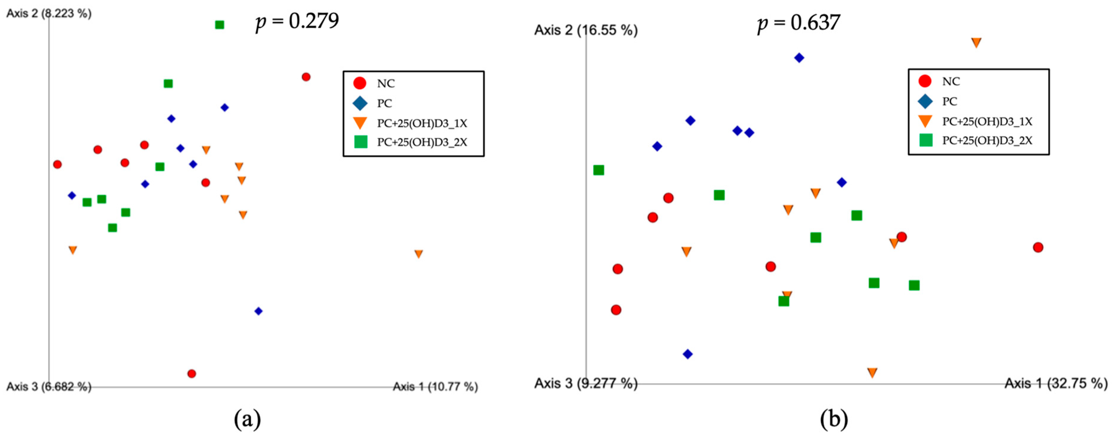
Alpha diversity (Figure 1) differed among treatments for species richness (Observed OTUs; Kruskal–Wallis, p = 0.028). Post hoc testing showed lower richness in the 25(OH)D3 1394 IU/kg group than the positive group (p < 0.05) and higher richness in the 25(OH)D3 2788 IU/kg group than the 25(OH)D3 1394 IU/kg group (p < 0.05). No overall difference was detected for Shannon diversity (p = 0.084), although pairwise comparison suggested that the negative control tended to have a higher Shannon index than the 25(OH)D3 1394 IU/kg group (p = 0.025). Pielou’s evenness differed among treatments (p = 0.042), with the negative group showing higher evenness than both 25(OH)D3 groups (1394 IU/kg: p = 0.012; 2788 IU/kg: p = 0.025). Faith’s phylogenetic diversity did not differ (p = 0.224). Beta diversity (Figure 2) based on PCoA showed no distinct clustering by treatment, and PERMANOVA confirmed non-significant differences for both unweighted UniFrac (p = 0.279) and weighted UniFrac (p = 0.637), indicating that supplementation affected within-community diversity metrics without overtly restructuring overall community composition.
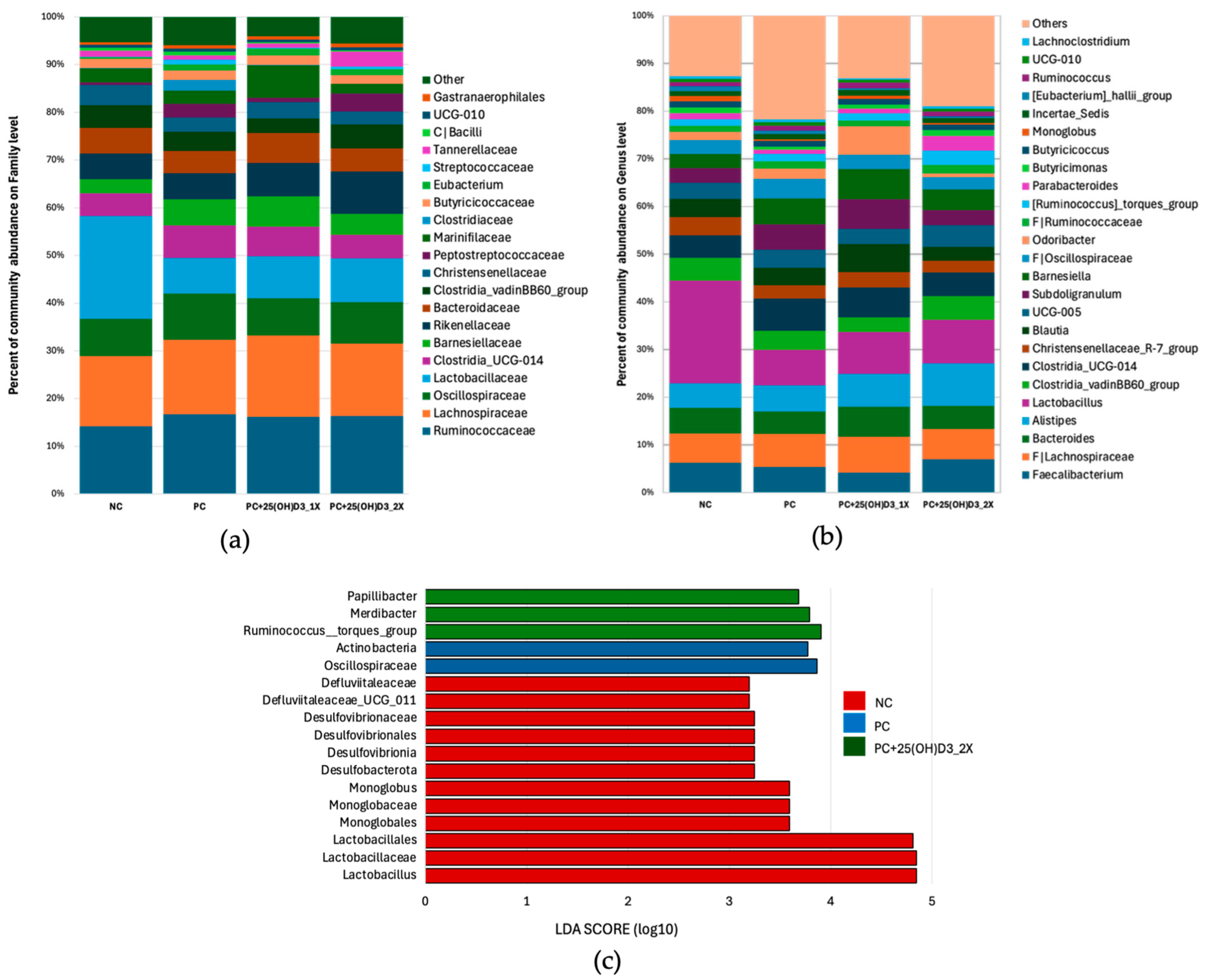
At the family/genus levels (Figure 3a,b), the negative group was characterized by higher relative abundance of Lactobacillaceae and its representative genus Lactobacillus than all supplemented groups. Vitamin D3 supplementation (positive) and diets containing 25(OH)D3 were associated with increased representation of families linked to fiber and bile-acid metabolism (e.g., Oscillospiraceae, Christensenellaceae), and with shifts in several genera. Specifically, the 25(OH)D3 2788 IU/kg group showed higher proportions of Alistipes and enrichment of fiber- and mucus-degrading taxa (Ruminococcus torques group, Papillibacter, Merdibacter), while the 25(OH)D3 1394 IU/kg group displayed relatively higher Barnesiella and Subdoligranulum; supplemented groups also tended to have more Clostridia UCG-014, Clostridia vadinBB60 group, and Christensenellaceae R-7 group. Consistent with these patterns, LEfSe (LDA cutoff > 3.0) identified discriminative taxa (Figure 3c): the negative group was enriched in Lactobacillus together with higher taxa (Lactobacillaceae, Lactobacillales), plus members of Monoglobaceae, Defluviitaleaceae, and Desulfovibrionales; the positive group was marked by Oscillospiraceae (and other Actinobacteria-related features); and the 25(OH)D3 2788 IU/kg group was marked by Ruminococcus torques group, Papillibacter, and Merdibacter.
The present findings indicate that dietary supplementation with vitamin D3 and 25(OH)D3 modulated cecal microbial ecology in a dose-dependent manner. Alpha diversity measures revealed that moderate inclusion of 25(OH)D3 (1394 IU/kg) reduced species richness and evenness, whereas higher inclusion (2788 IU/kg) restored richness to levels comparable to the positive group. These results suggest that 25(OH)D3 exerts selective pressures on microbial communities, potentially through modulation of intestinal epithelial function, antimicrobial peptide secretion, and mucosal immunity mediated by the vitamin D receptor (VDR) [6,31,32,33]. Importantly, although certain taxa showed localized changes in richness and evenness, beta diversity analyses confirmed that the overall community structure remained stable across treatments. This indicates that the effects of vitamin D3 and 25(OH)D3 represent localized adjustments within the microbial community rather than major restructuring.
Our taxonomic profiling showed that Lactobacillus was most abundant in the negative control group, while vitamin D3 and 25(OH)D3 supplementation reduced its dominance and enriched other functional taxa. Recent evidence indicates that vitamin D influences gut microbiota mainly through VDR signaling [34]. Impaired VDR expression or activation has been associated with reduced Lactobacillus populations and expansion of Proteobacteria, leading to a more pro-inflammatory environment [35]. Conversely, adequate vitamin D status or butyrate-mediated enhancement of VDR supports microbial balance and intestinal immune function [36]. Thus, the reduction in Lactobacillus observed here should be interpreted in context: while lower dominance may support greater diversity, Lactobacillus itself also contributes to host health, and its role likely depends on maintaining an appropriate balance within the community.
Supplementation with vitamin D3, and particularly 25(OH)D3, enriched several bacterial taxa associated with gut homeostasis and immunomodulatory functions. Notably, this included Oscillospiraceae, Ruminococcus torques group, and Papillibacter—genera reported in previous studies as important short-chain fatty acid (SCFA) producers and modulators of immune responses [37,38,39]. Such functions are generally linked with nutrient absorption, intestinal stability, and host metabolic health. Interestingly, the enrichment patterns differed between treatments: Oscillospiraceae was identified as a key biomarker in the vitamin D3 group, while the Ruminococcus torques group and Papillibacter were particularly elevated in the high-dose 25(OH)D3 group. Collectively, these microbial shifts are thought to enhance metabolic and immunomodulatory potential [40,41,42]. However, as our study did not directly measure SCFA concentrations, intestinal inflammation, or immune responses, these mechanisms remain uncertain and require confirmation in future studies.

4. Conclusions

This study demonstrated that dietary supplementation with 25-hydroxycholecalciferol [25(OH)D3], in addition to standard vitamin D3, improved growth performance of broiler chickens in a dose-dependent manner. Broilers fed diets with vitamin D3 or 25(OH)D3 showed improved growth performance compared with the negative control, while no significant differences were detected between the vitamin D3 and 25(OH)D3 groups. Linear responses suggested that potential benefits of 25(OH)D3 were more apparent during the early growth phase. Although tibia ash and bone-breaking strength were not significantly affected, linear response patterns indicated only numerical trends toward higher mineralization and skeletal integrity at greater supplementation levels.
Cecal microbiota analysis revealed that vitamin D3 and 25(OH)D3 selectively modulated microbial diversity and composition. While moderate inclusion reduced species richness and evenness, higher supplementation restored microbial richness and enriched taxa associated with fiber fermentation, bile acid metabolism, and short-chain fatty acid production. These microbial shifts, together with enhanced mineral utilization, likely contributed to the improvements in growth performance and skeletal development observed in this study.
Overall, the findings indicate that 25(OH)D3 is a more potent source of vitamin D than cholecalciferol alone, supporting growth efficiency, skeletal health, and microbial balance in broilers. From a practical standpoint, incorporating 25(OH)D3 into commercial diets—particularly at higher inclusion levels—may improve feed efficiency and bird welfare. Future research should refine optimal dosages and evaluate long-term impacts on immunity, carcass quality, and resilience under diverse production systems.

Author Contributions

Conceptualization, C.R. and C.B.; methodology, C.B.; software, R.P.; validation, C.R., C.B. and W.L.; formal analysis, R.P.; investigation, R.P.; resources, C.B.; data curation, R.P.; writing—original draft preparation, R.P.; writing—review and editing, C.R.; visualization, C.R.; supervision, C.R.; project administration, C.R., C.B. and W.L.; funding acquisition, C.B. All authors have read and agreed to the published version of the manuscript.

Funding

This research was funded by Huvepharma (Thailand) Co., Ltd.

Institutional Review Board Statement

The animal study protocol was approved by the Institutional Animal Care and Use Committee of Kasetsart University, Bangkok, Thailand (protocol code ACKU68-AGR-025).

Informed Consent Statement

Informed consent was obtained from the owner/representative of Sun Broiler Farm prior to the initiation of the study.

Data Availability Statement

The data presented in this study are available on request from the corresponding author upon reasonable request.

Conflicts of Interest

The authors declare no conflicts of interest. Huvepharma (Thailand) Co., Ltd. funded this study and supplied the vitamin D3 and 25(OH)D3 supplements. However, the company had no role in the study design, data collection, analysis, interpretation, or the decision to publish the results.

References

  1. Whitehead, C.C. Overview of bone biology in the egg-laying hen. Poult. Sci. 2004, 83, 193–199. [Google Scholar] [CrossRef] [PubMed]
  2. Shim, M.Y.; Karnuah, A.B.; Mitchell, A.D.; Anthony, N.B.; Pesti, G.M.; Aggrey, S.E. The effects of growth rate on leg morphology and tibia breaking strength, mineral density, mineral content, and bone ash in broilers. Poult. Sci. 2012, 91, 1790–1795. [Google Scholar] [CrossRef] [PubMed]
  3. Whitehead, C.C.; McCormack, H.A.; McTeir, L.; Fleming, R.H. High vitamin D3 requirements in broilers for bone quality and prevention of tibial dyschondroplasia and interactions with dietary calcium, available phosphorus and vitamin A. Br. Poult. Sci. 2004, 45, 425–436. [Google Scholar] [CrossRef]
  4. Bikle, D.D. Vitamin D metabolism, mechanism of action, and clinical applications. Chem. Biol. 2014, 21, 319–329. [Google Scholar] [CrossRef]
  5. DeLuca, H.F. Overview of general physiologic features and functions of vitamin D. Am. J. Clin. Nutr. 2004, 80, S1689–S1696. [Google Scholar] [CrossRef]
  6. Świątkiewicz, S.; Arczewska-Włosek, A.; Bederska-Łojewska, D. Efficacy of dietary vitamin D and its metabolites in poultry—Review and implications of the recent studies. World’s Poult. Sci. J. 2017, 73, 57–68. [Google Scholar] [CrossRef]
  7. Plum, L.A.; DeLuca, H.F. Vitamin D, disease and therapeutic opportunities. Nat. Rev. Drug Discov. 2010, 9, 941–955. [Google Scholar] [CrossRef]
  8. Holick, M.F.; Chen, T.C. Vitamin D deficiency: A worldwide problem with health consequences. Am. J. Clin. Nutr. 2008, 87, S1080–S1086. [Google Scholar] [CrossRef]
  9. Fritts, C.A.; Waldroup, P.W. Effect of source and level of vitamin D on live performance and bone development in growing broilers. J. Appl. Poult. Res. 2003, 12, 45–52. [Google Scholar] [CrossRef]
  10. Leyva-Jimenez, H.; Khan, M.; Gardner, K.; Abdaljaleel, R.A.; Yansoon, A.J.; Alsadwi, A.M.; Bailey, C.A. Developing a novel oral vitamin D3 intake bioassay to re-evaluate the vitamin D3 requirement for modern broiler chickens. Poult. Sci. 2019, 98, 3770–3776. [Google Scholar] [CrossRef]
  11. Bar, A.; Sharvit, M.; Noff, D.; Edelstein, S.; Hurwitz, S. Absorption and excretion of cholecalciferol and of 25-hydroxycholecalciferol and metabolites in birds. J. Nutr. 1980, 110, 1930–1934. [Google Scholar] [CrossRef]
  12. Guo, F.; Geng, Y.; Abbas, W.; Zhen, W.; Wang, S.; Huang, Y.; Guo, Y.; Ma, Q.; Wang, Z. Vitamin D3 nutritional status affects gut health of Salmonella-challenged laying hens. Front. Nutr. 2022, 9, 888580. [Google Scholar] [CrossRef] [PubMed]
  13. FASS. Guide for the Care and Use of Agricultural Animals in Research and Teaching, 4th ed.; Federation of Animal Science Societies: Champaign, IL, USA, 2020. [Google Scholar]
  14. Agricultural Commodity and Food Standards (ACFS). Guidance on the Application of Thai Agricultural Standard (TAS 6901-2017). ACFS: Bangkok, Thailand, 2017. Available online: https://acfs-backend.acfs.go.th/storage/ProductStandards/Files//20190614120531_637937.pdf (accessed on 15 August 2025).
  15. Aviagen. Ross 308 Broiler: Nutrition Specifications; Aviagen Group: Huntsville, AL, USA, 2019; Available online: https://en.aviagen.com/ (accessed on 24 August 2025).
  16. AOAC. Official Methods of Analysis, 15th ed.; Association of Official Analytical Chemists: Arlington, VA, USA, 1990. [Google Scholar]
  17. Kechin, A.; Boyarskikh, U.; Kel, A.; Filipenko, M. cutPrimers: A New Tool for Accurate Cutting of Primers from Reads of Targeted Next Generation Sequencing. J. Comput. Biol. 2017, 24, 1138–1143. [Google Scholar] [CrossRef] [PubMed]
  18. Bolyen, E.; Rideout, J.R.; Dillon, M.R.; Bokulich, N.A.; Abnet, C.C.; Al-Ghalith, G.A.; Alexander, H.; Alm, E.J.; Arumugam, M.; Asnicar, F.; et al. Reproducible, interactive, scalable and extensible microbiome data science using QIIME 2. Nat. Biotechnol. 2019, 37, 852–857. [Google Scholar] [CrossRef] [PubMed]
  19. Steel, R.G.D.; Torrie, J.H. Principles and Procedures of Statistics: A Biometrical Approach, 2nd ed.; McGraw-Hill: New York, NY, USA, 1980. [Google Scholar]
  20. Fleet, J.C. The role of vitamin D in the endocrinology controlling calcium homeostasis. Mol. Cell Endocrinol. 2017, 453, 36–45. [Google Scholar] [CrossRef]
  21. Wongdee, K.; Charoenphandhu, N. Vitamin D-enhanced duodenal calcium transport. Vitam. Horm. 2015, 98, 407–440. [Google Scholar] [CrossRef]
  22. Hutton, K.; Vaughn, M.; Litta, G.; Turner, B.; Starkey, J. Effect of vitamin D status improvement with 25-hydroxycholecalciferol on skeletal muscle growth characteristics and satellite cell activity in broiler chickens. J. Anim. Sci. 2014, 92, 411–420. [Google Scholar] [CrossRef]
  23. Yarger, J.G.; Saunders, C.A.; McNaughton, J.L.; Quarles, C.L.; Hollis, B.W.; Gray, R.W. Comparison of dietary 25-hydroxycholecalciferol and cholecalciferol in broiler chickens. Poult. Sci. 1995, 74, 1159–1167. [Google Scholar] [CrossRef]
  24. Wei, J.; Li, L.; Peng, Y.; Luo, J.; Chen, T.; Xi, Q.; Zhang, Y.; Sun, J. The Effects of Optimal Dietary Vitamin D3 on Growth and Carcass Performance, Tibia Traits, Meat Quality, and Intestinal Morphology of Chinese Yellow-Feathered Broiler Chickens. Animals 2024, 14, 920. [Google Scholar] [CrossRef]
  25. Norman, A.W. From vitamin D to hormone D: Fundamentals of the vitamin D endocrine system essential for good health. Am. J. Clin. Nutr. 2008, 88, S491–S499. [Google Scholar] [CrossRef]
  26. Christakos, S.; Dhawan, P.; Porta, A.; Mady, L.J.; Seth, T. Vitamin D and intestinal calcium absorption. Mol. Cell Endocrinol. 2011, 347, 25–29. [Google Scholar] [CrossRef]
  27. van de Peppel, J.; van Leeuwen, J.P.T.M. Vitamin D and gene networks in human osteoblasts. Front. Physiol. 2014, 5, 137. [Google Scholar] [CrossRef] [PubMed]
  28. Anderson, P.H.; Turner, A.G.; Morris, H.A. Vitamin D actions to regulate calcium and skeletal homeostasis. Clin. Biochem. 2012, 45, 880–886. [Google Scholar] [CrossRef] [PubMed]
  29. Chen, C.; Turner, B.; Applegate, T.J.; Litta, G.; Kim, W.K. Role of long-term supplementation of 25-hydroxyvitamin D3 on laying hen bone 3-dimensional structural development. Poult. Sci. 2020, 99, 5771–5782. [Google Scholar] [CrossRef] [PubMed]
  30. Li, D.; Ding, X.; Bai, S.; Wang, J.; Zeng, Q.; Peng, H.; Xuan, Y.; Zhang, K. Effects of Supplementation of 25-Hydroxyvitamin D3 as a Vitamin D3 Substitute on Performance, Bone Traits, and Egg Quality of Laying Hens from 1 Day to 72 Weeks of Age. Agriculture 2023, 13, 383. [Google Scholar] [CrossRef]
  31. Gombart, A.F. The Vitamin D–Antimicrobial Peptide Pathway and Its Role in Protection Against Infection. Future Microbiol. 2009, 4, 1151–1165. [Google Scholar] [CrossRef]
  32. Sun, J. Vitamin D and mucosal immune function. Curr. Opin. Gastroenterol. 2010, 26, 591–595. [Google Scholar] [CrossRef]
  33. Jiang, J.; Chen, G.; Shuler, F.D.; Wang, C.H.; Xie, J. Local sustained delivery of 25-hydroxyvitamin D3 for production of antimicrobial peptides. Pharm. Res. 2015, 32, 2851–2862. [Google Scholar] [CrossRef]
  34. Akimbekov, N.S.; Digel, I.; Sherelkhan, D.K.; Lutfor, A.B.; Razzaque, M.S. Vitamin D and the Host-Gut Microbiome: A Brief Overview. Acta Histochem. Cytochem. 2020, 53, 33–42. [Google Scholar] [CrossRef]
  35. Battistini, C.; Ballan, R.; Herkenhoff, M.E.; Saad, S.M.I.; Sun, J. Vitamin D Modulates Intestinal Microbiota in Inflammatory Bowel Diseases. Int. J. Mol. Sci. 2021, 22, 362. [Google Scholar] [CrossRef]
  36. He, L.; Liu, T.; Shi, Y.; Tian, F.; Hu, H.; Deb, D.K.; Chen, Y.; Bissonnette, M.; Li, Y.C. Gut epithelial vitamin D receptor regulates microbiota-dependent mucosal inflammation by suppressing intestinal epithelial cell apoptosis. Endocrinology 2018, 159, 967–979. [Google Scholar] [CrossRef]
  37. Liu, P.T.; Stenger, S.; Li, H.; Wenzel, L.; Tan, B.H.; Krutzik, S.R.; Ochoa, M.T.; Schauber, J.; Wu, K.; Meinken, C.; et al. Toll-like receptor triggering of a vitamin D–mediated human antimicrobial response. Science 2006, 311, 1770–1773. [Google Scholar] [CrossRef]
  38. Sun, B.; Hou, L.; Yang, Y. The development of the gut microbiota and short-chain fatty acids of layer chickens in different growth periods. Front. Vet. Sci. 2021, 8, 666535. [Google Scholar] [CrossRef]
  39. Pan, D.; Yu, Z. Intestinal microbiome of poultry and its interaction with host and diet. Gut Microbes 2014, 5, 108–119. [Google Scholar] [CrossRef]
  40. Yang, J.; Li, Y.; Wen, Z.; Liu, W.; Meng, L.; Huang, H. Oscillospira—A candidate for the next-generation probiotics. Gut Microbes 2021, 13, 1987783. [Google Scholar] [CrossRef]
  41. Fuentes, S.; den Hartog, G.; Nanlohy, N.M.; Wijnands, L.; Ferreira, J.A.; Nicolaie, M.A.; Pennings, J.L.A.; Jacobi, R.; de Wit, J.; van Beek, J.; et al. Associations of faecal microbiota with influenza-like illness in participants aged 60 years or older: An observational study. Lancet Healthy Longev. 2021, 2, e436–e445. [Google Scholar] [CrossRef]
  42. Zong, X.; Zhang, H.; Zhu, L.; Deehan, E.C.; Fu, J.; Wang, Y.; Jin, M. Auricularia auricula polysaccharides attenuate obesity in mice through gut commensal Papillibacter cinnamivorans. J. Adv. Res. 2023, 52, 203–218. [Google Scholar] [CrossRef] [PubMed]
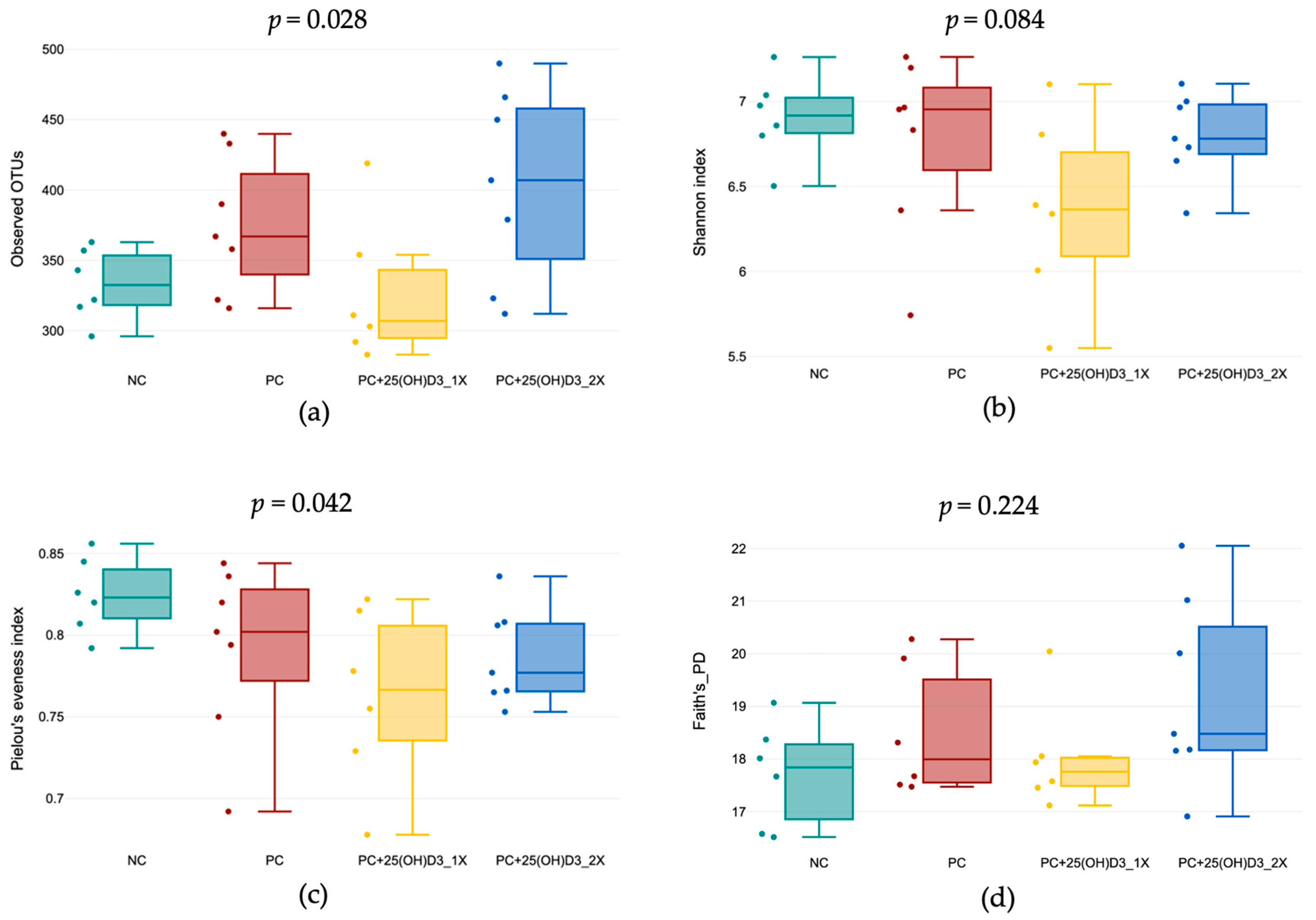
Figure 1. Alpha diversity of cecal microbiota in broilers. (a) Observed OTUs, (b) Shannon index, (c) Pielou’s evenness index, and (d) Faith’s phylogenetic diversity (PD) index.
Animals 15 02900 g001
Figure 2. The PCoA Emperor plots from (a) unweighted and (b) weighted UniFrac distances analyses of cecal microbiota in broilers.
Animals 15 02900 g002
Figure 3. Relative abundance of cecal microbiota in broilers: (a) family level, (b) genus level, and (c) LDA scores of taxonomic biomarkers identified by LEfSe (log10).
Animals 15 02900 g003
Table 1. The composition and the nutrient levels of the basal diets.
Ingredients (kg)StarterGrowerFinisher
Corn33.1149.1343.15
Broken rice10.0010.00-
Wheat10.00-20.00
Wheat bran2.00--
Full-fat soybean10.0010.0012.00
Soybean meal 44%31.4627.6320.16
Acid oil-0.252.15
Monodicalcium phosphate0.950.850.52
Limestone1.201.050.70
Sodium bicarbonate0.200.200.30
Choline0.030.030.00
DL-methionine0.360.300.26
L-Lysine0.180.130.14
L-threonine0.100.070.06
Phytase0.010.010.01
NSP Enzyme0.010.010.01
Salt0.250.200.20
Mineral Premix 10.100.100.10
Vitamin Premix 20.040.040.04
Total (kg)100.00100.00100.00
Nutrients by calculation
AME, Kcal/kg3000.003100.003200.00
Crude protein, %23.5021.5019.50
Fiber, %3.102.923.05
Fat, %4.134.656.95
Methionine, %0.560.510.47
Methionine + Cystine, %1.080.990.90
Lysine, %1.441.291.15
Threonine, %0.970.880.78
Valine, %1.111.000.89
Isoleucine, %0.970.890.80
Arginine, %1.521.371.21
Tryptophan, %0.230.210.18
Calcium, %0.960.880.78
Total phosphorus, %0.720.670.62
Available phosphorus, %0.480.440.39
Na, %0.230.230.20
DEB, mEq/kg292.63266.10233.49
NSP: Non-starch polysaccharides; AME: Apparent Metabolizable Energy; 1 The mineral premix supplied the following per kg of complete feed: Cu, 16 mg; Zn, 120 mg; Fe, 20 mg; Mn, 120 mg; I, 1.25 mg; Selenium, 0.30 mg. 2 The vitamin premix supplied the following per kg of complete feed: vitamin A, 11,500 IU; vitamin K3, 3.60 mg; vitamin B1, 4 mg; vitamin B2, 8 mg; vitamin B12, 0.025 mg; vitamin E, 65 IU; biotin, 0.28 mg; folic acid, 2.00 mg; pantothenic acid, 25 mg; niacin, 65 mg (vitamin D3 not included). DEB: Dietary electrolyte balance, calculated as Na + K − Cl (mEq/kg diet).
Table 2. Vitamin D3 and 25(OH)D3 supplementation in treatment diets (per kg).
TreatmentsStarterGrowerFinisher
NCNNN
PCVit D3 5000 IUVit D3 4500 IUVit D3 4000 IU
PC+25(OH)D3_1XVit D3 5000 IU
+25(OH)D3 1394 IU
Vit D3 4500 IU
+25(OH)D3 1394 IU
Vit D3 4000 IU
+25(OH)D3 1394 IU
PC+25(OH)D3_2XVit D3 5000 IU
+25(OH)D3 2788 IU
Vit D3 4500 IU
+25(OH)D3 2788 IU
Vit D3 4000 IU
+25(OH)D3 2788 IU
N = Not supplementing vitamin D3 in premix. The NC premix did not contain vitamin D3, so any vitamin D in this diet was provided only by the feed ingredients.
Table 3. Effects of vitamin D3 and 25(OH)D3 supplementation on growth performance of broiler chickens.
ItemNCPCPC+PC+SEMp-Value
25(OH)D3_1X25(OH)D3_2X ANOVALQ
Starter phase (0–11 days)
BW (g/b)294.45 b ± 19.34306.98 a ± 14.47310.13 a ± 8.41314.91 a ± 10.242.06<0.01<0.010.30
BWG (g/b)251.55 b ± 19.34264.09 a ± 14.47267.23 a ± 8.41272.01 a ± 10.242.06<0.01<0.010.30
ADG (g/b)22.87 b ± 1.7624.01 a ± 1.3224.29 a ± 0.7624.73 a ± 0.930.19<0.01<0.010.30
FI (g/b)332.69 ± 15.19348.03 ± 21.34346.41 ± 14.82346.12 ± 23.952.630.140.100.13
FCR1.33 ± 0.061.32 ± 0.061.30 ± 0.041.27 ± 0.080.010.110.020.65
Mortality (%)0.84 ± 2.140.42 ± 1.570.00 ± 0.000.42 ± 1.570.200.560.370.31
Grower phase (12–30 days)
BW (g/b)1719.05 b ± 50.491784.05 a ± 42.571764.88 a ± 64.561803.95 a ± 74.068.78<0.01<0.010.42
BWG (g/b)1408.93 b ± 47.241469.14 a ± 42.051470.43 a ± 52.911496.96 a ± 66.708.15<0.01<0.010.24
ADG (g/b)74.15 b ± 2.4977.32 a ± 2.2177.39 a ± 2.7878.79 a ± 3.510.43<0.01<0.010.24
FI (g/b)2120.73 ± 86.192168.44 ± 59.972116.09 ± 84.312151.33 ± 88.3110.860.270.680.77
FCR1.51 a ± 0.051.48 ab ± 0.051.44 b ± 0.051.44 b ± 0.070.01<0.01<0.010.36
Mortality (%)3.78 ± 2.923.78 ± 4.954.20 ± 4.862.94 ± 4.470.570.890.690.59
Finisher phase (31–40 days)
BW (g/b)2793.03 b ± 122.262913.80 a ± 83.912870.30 a ± 72.812934.38 a ± 78.8213.96<0.01<0.010.25
BWG (g/b)1073.98 ± 103.451122.61 ± 63.481105.43 ± 56.111130.43 ± 79.7210.530.240.110.57
ADG (g/b)107.40 ± 10.35112.26 ± 6.35110.54 ± 5.61113.04 ± 7.971.050.240.110.57
FI (g/b)1864.01 ± 83.201863.97 ± 118.421900.74 ± 87.471894.34 ± 64.0911.970.580.240.90
FCR1.75 ± 0.141.66 ± 0.111.72 ± 0.111.68 ± 0.120.020.240.340.49
Mortality (%)2.52 ± 3.802.10 ± 2.923.36 ± 5.012.10 ± 2.920.490.791.000.68
Overall (0–40 days)
BW (g/b)2793.03 b ± 122.262913.80 a ± 83.912870.30 a ± 72.812934.38 a ± 78.8213.96<0.01<0.010.25
BWG (g/b)2750.14 b ± 122.262870.91 a ± 83.912827.41 a ± 72.812891.48 a ± 78.8213.96<0.01<0.010.25
ADG (g/b)68.75 b ± 3.0671.77 a ± 2.1070.69 a ± 1.8272.29 a ± 1.970.35<0.01<0.010.25
FI (g/b)4317.42 ± 133.624380.43 ± 122.004363.24 ± 117.044391.79 ± 105.7516.060.380.160.59
FCR1.57 a ± 0.041.53 b ± 0.031.54 b ± 0.031.52 b ± 0.030.01<0.01<0.010.27
Mortality (%)7.56 ± 6.295.58 ± 6.107.56 ± 6.705.46 ± 4.870.790.700.520.90
ab Means with different superscripts within the same row are significantly different (p < 0.05). Values are presented as mean ± standard deviation (mean ± SD). SEM = standard error of means. Orthogonal contrasts: L = Linear; Q = Quadratic.
Table 4. Effects of vitamin D3 and 25(OH)D3 supplementation on bone mineralization and strength of broiler chickens.
ItemNCPCPC+PC+SEMp-Value
25(OH)D3_1X25(OH)D3_2X ANOVALQ
Tibia ash (%)41.30 ± 1.7042.08 ± 2.4042.44 ± 1.3842.52 ± 1.510.200.510.080.49
Bone-breaking strength (N)30.26 ± 5.2332.03 ± 6.6333.49 ± 4.8735.86 ± 7.040.930.210.060.89
Data are presented as mean ± standard deviation (mean ± SD). SEM = standard error of means. Orthogonal contrasts: L = Linear, Q = Quadratic.
Disclaimer/Publisher’s Note: The statements, opinions and data contained in all publications are solely those of the individual author(s) and contributor(s) and not of MDPI and/or the editor(s). MDPI and/or the editor(s) disclaim responsibility for any injury to people or property resulting from any ideas, methods, instructions or products referred to in the content.