Review

Selected Pathologies of the Male Genital Organs in Bulls, Including Frequency, Significance, and Risk Factors: A Review

by
Aleksander F. Butkiewicz
1,*,
Maciej Zdun
1,2 and
Jędrzej M. Jaśkowski
3
1
Department of Basic and Preclinical Sciences, Institute of Veterinary Medicine, Faculty of Biological and Veterinary Sciences, Nicolaus Copernicus University, 87-100 Torun, Poland
2
Department of Animal Anatomy, Poznan University of Life Sciences, Wojska Polskiego 71C, 60-625 Poznan, Poland
3
Department of Diagnostics and Clinical Sciences, Institute of Veterinary Medicine, Faculty of Biological and Veterinary Sciences, Nicolaus Copernicus University, 87-100 Torun, Poland
*
Author to whom correspondence should be addressed.
Animals 2025, 15(19), 2804; https://doi.org/10.3390/ani15192804
Submission received: 28 July 2025 / Revised: 12 September 2025 / Accepted: 23 September 2025 / Published: 25 September 2025
(This article belongs to the Special Issue Reproductive Diseases in Ruminants)

Abstract

Simple Summary

This study provides an overview of the most common genital pathologies in bulls, including their frequency, significance, and risk factors. These pathologies can cause infertility, reduce semen quality, impair copulation, and lead to reluctance to copulate; they also pose a risk of transmitting infectious diseases. This study discusses selected pathologies of the male genital tract, such as orchitis, testicular hypoplasia, testicular degeneration, testicular hydrocele, malignant tumors, spermatocele, penile papillomatosis, penile hematoma, and other diseases that occur with varying frequency in cattle herds. Information is provided on the incidence of these diseases, their causes, and treatment methods. Understanding these issues is important for breeders, herd personnel, and veterinarians. The study also discusses the diagnosis and treatment of these diseases. Measures must be taken to minimize the incidence of genital defects in bulls to improve breeding efficiency and animal health.

Abstract

Bulls can develop various conditions affecting the penis, testicles, and associated structures that reduce fertility, spread disease, and increase costs for farmers. This review synthesizes data from farm fertility examinations and slaughterhouse findings to illustrate the prevalence of these conditions, their causes (including inherited defects, infections, and injuries), their impacts on breeding, and potential interventions. Conditions discussed include orchitis, testicular hypoplasia, testicular degeneration, testicular hydrocele, malignant tumors, spermatocele, penile papillomatosis, and penile hematoma. Lameness and systemic illnesses can also impair a bull’s breeding ability. Regular fertility examinations assessing overall health, testicle size, semen motility, and sperm morphology, coupled with timely medical or surgical treatment, can improve reproductive outcomes. Maintaining national records of birth defects and health histories could aid in preventing the transmission of deleterious traits. Reducing these disorders enhances animal welfare, protects herd fertility and the food supply, and supports the economic viability of cattle producers.

1. Introduction

Genital malformations are common defects found in bulls; however, there are no accurate data on the prevalence of these pathologies. Nonetheless, tracking their incidence in herds, identifying potential risk factors, and preventing and eliminating these problems are important for ensuring livestock welfare and increasing economic returns for breeders. To avoid costs and unnecessary animal suffering, a nationwide register of congenital disabilities in bulls should be introduced, including data on each bull’s genetic profile and genealogy, in order to eliminate the potential transmission of defective traits to offspring. Such registries exist, for example, in human medicine [1].
Pathologies of the genital organs in bulls are classified as acquired or congenital lesions. Penile frenulum disorders, congenital penile deformities, stuttering, bacterial and viral lesions, and mechanical trauma are among the most common pathologies of the bull reproductive system [2]. These diseases can cause infertility, reduce semen quality, lead to genitourinary pathologies, and limit the economic potential of breeding. Understanding the causes, symptoms, and treatment of these diseases is important for breeders, herd personnel, veterinarians, and animal technicians [3].
In young bulls, the bull breeding soundness exam (BBSE) is performed based on criteria developed by the Society for Theriogenology [4,5]. The evaluation is conducted at 12–14 months of age and includes a physical examination, measurement of testicular circumference, analysis of semen motility, and assessment of sperm morphology. A bull found to have pathologies is classified as unsatisfactory in terms of reproductive capacity. Typically, estimation of the BBSE is sufficient to identify bulls exhibiting undesirable reproductive traits [6]. The results of such tests are a valuable source of information about defects and diseases of the reproductive system in young bulls. A second source of data is post-mortem examination of bulls of various ages. Under field conditions, a small proportion of pathological changes are sometimes reported to the veterinarian or result in the bull being transferred to a clinic.
This review provides an overview of selected pathologies affecting the male reproductive organs in cattle, with a particular focus on their prevalence (Table 1 and Table 2), diagnostic approaches, therapeutic interventions, and preventive measures.

2. Testicular and Epididymal Pathologies

2.1. Orchitis

It has been estimated that orchitis affects about 1.94–3.3% of bulls [15]. In young Nellore bulls, the incidence is 2.04% [16]. A more recent pre-slaughter study conducted in 2014–2016 found that orchitis affected 7.1% of bulls [17]. The frequency of orchitis ranges from 2% to 4.4% in post-slaughter studies [11,17]. Initially, testicular inflammation has an acute course before becoming chronic and leading to secondary testicular atrophy [8,18]. Etiological factors are classified as infectious, autoimmune, allergic, and secondary infections resulting from mechanical trauma to the testes [19,20]. More recently, orchitis due to invasion by Trypanosoma evansi (a parasitic Trypanosoma transmitted by certain insects) has been described [20], as well as besnoitiosis, which is endemic in sub-Saharan areas. Cysts of Besnoitia besnoiti have been found in the testes, epididymis, ampullae, blood vessel walls, and striatum, which can interfere with spermatogenesis [21]. In bulls, its invasion can lead to testicular atrophy with azoospermia. Granulomatous orchitis has also been reported following experimental infection of bulls with chlamydophilosis [22].
Unilateral testicular inflammation has been reported in a Holstein Friesian bull infected with Salmonella typhimurium [23]. Treatment of orchitis in bulls requires prompt initiation of targeted antibiotic therapy (except in cases of autoimmune origin), combined with non-steroidal anti-inflammatory drugs, in order to reduce the risk of damage to the spermatogenic epithelium and secondary deterioration of semen quality. In clinical practice, depending on the severity of the lesions, pharmacological treatment, surgical intervention (including unilateral castration), or culling may be applied. Additionally, cold compresses can be used to alleviate inflammatory symptoms. Each case of orchitis requires individual assessment and determination of the cause, as chronic inflammation leads to irreversible damage to the testes and elimination of the bull from breeding [20,24,25].

2.2. Hypoplasia Testis

Testicular hypoplasia is a congenital condition, most often of hereditary origin, which may be partial or complete. The only effective method of preventing its spread is the elimination of individuals carrying defective genes. Because the symptoms of hypoplasia can vary greatly, making an accurate diagnosis requires collecting detailed information, including the case history, pedigree data, results of clinical examination of the reproductive organs and semen, and macroscopic and microscopic post-mortem findings [26,27,28].
According to various sources, rates of testicular hypoplasia range from 0.2% of slaughtered bulls [29] to 2.7% and 3.6% of young and older bulls of various meat breeds [30]. It is most common in the Brahmann breed, occurring in 8.6% of bulls [11,30]. Belgian Blue bulls are also prone to hypoplasia and associated poor sperm quality [31]. Testicular hypoplasia has been observed in bulls permanently infected with the BVD virus [32]. Furthermore, it occurs in bulls with a 61 XXY karyotype and in those with XX/XY chimerism [33,34,35].

2.3. Degeneratio Testis

Degeneration of the testicles is most often the result of trauma. It can be caused by various thermal or toxic factors, systemic diseases associated with fever, tumors, and nutritional imbalances. Progressive degeneration of the testes and epididymides has been observed in bulls experimentally infected with Trypanosoma vivax (parasitic flagella carried by tse-se flies). Epididymal sperm reserves decreased from 36% to 4% and 0% at 14, 28, and 56 days after infection [36]. Migbaru et al. [11] found that the frequency of testicular degeneration in bulls after slaughter was 8.1%. Degenerative changes can be unilateral or bilateral. The more severe and longer-lasting the testicular injury, the greater the degree of degeneration. Mild trauma to the testes can cause a temporary increase in sperm defects, while prolonged disorders result in progressive degeneration. The testes can partially self-regenerate depending on the extent of damage and the quality of the regeneration process. If the cause is unknown, testicular degeneration is classified as idiopathic. This type of disorder has been observed primarily in older bulls. Degeneration leads to decreased sperm production and lower testosterone levels. Testicular circumference is reduced, and structural changes can be determined by palpation. If the underlying cause is removed, gradual recovery is possible within three to four months [37]. A successful case of treating a bull with bilateral testicular degeneration using gonadoliberin has been described [38].
Ultrasonography can be successfully used for diagnosis. While this method allows for the detection of testicular degeneration, it usually does not enable determination of the underlying cause. In a study by Moran et al. in 2025 [39], ultrasonography was used to diagnose testicular degeneration in Limangus bulls that were culled due to poor reproductive performance.

2.4. Hydrocele Testis and Cysts Testes

Testicular hydrocele is very rare. In young bulls, it is recorded in 0.78–1.02% of individuals [15,16]. There are some breed-related differences in the incidence of hydrocele, with cases occurring in about 1% of bulls [40]. Hydrocele has been reported in Holstein Friesian [41], Angus [42], Ongole Guernsey [43], Sahival [44], and other breeds. In calves, it is rare. Lesions can undergo gradual resorption and may affect semen quality. An interesting case of testicular hydrops was described more than a century ago. The initial examination revealed a large hydrops of the left scrotal sac, with the scrotum extending below the ankles.
After thorough examination, fluid aspiration was performed, and a twenty-five per cent solution of tui extract was introduced into the resulting cavity. Ten days later, the scrotum remained a similar size but was much firmer. Surgical intervention was then undertaken, and after removal of the diseased fibrous tissues, the cavity was rinsed with Lugol’s fluid and packed with gauze for 24 h. The next step included irrigation with a 1:2000 dichloride solution. Within four weeks, the scrotal wound had healed, and the scrotum regained its normal size and shape. Eight months later, a tuberculin test was performed in the bull’s herd of origin. The bull with the hydrocele tested positive, suggesting that the hydrocele may have been caused by tuberculosis of the testis or its appendages [45].
Testicular cysts are even rarer. They have been detected within the testicular parenchyma of bulls during ultrasound examinations and are often associated with infertility [46]. Recently, a case of congenital testicular pseudocyst (cystis spuria testiculi congenita) was described in a three-week-old calf [47], representing the largest recorded so far in a bull. The cyst was successfully removed. Castration was performed using the bloody method according to Muir, involving removal of the testis along with the common vaginal tunic (Figure 1).
In cases of hydrocele and testicular cysts, surgical treatment is the method of choice [47,48].

2.5. Tumores Neoplasmatices

Neoplastic lesions of the testes are rare in bulls. The most common testicular neoplasm is the Sertole cell tumor. Shorthorn bulls have a relatively high predisposition to this type of cancer [49]. Very rarely, lesions include mesothelioma, hamartoma vascularis, and sarcoma lymphaticum [49]. Only four cases of testicular yolk sac tumors other than seminomas have been reported in bulls. One of these was recently described in a 32-day-old bull, which presented with significant testicular enlargement [50].
The diagnosis of testicular neoplasms in bulls relies primarily on clinical and ultrasonographic examination, whereas definitive confirmation is only possible through histopathology. Due to the exceptional rarity of these lesions, diagnoses are most often made post mortem, while antemortem diagnostic approaches remain poorly developed. Standardized diagnostic criteria for bovine populations are also lacking. From a practical standpoint, testicular neoplasms in bulls are of lesser importance compared with other testicular pathologies, which occur more frequently and have a more pronounced impact on fertility. Nevertheless, they should not be disregarded, as they may result in unilateral or bilateral testicular atrophy, deterioration of semen quality, and ultimately exclusion of bulls from breeding programs. For this reason, they should be considered in the differential diagnosis of testicular lesions [49].
In summary, testicular neoplasms in bulls are rare, and the available knowledge is based almost exclusively on case reports. Population-level studies are lacking, which limits the ability to assess their true prevalence and clinical relevance. Further research is needed to clarify their pathogenesis, improve diagnostic methods, and determine their potential impact on fertility and breeding value in bulls.

2.6. Cryptorchidism

Cryptorchidism in bulls is a rare but significant reproductive disorder, documented in post-mortem studies with an incidence of approximately 2.6% [11]. The majority of cases (around 90%) are unilateral, while the remaining 10% are bilateral. Analyses indicate that the left testicle is more commonly retained than the right (69% vs. 31%) [51], and subcutaneous cryptorchidism occurs more frequently than abdominal cryptorchidism [52]. In 66% of cases, the retained testis was located in the inguinal canal [51].
Breed is considered an important risk factor; Polled Hereford and Shorthorn bulls show a higher predisposition to this condition [51]. Similarly, studies have shown that cryptorchidism occurs more frequently in Sokoto Gudali bulls compared to Red Bororo bulls and their crossbreeds [52]. Furthermore, unilateral cryptorchidism is observed more often in bulls with Robertsonian translocation 1/26, suggesting a possible genetic basis [53].
The clinical significance of cryptorchidism includes not only potential fertility problems but also an increased risk of testicular tumors in the retained gonads, as well as challenges in breeding program management. The literature describes a successful laparoscopic method for removal of cryptorchid testes in a standing bull, providing a less invasive and potentially safer alternative to traditional surgery [54].

2.7. Epididymitis

Epididymitis in bulls often occurs concurrently with orchitis and is characterized by a similar clinical presentation and potential complications. Clinical signs may include swelling and tenderness of the epididymis, occasional fever, and, in chronic cases, testicular atrophy and reduced semen quality. Kouamo and Eta [13] reported a prevalence of 2–4% in slaughtered bulls, indicating a moderate but significant occurrence in breeding populations. Together with seminal vesiculitis, epididymitis is among the most common causes of accessory gland inflammation in bulls [55].
The etiology of epididymitis is diverse. It is often a consequence of bacterial or viral infections, with Chlamydophila spp. being one documented pathogen responsible for chlamydiosis in cattle [56]. Post-mortem studies have estimated the incidence of epididymitis at 3.7% [17]. Pharmacological treatment is frequently suboptimal, which may result from delayed diagnosis, poor drug penetration into the epididymal tissue, or inappropriate antibiotic selection [55].
The consequences of chronic or inadequately treated epididymitis can be severe. In some cases, permanent damage to the spermatic ducts occurs, leading to impaired sperm transport and reduced fertility in bulls [57]. Therefore, early diagnosis, appropriate treatment, and monitoring of semen quality are essential in breeding practice to minimize reproductive and economic losses.

2.8. Spermatocele

Spermatoceles are benign lesions of the epididymis that can disrupt the passage of sperm through its structures and, consequently, affect the reproductive function of bulls [58]. These lesions are most commonly located in the head of the epididymis and result from damage to the seminiferous tubules. The most frequent cause is a congenital developmental defect known as aplasia, characterized by underdevelopment of the organ despite proper formation of its primordium. Unlike the testis, which develops from the genital ridge, the epididymis arises from the Wolffian ducts, making it more susceptible to congenital malformations. These can include aplasia of parts of the epididymis, the vas deferens, and the seminal vesicles, potentially leading to impaired sperm transport and reduced fertility [59,60].
In some cases, spermatoceles arise spontaneously in the head of the epididymis without an apparent cause. They may also develop secondarily due to infections, such as Brucella abortus or Histophilus somni. However, the literature indicates that most infection-related spermatoceles occur in the body or tail of the epididymis, suggesting differences in the pathogenesis between congenital and acquired lesions [61].
Unilateral spermatoceles generally do not result in complete infertility, provided the contralateral testis is normal and produces semen of adequate quality. Nevertheless, total ejaculate volume is often reduced, which may affect reproductive efficiency. Detection of spermatoceles relies on palpation and ultrasonographic examination, which allows accurate assessment of the lesion’s location, size, and structure [61].

2.9. Hernia Scrotalis

Hernia scrotalis is a variant of inguinal hernia in which the hernia sac is located in the scrotum. Scrotal hernias are primarily encountered by veterinarians and pig breeders [62]. Its incidence in cattle is low; according to some researchers, it is 0.87% [15]. Treatment requires surgical intervention, with three main approaches. The first technique is similar to the one commonly used in pigs but unfortunately involves unilateral or bilateral castration by repositioning the hernia sac and then suturing the canal. The second method involves a laparotomy, or opening of the abdominal cavity, and reduction of the hernia sac from the inside by retracting the intestine and suturing the hernia ring in the peritoneal cavity. This technique presents some difficulties, such as suturing “blindly” and the need to operate with one hand. The third method involves an incision over the inguinal canal and repositioning the intestines. The procedure is followed by suturing of the peritoneum [63].

3. Penile and Foreskin Pathologies

3.1. Fibropapillomatosis

Penile papillomatosis is a relatively common lesion in young bulls [64]. It is caused by the double-stranded DNA bovine papillomavirus (BPV) of the Papillomaviridae family. Infection results in the appearance of characteristic cauliflower-shaped warts in the glans region of the penis [65,66]. The virus is thought to enter the penile skin through minor abrasions that occur during interactions with other males. The virus causes neoplastic growth of fibroblasts but does not cause metastasis. Penile papillomatosis often develops in several bulls from the same production group, but no visible lesions are observed on other parts of the body. Other neoplastic lesions of the penis in cattle are extremely rare and include squamous papilloma, squamous cell carcinoma, and transmissible venereal tumor [67,68,69,70,71].
Although these lesions are benign and non-metastatic, their occurrence can significantly affect the reproductive performance of the herd. Without proper veterinary oversight, untreated cases may facilitate the spread of the virus among young bulls, representing an epidemiological risk. Furthermore, these lesions can cause discomfort and stress in affected animals, potentially impacting overall condition and reproductive behavior.
Papillae on the penile surface make copulation and semen collection difficult. An infected bull serves as a vector for the virus; urination and defecation pose a potential threat to other animals. In addition, semen collected from an infected individual can transmit papillomatosis to the recipient [72]. Stocco dos Santos et al. [73] reported symptoms of BPV following a blood transfusion from an infected individual to a healthy one.
The treatment of choice is surgical resection of the affected parts of the penis using a catheter to precisely locate the external urethral outlet and avoid damage. Penile papillomatosis can recur during periods of active disease. To reduce the risk of recurrence, complete removal of the papillae and the surrounding tissues is recommended. As a prophylactic measure or to reduce the recurrence of lesions, commercial or autologous vaccines against bovine papillomavirus are recommended. Bulls treated for penile fibropapillomatosis should be examined by a veterinarian to assess healing and check for recurrence at least four weeks after treatment before being allowed to breed [2].

3.2. Penile Hematoma

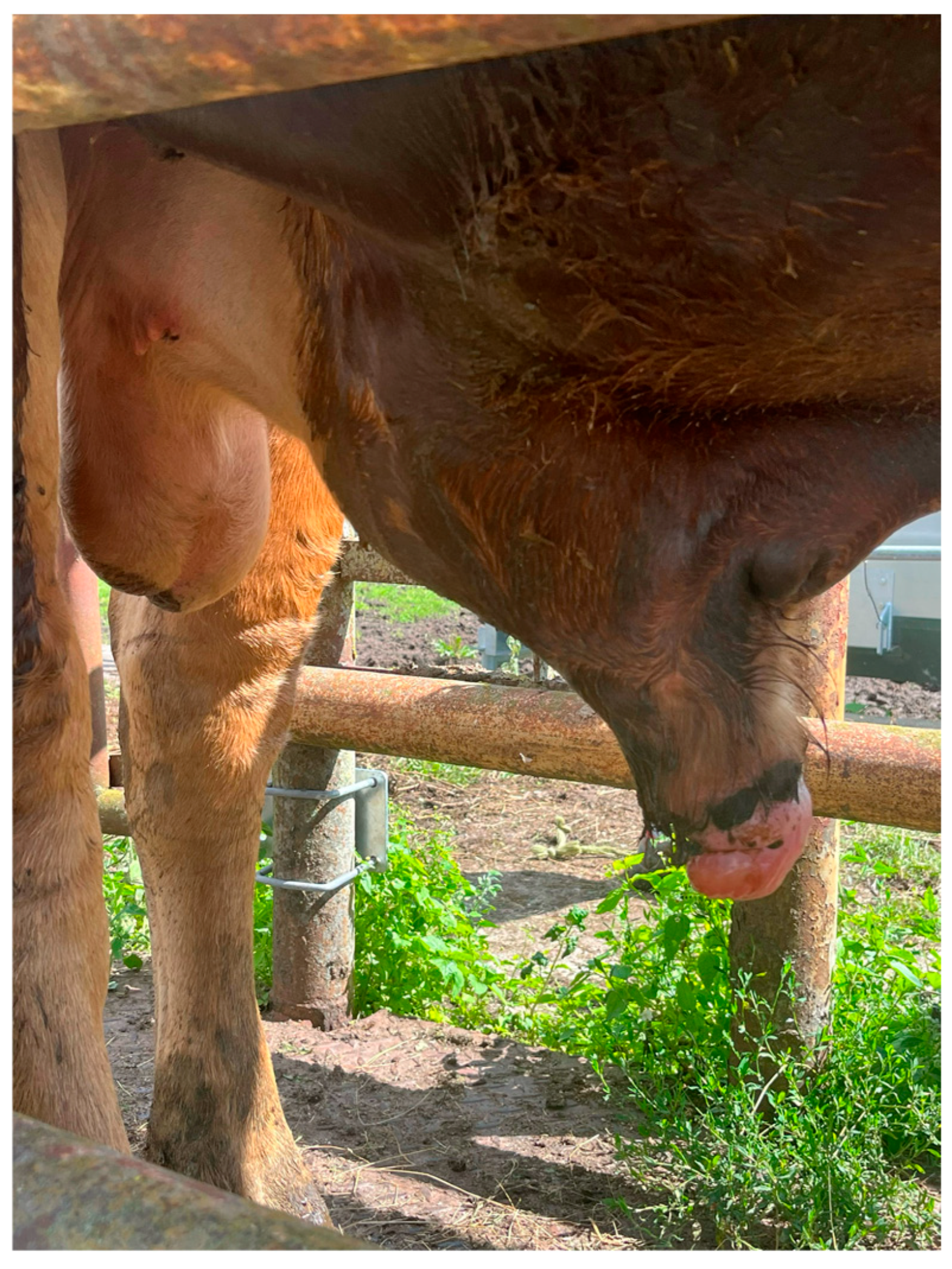
Penile hematoma is another challenge in veterinary clinical practice. It affects 7–14.9% of bulls at slaughter [13,17]. It usually occurs during the rupture of the penile tunica albuginea, e.g., during mechanical trauma or copulation. Most ruptures are localized on the dorsum of the penis [74]. Injury is usually followed by penile swelling (Figure 2). Two types of therapeutic management are described: conservative treatment, which involves antibiotic therapy and isolation of the bull to prevent contact with other animals, and surgical treatment, which involves removal of the hematoma within seven days of injury [2,75].

3.3. Penile Hair Ring

Penile hair ring occurs sporadically [76]. Cases have been reported in young 12–14-month-old bulls kept in the same enclosure [77]. Under such conditions, mutual flopping can occur, leading to the formation of a ring of another individual’s hair or their own around the bull’s copulatory organ. Many cases of penile hair ring resolve spontaneously, but some require surgical intervention [76]. After removal, the ring-shaped lesion may persist for some time. In severe cases, there is a risk of organ necrosis [2,78].

3.4. Penile Tuberculosis

Cases of penile tuberculosis have also been described [79,80,81,82]. This zoonosis is caused by Mycobacterium tuberculosis var. bovis [83]. Vielmo et al. (2020) described a bull with a thickened, hard prepuce with a pale inner surface [82]. Multifocal nodules on the preputium ranging from 0.5 to 5 cm in diameter were also observed. This is a unique case, as bovine tuberculosis mainly involves the lungs, the liver, and the tracheobronchial, mediastinal, retropharyngeal, mandibular, mesenteric, and pre-scapular lymph nodes [84]. Penile tuberculosis in cattle most likely results from mating females with uterine tuberculosis [85,86].

3.5. Tumors of the Copulatory Organ

Cases of penile fibrosarcoma have been reported [87,88]. This is a particularly unusual type of neoplasm in cattle originating from fibrous connective tissue [89,90,91]. In the case described, the size-differentiated, confined, and non-pigmented tumors were located on the glans penis. They consisted of densely packed and irregular connective tissue with many fibroblasts. Symptoms included painful urination, hematuria, and weight loss [87].

3.6. Frenulum Preputii Persistens

The literature on the etiology of this pathology is limited. At present, it is so rare that its significance is considered minor, and some authors [18,92,93] do not attach particular importance to it. Some publications suggest a genetic basis with recessive inheritance, and affected bulls should not be used for breeding [94,95]. Its incidence, assessed post-maturity in bulls, 46% of which were post-maturity, was 0.5% [29].
A persistent frenulum (i.e., retention of the original attachment) is considered pathological in bulls older than eleven months [93]. This condition is treated surgically. The frenulum should be ligated from the temporal and caudal sides to reduce potential bleeding, followed by resection of the foreskin frenulum [2,95].

3.7. Phimosis and Paraphimosis

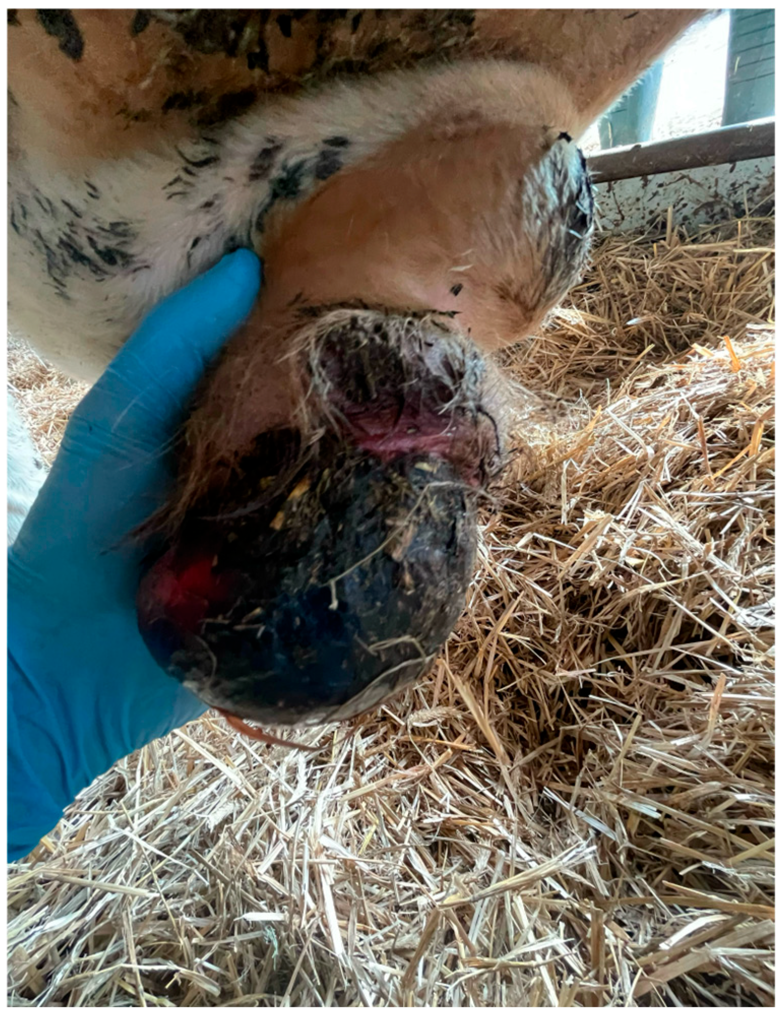
Phimosis occurs when the penile prepuce cannot be retracted from the glans penis. The condition can be acquired or hereditary. Phimosis can cause pain and discomfort during copulation and bacterial infections of the urethra, preputium, penis, and bladder due to impaired urination [96]. When a stool diagnosis is made, the most common procedure is surgical plication of the external orifice of the foreskin to widen it. The situation becomes more complicated with paraphimosis, a blockage of the prepuce in the retracted position, leading to swelling and impaired blood supply to the glans and the preputium (Figure 3). In most cases, arterial vascularization remains intact for hours or days, although necrosis of the surrounding tissues can occur, sometimes requiring amputation of the penile glans [96,97].
There are three main causes and mechanisms of paraphimosis in bulls. The first is swelling of the preputium, which can result from trauma to the penis or the preputium, as well as diseases that cause abdominal swelling. The second mechanism is damage to penile innervation, which can result from spinal cord injury, mechanical damage, or penile hematoma. The third cause is penile paralysis, which can result from priapism (prolonged non-physiological erection) or the use of acepromazine/phenothiazine as a sedative [98,99,100,101]. Treatment is surgical and involves widening the preputial orifice to free the tissues. A purse-string closure can be used after penile repositioning [96].

3.8. Other Penile Defects

Other defects occur occasionally. A corkscrew penis was described in an aged Angus bull [102]. Congenital penile urethral aplasia was described in a 4-day-old bull [103]. Treatment included a permanent perineal urethrostomy and post-operative antibiotic and fluid therapy. Ventral deviation is much less common [2]. Another rare cause of impotence is a congenitally short penis. Diagnosis is based on measuring the length of the penis from the atrium of the preputium to the base of the penis at the blockage of the n. pudendus, with a value above 25 cm considered normal in adults [104]. Young bulls may occasionally have difficulty achieving an erection due to congenital vascular fistulas in the corpora cavernosa. During attempted erection, the free part of the penis may appear bluish, caused by an influx of blood through the subcutaneous vessels. Treatment of such fistulas is usually difficult. Vascular fistulas can also develop as a result of trauma. Attempting to heal the penis after trauma may lead to fistulas that prevent the penis from maintaining normal blood pressure during the erection. Contrast cavernosography can be used to confirm penile vascular malformations, and vascular leaks can be identified using a contrast agent that exits the corpora cavernosa [2,105,106,107].

4. Disorders of the Accessory Sex Glands

Pathologies of the accessory genital glands in cattle are described only to a limited extent in the literature, particularly in recent publications. The notable exception is vesicular adenitis. This limited coverage may result from the perception that other conditions are not considered major breeding problems, as well as decreased interest among contemporary researchers, as most available reports are relatively dated. More publications have addressed the physiology of the accessory genital glands and semen collection [108,109,110,111]. In domestic cattle, three principal accessory genital glands are recognized: the prostate, the bulbourethral glands, and the seminal vesicles [112]. Abnormalities of the prostate and the bulbourethral glands in bulls are considered rare [113,114].
In a large-scale study by Bagshaw and Ladds [115] involving 521 bulls, accessory genital glands were collected post-mortem at a slaughterhouse, and numerous abnormalities and pathological changes were identified. In nine bulls, malformations of the mesonephric duct were observed, including aplasia, hypoplasia, or adhesions of the seminal vesicles and ampullae of the vas deferens. Unilateral aplasia of the bulbourethral glands occurred in four cases, and hypoplasia was found in four older bulls.
Melanosis (the presence of black melanin spots) was diagnosed in five animals. Cysts of the seminal vesicles exceeding 4 mm in diameter were found in eight bulls; in five cases, the glands were macroscopically normal, and in three they were enlarged; histological examination frequently revealed concurrent inflammation. Prostate cysts measuring 0.5 cm in diameter were recorded in one bull, and bulbourethral gland cysts >0.5 cm were found in six bulls, one of which exhibited inflammatory changes.
Vesiculitis was diagnosed in 47 bulls (9.0%), of which 14 cases (2.7%) would have been detectable via rectum examination, underscoring the higher sensitivity of post-mortem evaluation. Of these cases, 17 were chronic interstitial (unilateral, n = 8; bilateral, n = 9), frequently accompanied by adhesions, abscesses (n = 4), cysts (n = 4), calculi (n = 5), and concurrent ampullitis (n = 11). The remaining 30 cases were degenerative (unilateral, n = 13; bilateral, n = 17), sometimes associated with abscesses (n = 1), cysts (n = 2), calculi (n = 4), and ampullitis (n = 6).
Ampullitis was detected in 20 bulls, including 10 with concurrent vesiculitis; pathological changes included glandular enlargement, increased firmness, and occasional fibrosis. Inflammation of the bulbourethral glands was recorded in one bull, characterized by plasma cell infiltration.
Cysts within the urogenital fold (cystic uterus masculinus) were found in 34 bulls (6.5%), most frequently in the Brahman breed (18%). The study concluded that no single factor or pathology could be definitively linked to infertility in the examined bulls [16].
In another study of the bulbourethral glands in Bos indicus cattle and crossbreeds, adenitis was identified in four bulls (1.2%). In two cases, unilateral chronic interstitial inflammation was present, one of which was associated with degenerative vesiculitis. The remaining cases were bilateral; in one, a foreign body was located in the duct of the left gland, and, in another, multiple calculi and chronic, active diffuse inflammation were observed. The authors hypothesized that the foreign body may have migrated proximally from the prepuce or distal urethra. The impact of these lesions on fertility remains uncertain [116].
Congenital abnormalities of the accessory genital glands may predispose bulls to inflammatory conditions, particularly in the presence of ductal outlet disorders at the colliculus seminalis. Anomalies, such as segmental aplasia or hypoplasia of the mesonephric ducts, are generally not associated with inflammation, although hypoplasia has occasionally led to inflammatory changes. Early detection of defects—ideally, during puberty—is important in the selection of breeding bulls [2].
Vesiculitis occurs in 1–10% of bulls and can significantly impair fertility. Causative agents include bacteria, chlamydia, mycoplasmas, and ureaplasmas; the most commonly isolated pathogens are Trueperella pyogenes, Histophilus somni, and Brucella abortus (in regions where brucellosis persists). Routes of infection include ascending, descending, local, and hematogenous spread. Congenital defects of the colliculus seminalis and umbilical abscesses in calves increase disease risk [2].
Treatment options include antibiotic therapy, with tulathromycin reported as the most effective agent, administered locally or systemically. Other options include chemical ablation using formalin and surgical excision of the glands in chronic cases [2,117]. Preventive measures involve sound breeding practices, balanced nutrition, deworming, and genetic monitoring, as the pathogenesis is still not fully understood. Tilmicosin may also be effective [118]. Direct intraglandular injection of ceftiofur or penicillin is possible but technically challenging [119].
Recent experimental approaches have included the injection of stem cells into the seminal vesicles. Allogeneic mesenchymal stem cells (MSCs), administered directly into the vesicles of bulls with chronic vesiculitis, improved semen quality and promoted glandular regeneration. In a study of 12 bulls, MSC therapy resulted in increased sperm motility, greater semen production, and complete elimination of leukocytes from semen, confirming both the efficacy and safety of this method [120].
A surgical technique for removal of the seminal vesicles has also been developed; however, its success is not guaranteed and the procedure should be reserved as a last resort [121].

5. Sterilitas Secundaria

There are a number of pathologies that are unrelated to the reproductive system but significantly affect a bull’s willingness to copulate and the quality of semen. Such pathologies include laminitis, fractures, interdigital hyperplasia, sole ulcer, arthritis, spinal degeneration and other diseases that affect the animal’s welfare and perception of potential discomfort or pain [122,123,124,125,126,127,128,129,130,131,132,133,134,135,136,137,138,139].
The introduction of registries for congenital genital defects in bulls is important for animal welfare and offers potential economic benefits for breeders. Although these defects are common, accurate data on their incidence are lacking. Pathologies of the bull’s reproductive system, such as orchitis, testicular hypoplasia, testicular degeneration, and testicular hydrocele, can lead to infertility, reduced semen quality, and decreased economic potential in breeding. The Bull Breeding Soundness Evaluation, which includes physical examination, testicular circumference measurement, assessment of semen motility, and sperm morphology analysis, is crucial for evaluating the reproductive condition of bulls. The results provide valuable information on defects and diseases of the reproductive system in young bulls. Proper identification of these pathologies, as well as understanding their causes, symptoms, and treatment methods, is essential for breeders, herd personnel, veterinarians, and animal technicians.

6. Conclusions

In summary, comprehensive understanding, early detection, and effective treatment of congenital and acquired disorders of the reproductive organs in bulls are essential for ensuring animal welfare, optimizing reproductive performance, and minimizing economic losses in cattle breeding. The diversity of pathologies within the reproductive system highlights the need for systematic veterinary assessment, precise diagnostics, and timely interventions.
Future research should focus on further exploring the genetic and molecular bases of these disorders, developing minimally invasive therapeutic methods, and investigating preventive measures, such as vaccinations against infectious etiologies. The establishment of national and international registries of reproductive defects would provide valuable epidemiological data, support selective breeding programs, and improve herd management strategies. Integrating such registries with modern breeding technologies could enable predictive monitoring and early intervention, further enhancing reproductive outcomes.
Moreover, modern cattle breeding researchers should aim to utilize artificial intelligence; for instance, by developing programs that are capable of predicting the occurrence of congenital defects. Ultimately, a multidisciplinary approach combining veterinary medicine, genetics, advanced diagnostics, and herd management will be key to improving health and reproduction. These initiatives may contribute to increasing breeding efficiency, reducing animal suffering, and striving for excellence in genetic selection.

Author Contributions

Conceptualization, A.F.B., M.Z. and J.M.J.; validation, A.F.B., M.Z. and J.M.J.; writing, A.F.B.; writing—review and editing, J.M.J.; visualization, A.F.B. and M.Z.; supervision, J.M.J. All authors have read and agreed to the published version of the manuscript.

Funding

This research received no external funding.

Institutional Review Board Statement

Not applicable.

Informed Consent Statement

Not applicable.

Data Availability Statement

The data presented in this study are available on request from the corresponding author.

Conflicts of Interest

The authors declare no conflicts of interest.

References

  1. Latos-Bieleńska, A.; Materna-Kiryluk, A.; PRCM Working Group. Polish registry of congenital malformations—Aims and organization of the registry monitoring 300 000 births a year. J. Appl. Genet. 2005, 46, 341–348. [Google Scholar] [PubMed]
  2. Wolfe, D.F. Review: Abnormalities of the bull—Occurrence, diagnosis and treatment of abnormalities of the bull, including structural soundness. Animal 2018, 12, 148–157. [Google Scholar] [CrossRef]
  3. Hopper, R.M. Bovine Reproduction, 2nd ed.; John Wiley and Sons Ltd.: Starkville, MS, USA, 2021; 1232p. [Google Scholar]
  4. Engelken, T. The development of beef breeding bulls. Theriogenology 2008, 70, 573–575. [Google Scholar] [CrossRef]
  5. Society for Theriogenology. Veterinary Professionals Dedicated to Animal Reproduction. Available online: https://www.therio.org/ (accessed on 15 July 2023).
  6. Kastelic, J.P.; Thundathil, J.C. Breeding soundness evaluation and semen analysis for predicting bull fertility. Reprod. Domest. Anim. 2008, 43, 368–373. [Google Scholar] [CrossRef]
  7. Kouamo, J.; Nyonga, G.V. Gross reproductive organs abnormalities in bulls of northern regions of Cameroon. J. Infertil. Reprod. Biol. 2022, 10, 41–47. [Google Scholar]
  8. Bousmaha, F.; Benchaib Khoudja, F. Comparative and pathological study of the testis and epididymis in rams, bucks and bulls of Algeria. Asian J. Anim. Vet. Adv. 2012, 7, 950–959. [Google Scholar] [CrossRef]
  9. Silva, C.; Delgado, R.; Magaña, J.; Reyes, A. Abnormalities of testicular and scrotal development in bulls of three breeds in southeast México. Av. En Investig. Agropecu. 2008, 12, 21–31. [Google Scholar]
  10. McGowan, M.R.; Bertram, J.D.; Fordyce, G.; Fitzpatrick, L.A.; Miller, R.G.; Jayawardhana, G.A.; Doogang, V.J.; De Faveri, J.; Holroyd, R.G. Bull selection and use in northern Australia. 1. Physical traits. Anim. Reprod. Sci. 2002, 71, 25–37. [Google Scholar] [CrossRef]
  11. Migbaru, K.; Sisay, G.; Kasa, T. Study on Gross Testicular Disorders of Bulls Slaughtered at Addis Ababa Abattoirs Enterprise. J. Reprod. Fertil. 2014, 5, 45–49. [Google Scholar]
  12. Gemeda, A.E. Gross testicular abnormalities in indigenous breeds of bulls in Eastern Ethiopia. JAVAR 2017, 4, 200–206. [Google Scholar] [CrossRef]
  13. Kouamo, J.; Eta, L.E. Prevalence and associated risk factors of genital abnormalities in bulls slaughtered at the SODEPA Industrial Abattoir, Douala, Cameroon. J. Infertil. Reprod. Biol. 2021, 9, 136–141. [Google Scholar]
  14. Uyar, A.; Uslu, B.A.; Yurdakul, İ. Evaluation on the histopathology of testes anomalies in the bulls slaughtered at the city of Van. DUVETFD 2019, 12, 30–36. [Google Scholar]
  15. Kandiwa, E.; Nyirakunzimana, L.; Habarugira, G.; Mushonga, B.; Samkange, A. A 4-year study of the proportional distribution of male reproductive organ abnormalities in cattle slaughtered at Nyagatare abattoir. Vet. Med. Sci. 2017, 3, 179–186. [Google Scholar] [CrossRef] [PubMed]
  16. Oliveira, A.P.L.; Lobo Júnior, A.R.; Castilho, E.F.; Guimarães, J.D.; Melo, T.V.; Mota, D.A.; Siqueira, J.B. Reproductive disorders affecting 21-month-old bulls assessed by andrological examination. Rev. Fac. Cienc. Agrar. 2020, 41, 3199–3209. [Google Scholar] [CrossRef]
  17. Amaer, E.; Habtemariam, K.; Samulel, A.; Shimellis, D.; Belina, D. Incidence of gross pathological conditions of the reproductive organs in bulls. Indian J. Vet. Pathol. 2016, 40, 305–311. [Google Scholar]
  18. Boryczko, Z. Zaburzenia płodności. In Fizjologia i Patologia Rozrodu Bydła, 1st ed.; Boryczko, Z., Bostedt, H., Jaśkowski, J.M., Eds.; University of Nicolaus Copernicus: Toruń, Poland, 2021; p. 86. [Google Scholar]
  19. Parsonson, I.M.; Winter, J.; McEntee, K. Allergic epididymo-orchitis in guinea pigs and bulls. Vet. Pat. 1971, 8, 333–351. [Google Scholar] [CrossRef]
  20. Sivahoth, S.; Sudnakara Reddy, B. Rare report of orchitis in a bull due to Trypanosoma evansi infection. J. Parasit. Dis. 2019, 43, 28–30. [Google Scholar]
  21. González-Huecas, M.; Ferre, I.; Jiménez-Meléndez, A.; Criado, F.; Gutiérrez-Expósito, D.; Ortega-Mora, L.M.; Álvarez-Garcia, G. Vascular wall injury and inflammation are key pathogenic mechanisms responsible for early testicular degeneration during acute besnoitiosis in bulls. Parasit Vectors 2020, 13, 113. [Google Scholar] [CrossRef]
  22. Jaśkowski, L.; Sadowski, J.M.; Hoffmann-Woźniak, K.; Wieczór, H.; Truszczyński, M. Experimental chlamydiosis in bulls. Effects of unilateral intratesticular inoculation. Bull. Vet. Inst. Puławy 1979, 23, 14–20. [Google Scholar]
  23. Machmoud, M.A.M.; Megahed, G.; Yousef, M.S.; Zakib Ali, F.A.; Zako, R.S.; Abdelhafeza, H.H. Salmonella Typhimurium triggered unilaateral epididimo-orchitis and splenomegaly in a holstein bulls Assiut, Egypt: A case report. Pathogens 2020, 9, 314. [Google Scholar] [CrossRef]
  24. Mahesh, V.; Rashmitha, H.; Preetham Kumar, S.V.; Shivaraju, E.; Nagaraja, B.N. Surgical management of orchitis in Gir bullock by scrotal ablation technique. J. Livest. Sci. 2022, 13, 38–40. [Google Scholar] [CrossRef]
  25. Youngquist, R.S.; Threlfall, W.R. Current Therapy in Large Animal Theriogenology, 2nd ed.; Elsevier Health Sciences: London, UK, 2006; 1088p. [Google Scholar]
  26. Neves, H.H.; Vargas, G.; Brito, L.F.; Schenkel, F.S.; Albuquerque, L.G.; Carvalheiro, R. Genetic and genomic analyses of testicular hypoplasia in Nellore cattle. PLoS ONE 2019, 14, e0211159. [Google Scholar] [CrossRef]
  27. Salvador, D.F.; Salvador, S.C. Testicular hypoplasia in cattle: Causes and consequences. Ciência Anim. 2021, 31, 67–79. [Google Scholar]
  28. Barth, A.D. Congenital and acquired abnormalities of the scrotum, testes and epididymides of bulls. Large Anim. Vet. 2006, 6, 1–6. [Google Scholar]
  29. Saunders, P.J.; Ladds, P.W. Congenital and developmental anomalies of the genital of slaughtered bulls. Aust. Vet. J. 1978, 54, 10–13. [Google Scholar] [CrossRef] [PubMed]
  30. Chenoweth, P.J.; Osborne, H.G. Breed differences in abnormalities of the reproductive organs of young beef bulls. Aust. Vet. J. 1978, 54, 463–468. [Google Scholar] [CrossRef]
  31. Hoflack, G.; Van der Broeck, W.; Maes, D.; Van Damme, K.; Opsomer, G.; Duchateau, L.; de Kruif, A.; Rodriguez-Martinez, H.; Van Soom, A. Testicular dysfunction is responsible for low sperm quality in Belgian Blue bulls. Theriogenology 2008, 69, 232–233. [Google Scholar] [CrossRef]
  32. Borel, N.; Janett, F.; Teankum, K.; Zlinszky, K.; Iten, C.; Hilbe, M. Testicular hypoplasia in a bull persistently infected with bovine diarrhoea virus. J. Comp. Pathol. 2007, 137, 169–173. [Google Scholar] [CrossRef]
  33. Dunn, H.O.; Lein, D.H.; McKentee, K. Testicular hypoplasia in a Hereford bull with 61,XXY karyotype: The bovine counterpart of human Klinefelter’s syndrome. Cornell Vet. 1980, 70, 137–146. [Google Scholar]
  34. Bongso, T.A.; Jainudeen, M.R.; Lee, Y.J. Testicular hypoplasia in a bull with XX/XY chimerism. Cornell Vet. 1981, 71, 376–382. [Google Scholar]
  35. Krumrych, W.; Jaśkowski, J.M.; Gehrke, M.; Zbylut, E.N. Legitimacy of systematic karyotype evaluation of cattle qualified for reproduction. J. Bull. Vet. Inst. Puławy 2002, 46, 307–315. [Google Scholar]
  36. Adamu, S.; Fatihu, M.Y.; Useh, N.M.; Mamman, M.; Sekoni, V.O.; Esievo, K.A.N. Sequential testicular and epididymal damage in Zebu bulls experimentally infected with Trypanosoma vivax. Vet. Parasitol. 2007, 143, 29–34. [Google Scholar]
  37. Koziol, J.; Palmer, C. Pathophysiology, diagnosis, and management of testicular degeneration in the bull. Clin. Theriogenology 2023, 15, 1–7. [Google Scholar] [CrossRef]
  38. Contri, A.; Gloria, A.; Wegher, L.; Carluccio, A. Successful use of gonadotropin-releasing hormone (GnRH) analog for the treatment of tertiary hypogonadism (GnRH deficiency) in a 5-year-old Belgian Blue bull. Vet. Q. 2012, 32, 51–54. [Google Scholar] [PubMed]
  39. Moran, K.; Picco, R.; Morrell, E.; Verna, A.; Zapata, L.; Moiraghi, L.; Bilbao, G.; Bartolomé, J. Testicular degeneration, fibrosis, and mineralization in Limangus bulls. Clin. Theriogenology 2025, 17, 1–5. [Google Scholar] [CrossRef]
  40. Hassan, M.F. Biometrical and pathological studies on testes of cow bulls. In Proceedings of the First International Congress of Veterinary Phatmacology & Pharmaceutical Sciences (1st ICVPS), Teheran, Iran, 4–5 October 2008. [Google Scholar]
  41. Abu-Seida, A.M. Ultrasonographic diagnosis of some scrotal swellings in the bulls. Pak. Vet. J. 2012, 32, 378–381. [Google Scholar]
  42. McClure, M.; King, H. Scrotal hydrocele in an Angus bull. In Proceedings of the SFT Theriogenology Annual Conference, Online, 22–25 July 2020; TherioConference; Society for Theriogenology: Sacramento, CA, USA, 2020. [Google Scholar]
  43. Singh, K.B. Hydrocele in an Ongole bull. Indian Vet. J. 1985, 62, 529–530. [Google Scholar]
  44. Dalal, J.; Kumar, A.; Gupta, G. Management of unilateral chronic orchitis and hydrocele in a Sahiwal Bull. Indian Vet. J. 2017, 94, 68–69. [Google Scholar]
  45. Sindh, R.S. Hydrocele in a yearling bull. The Vet. J. 1916, 72, 310. [Google Scholar]
  46. Ali, K.M.; Ahmad, N.; Akhtar, N.; Ali, S.; Ahmad, M.; Younis, M. Ultrasound imaging testes and epididymitis of normal and infertile breeding bulls. Pak. Vet. J. 2011, 31, 345–350. [Google Scholar]
  47. Jaśkowski, J.M.; Kaptur, M.D.; Jaśkowski, B.M.; Kulynych, S.; Gehrke, M. A premier case report of a congenital pseudocyst of the testis (cystis spuria testiculi congenita) in a three-week-old calf. BMC Vet. Res. 2025, 21, 306. [Google Scholar] [CrossRef]
  48. Prado, T.M.; Dawson, L.J.; Schumacher, J. Surgical procedures of the genital organs of bulls. Vet. Clin. N. Am. Food. Anim. Pract. 2016, 32, 701–725. [Google Scholar] [CrossRef]
  49. Luby, C.D.; Middleton, J.R.; Youngquist, R.S.; Kim, D.Y.; Evans, A.T. Theriogenology question of the month. Sertoli cell tumor. J. Am. Vet. Med. Assoc. 2007, 231, 1503–1505. [Google Scholar] [CrossRef]
  50. Schiendewolffs, L.; Dierks, C.; Heppelmann, M.; Gähle, M.; Piechotta, M.; Bieneke, A.; Brehem, L.; Distl, O. Testicular yolk sac tumor and impaired spermatogenesis in a Holstein Friesian calf. Syst. Biol. Reprod. Med. 2015, 61, 314–319. [Google Scholar]
  51. Jean, G.; Gaughan, E.M.; Constable, P.D. Cryptorchidism in North American cattle: Breed predisposition and clinical findings. Theriogenology 1992, 38, 951–958. [Google Scholar] [CrossRef]
  52. Ayodeji, A.A.; Suwaiba, W. Cryptorchidism among breeds of bulls in a semi-arid region of Nigeria. Maced. Vet. Rev. 2013, 36, 123–128. [Google Scholar]
  53. Miyake, Y.; Kaneda, Y. A new type of Robertsonian translocation (1/26) in a bull with unilateral cryptorchidism, probably occurring de novo. Jpn. J. Vet. Sci. 1987, 49, 1015–1019. [Google Scholar] [CrossRef] [PubMed]
  54. Kaneko, Y.; Torisu, S.; Kitahara, G.; Hidaka, Y.; Satoh, H.; Asanuma, T.; Mizutani, S.; Osawa, T.; Naganobu, K. Laparoscopic cryptorchidectomy in standing bulls. J. Vet. Med. Sci. 2015, 77, 631–635. [Google Scholar] [CrossRef] [PubMed]
  55. Van Camp, S.D. Common causes of infertility in the bull. Vet. Clin. N. Am. Food Anim. Pract. 1997, 13, 203–231. [Google Scholar] [CrossRef]
  56. Pagliarani, S.; Johnston, S.A.; Beagly, K.W.; Dief, H.; Palmieri, C. The occurrence and pathology of chlamydiosis in the male reproductive tract of non-human mammals: A review. Theriogenology 2020, 154, 152–160. [Google Scholar] [CrossRef]
  57. McEntee, K. Reproductive Pathology of Domestic Mammals, 1st ed.; Academic Press: Cambridge, MA, USA, 2012; 400p. [Google Scholar]
  58. Dorne, M. Spermatocele. J. Urol. 1926, 15, 389–396. [Google Scholar] [CrossRef]
  59. Stachura, J.; Domagała, W. Patologia Znaczy Słowo o Chorobie, 3rd ed.; Polska Akademia Umiejętności: Kraków, Poland, 2016; 676p. [Google Scholar]
  60. Threlfall, W.R.; Robertson, J.T.; Munsterman, A.S.; Oglesbee, M.J.; Hubbell, J.A. Theriogenology question of the month. Seminoma, spermatocele, sustentacular cell tumor. J. Am. Vet. Med. Assoc. 2005, 226, 1649–1650. [Google Scholar] [CrossRef] [PubMed]
  61. Statham, J. Differential diagnosis of scrotal enlargement in bulls. Practice 2010, 32, 200–206. [Google Scholar] [CrossRef]
  62. Grindflek, E.; Moe, M.; Taubert, H.; Similander, H.; Lien, S.; Moen, T. Genome-wide linkage analysis of inguinal hernia in pigs using affected sib pairs. BMC Genet. 2006, 7, 25. [Google Scholar] [CrossRef]
  63. Ebert, E.F. Surgical repair of scrotal hernia in the bull. Can. Vet. J. 1960, 1, 441–443. [Google Scholar]
  64. Baba, A.I. Oncologie Comparată; Editura Academiei Române: Bucureşti, Romania, 2002; 684p. [Google Scholar]
  65. Knipe, D.M.; Howley, P.M. Fields Virology, 6th ed.; Lippincott Williams & Wilkins: Philadelphia, PA, USA, 1996; 2664p. [Google Scholar]
  66. Campo, M.S. Papillomavirus Research: From Natural History to Vaccine and Beyond, 1st ed.; Caister Academic Press: Glasgow, UK, 2006; 424p. [Google Scholar]
  67. McEntee, K. Fibropapilloma of the external genitalia of cattle. Cornell Vet. 1950, 40, 304–312. [Google Scholar]
  68. Batamuzi, E.K.; Kassuku, A.A.; Agger, J.F. Risk factors associated with canine transmissible venereal tumour in Tanzania. Prev. Vet. Med. 1992, 13, 13–17. [Google Scholar] [CrossRef]
  69. Borzacchiello, G.; Roperto, F. Bovine papillomaviruses, papillomas and cancer in cattle. Vet. Res. 2008, 39, 1. [Google Scholar]
  70. Patnaik, A.K.; Matthiesen, D.T.; Zarvie, D.A. Two cases of canine penile neoplasm: Squamous cell carcinoma and mesenchymal chondrosarcoma. J. Am. Anim. Hosp. Assoc. 1987, 24, 403–406. [Google Scholar]
  71. Kennedy, P.C.; Cullen, J.M.; Edwards, J.F.; Goldschmidt, M.H.; Larsen, S.; Munson, L.; Nielsen, S. Histological Classification of Tumors of the Genital System of Domestic Animals, 2nd ed.; Armed Forces Institute of Pathology: Washington, DC, USA, 1998; 79p. [Google Scholar]
  72. Lindsey, C.L.; Almeida, M.E.; Vicari, C.F.; Carvalho, C.; Yaguiu, A.; Freitas, A.C.; Beçak, W. Stocco dos Santos RC. Bovine papillomavirus DNA in milk, blood, urine, semen, and spermatozoa of bovine papillomavirus-infected animals. Genet. Mol. Res. 2009, 8, 310–318. [Google Scholar] [CrossRef]
  73. Stocco dos Santos, R.C.; Lindsey, C.J.; Ferraz, O.P.; Pinto, J.R.; Mirandola, R.S.; Benesi, F.J.; Birgel, E.H.; Pereira, C.A.; Beçak, W. Bovine papillomavirus transmission and chromosomal aberrations: An experimental model. J. Gen. Virol. 1998, 79, 2127–2135. [Google Scholar] [CrossRef]
  74. Beckett, S.D.; Reynolds, T.M.; Walker, D.F.; Hudson, R.S.; Purohit, R.C. Experimentally induced rupture of corpus cavernosum penis of the bull. Am. J. Vet. Res. 1974, 35, 765–767. [Google Scholar] [CrossRef]
  75. Wolfe, D.F.; Beckett, S.D.; Carson, R.L.; Powe, T.A.; Pugh, D.G. Acquired conditions of the penis and prepuce. In Large Animal Urogenital Surgery, 1st ed.; Wolfe, D.F., Moll, H.D., Eds.; Williams and Wilkins: Baltimore, MD, USA, 1998; pp. 237–272. [Google Scholar]
  76. Akin, I. A Case of penile hair ring in a young bull. Anim. Health Prod. Hyg. 2013, 2, 212. [Google Scholar]
  77. Armstrong, C.L.; Koziol, J.H. Understanding a breeding soundness evaluation and factors that impact bull fertility. In Proceedings of the Applied Reproductive Strategies in Beef Cattle, San Antonio, TX, USA, 30–31 August 2022. Beef Reproduction Task Force. [Google Scholar]
  78. Wolfe, D.F.; Carson, R.L. Juvenile anomalies of the penis and prepuce: Bulls. In Large Animal Urogenital Surgery, 1st ed.; Wolfe, D.F., Moll, H.D., Eds.; Williams and Wilkins: Baltimore, MD, USA, 1998; pp. 233–235. [Google Scholar]
  79. Trotter, A.M. Tuberculosis of the penis of a bull. J. Com. Pathol. 1903, 16, 252. [Google Scholar] [CrossRef]
  80. Thoen, C.O.; Himes, E.M.; Stumpff, C.D.; Parks, T.W.; Sturkie, H.N. Isolation of Mycobacterium bovis from the prepuce of a herd bull. Am. J. Vet. Res. 1977, 30, 877–878. [Google Scholar] [CrossRef]
  81. Foster, R.A. Male genital system. In Jubb, Kennedy and Palmer’s Pathology of Domestic Animals, 6th ed.; Maxie, M.G., Ed.; Elsevier: St. Louis, MO, USA, 2016; Volume 3, pp. 465–510. [Google Scholar]
  82. Vielmo, A.; Lopes, B.C.; Panziera, W.; Bianchi, R.M.; Mayer, F.Q.; Vielmo, L.A.; Barros, C.S.L.; Driemeier, D. Penile Tuberculosis in a bull. J. Comp. Pathol. 2020, 180, 5–8. [Google Scholar] [CrossRef] [PubMed]
  83. Riojas, M.A.; McGough, K.J.; Rider-Riojas, C.J.; Rastogi, N.; Hazbón, M.H. Phylogenomic analysis of the species of the Mycobacterium tuberculosis complex demonstrates that Mycobacterium africanum, Mycobacterium bovis, Mycobacterium caprae, Mycobacterium microti and Mycobacterium pinnipedii are later heterotypic synonyms of Mycobacterium tuberculosis. Int. J. Syst. Evol. Microbiol. 2018, 68, 324–332. [Google Scholar] [CrossRef] [PubMed]
  84. Terefe, D. Gross pathological lesions of bovine tuberculosis and efficiency of meat inspection procedure to detect-infected cattle in Adama municipal abattoir. J. Vet. Med. Anim. Health 2014, 6, 48–53. [Google Scholar]
  85. Domingo, M.; Vidal, E.; Marco, A. Pathology of bovine tuberculosis. Res. Vet. Sci. 2014, 97, 20–29. [Google Scholar] [CrossRef]
  86. Schlafer, D.H.; Foster, R.A. Female Genital System. In Jubb, Kennedy and Palmer’s Pathology of Domestic Animals, 6th ed.; Maxie, M.G., Ed.; Elsevier: St. Louis, MO, USA, 2016; Volume 3, pp. 358–464. [Google Scholar]
  87. Hesaraki, S.; Abedi, G.; Rismanchi, S. Penile fibrosarcoma tumor in a bull. Iran J. Vet. Res. 2010, 11, 283–286. [Google Scholar]
  88. Pandit, G.A.; Kudrimoti, J.K.; Kokandakar, H.R.; Bhople, K.S. Fibrosarcoma of penis—A case report. Indian J. Pathol. Microbiol. 2004, 47, 389–390. [Google Scholar] [PubMed]
  89. Smirnov, A.V. Fibrosarcoma: Immunohistochemical study of the extracellular matrix. Arkh. Pathol. 1988, 50, 17–24. [Google Scholar]
  90. Fletcher, C.D.M.; Unni, K.K.; Mertens, F. Pathology and Genetics of Tumors of Soft Tissue and Bone, 1st ed.; IARC Press: Lyon, France, 2002; pp. 100–102. [Google Scholar]
  91. Van den Top, J.G.B.; De Heer, N.; Klein, W.R.; Ensink, J.M. Penile and preputial tumours in the horse: A retrospective study of 114 affected horses. J. Equine Vet. 2008, 40, 528–532. [Google Scholar] [CrossRef] [PubMed]
  92. Hudson, R.S.; Wolfe, D.F. Physical defects which limit breeding soundness of bulls. In American Association of Bovine Practitioners Conference Proceedings, Oklahoma City, OK, USA, 28 November–1 December 1983; American Association of Bovine Practitioners: Ashland, OH, USA, 1983. [Google Scholar]
  93. Ashdown, R.R. Persistence of the penile frenulum in young bulls. Vet. Rec. 1962, 74, 1464–1468. [Google Scholar]
  94. Elmore, R.G.; Breuer, J.; Youngquist, R.S.; Lasley, J.F.; Bierschwald, C.J. Breeding soundness examinations in 18 closely related inbred Angus bull. Theriogenology 1978, 10, 355–363. [Google Scholar] [CrossRef]
  95. Elmore, R.G. Surgical repair of bovine persistent penile frenulum. Vet. Med. Small Anim. Clin. 1981, 76, 701–704. [Google Scholar]
  96. Fesseha, H. Acute paraphimosis in bull and its surgical management. Prac. Clin. Investig. 2020, 3, 68–71. [Google Scholar]
  97. Reynard, J.M.; Barau, J.M. Reduction of paraphimosis the simple way: The Dundee technique. Br. J. Urol. Intern. 1999, 83, 859–860. [Google Scholar] [CrossRef]
  98. Choe, J.M. Paraphimosis: Current treatment options. Am. Fam. Physician 2000, 62, 2623–2626. [Google Scholar]
  99. Adeola, B.S.; Enobong, H. Surgical management of paraphimosis in dog: A case report. Glob. Vet. 2016, 16, 49–51. [Google Scholar]
  100. Fossum, T. Small Animal Surgery, 3rd ed.; Elsevier: Philadelphia, PA, USA, 2007; 1632p. [Google Scholar]
  101. Hopper, R.M. Management of male reproductive tract injuries and disease. Vet. Clin. N. Am. Food. Anim. Pract. 2016, 32, 497–510. [Google Scholar] [CrossRef]
  102. McDiarmid, J.J. “Corkscrew penis” and other breeding abnormalities in beef bulls. N. Z. Vet. J. 2011, 23, 35–36. [Google Scholar] [CrossRef]
  103. Javdani Gandomani, M.; Dehghani Nazhvani, S.; Raayat Jahromi, A.R. Congenital penile urethral aplasia in a 4-day-old bull calf. Iran J. Vet. Res. 2009, 10, 87–89. [Google Scholar]
  104. Gilbert, R.O. The diagnosis of short penis as a cause of impotentia coeundi in bulls. Theriogenology 1989, 32, 805–815. [Google Scholar] [CrossRef]
  105. Young, S.; Hudson, R.; Walker, D.F. Impotence in bulls due to vascular shuns from the corpus cavernosum penis. J. Am. Vet. Med. Assoc. 1977, 171, 643–648. [Google Scholar] [CrossRef]
  106. Hopper, R.M.; Wolfe, D.F. Restorative surgery of the penis. In Bovine Reproduction, 2nd ed.; Hopper, R.M., Ed.; John Wiley and Sons Ltd.: Starkville, MS, USA, 2021; pp. 210–229. [Google Scholar]
  107. Moll, H.D.; Wolfe, D.F.; Hathcock, J.T. Caversonography for diagnosis of erection failure in bulls. Compend. Contin. Educ. Vet. 1993, 161, 1160–1164. [Google Scholar]
  108. Lima, U.A.; Freire, L.Q.B.; Lima, W.S.M.; Sancler-Silva, Y.F.R.; Mesquita, E.P.; Oliveira, D.; Torres, M.B.A.M.; Carneiro, G.F. Characterization of vesicular glands, study of vesiculitis and its association with other accessory sex glands in bulls. Arq. Bras. Med. Vet. Zootec. 2024, 76, 7–15. [Google Scholar] [CrossRef]
  109. Sánchez, J.M.; Rabaglino, M.B.; Bagés-Arnal, S.; McDonald, M.; Behura, S.K.; Spencer, T.E.; Longeran, P.; Fernandez-Fuertes, B. Sperm exposure to accessory gland secretions alters the transcriptomic response of the endometrium in cattle. Theriogenology 2024, 218, 26–34. [Google Scholar] [CrossRef]
  110. Sethi, M.; Mohanty, T.K.; Shah, N.; Bhakat, M.; Kumar, N.; Baithalu, R.K. Understanding the Crucial Role of Seminal Plasma Exosomes in Bull Fertility: A Review. Reprod. Domest. Anim. 2024, 59, e70000. [Google Scholar] [CrossRef] [PubMed]
  111. Ferrer, M.S.; Palomares, R.; Heins, B.; Xavier, P.; Fyke, H.; Hurley, D.J.; Gordon, J. Analytical and clinical validation of diagnostic tests for the detection of leucospermia in beef bulls. Theriogenology 2024, 230, 46–53. [Google Scholar] [CrossRef] [PubMed]
  112. International Committee on Veterinary Gross Anatomical Nomenclature. In Nomina Anatomica Veterinária, 6th ed.; Editorial Committee: Hanover, Germany, 2017.
  113. McGowan, M.; Holland, M.K.; Boe-Hansen, G. Review: Ontology and endocrinology of the reproductive system of bulls from fetus to maturity. Animal 2018, 12, 19–26. [Google Scholar] [CrossRef]
  114. Campero, C.M.; Bagshaw, P.A.; Ladds, P.W. Lesions of presumed congenital origin in the accessory sex glands of bulls. Aust. Vet. J. 1989, 66, 80–85. [Google Scholar]
  115. Bagshaw, P.A.; Ladds, P.W. A study of the accessory sex glands of bulls in abattoirs in Northern Australia. Aust. Vet. J. 1974, 50, 489–495. [Google Scholar]
  116. Campero, C.M.; Ladds, P.W.; Thomas, A.D. Pathological findings in the bulbourethral glands of bulls. Aust. Vet. J. 1988, 65, 241–244. [Google Scholar] [CrossRef]
  117. Rovay, H.; Barth, A.D.; Chirino-Trejo, M.; Martínez, M.F. Update on treatment of vesiculitis in bulls. Theriogenology 2008, 70, 495–503. [Google Scholar] [CrossRef]
  118. Martínez, M.F.; Barth, A.D. Early detection and treatment of vesicular adenitis in bulls. Anim. Reprod. Sci. 2007, 101, 252–256. [Google Scholar] [CrossRef] [PubMed]
  119. Martínez, M.F.; Arteaga, A.A.; Barth, A.D. Intraglandular injection of antibiotics for the treatment of vesicular adenitis in bulls. Anim. Reprod. Sci. 2008, 104, 201–211. [Google Scholar] [CrossRef] [PubMed]
  120. Severo, N.C.; de Assumpção, T.I.; Peixer, M.A.S.; da Cunha Xavier, M.; Malard, P.F.; Brunel, H.D.S.S.; Lançoni, R. Effectiveness of intraglandular allogeneic mesenchymal stem cell administration for treating chronic vesicular adenitis in bulls. Theriogenology 2025, 241, 117419. [Google Scholar] [CrossRef]
  121. Shah, B.A.; Hopwood, M.L.; Faulkner, L.C. Seminal vesiculectomy in bulls. J. Reprod. Fertil. 1968, 16, 171–177. [Google Scholar]
  122. Ahola, J.K.; Foster, H.A.; VanOverbeke, D.L.; Jensen, K.S.; Wilson, R.L.; Glaze, J.B.; Fife, T.E.; Gray, C.W.; Nash, S.A.; Panting, R.R.; et al. Survey of quality defects in market beef and dairy cows and bulls sold through livestock auction markets in the Western United States: I. Incidence rates. J. Anim. Sci. 2011, 89, 1474–1483. [Google Scholar] [CrossRef]
  123. Bartels, J.E. Femoral-tibial osteoarthrosis in the bull: I. Clinical survey and radiological interpretation. J. Am. Vet. Rad. Soc. 1975, 16, 159–173. [Google Scholar] [CrossRef]
  124. Berry, S.L. Diseases of the digital soft tissues. Vet. Clin. N. Am. Food Anim. Pract. 2001, 17, 129–142. [Google Scholar] [CrossRef] [PubMed]
  125. Hopkins, F.M.; Spitzer, J.C. The new Society for Theriogenology breeding soundness evaluation system. Vet. Clin. N. Am. Food Anim. Pract. 1997, 13, 283–293. [Google Scholar] [CrossRef] [PubMed]
  126. Desrochers, A. Diagnosis and prognosis of common disorders involving the proximal limb. Vet. Clin. N. Am. Food Anim. Pract. 2017, 33, 251–270. [Google Scholar] [CrossRef] [PubMed]
  127. Ducharme, N.G. Stifle injuries in cattle. Vet. Clin. N. Am. Food Anim. Pract. 1996, 12, 59–84. [Google Scholar] [CrossRef]
  128. Frithian, D.C.; Kelly, M.A.; Mow, V.C. Material properties and structure- function relationships in the menisci. Clin. Orthop. Relat. Res. 1990, 252, 19–31. [Google Scholar] [CrossRef]
  129. Gentile, A.; Testoni, S. Inherited diseases of cattle: A selected review. Slov. Vet. Zb. 2006, 43, 17–29. [Google Scholar]
  130. Greenough, P.R. Lameness in Cattle, 3rd ed.; W.B. Saunders: Philadelphia, PA, USA, 1972; 336p. [Google Scholar]
  131. Greenough, P. Pododermatitis circumscripta (ulceration of the sole) in cattle. Agri Prac. 1987, 6, 17–22. [Google Scholar]
  132. Hoblet, K.H.; Weiss, W. Metabolic hoof horn disease claw horn disruption. Vet. Clin. N. Am. Food Anim. Pract. 2001, 17, 111–127. [Google Scholar] [CrossRef]
  133. Lischer, C.; Ossent, P.; Raben, M.; Geyer, H. Suspensory structures and supporting structures of the third phalanx of cows and their relevance to development of typical sole ulcers (Rusterholz ulcers). Vet. Rec. 2002, 151, 694–698. [Google Scholar]
  134. Nelson, D.R.; Huhn, J.C.; Knelle, S.K. Peripheral detachment of the medial meniscus with injury to the medial collateral ligament in 50 cattle. Vet. Rec. 1990, 127, 59–66. [Google Scholar]
  135. Pentecost, R.; Niehaus, A. Cranial cruciate ligament, meniscus, upward fixation of the patella. Vet. Clin. N. Am. Food Anim. Pract. 2014, 30, 265–281. [Google Scholar]
  136. Persson, Y.; Söderquist, L.; Ekman, S. Joint disorder; a contributory cause to reproductive failure in beef bulls? Acta Vet. Scand. 2007, 49, 31. [Google Scholar] [CrossRef] [PubMed]
  137. Purohit, R.C.; Hudson, R.S.; Riddell, M.G.; Carson, R.L.; Wolfe, D.F.; Walker, D.F. Thermography of the bovine scrotum. Am. J. Vet. Res. 1985, 46, 2388–2392. [Google Scholar] [CrossRef]
  138. Reiland, S.; Stromberg, B.; Olsson, S.E.; Dreimanis, I.; Olsson, I.G. Osteochondrosis in growing bulls. Pathology, frequency and severity on different feedings. Acta Radiol. 1978, 358, 179–196. [Google Scholar]
  139. Weisbrode, S.E.; Monke, D.R.; Dodaro, S.T.; Hull, B.L. Osteochondrosis, degenerative joint disease, and vertebral osteophytosis in middle-aged bulls. J. Am. Vet. Med. Assoc. 1982, 181, 700–705. [Google Scholar] [CrossRef]
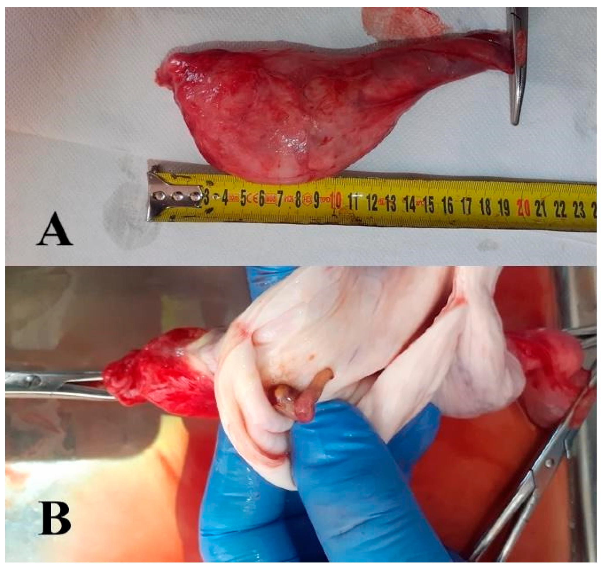
Figure 1. Congenital pseudocyst of the testis (cystis spuria testiculi congenita) in a three-week-old calf. (A)—View of the side surface. (B)—View after dissection.
Animals 15 02804 g001
Figure 2. Penile swelling caused by mechanical trauma. The photograph shows eversion of the prepuce due to the inability to retract the penis. The injury occurred during copulation. Conservative management included antibiotic therapy, steroidal anti-inflammatory agents, and anti-edematous medications. Sexual rest was recommended. The treatment resulted in a favorable outcome for the animal. (photo A.F. Butkiewicz).
Animals 15 02804 g002
Figure 3. Mechanical injury of the prepuce accompanied by its eversion. The preputial orifice exerts pressure on its external portion. (photo A.F. Butkiewicz).
Animals 15 02804 g003
Table 1. Selected pathologies of the male genital organs in bulls.
PathologyEtiological FactorType of DefectTreatmentEstimated
Frequency [%]
OrchitisInfectious, autoimmune, allergic, mechanical traumaAcquiredAntibiotic therapy1.94–7.1%
Hypoplasia testisBVD virus, genetic mutationCongenital. acquiredCull, vaccines0.2–3.6%
Degeneratio testisTrauma, tumours, deficiency of nutrients, Trypanosoma vivax, idiopathicAcquiredCull, GnRH8.1%
Hydrocele testisTrauma, tumours, orchitis, epididymitisAcquiredSurgical, self-treatment1%
CryptochidismGenetic mutationCongenitalSurgical2.6%
EpididymitisInfectious, autoimmune, allergic, mechanical trauma, chlamydophilia,AcquiredAntibiotic therapy, cull3.7%
Hernia scrotalis?Congenital. acquiredSurgical0.87%
Penile papillomatosisBPV virusAcquiredSurgical, vaccines?
Penile hematomaTraumaAcquiredAntibiotic therapy, surgical7–14.9%
Penile hair ringTraumaAcquiredSelf-treatment, surgical?
Frenulum preputii persistensGenetic mutationCongenitalSurgical0.5%
Phimosis and paraphimosisTrauma, genetic mutation, paralysisCongenital. acquiredSelf-treatment, surgical?
Table 2. Prevalence of selected reproductive system pathologies in bulls across different regions, breeds, and studies.
PathologyCountry/RegionBreedFrequency (%)Authors
HypoplasiaCameroonLocal breeds5.8Kouamo & Nyonga 2022 [7]
AlgeriaLocal breeds0.66Bousmaha & Khoudja 2012 [8]
MexicoBrahman3.45Silva et al., 2008 [9]
MexicoNelore1.87Silva et al., 2008 [9]
MexicoBrown Swiss3.1Silva et al., 2008 [9]
AustraliaSanta Gertrudis1.4McGowan et al., 2002 [10]
AustraliaBrown Swiss3.1McGowan et al., 2002 [10]
EthiopiaLocal breeds3.6Migbaru et al., 2014 [11]
EthiopiaLocal breeds18.8Gemeda 2017 [12]
CameroonLocal breeds8.9Kouamo & Eta
2021 [13]
TurkeyMixed1.67Uyar et al., 2019 [14]
Testicular degenerationCameroonLocal breeds2.5Kouamo & Nyonga 2022 [7]
EthiopiaLocal breeds6.5Gemeda 2017 [12]
EthiopiaLocal breeds8.1Migbaru et al., 2014 [11]
CameroonLocal breeds1.0Kouamo & Eta
2021 [13]
CryptorchidismCameroonLocal breeds1.3Kouamo & Nyonga 2022 [7]
EthiopiaLocal breeds1.0Gemeda 2017 [12]
MexicoBrahman0.49Silva et al., 2008 [9]
MexicoBrown Swiss0.92Silva et al., 2008 [9]
EthiopiaLocal breeds2.6Migbaru et al., 2014 [11]
TurkeyMixed0.44Uyar et al., 2019 [14]
Hematoma (testicles/penile)CameroonLocal breeds1.9Kouamo & Nyonga 2022 [7]
CameroonLocal breeds14.9Kouamo & Eta
2021 [13]
EthiopiaLocal breeds9.0Gemeda 2017 [12]
EthiopiaLocal breeds2.1Migbaru et al., 2014 [11]
OrchitisCameroonLocal breeds0.5Kouamo & Nyonga 2022 [7]
CameroonLocal breeds7.0Kouamo & Eta
2021 [13]
EthiopiaLocal breeds4.4Migbaru et al., 2014 [11]
TurkeyMixed1.23Uyar et al., 2019 [14]
EpididymitisCameroonLocal breeds0.2Kouamo & Nyonga 2022 [7]
CameroonLocal breeds2.0Kouamo & Eta
2021 [13]
EthiopiaLocal breeds3.4Migbaru et al., 2014 [11]
AustraliaSanta Gertrudis3.0McGowan et al., 2002 [10]
EthiopiaLocal breeds2.5Gemeda 2017 [12]
Disclaimer/Publisher’s Note: The statements, opinions and data contained in all publications are solely those of the individual author(s) and contributor(s) and not of MDPI and/or the editor(s). MDPI and/or the editor(s) disclaim responsibility for any injury to people or property resulting from any ideas, methods, instructions or products referred to in the content.