Communication

Molecular Detection of Leishmania (V.) braziliensis and Leishmania (M.) martiniquensis Infecting Domestic Animals from Panama, Central America

by
Vanessa Pineda
1,†,
Jose E. Calzada
1,2,†,
Santiago Montilla
3,
Indra Rodríguez
4,
Erika Howard
5,
Alicia I. Torres
6,
Vanessa Vasquez
1,
Adelys Reina
1,
Azael Saldaña
7,* and
Kadir González
1,8,*
1
Departamento de Investigación en Parasitología, Instituto Conmemorativo Gorgas de Estudios de la Salud, Panama 0816-02593, Panama
2
Facultad de Medicina Veterinaria, Universidad de Panama, Panama 0816-03366, Panama
3
Estación Biomédica Experimental, Instituto Conmemorativo Gorgas de Estudios de la Salud, Panama 0816-02593, Panama
4
Clínica Veterinaria Mundo Animal, Panama 0816-03352, Panama
5
Clínica de Diagnóstico Integral Veterinario (CADIV), Panama 0816-46686, Panama
6
Departamento de Clínicas y Cirugías Veterinarias, Facultad de Medicina Veterinaria, Complejo Hospitalario Veterinario de Corozal, Universidad de Panamá, Panama 0816-03366, Panama
7
Centro de Investigación y Diagnóstico de Enfermedades Parasitarias (CIDEP), Facultad de Medicina, Universidad de Panamá, Panama 0816-03366, Panama
8
Departamento de Microbiología Humana, Facultad de Medicina, Universidad de Panamá, Panama 0816-03366, Panama
*
Authors to whom correspondence should be addressed.
These authors contributed equally to this work.
Animals 2025, 15(18), 2677; https://doi.org/10.3390/ani15182677
Submission received: 1 July 2025 / Revised: 31 July 2025 / Accepted: 4 August 2025 / Published: 12 September 2025
(This article belongs to the Special Issue Leishmania Infection in Animals)

Simple Summary

Cutaneous leishmaniasis is a disease transmitted by sandflies that mainly affects the skin. It is quite common in humans from Panama, but there has been little research on its presence in domestic animals. In this study, we evaluated twelve domestic animals with suspected leishmaniasis lesions between 2021 and 2025. Six of them (50%) tested positive: five dogs and one horse. Remarkably, three of the dogs were infected with Leishmania (V.) braziliensis, marking the first molecularly confirmed cases in dogs in Panama and Central America. Two other dogs had Leishmania (L.) infantum, but these cases were imported from other countries. The horse was infected with Leishmania (M.) martiniquensis, a species not previously reported in horses from this region. These findings suggest that more types of Leishmania can affect animals in Panama than we previously realized. This is especially important because animals might play a role in spreading the disease. So, it is essential to keep an eye on both people and animals, particularly in rural areas, and to consider leishmaniasis if pets develop skin wounds. We also need more research to understand how the disease spreads and how we can protect the health of both humans and animals in the future.

Abstract

Cutaneous leishmaniasis (CL) is a vector-borne zoonotic disease affecting the skin and mucous membranes of animals and humans. While CL is commonly diagnosed and studied in humans in Panama, limited information exists on its occurrence in domestic animals and their potential role as reservoirs. In this study, samples from twelve domestic animals (ten dogs and two horses) with suspected CL lesions were collected between 2021 and 2025 in endemic regions of Panama and evaluated using multiple diagnostic methods. Leishmania infection was confirmed in six of them (50%): five dogs and one horse. Three dogs were infected with Leishmania (Viannia) braziliensis, representing the first molecularly confirmed cases of this species in dogs from Panama and Central America. Two dogs tested positive for Leishmania (Leishmania) infantum, though epidemiological evidence suggests these were imported cases. Notably, Leishmania (Mundinia) martiniquensis was identified in a horse, marking the first report of this species in equines in Central America. These findings indicate a broader diversity of Leishmania species circulating in domestic animals than previously recognized and highlight their potential role in sustaining transmission cycles. The study underscores the need for enhanced surveillance of animal reservoirs to better understand the epidemiology and public health risks of CL in Panama.

1. Introduction

American cutaneous leishmaniasis (ACL) is a common but neglected tropical zoonotic disease in Central America. Mammalian reservoirs play a crucial role in its epidemiology by serving as infection sources for sandflies, which act as vectors transmitting Leishmania spp. to humans [1,2]. In this region, four species within the subgenus Viannia (L. (V.) braziliensis, L. (V.) panamensis, L. (V.) guyanensis, and L. (V.) naiffi) and one within the subgenus Leishmania (L. (L.) mexicana) have been identified as etiological agents of ACL in humans [3,4].
In Panama, the southernmost country of Central America, ACL is endemic in both its cutaneous and mucosal clinical forms. Recent epidemiological reports place Panama among the countries in the Americas with the highest incidence and transmission risk of ACL [3]. Leishmania (V.) panamensis is the predominant etiological agent of human ACL in the country, responsible for over 95% of reported cases [5,6,7,8]. However, the recent implementation of molecular techniques has led to the sporadic detection of other Leishmania species in human cutaneous lesions, indicating their circulation in endemic areas, albeit at low frequencies [5,6,9].
Canine and equine leishmaniasis have been reported throughout the Americas, involving different Leishmania subgenera (Viannia, Leishmania, and Mundinia) [10,11,12,13,14,15,16]. However, in contrast to the well-studied epidemiology of human ACL, information on Leishmania species infecting domestic animals in Panama remains limited. Dogs and horses, frequently found in endemic rural and deforested areas, may play a role in parasite transmission due to their proximity to humans and vector exposure. Environmental changes such as deforestation and land use shifts can alter vector dynamics, affect parasite reservoirs, and increase human risk. Identifying Leishmania species in domestic animals is therefore key for understanding their role in maintaining transmission cycles and for assessing zoonotic risks [17,18]. This study presents the first molecular evidence of L. (V.) braziliensis and L. (M.) martiniquensis infections in naturally infected local dogs and a horse with cutaneous lesions from endemic areas in Panama.

2. Materials and Methods

2.1. Study Design

Samples from domestic animals (blood, serum, and/or biopsies) with a clinical suspicion of leishmaniasis were analyzed at the Instituto Conmemorativo Gorgas de Estudios de la Salud (ICGES) from 2021 to 2025. These samples were referred to the ICGES by private veterinary clinics for confirmation of the initial diagnosis. The ICGES is the Reference Center for leishmaniasis diagnosis in Panama. The suspected samples originated from ten dogs (Canis lupus familiaris) and two horses (Equus ferus caballus) from endemic regions of cutaneous leishmaniasis (CL) in the country, within the areas of Panama (Panama City), Panama Oeste (La Chorrera), Panama Este (Chepo), Panama Norte (Chilibre), and Colon (Escobal). Samples were stored at −20 °C until processed, depending on the type of sample.

2.2. Serological Analysis

Serum samples were analyzed for the detection of Leishmania antibodies using two serological methods. The indirect immunofluorescence assay (IFA) detects anti-Leishmania antibodies by incubating serum samples with antigen-coated slides, followed by binding of a fluorescent secondary antibody and microscopic visualization. The antigen was obtained from L. (V.) panamensis promastigotes, and we used an anti-Dog IgG (whole molecule)—FITC antibody produced in rabbit (Sigma: F7884, Saint Louis, MO, USA). A titer of ≥1:64 was considered positive, based on prior validation studies in dogs from Panama [19]. The second method was a rapid immunochromatographic test (DPP®, Bio Manguinhos, Rio de Janeiro, Brazil), which detects antibodies against the rK39 recombinant antigen using lateral flow technology and is commonly used for the diagnosis of canine visceral leishmaniasis (CVL). Results were interpreted visually according to the manufacturer’s instructions.

2.3. Molecular Analysis

2.3.1. DNA Extraction

DNA was extracted from blood samples using the QIAamp DNA Blood Mini Kit (Qiagen, Redwood City, CA, USA). For biopsies, the Wizard™ Genomic DNA Purification Kit (Promega: A1120, Madison, WI, USA) was used, following the manufacturer’s instructions.

2.3.2. PCR

Four conventional PCR tests were performed for the detection of Leishmania (Table 1): (a) kDNA Viannia genus-specific PCR: targeting the kinetoplast minicircle region for the detection of Leishmania Viannia subgenus including L. braziliensis complex [20]; (b) Hsp-70 gene PCR: used for the detection and characterization of Leishmania Viannia and Mundinia subgenus [21]; (c) L150/L151-PCR: for kDNA minicircle gene specific to the Leishmania genus [22]; and (d) internal transcribed spacer 1 (ITS1) region for the rRNA gene for the detection of L. (M.) martiniquensis [23,24,25,26].

2.4. Sequencing

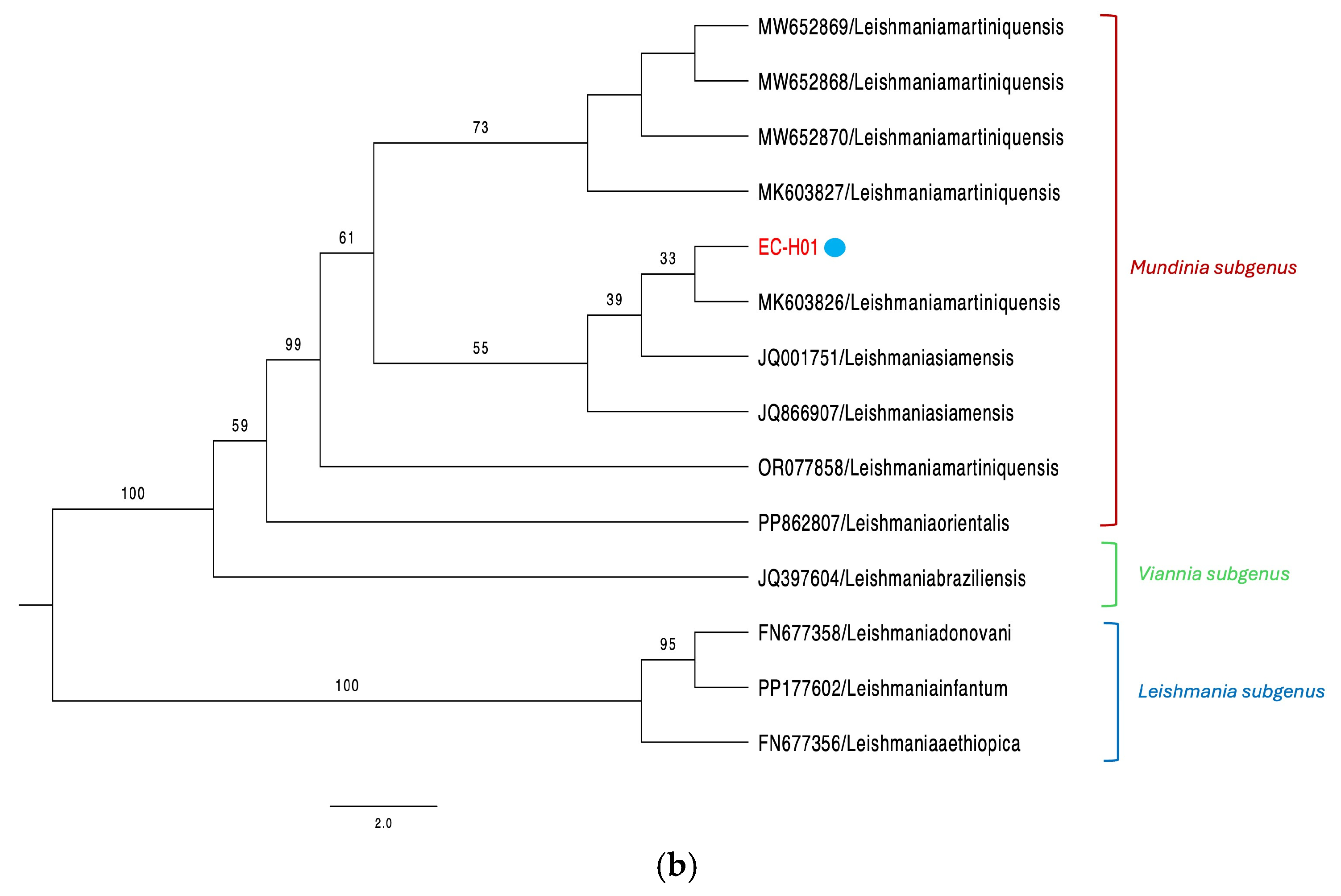
Hsp70-PCR products were sequenced using the Sanger technique. In the case of the horse, amplification and Sanger sequencing of the ITS1 region of the rRNA gene (379 bp product) were performed to confirm L. (M.) martiniquensis infection. Although the methodology referenced [25] was originally developed for Old World Leishmania species, it was suitable for L. (M.) martiniquensis due to its phylogenetic placement within the Mundinia subgenus, which clusters near Old World species [23,24,26]. ITS1 sequences were analyzed by BLAST v2.15.0 and phylogenetic reconstruction to establish species identity. A second molecular approach (Hsp-70 gene sequencing) was used to confirm this result [23,24,25,26]. Sanger sequencing reactions were performed using the Applied Biosystems™ BigDye Terminator v3.1 cycling kit (Thermo Fisher Scientific: 4337454, Vilnius, Lithuania). Reaction products were purified with the BigDye XTerminator kit (Applied Biosystems, Waltham, MA, USA) and analyzed using the 3130xl Genetic Analyzer sequencer (Applied Biosystems, Foster, CA, USA). Resulting sequences were edited using Sequencher software v4.6 and their homology with those available in GenBank database was assessed by performing a BLAST search from the National Center for Biotechnology Information Database (http://www.ncbi.nlm.nih.gov/BLAST/, accessed on 22 July 2025). Sequence alignment and phylogenetic analyses were performed as described [5]. A consensus tree was summarized from sampled trees and visualized with FigTree v.1.4.4. To create the phylogenetic tree of the Hsp-70 gene with the sequences of L. (V.) braziliensis and L. (M.) martiniquensis, the best-fit model TN+F+I and an ultra-fast bootstrap with 1000 replicates were used. In the case of the ITS1 gene with the sequence of L. (M.) martiniquensis, the best-fit model HKY+F+I and an ultra-fast bootstrap with 1000 replicates were employed. Representative sequences obtained from NCBI GenBank of Leishmania and Endotrypanum were used for phylogenetic analysis with the Hsp-70 gene, and existing sequences of Leishmania were used for phylogenetic analysis with the ITS1 gene. Nucleotide sequences are available in NCBI GenBank under the accession numbers PV658270 (ITS1) and PV844900-PV844903 (Hsp70).

2.5. Ethical Statement

ICGES is the national reference laboratory for Leishmania diagnosis. As leishmaniasis is a notifiable disease in Panama, samples from this study were analyzed as part of the surveillance activities approved in the National Commission for the Control and Prevention of Tropical Neglected Diseases. The use of animal biological samples for this study was granted exemption (008/CIUCAL-ICGES25) from the Comité Institucional para el Uso y Cuidado de Animales de Laboratorio (CIUCAL-ICGES). Samples were coded and anonymized to protect the confidentiality of the study subjects and owners.

3. Results

Between 2021 and 2025, twelve domestic animals, including ten dogs and two horses from endemic areas of CL, were tested for clinical suspicion of leishmaniasis. Leishmania infection was confirmed by serological testing and/or conventional PCR in 50% of cases (6/12), with one horse and five dogs identified (Table 2). Of the canine cases, three were classified as CL and two as CVL, while the horse case was diagnosed with CL.

3.1. Canine Cases

Of the 10 canine cases analyzed, three local dogs were diagnosed with CL (CF-W01, CF-P02, CF-L03) by clinical and laboratory tests. All three dogs had ulcerated lesions on their noses and had no travel history to another country (Figure S1). The remaining dogs were tested for clinical suspicion of CVL (7/10), with two testing positive for Leishmania (CF-J07, CF-A09) via serological (DPP rk39 and IFA) and molecular (kDNA, Hsp-70) tests. One CVL-positive dog had a travel history to Brazil and exhibited overt clinical signs compatible with leishmaniasis, including dermatological alterations. The second dog diagnosed with CVL had traveled to Spain and presented an enlarged spleen as the only clinical sign consistent with the infection. The remaining five dogs with suspected CVL presented one or more of the following non-specific clinical signs: weight loss, lethargy, enlarged lymph nodes, and different types of skin lesions. Leishmania infections in the three CL cases were characterized as L. (V.) braziliensis by Hsp70-RFLP and Sanger sequencing (Figure 1). In the two CVL cases, Hsp70-RFLP and sequencing were not possible due to insufficient amplified DNA.

3.2. Equine Cases

Of the two horses tested for clinical suspicion of CL, one tested positive for Leishmania (EC-H01) using molecular methodologies involving the Hsp-70 and kDNA genes. This sample was further characterized as L. (M.) martiniquensis by sequencing the Hsp-70 and ITS1 genes (Figure 1). The horse exhibited nodular and ulcerated lesions on the ears, originated from a CL endemic area, and had no history of travel outside the country. The CL-negative horse had an ulcerated lesion with crust, purulent discharge, and intense itching. This horse was diagnosed with cutaneous habronemiasis, a parasitic disease of horses frequently prevalent in tropical climates caused by Habronema larvae transmitted by flies (Musca domestica and Stomoxys calcitrans), which cause skin, eye, or gastric lesions. Gross lesions are manifest as granulomatous ulcers that can be confused with ulcerated lesions caused by Leishmania parasites [27,28].

4. Discussion

In the Americas, wild animal reservoirs play a crucial role in maintaining the zoonotic transmission cycle of Leishmania species indigenous to the Neotropical region [1,2,3]. The exception is L. (L.) infantum, introduced to the continent during the colonial era [29], which is primarily maintained in a zoonotic transmission cycle where dogs serve as the main reservoir hosts [10]. In Panama, L. (L.) infantum has only been recently detected in dogs imported from the Mediterranean Basin and South America [30]. In this study, we report a case of L. (L.) infantum infection in a local dog from a recognized ACL-endemic region of the country. However, further investigation of its travel history revealed that although the dog was born in Panama, it lived for a short time in Zaragoza, Spain before returning, confirming it as a new imported case.
Although domestic animals are frequently exposed to Leishmania Viannia species in endemic areas of Panama [19], their role as reservoir hosts in the zoonotic transmission of ACL remains unclear. Canine CL cases were documented in the 1970s in rural communities of central Panama [31]. Although the infecting Leishmania species were not identified using laboratory methods, the authors hypothesized L. (V.) braziliensis as the etiological agent based on ecological and epidemiological evidence. Using molecular techniques, here we detected L. (V.) braziliensis infections in three local dogs from recognized ACL-endemic areas of Panama, where L. (V.) panamensis is the predominant species. These three dogs presented cutaneous and mucosal ulcers consistent with L. (V.) braziliensis infections [32] (Figure S1). While L. (V.) braziliensis infections in humans have been reported at low frequencies in Panama [5], these findings represent the first molecularly documented cases of L. (V.) braziliensis in dogs from the country and, more broadly, from any Central American nation where human infections by this species have been recorded. In South America, L. (V.) braziliensis is the most common causative agent of ACL in dogs [10,32]. These findings suggest that the co-circulation of L. (V.) braziliensis in ACL-endemic regions of Panama may be more frequent than previously reported. Further research is required to assess the epidemiological significance of L. (V.) braziliensis infections in dogs, including their potential role as competent reservoirs in the transmission cycle.
However, the epidemiological significance of L. (V.) braziliensis infections in dogs in Panama requires cautious interpretation. The small number of cases limits generalization, and the mere presence of infection does not confirm reservoir competence. Factors such as parasitemia levels, duration of infection, and the ability of local vectors to acquire and transmit the parasite from dogs remain to be elucidated.
It is also important to consider the possibility of L. (Viannia) hybrid strains in Panama. A previous study conducted in central Panama detected molecular signatures suggestive of L. (V.) braziliensis/L. (V.) panamensis hybrids in human cutaneous lesions [5]. Although phylogenetic analysis demonstrated the presence of L. (V.) braziliensis/L. (V.) guyanensis hybrids, we cannot rule out the possibility of the presence of both hybrids in the country. Such hybrids may complicate molecular identification, particularly when using single-locus markers such as Hsp-70. Although our Hsp-70-based phylogeny grouped the sequences within the L. (V.) braziliensis clade, we cannot rule out the presence of genetic introgression or hybridization with L. (V.) panamensis, especially given the geographic overlap of these species in endemic areas. Future studies using multilocus genotyping or whole-genome sequencing will be essential to clarify this issue.
In the Americas, natural Leishmania infections in horses have been frequently documented, with L. (V.) braziliensis and L. (L.) infantum being the most detected species [15,33]. However, in recent years, L. (M.) martiniquensis has emerged as an important species infecting equine [34,35]. Given their frequent exposure to Leishmania-infected sandflies in endemic regions and their close interactions with humans and other domestic animals, assessing Leishmania infections in horses is essential to elucidate their epidemiological role, zoonotic potential, and clinical significance.
In Panama, several species of Lutzomyia sand flies have been implicated in the transmission of L. (V.) panamensis and L. (V.) braziliensis, particularly Lutzomyia gomezi, L. panamensis, and L. trapidoi [8,36,37]. However, the vector responsible for the transmission of L. (M.) martiniquensis remains unknown. In other endemic areas such as Southeast Asia, Sergentomyia spp. and Phlebotomus spp. have been suggested as potential vectors of Mundinia species [34]. Given the presence of L. (M.) martiniquensis in a horse from Panama without a travel history, and its phylogenetic confirmation, it is plausible that local phlebotomine species may be involved in its transmission. Entomological investigations are needed to identify potential vector species and assess their infection rates with Mundinia parasites in Central America.
In this study, we identified a horse with cutaneous lesions naturally infected with L. (M.) martiniquensis using two molecular markers (Figure 1). The horse was born and raised in Panama and had no travel history outside the country, providing strong evidence that the infection was locally acquired. This represents the first confirmed report of L. (V.) martiniquensis infecting a horse in Central America. A recent study from the neighboring country of Costa Rica reported five equine cases of leishmaniasis with cutaneous lesions diagnosed through immunohistochemical detection; however, the causative Leishmania species in those infections were not identified [38].
One limitation of our study is the use of single-locus markers (Hsp-70 and ITS1) for phylogenetic reconstruction, which may lead to lower bootstrap support in certain branches of the trees, as observed in Figure 1. While these genes are commonly used for Leishmania species identification [21,35,39,40], they may not provide sufficient resolution for deeper evolutionary relationships. The use of multilocus or whole-genome sequencing approaches would improve phylogenetic robustness and should be considered in future studies. Nevertheless, the consistent results across two genetic markers and the high sequence identity with reference strains support the accuracy of our species-level identifications. In addition to the molecular findings, the clinical and epidemiological context strengthens our identification. The cutaneous lesion characteristics observed in the horse, along with its origin in a tropical region, are consistent with previously reported cases of leishmaniasis caused by L. (M.) martiniquensis [34,35]. Taken together, the molecular, clinical, and epidemiological evidence strongly supports L. (M.) martiniquensis as the etiological agent in this case.
The detection of L. (M.) martiniquensis in a horse in Panama underscores the need to expand epidemiological surveillance in domestic animals and potential vectors in the region. It is essential to investigate whether its emergence in Panama is linked to environmental factors such as deforestation and climate change, which may be facilitating the expansion of its vectors and reservoirs. Additionally, this finding raises important questions regarding the possible circulation of L. (M.) martiniquensis in humans in Panama, which may have gone undiagnosed due to limitations in conventional detection methods. Enhanced molecular and epidemiological studies are necessary to better understand the distribution, transmission dynamics, and potential public health implications of L. (M.) martiniquensis in the region.

5. Conclusions

Our findings provide preliminary molecular evidence of the diversity of Leishmania species infecting domestic animals in Panama, including the first molecular documentation of L. (V.) braziliensis in local dogs and L. (M.) martiniquensis in a horse. While these results expand our understanding of potential animal involvement in the transmission cycle of cutaneous leishmaniasis, the analyses were based on two single-locus markers, which generally provided lower phylogenetic resolution. Consequently, species identification should be interpreted with caution. Future studies using multilocus genotyping or genomic approaches are essential to confirm these findings and to explore the possibility of hybrid strains or cryptic diversity. Nonetheless, our results underscore the importance of strengthening surveillance efforts in domestic animals under a One Health framework to better understand the eco-epidemiology of Leishmania transmission in Central America.

Supplementary Materials

The following supporting information can be downloaded at https://www.mdpi.com/article/10.3390/ani15182677/s1, Figure S1: Ulcerated lesions on the nose of CL dogs infected with L. (V.) braziliensis. A: CF-W01, B: CF-L03, C: CF-P02.

Author Contributions

Conceptualization, J.E.C., K.G. and A.S.; methodology, V.P., V.V., A.R. and K.G.; software, K.G. and V.V.; validation, V.P., J.E.C., K.G. and A.S.; formal analysis, V.P., S.M., I.R., E.H. and A.I.T.; investigation, S.M., J.E.C., K.G., A.S. and V.P.; resources, J.E.C., A.S. and K.G.; data curation, V.P., V.V. and K.G.; writing—original draft preparation, V.P. and J.E.C.; writing—review and editing, K.G. and A.S.; visualization, S.M., I.R., E.H., A.I.T. and K.G.; supervision, A.S. and J.E.C.; project administration, K.G.; funding acquisition, J.E.C., A.S. and K.G. All authors have read and agreed to the published version of the manuscript.

Funding

This research was funded by the Sistema Nacional de Investigación (SNI-SENACYT), Panama, awarded to K.G., J.E.C. and A.S.; and through the Ministerio de Economia y Finanzas de Panama grant number MEF 3.04.20. This work also received administrative and financial support from the Gorgas Memorial Institute for Health Studies.

Institutional Review Board Statement

The use of animal biological samples for this study was granted exemption (008/CIUCAL-ICGES25) from the Comité Institucional para el Uso y Cuidado de Animales de Laboratorio of the Instituto Conmemorativo Gorgas de Estudios de la Salud (CIUCAL-ICGES).

Informed Consent Statement

The use of clinical samples, as well as the written informed consent, was granted exemption by the CIUCAL-ICGES (008/CIUCAL-ICGES25). ICGES is the national reference laboratory for Leishmania diagnosis. As leishmaniasis is a notifiable disease in Panama, samples from this study were analyzed as part of the surveillance activities approved in the National Commission for the Control and Prevention of Tropical Neglected Diseases. Samples were coded and anonymized to protect the confidentiality of the study subjects and owners.

Data Availability Statement

The sequences used to support the findings of this study are available from GenBank under the accession numbers: PV658270 and PV844900-PV844903.

Acknowledgments

We thank private veterinarians for their favorable cooperation in sharing information and biological samples from the clinical cases. We also thank Maria Chen for her advice in the preparation of the phylogenetic trees. This study was possible thanks to the support of SNI-SENACYT, Panama, awarded to Azael Saldaña, Kadir González, and José E. Calzada. During the preparation of this manuscript, the authors used Grammarly, version 1.127.0.0, as a grammar assistance tool for correcting manuscript text writing, and Mendeley Reference Manager Version 2.135.0 for managing bibliographic references in the manuscript. The authors have reviewed and edited the output and take full responsibility for the content of this publication.

Conflicts of Interest

The authors declare no conflicts of interest.

Abbreviations

The following abbreviations are used in this manuscript:
ACLAmerican cutaneous leishmaniasis
IFAIndirect immunofluorescence assay
CLCutaneous leishmaniasis
CVLCanine visceral leishmaniasis

References

  1. Pan American Health Organization. Plan of Action to Strengthen the Surveillance and Control of Leishmaniasis in the Americas 2023–2030; Pan American Health Organization: Washington, DC, USA, 2024. [Google Scholar]
  2. Pan American Health Organization. Manual of Procedures for Leishmaniasis Surveillance and Control in the Region of the Americas; Pan American Health Organization: Washington, DC, USA, 2024. [Google Scholar]
  3. Pan American Health Organization. Leishmaniasis: Epidemiological Report for the Americas; Pan American Health Organization: Washington, DC, USA, 2024. [Google Scholar]
  4. Lainson, R. The Neotropical Leishmania Species: A Brief Historical Review of Their Discovery, Ecology and Taxonomy. Rev Pan-Amaz. Saude 2010, 1, 25–33. [Google Scholar] [CrossRef]
  5. Miranda, A.; Del, C.; González, K.A.; Samudio, F.; Pineda, V.J.; Calzada, J.E.; Capitan-Barrios, Z.; Jiménez, A.; Castillo, J.; Mendoza, Y.; et al. Molecular Identification of Parasites Causing Cutaneous Leishmaniasis in Panama. Am. J. Trop. Med. Hyg. 2021, 104, 1326. [Google Scholar] [CrossRef]
  6. Davila, M.; Pineda, V.; Calzada, J.E.; Saldaña, A.; Samudio, F. Evaluation of Cytochrome b Sequence to Identify Leishmania Species and Variants: The Case of Panama. Mem. Inst. Oswaldo Cruz. 2021, 116, e200572. [Google Scholar] [CrossRef]
  7. Vásquez, A.; Paz, H.; Alvar, J.; Perez, D.; Hernandez, C. Informe Final: Estudios Sobre La Epidemiología de la Leishmaniasis Panamá, en la Parte Occidental de la República de Panamá; Instituto Conmemorativo Gorgas de Estudios de la Salud-MINSA: Panama, Panama, 1998. [Google Scholar]
  8. de Vasquez, A.M.; Christensen, H.A.; Petersen, J.L. Short Report Epidemiologic Studies on Cutaneous Leishmaniasis in Eastern Panama. Am. J. Trop. Med. Hyg. 1999, 60, 54–57. [Google Scholar] [CrossRef] [PubMed]
  9. Reina, A.M.; Mewa, J.C.; Calzada, J.E.; Saldaña, A. Characterization of Leishmania spp. Causing Cutaneous Lesions with a Negative Parasitological Diagnosis in Panama. Trop. Med. Infect. Dis. 2022, 7, 282. [Google Scholar] [CrossRef]
  10. Dantas-Torres, F. Canine Leishmaniasis in the Americas: Etiology, Distribution, and Clinical and Zoonotic Importance. Parasit. Vectors 2024, 17, 198. [Google Scholar] [CrossRef]
  11. Barrera, C.; Le Pont, F.; Dereure, P.J.; Barrera, C.; Guerrini, F.; Martini, A.; Echeverria, R.; Guderianz, R.; Pont, F.L. Infestation Naturelle Du Chien Par Leishmania Panamensis. Ann. Soc. Belg. Med. Trop. 1994, 74, 29–33. [Google Scholar]
  12. Kent, A.; Ramkalup, P.; Mans, D.; Schallig, H. Is the Dog a Possible Reservoir for Cutaneous Leishmaniasis in Suriname? J. Trop. Med. 2013, 1, 324140. [Google Scholar] [CrossRef]
  13. Santaella, J.; Ocampo, C.B.; Saravia, N.G.; Méndez, F.; Góngora, R.; Gomez, M.A.; Munstermann, L.E.; Quinnell, R.J. Leishmania (Viannia) Infection in the Domestic Dog in Chaparral, Colombia. Am. J. Trop. Med. Hyg. 2011, 84, 674–680. [Google Scholar] [CrossRef]
  14. Medkour, H.; Davoust, B.; Dulieu, F.; Maurizi, L.; Lamour, T.; Marié, J.L.; Mediannikov, O. Potential Animal Reservoirs (Dogs and Bats) of Human Visceral Leishmaniasis Due to Leishmania infantum in French Guiana. PLoS Negl. Trop. Dis. 2019, 13, e0007456. [Google Scholar] [CrossRef]
  15. Ratzlaff, F.R.; Osmari, V.; da Silva, D.; de Paula Vasconcellos, J.S.; Pötter, L.; Fernandes, F.D.; de Mello Filho, J.A.; de Avila Botton, S.; Vogel, F.S.F.; Sangioni, L.A. Identification of Infection by Leishmania spp. in Wild and Domestic Animals in Brazil: A Systematic Review with Meta-Analysis (2001–2021). Parasitol. Res. 2023, 122, 1605–1619. [Google Scholar] [CrossRef]
  16. Belo, V.S.; Struchiner, C.J.; Werneck, G.L.; Barbosa, D.S.; de Oliveira, R.B.; Neto, R.G.T.; da Silva, E.S. A Systematic Review and Meta-Analysis of the Factors Associated with Leishmania infantum Infection in Dogs in Brazil. Vet. Parasitol. 2013, 195, 1–13. [Google Scholar] [CrossRef]
  17. Yamada, K.; Valderrama, A.; Gottdenker, N.; Cerezo, L.; Minakawa, N.; Saldaña, A.; Calzada, J.E.; Chaves, L.F. Macroecological Patterns of American Cutaneous Leishmaniasis Transmission across the Health Areas of Panamá (1980–2012). Parasite Epidemiol. Control 2016, 1, 42–55. [Google Scholar] [CrossRef] [PubMed]
  18. Hage, R.; Dos, S.; Nunes e Silva, S.V.; Bohm, B.C.; Lima, J.V.; Bruhn, N.C.P.; Menezes, G.R.; Bruhn, F.R.P. Spatiotemporal Relationship between Agriculture, Livestock, Deforestation, and Visceral Leishmaniasis in Brazilian Legal Amazon. Sci. Rep. 2024, 14, 21542. [Google Scholar] [CrossRef] [PubMed]
  19. Calzada, J.E.; Saldaña, A.; González, K.; Rigg, C.; Pineda, V.; Santamaría, A.M.; Rodríguez, I.; Gottdenker, N.L.; Laurenti, M.D.; Chaves, L.F. Cutaneous Leishmaniasis in Dogs: Is High Seroprevalence Indicative of a Reservoir Role? Parasitology 2015, 142, 1202–1214. [Google Scholar] [CrossRef] [PubMed]
  20. Vergel, C.; Walker, J.; Saravia, N.G. Amplification of human DNA by primers targeted to Leishmania kinetoplast DNA and post-genome considerations in the detection of parasites by a polumerase chain reaction. Am. J. Trop. Med. Hyg. 2005, 72, 423–429. [Google Scholar] [CrossRef]
  21. Montalvo, A.M.; Fraga, J.; Maes, I.; Dujardin, J.-C.; Van der Auwera, G. Three New Sensitive and Specific Heat-Shock Protein 70 PCRs for Global Leishmania Species Identification. Eur. J. Clin. Microbiol. Infect. Dis. 2012, 31, 1453–1461. [Google Scholar] [CrossRef]
  22. Marques, M.J.; Volpini, Â.C.; Machado-Coelho, G.L.L.; Machado-Pinto, J.; da Costa, C.A.; Mayrink, W.; Genaro, O.; Romanha, A.J. Comparison of Polymerase Chain Reaction with Other Laboratory Methods for the Diagnosis of American Cutaneous Leishmaniasis. Diagn. Microbiol. Infect. Dis. 2006, 54, 37–43. [Google Scholar] [CrossRef]
  23. Songumpai, N.; Promrangsee, C.; Noopetch, P.; Siriyasatien, P.; Preativatanyou, K. First Evidence of Co-Circulation of Emerging Leishmania martiniquensis, Leishmania orientalis, and Crithidia sp. in Culicoides Biting Midges (Diptera: Ceratopogonidae), the Putative Vectors for Autochthonous Transmission in Southern Thailand. Trop. Med. Infect. Dis. 2022, 7, 379. [Google Scholar] [CrossRef]
  24. Srivarasat, S.; Brownell, N.; Siriyasatien, P.; Noppakun, N.; Asawanonda, P.; Rattanakorn, K.; Preativatanyou, K.; Kumtornrut, C. Case Report: Autochthonous Disseminated Cutaneous, Mucocutaneous, and Visceral Leishmaniasis Caused by Leishmania martiniquensis in a Patient with HIV/AIDS from Northern Thailand and Literature Review. Am. J. Trop. Med. Hyg. 2022, 107, 1196–1202. [Google Scholar] [CrossRef]
  25. Spanakos, G.; Piperaki, E.T.; Menounos, P.G.; Tegos, N.; Flemetakis, A.; Vakalis, N.C. Detection and Species Identification of Old World Leishmania in Clinical Samples Using a PCR-Based Method. Trans. R Soc. Trop. Med. Hyg. 2008, 102, 46–53. [Google Scholar] [CrossRef]
  26. Jariyapan, N.; Bates, M.D.; Bates, P.A. Molecular Identification of Two Newly Identified Human Pathogens Causing Leishmaniasis Using PCR-Based Methods on the 30 Untranslated Region of the Heat Shock Protein 70 (Type i) Gene. PLoS Negl. Trop. Dis. 2021, 15, e0009982. [Google Scholar] [CrossRef]
  27. Barlaam, A.; Traversa, D.; Papini, R.; Giangaspero, A. Habronematidosis in Equids: Current Status, Advances, Future Challenges. Front. Vet. Sci. 2020, 7, 358. [Google Scholar] [CrossRef]
  28. Traversa, D.; Otranto, D.; Iorio, R.; Carluccio, A.; Contri, A.; Paoletti, B.; Bartolini, R.; Giangaspero, A. Identification of the Intermediate Hosts of Habronema microstoma and Habronema muscae under Field Conditions. Med. Vet. Entomol. 2008, 22, 283–287. [Google Scholar] [CrossRef]
  29. Schwabl, P.; Boité, M.C.; Bussotti, G.; Jacobs, A.; Andersson, B.; Moreira, O.; Freitas-Mesquita, A.L.; Meyer-Fernandes, J.R.; Telleria, E.L.; Traub-Csekö, Y.; et al. Colonization and Genetic Diversification Processes of Leishmania infantum in the Americas. Commun. Biol. 2021, 4, 139. [Google Scholar] [CrossRef] [PubMed]
  30. Terrero, I.; Pineda, V.; Vásquez, V.; Miranda, A.; Saldaña, A.; Calzada, J.E.; González, K. First Report of Imported Canine Visceral Leishmaniasis Cases in Panama, Central America: Public Health Implications. Vet. Parasitol. Reg. Stud. Rep. 2022, 32, 100745. [Google Scholar] [CrossRef] [PubMed]
  31. Herrer, A.; Christensen, H.A. Natural Cutaneous Leishmaniasis among Dogs in Panama. Am. J. Trop. Med. Hyg. 1976, 25, 59–63. [Google Scholar] [CrossRef] [PubMed]
  32. Lago, J.; Silva, J.A.; Borja, L.; Fraga, D.B.M.; Schriefer, A.; Arruda, S.; Lago, E.; Carvalho, E.M.; Bacellar, O. Clinical and Histopathologic Features of Canine Tegumentary Leishmaniasis and the Molecular Characterization of Leishmania braziliensis in Dogs. PLoS Negl. Trop. Dis. 2019, 13, e0007532. [Google Scholar] [CrossRef]
  33. Mhadhbi, M.; Sassi, A. Infection of the Equine Population by Leishmania Parasites. Equine Vet. J. 2020, 52, 28–33. [Google Scholar] [CrossRef]
  34. Ahmadi, S.; Hataminejad, M.; Rahimi Esboei, B.; Hosseini, S.A.; Fakhar, M. An Update on Leishmania martiniquensis Infections: Transmission, Clinical Characteristics, and Treatment. Parasite Epidemiol. Control 2024, 27, e00386. [Google Scholar] [CrossRef]
  35. Mendes, A.A.V.; Filgueira, C.P.B.; De Freitas Campos Miranda, L.; De Almeida, A.B.; Cantanhêde, L.M.; Fagundes, A.; Pereira, S.A.; Menezes, R.C.; Cupolillo, E. First Report of Leishmania (Mundinia) martiniquensis in South American Territory and Confirmation of Leishbunyavirus Infecting This Parasite in a Mare. Mem. Inst. Oswaldo Cruz 2023, 118, e220220. [Google Scholar] [CrossRef]
  36. Valderrama, A.; Tavares, M.G.; Andrade Filho, J.D. Anthropogenic Influence on the Distribution, Abundance and Diversity of Sandfly Species (Diptera: Phlebotominae: Psychodidae), Vectors of Cutaneous Leishmaniasis in Panama. Mem. Inst. Oswaldo Cruz 2011, 106, 1024–1031. [Google Scholar] [CrossRef] [PubMed]
  37. Christensen, H.A.; Fairchild, G.B.; Herrer, A.; Johnson, C.M.; Young, D.G.; de Vásquez, A.M. The Ecology of Cutaneous Leishmaniasis in the Republic of Panama. J. Med. Entomol. 1983, 20, 463–484. [Google Scholar] [CrossRef] [PubMed]
  38. Ortega-García, M.V.; Salguero, F.J.; García, N.; Domínguez, M.; Moreno, I.; Berrocal, A. Equine Infection with Leishmania spp. in Costa Rica: Study of Five Cases. Vet. Med. Sci. 2021, 7, 2234–2239. [Google Scholar] [CrossRef] [PubMed]
  39. Fraga, J.; Montalvo, A.M.; De Doncker, S.; Dujardin, J.C.; Van der Auwera, G. Phylogeny of Leishmania Species Based on the Heat-Shock Protein 70 Gene. Infect. Genet. Evol. 2010, 10, 238–245. [Google Scholar] [CrossRef]
  40. Carbonara, M.; Mendoza-Roldan, J.A.; Bezerra-Santos, M.A.; de Abreu Teles, P.P.; Lia, R.P.; Locantore, F.; Iatta, R.; Volf, P.; Otranto, D. Leishmania spp. in Equids and Their Potential Vectors in Endemic Areas of Canine Leishmaniasis. PLoS Negl. Trop. Dis. 2024, 18, e0012290. [Google Scholar] [CrossRef]
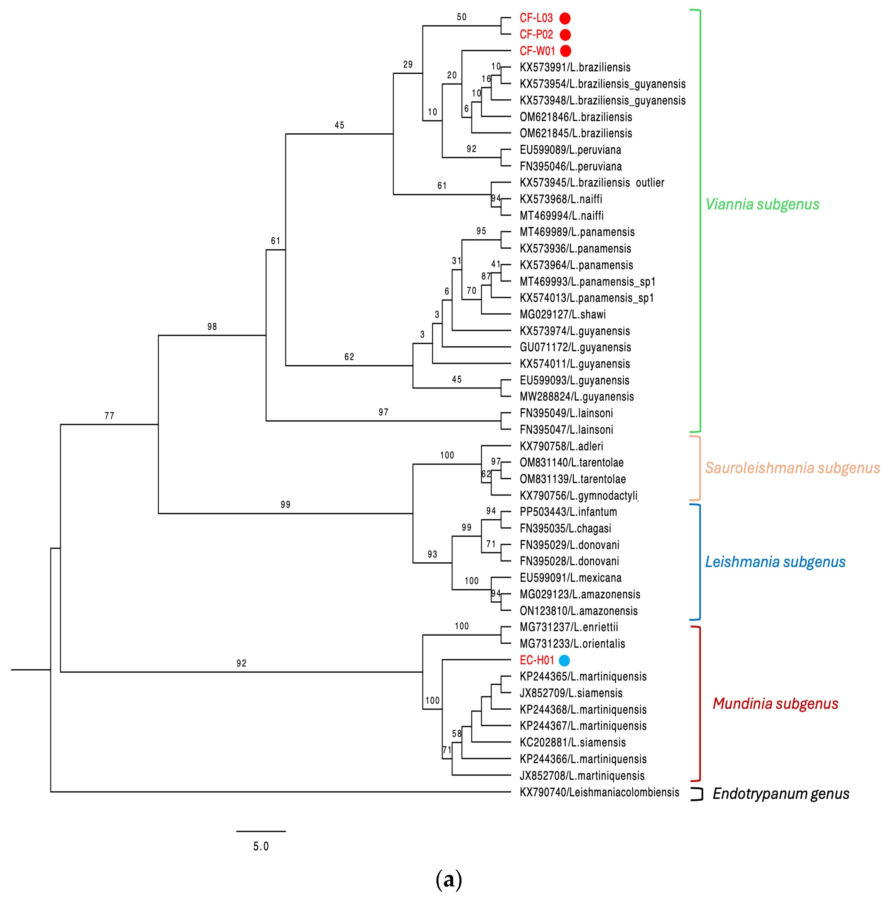
Figure 1. (a) Phylogenetic tree with the Hsp-70 gene sequences of L. (V.) braziliensis (red circle) and L. (M.) martiniquensis (blue circle). The best-fit model TN+F+I and an ultra-fast bootstrap with 1000 replicates were used. (b) Phylogenetic tree with the ITS1 gene sequence of L. (M.) martiniquensis (blue circle) obtained from samples of domestic animals with CL. The best-fit model, HKY+F+I, and an ultra-fast bootstrap with 1000 replicates were used.
Animals 15 02677 g001aAnimals 15 02677 g001b
Table 1. PCR protocols used to identify Leishmania parasites from domestic animals.
PCRPrimersPrimers
Concentrations
DNA Used per ReactionPCR ConditionsPCR Products Size
kDNA
Viannia
Vergel et al., 2005 [20]
B1:
5′-GGGGTTGGTGTAATATAGTGG-3′
LV:
5′-ATTTTTGAACGGGGTTTCTG-3′
0.6 μm/L
each one
5 μL5 cycles of
95 °C × 6 min
95 °C × 30 seg
64.5 °C × 2 min
72 °C × 1 min
35 cycles of
95 °C × 30 seg
64 °C × 1 min
72 °C × 1 min
72 °C × 10 min
750 bp
Hsp-70
Montalvo et al., 2012
[21]
Hsp70-F25:
5′-GGACGCCGGCACGATTKCT-3′
Hsp70-R1310:
5′-CCTGGTTGTTGTTCAGCCACTC-3′
0.6 μm/L
each one
5 μL94 °C × 5 min
33 cycles of
94 °C × 30 seg
61 °C × 1 min
72 °C × 3 min
72 °C × 10 min
1286 bp
kDNA L150/151
Marques et al., 2006 [22]
L-150:
5′-GGG(G/T)AGGGGCGTTCT(G/C)CGAA-3′
L-151:
5′-(G/C)(G/C)(G/C)A/C)CTAT(A/T)TTACACCAACCCC-3′
0.5 μm/L
each one
5 μL94 °C × 4 min
33 cycles of
94 °C × 30 seg
52.3 °C × 30 seg
72 °C × 30 seg
72 °C × 10 min
120 bp
ITS1
Spanakos et al., 2008 [25]
LeR:
5′-CCAAGTCATCCATCGCGACACG-3′
LeF
5′-TCCGCCCGAAAGTTCACCGATA-3′
1.0 μm/L
each one
5 μL95 °C × 5 min
40 cycles of
95 °C × 1 min
65 °C × 1 min
72 °C × 1 min
72 °C × 10 min
379 bp
Table 2. Detection of Leishmania in domestic animals, including diagnostic results, species identification, and region. N/A: Not available or not applicable.
IDAnimal SpeciesSample TypeRegionDPP rk39IFIPCR kDNA *PCR Hsp-70Leishmania SpeciesGenBank
Accession
CF-W01DogBiopsyColonN/AN/A++L. (V.) braziliensisPV844902
CF-P02DogBiopsyColonN/AN/A++L. (V.) braziliensisPV844901
CF-L03DogBiopsyPanama Este N/AN/A++L. (V.) braziliensisPV844903
CF-C04DogBloodPanama----N/AN/A
CF-A05DogSerum/
Blood
Panama Oeste----N/AN/A
CF-S06DogSerum/
Blood
Panama Oeste----N/AN/A
CF-J07DogSerum/
Blood
Panama++++N/AN/A
CF-F08DogSerum/
Blood
Panama----N/AN/A
CF-A09DogSerum/
Blood
Panama Norte++++N/AN/A
CF-S10DogSerum/
Blood
Panama Norte----N/AN/A
EC-H01HorseBiopsyPanama OesteN/AN/A++L. (M.)
martiniquensis
PV658270
PV844900
EC-H02HorseBiopsyPanama EsteN/AN/A--N/AN/A
* PCR kDNA includes Viannia genus-specific PCR and L150/L151 PCR. +: Positive. -: Negative
Disclaimer/Publisher’s Note: The statements, opinions and data contained in all publications are solely those of the individual author(s) and contributor(s) and not of MDPI and/or the editor(s). MDPI and/or the editor(s) disclaim responsibility for any injury to people or property resulting from any ideas, methods, instructions or products referred to in the content.