1. Introduction
The uterus of mammals, including the uterine tubes and the cranial vagina, develops from paired paramesonephric (Müllerian) ducts [
1,
2,
3]. It is widely accepted that these ducts originate from the cephalolateral invagination of the coelomic epithelium to the upper regions of the mesonephroi, followed by the caudal elongation of the resulting epithelial tubes [
3,
4,
5,
6].
In domestic animals, prenatal uterine development follows variable patterns, as the uterine and uterovaginal segments of the paramesonephric ducts may fuse completely, partially, or minimally [
2]. In the domestic cat, the long uterine horns derive from the middle uterine segments of the paramesonephric ducts, whereas the short uterine body develops from the uterovaginal canal [
6].
Various aspects of uterine development have been investigated in rodents, domestic animals, and humans. The histogenesis of the uterine wall has been described in mice and rats [
7], sheep [
7,
8,
9], cattle [
9], pigs [
7,
10], and humans [
11,
12,
13]. However, studies on uterine development in carnivores, including cats, remain limited. Using light microscopy, Inomata et al. [
14] described the development of the paramesonephric and mesonephric ducts in male and female domestic cats with crown–rump length (CRL) ranging from 0.8 to 10.5 cm, aiming to identify the timing of sexual differentiation of these structures. Additionally, Merlo et al. [
15] characterized the development of uterine glands in domestic cats from late gestation to puberty using light microscopy.
In our previous morphological and three-dimensional reconstruction study of domestic cat fetuses, we identified the stages of reproductive system development and determined the spatial and temporal differentiation of the paramesonephric duct rudiments into uterine tubes and uterine horns [
6]. We found that the first stage of feline internal reproductive organ development lasts from day 26 to 44 p.c., and it involves the formation of the tubal and uterine segments of the paramesonephric ducts. The second stage continues until the end of the prenatal period and entails the histodifferentiation of the paramesonephric duct wall, following the structural patterns assigned to the uterine tubes and the uterus. These studies also demonstrated changes in the topographical relationships between the developing female reproductive tract, the ovaries, and the mesonephros and indicated conversions in the shape of the paramesonephric ducts, which, after day 54 p.c., transform into the uterus, shifting from a U-shaped to a W-shaped configuration. The final shape and topography of the uterus in the domestic cat are established by the third month of postnatal development when the uterine horns straighten and, together with the uterine body, form a V-shaped structure within the abdominal cavity [
6].
In another study, we described the sequence of ultrastructural changes in the endometrial epithelium of the paramesonephric ducts and developing uterine horns in domestic cats [
16]. The study identified two primary stages in uterine epithelium development. The first stage, lasting until day 42 p.c., involves the formation of the pseudostratified epithelium of the paramesonephric duct. The second stage concerns the development of the endometrial epithelium, which remains pseudostratified with variable height by the end of the prenatal period and only transforms into simple columnar epithelium after birth [
16]. The postnatal development of the domestic cat’s uterine horns has also been documented, with a particular focus on changes in the endometrium and its maturation necessary for reproductive function [
17].
The present study aimed to describe the prenatal histodifferentiation of the uterine wall in domestic cats during the development of the uterine segments of the paramesonephric ducts and the uterine horns, with particular emphasis on the structural maturity of the endometrium and myometrium at birth. The findings are discussed in the context of both the prenatal development of the uterine tubes and postnatal microstructural changes in the uterine horns. Expanding knowledge of uterine development in domestic cats—one of the most widespread and socially integrated companion species—is essential for advancing our understanding of feline reproductive physiology. This study contributes valuable new insights into uterine wall histodifferentiation, enriching the field of veterinary science and comparative embryology and offering a foundation for future cross-species research. Moreover, a better understanding of embryonic development in the feline reproductive system has practical implications for veterinary diagnostics, particularly in identifying developmental abnormalities.
2. Materials and Methods
Studies were conducted on uteri collected from 60 euthanized fetuses of domestic cats aged 28–63 days p.c. (post-conception). In fetuses older than 33 days p.c., euthanasia was performed using Morbital (pentobarbital sodium and pentobarbitone; Biowet, Puławy, Poland). The fetuses were obtained postmortem from wild living female cats subjected to surgical ovariohysterectomy for clinical reasons, provided by veterinary clinics in Poznan (Poland). The age of fetuses was estimated according to the crown–rump length (CRL) and growth curve of Evans and Sack [
18]. The fetuses were measured from the tip of the head to the root of the tail. The measurement was taken along the natural curve of the spine using a string. This study followed the guidelines of Directive 2010/63/EU on the protection of animals used for scientific purposes. According to Polish law, the experiments conducted within this study did not require approval from the Local Ethical Committee for Experiments on Animals.
For light microscopy (LM), cross-sections of the uterine horns were collected from 30 fetuses aged 28, 33, 42, 51, and 63 days p.c. Tissues were fixed for 24–48 h in 4% formalin or Bouin solution, dehydrated for 30 min in a graded series of ethanol (50–96%), transferred to methyl benzoate, and then placed for one hour at 60 °C in Paraplast
®. Afterward, the samples were embedded in Paraplast blocks, and the serial sections of 4.5 µm were cut using an RM 2055 microtome (LEICA, Wetzlar, Germany). To remove Paraplast, slides were placed for 15 min in xylene and then stained with the Masson–Goldner trichrome method and with Orcein or Orcein–Picroindigocarmin staining [
19,
20]. Observations of the histological slides were made using an Axioskop 2 plus microscope (ZEISS, Oberkochen, Germany) and KS400 image analysis system v. 3.0 (ZEISS, Oberkochen, Germany). The morphometric analysis was performed using MultiScan v. 18.03 software (CSS, Warszawa, Poland). For each developmental stage (i.e., days 28, 33, 42, 51, and 63 p.c.), six individuals were selected for analysis. To estimate the thickness of the wall and its individual layers, 30 measurements were taken on each day of the study, while 42 measurements were taken to determine the height of the epithelium. The larger number of measurements taken from the uterine horn epithelium was due to its variable height. The morphometric data are presented in
Table 1.
For scanning electron microscopy (SEM), cross-sections of the uterine horns were collected from 24 fetuses aged between 28 and 63 days p.c. The Paraplast® sections of 10–20 µm were used for the SEM observations. The slides were placed for 15 min in xylene and then dehydrated in 96% ethanol for 2 min. Then, the slides were dried at room temperature, mounted on aluminum stubs covered with carbon tape, and then gold-sputtered for 1 min in Sputter Coater S 150B (BOC Edwards, Crawley, England). The preparations were observed and documented with a scanning electron microscope LEO 435 VP (ZEISS, Oberkochen, Germany) at an accelerating voltage of 10–15 kV.
For the transmission electron microscopy (TEM), cross-sections of the uterine horns were taken from 6 fetuses aged 33, 42, 55, and 63 days p.c. The tissues were fixed for an hour in 2.5% glutaraldehyde in cacodylate buffer (pH = 7.2) at 0–4 °C and for 40 min in 4% osmium tetroxide solution in cacodylate buffer (pH = 7.2) at 4 °C. After fixation, samples were dehydrated for 15 min in a graded series of ethanol (30–96%) and placed for 5 min in a 1:1 mixture of ethanol–propylene oxide and for 5 min in propylene oxide. Dehydrated tissues were incubated for 12 h in three changes of the solution of propylene oxide with Epon 812 (proportions: 3:1, 1:1, 1:3) and then embedded in Epon 812 epoxy resin with 2% DMP–30. The polymerization of the resin was conducted at 37 °C, 45 °C, and 60 °C. Epon blocks were cut into 2.5 µm semithin sections and stained with 1% toluidine blue. The ultracut microtome (LEICA, Wetzlar, Germany) was used to cut ultrathin sections of 70 nm, which were then counterstained with 2% aqueous uranyl acetate and lead citrate. The electron micrographs were observed and documented under the transmission electron microscope JEM1200 EX II (JEOL, Tokyo, Japan).
4. Discussion
The results presented in this study demonstrate the sequence of structural changes in the wall of the developing uterine horns, from the primordial uterine segments of the paramesonephric ducts to the end of the prenatal period. Observations using light microscopy (LM), scanning electron microscopy (SEM), and transmission electron microscopy (TEM) enabled us to describe the histodifferentiation of the uterine wall from mesenchyme into the endometrial loose connective tissue and the myometrial smooth muscle tissue.
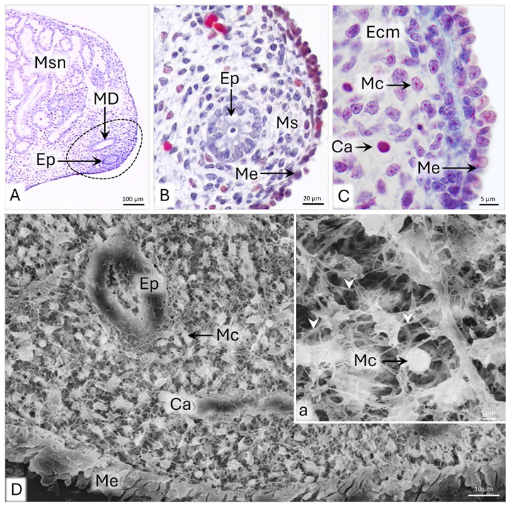
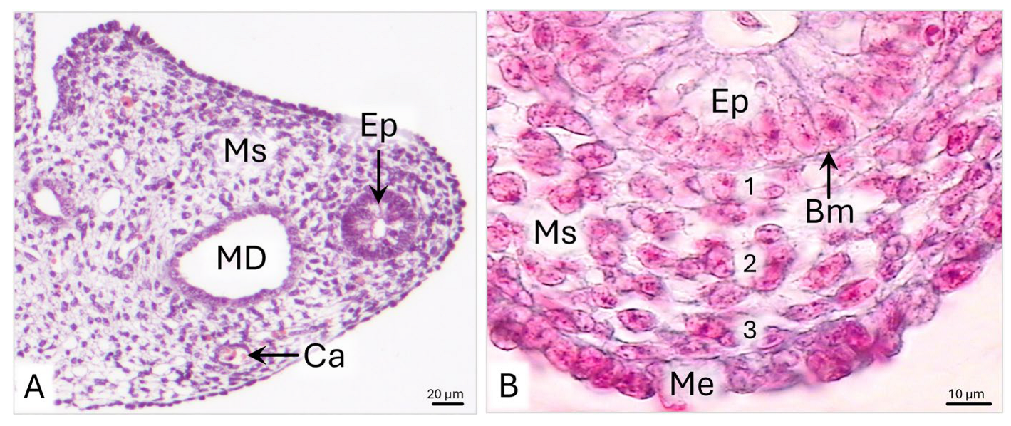
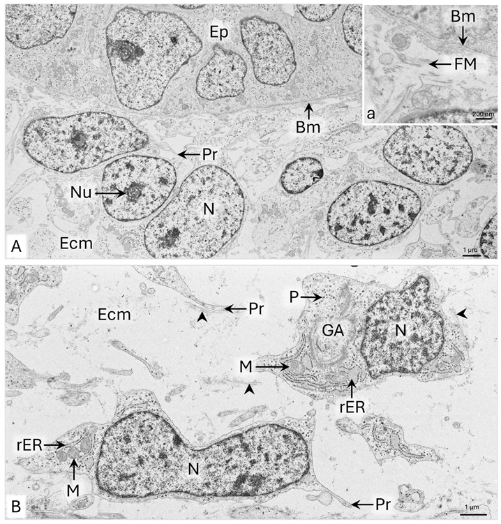
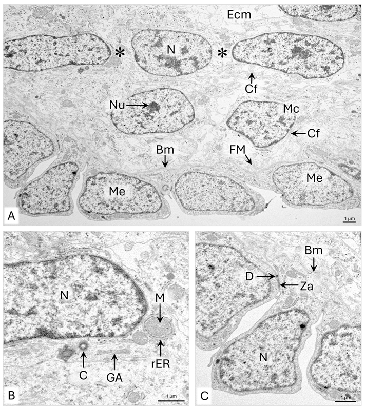
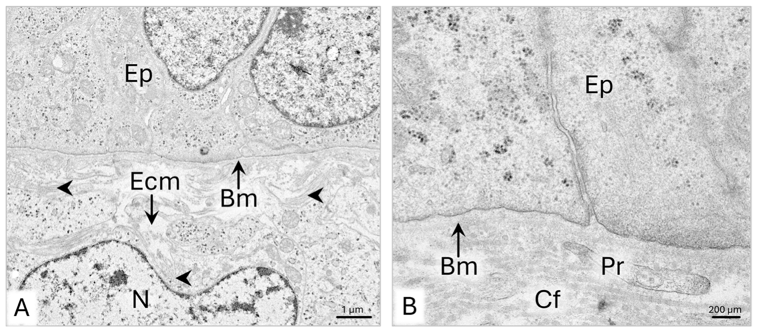
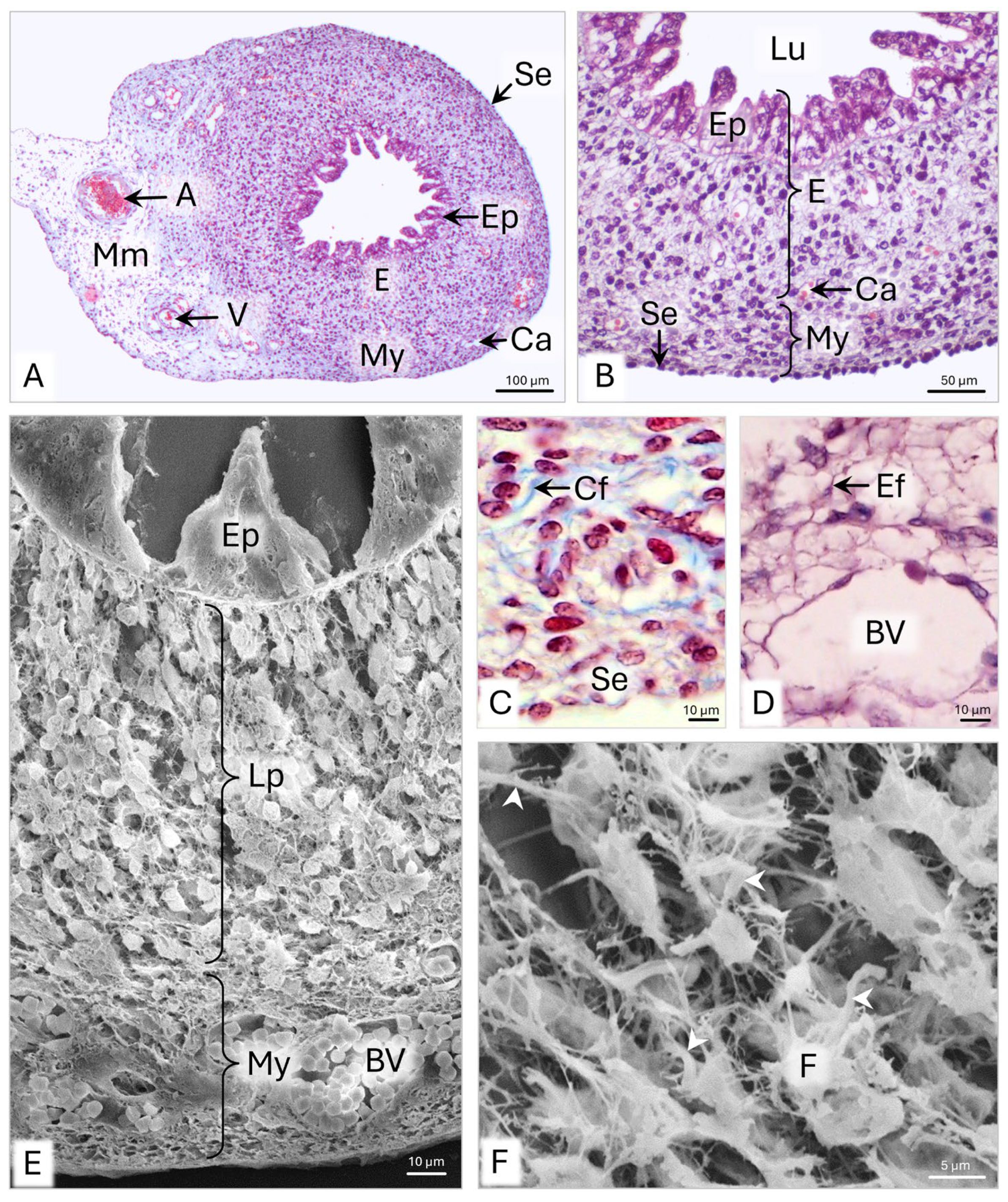
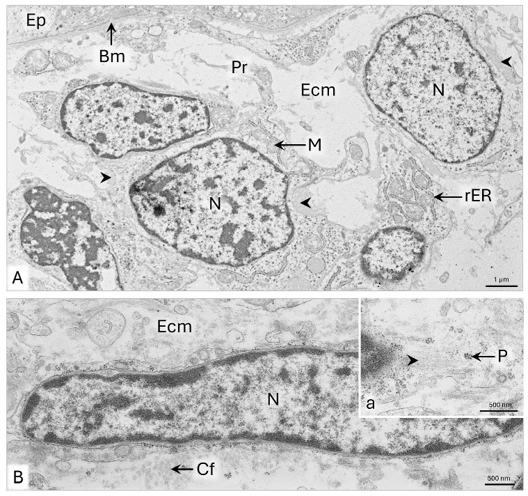
The differentiation of uterine mesenchyme into endometrial connective tissue and myometrial smooth muscle begins between days 28 and 33 p.c. As revealed by our observations, on day 28 p.c., the uterine horns of the domestic cat appear as rudimentary uterine segments of the paramesonephric ducts, characterized by an embryonic wall composed of a simple epithelium surrounded by mesenchyme. At this stage, the only indicator of mesenchymal cell activity is the bluish staining of the amorphous extracellular matrix in histological slides, suggesting the secretion of proteoglycans. By day 33 p.c., the wall of the uPD loses its embryonic features: the epithelium becomes pseudostratified, and the mesenchymal cells differentiate into fibroblasts. In addition to proteoglycans, these cells secrete fine fibrous material into the intercellular space, particularly accumulating beneath the epithelial basement membrane. Collagen fibrils are also deposited on the surfaces of fibroblasts. In the peripheral region of the wall, elongated mesenchymal cells assume a circular arrangement and become precursors of future myoblasts.
According to the literature, early mesenchymal differentiation in the uterine wall is characteristic of humans and livestock, species with relatively long gestation periods [
7,
8,
12]. In humans, fibroblasts and smooth muscle cells differentiate by the 20th week of gestation [
7,
12,
21], while in sheep, this process begins around day 55 of prenatal development [
8]. In contrast, in rodents, the uterine wall mesenchyme remains undifferentiated until the neonatal period [
7,
22].
In the domestic cat, the development of loose connective and smooth muscle tissue in the uPD, beginning before day 33 p.c., leads to the formation of the endometrium and myometrium. During endometrial development, the epithelial structure changes, and fibroblasts organize into patterns that correspond to the future basal and functional layers of the mucosal lamina propria. Between days 42 and 51 p.c., the uterine epithelium becomes pseudostratified and displays variable height. Over the prenatal development, this epithelium increases in height nearly threefold, and by the end of the prenatal period, its surface exhibits marked undulations. Previous studies have shown that the simple columnar structure of the uterine epithelium in domestic cats is established approximately one month after birth, coinciding with the appearance of uterine gland primordia in the mucosal lamina propria [
17].
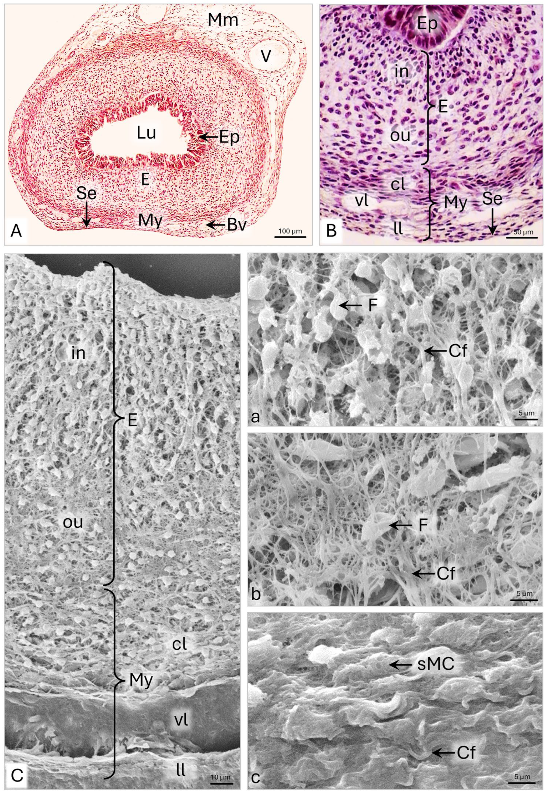
The formation of the endometrial lamina propria begins after day 33 p.c., when fibroblasts arrange into columns and align radially around the uterine epithelium. Initially, the alignment of fibroblast columns is perpendicular to the epithelium, shifting to an oblique orientation by day 51 p.c. The distinct division of the endometrium into basal and functional layers becomes apparent toward the end of the prenatal period, evidenced by differences in fibroblast density and spatial distribution. The inner functional layer contains fibroblasts arranged in columns and has up to 60% more cells than the outer layer, where fibroblasts are irregularly distributed.
A double-layered endometrium during prenatal development has also been reported in sheep and humans [
7,
8,
12]. In contrast, studies in pigs have shown that the endometrium does not divide into layers until after birth; distinct subepithelial tissue areas become apparent only about a week postpartum [
7,
10].
SEM observations revealed that, in the domestic cat, collagen fibers progressively accumulate in the extracellular matrix, aligning with the cellular organization of the mucosal layers. By the end of the prenatal period, thick collagen fiber bundles reflect a columnar arrangement of fibroblasts in the inner layer and a more irregular pattern in the outer layer. Our previous research on postnatal uterine development in domestic cats showed that this specific organization of fibroblasts and collagen bundles in the functional layer of the endometrium is maintained for the first 3 to 4 weeks after birth. However, as uterine glands begin to invade the lamina propria, this arrangement gradually becomes less distinct [
17]. These findings suggest that the columnar organization of fibroblasts, along with associated collagen fibers and capillaries, may provide structural guidance for developing glandular tubules. This arrangement likely offers both a stable connective tissue framework and effective vascular support.
Morphometric analysis revealed that between days 42 and 63 p.c., the thickness of the endometrial lamina propria in the domestic cat increases more than fourfold. Notably, the endometrium remains smooth until the end of the prenatal period. Mucosal folds begin to form 3 to 4 weeks after birth and continue developing at least until the third month of life. This process is likely related to the need for creating sufficient space in the functional layer of the endometrium to accommodate the expanding uterine glands [
17].
The formation of myometrial smooth muscle tissue begins on day 33 p.c. with the differentiation of the first myoblasts, which appear in a circular arrangement at the periphery of the uPD wall. By day 42 p.c., the myometrium becomes recognizable. At this stage, it is more than twice as thin as the endometrial lamina propria and consists of several rows of circularly arranged smooth muscle cells interspersed with fibroblasts. Subsequent development of the myometrium involves an increase in the number of myocyte layers. By the end of the prenatal period, the myometrium undergoes nearly a sixfold increase in thickness and occupies approximately one-third of the total uterine wall.
The characteristic microstructure of the myometrium, comprising three distinct layers, can be identified in the domestic cat at the end of the prenatal period. Between days 51 and 63 p.c., the myometrium divides into sublayers; however, their relative proportions differ substantially from those observed in the adult uterus. Just prior to birth, the predominant layer is the inner circular muscle layer, composed of several rows of smooth muscle cells. In contrast, the outer longitudinal layer and the vascular interlayer remain poorly developed. As described in our previous work, postnatal development of the myometrium in the domestic cat involves a progressive thickening of each layer, with particularly rapid growth of the outer longitudinal layer. In adult specimens, this layer is approximately 40% thicker than the inner circular layer [
17]. Additionally, during the first three months after birth, the vascular layer expands and differentiates into a distinct intermediate myometrial layer [
17].
The developmental pattern of the myometrium in the domestic cat resembles that observed in rodents, pigs, and humans, where differentiation of smooth muscle cells begins at the periphery of the uterine wall, with the inner circular layer forming first [
10,
21,
22].
A comparative analysis of the prenatal development of the uterine horn and uterine tube walls in the domestic cat reveals both similarities and differences in the rate and pattern of tissue differentiation [
23]. As both organs originate from a common primordium, they initially develop from a simple cylindrical epithelium surrounded by mesenchyme. A shared feature in their epithelial development is the transformation into a pseudostratified epithelium with variable height. In both organs, this pseudostratified form is temporary. In the uterine tube, it transforms into a simple ciliated columnar epithelium before birth, whereas in the uterus, this change occurs only postnatally [
23].
Mesenchymal differentiation into loose connective tissue begins earlier in the uterine tube—around day 27 p.c.—than in the uterus. Moreover, the lamina propria of the uterine tube’s infundibulum begins to fold around day 40 p.c., while in the uterus, such folding occurs postnatally [
23]. Smooth muscle cells also appear earlier in the uterine tube, specifically in the ampulla, around day 40 p.c. However, at birth, the uterine myometrium is more developed. In contrast, the tunica muscularis of the uterine tube still consists of only a single layer of circularly arranged smooth muscle cells [
23].
Despite differences in the timing of tissue layer development, both the uterine tube and uterus remain physiologically inactive at birth, as indicated by their incomplete structural organization. Maturation and the acquisition of adult characteristics occur during the first months of life. Functional maturity of the uterine tube is marked by the presence of labyrinthine mucosal folds lined with ciliated epithelium, while in the uterus, it is evidenced by the formation of uterine glands and the thickening of the muscular layer.
