Article

NOTCH1 as a Negative Regulator of Avian Adipocyte Differentiation: Implications for Fat Deposition

1
College of Life Sciences, Shanxi Agricultural University, Jinzhong 030801, China
2
College of Animal Science, Shanxi Agricultural University, Jinzhong 030801, China
3
National Engineering Laboratory for Animal Breeding and MARA Key Laboratory of Animal Genetics, Breeding and Reproduction, College of Animal Science and Technology, China Agricultural University, Beijing 100193, China
*
Author to whom correspondence should be addressed.
These authors contributed equally to this work.
Animals 2024, 14(4), 585; https://doi.org/10.3390/ani14040585
Submission received: 9 January 2024 / Revised: 2 February 2024 / Accepted: 7 February 2024 / Published: 9 February 2024

Abstract

:

Simple Summary

Excessive abdominal fat deposition significantly impacts feed efficiency and meat yield of chicken, which is closely associated with adipocyte differentiation (adipogenesis). Adipogenesis is regulated by various transcription factors and signaling molecules. Our previous transcriptome study found that the NOTCH signaling pathway is significantly enriched during avian adipocyte differentiation. However, in previous studies, NOTCH1 exerts both positive and negative effects on adipogenesis. Here, we perturbed NOTCH1 expression in chicken adipocytes using different strategies and investigated the effects of NOTCH1 on gene expression and adipogenesis. This study demonstrated that NOTCH1 is a key regulator of avian adipogenesis and provides a potential target for molecular breeding to reduce abdominal fat deposition in chicken.

Abstract

The NOTCH signaling pathway plays a pivotal role in diverse developmental processes, including cell proliferation and differentiation. In this study, we investigated whether this signaling molecules also contribute to avian adipogenesis. Using previous mRNA-seq datasets, we examined the expression of 11 signaling members during avian adipocyte differentiation. We found most members are down-regulated throughout differentiation (p < 0.05). As a representative, NOTCH1 was decreased in cultured chicken abdominal adipocytes during adipogenesis at mRNA and protein levels (p < 0.05). Moreover, using an overexpression plasmid for NOTCH1’s intracellular domain (NICD1), as well as siRNA and DAPT to activate or deplete NOTCH1 in cells, we investigated the role of NOTCH1 in avian adipogenesis. Our findings illuminate that NOTCH1 activates the expression of HES1 and SOCS3 while it decreases NR2F2 and NUMB (p < 0.05), as well as inhibits oleic acid-induced adipocyte differentiation (p < 0.01). We further demonstrate that HES1, a downstream transcription factor activated by NOTCH1, also significantly inhibits adipogenesis by suppressing PPARγ and C/EBPα (p < 0.01). Collectively, these findings establish NOTCH1 as a negative regulator of avian adipocyte differentiation, unveiling NOTCH signaling as a potential target for regulating avian fat deposition.

1. Introduction

Owing to the intensive genetic selection for growth rate and weight, poultry have experienced excessive abdominal fat accumulation, which adversely affects feed efficiency and meat production [1]. Fat tissue functions as a crucial energy storage organ, playing pivotal roles in sustaining the metabolic health of avian species [2]. However, dysfunctional abdominal fat tissue is usually correlated with metabolic disorders, which may lead to fatty liver disease and more mortality [3]. Hence, a thorough investigation into the molecular regulatory mechanisms governing fat development can contribute to the genetic improvement of avian abdominal fat deposition.
The development of adipose tissue results from both an increase in the number of adipocytes (hyperplasia) and the promotion of adipocyte differentiation (hypertrophy) [4]. Excessive fat deposition is closely linked with adipocytes differentiation during animal growth [5]. The process of adipocyte differentiation and lipid accumulation is governed by a meticulously regulated cascade of transcription factors, which includes members of the CCAAT/enhancer binding protein (C/EBP) family (C/EBPα, β, and δ), along with the nuclear receptor peroxisome proliferator activated receptor γ (PPARγ) [6,7]. In addition, many signaling pathways, such as WNT [8], have been reported to be involved in regulating adipocyte differentiation. Recently, our group utilized the established chicken abdominal adipocyte model (ICP1 cells) to analyze mRNA expression at different stages of differentiation, and identified the function of MYOD1 in avian adipogenesis [9]. Moreover, we also explored the differentially expressed genes during duck subcutaneous adipocyte differentiation process through mRNA-seq and mapped an elaborate network of potential transcription factors that regulate differentiation [10]. Notably, NOTCH signaling was enriched significantly in the early regulatory annotation of the differentiation of both types of avian adipocytes.
NOTCH signaling plays a conserved role in connecting cell membrane events with transcriptional regulation, impacting cell fate decisions in multicellular organism development [11]. Four transmembrane NOTCH receptors (NOTCH1–4) and the five canonical ligands (DLL1, DLL3, DLL4, JAGGED1, and JAGGED2), along with the two non-canonical ligands (DLK1 and DLK2), constitute the NOTCH signaling [12]. The NOTCH receptor–ligand interaction leads to the release of NOTCH intracellular domain (NICD) within signal–receiving cells, which then enters the nucleus and interacts with RBP–Jκ to regulate gene expression, including the HES1 and HES–related family bHLH transcription factor with YRPW motif 1, 2 (HEY1, HEY2) [13,14]. However, current genome versions of chicken (GRCg7b) and duck (CAU_wild_1.0) lack gene annotations for DLL3, NOTCH3, and NOTCH4. Moreover, recent evidence has found that NOTCH signaling could activate Suppressor of Cytokine Signaling 3 (SOCS3) in macrophages [15] and repressed by Nuclear Receptor Subfamily 2, Group F, Member 2 (NR2F2) and NUMB Endocytic Adaptor Protein (NUMB) in endothelial cells and cardiac cells [16,17]. However, little is known about the role of the NOTCH signaling in avian adipocytes.
NOTCH1 is well known to regulate various cell fates, including self–renewal, proliferation, and differentiation programs in different cells, such as muscle stem cells [18], primordial germ cells, and spermatogonial stem cells [19]. Conflicting reports exist regarding the role of NOTCH1 in adipogenic regulation. Ross et al. initially found that decreased expression of NOTCH1 or inhibition of NOTCH signaling by soluble JAG1 inhibits the differentiation of 3T3–L1 adipocytes [20]. However, subsequent studies argued that NOTCH1 is dispensable for adipocyte specification and differentiation [21]. More recently, Song et al. reported that blocking NOTCH1 promotes adipogenic differentiation of mesenchymal stem cells [22]. Given these contradictory findings, investigating the role of NOTCH1 in avian adipocytes is valuable.
To elucidate the involvement of NOTCH signaling in avian adipocyte differentiation, exploring the effect of NOTCH1 on gene expression and adipogenesis. The insights gained from this research could lay a solid foundation for constructing a regulatory network of avian adipocyte differentiation and advancing our understanding of poultry fat deposition mechanisms.

2. Materials and Methods

2.1. Cell Culture

Immortalized chicken preadipocytes 1 (ICP1) was obtained from the Poultry Breeding Lab of Northeast Agricultural University (Supplementary Figure S1A–D) [23], and then cultured in DMEM/F12 (11320033, Gibco, Grand Island, NY, USA) supplemented with 10% FBS (10099141C, Gibco, Grand Island, NY, USA), and 1% penicillin/streptomycin (15140122, Invitrogen, Carlsbad, CA, USA) at 37 °C in a humidified atmosphere with 5% carbon dioxide. Cells from passage three to five were used for the following experiments.

2.2. Construction, Amplification, and Purification of Plasmids

For NOTCH1 intracellular domain (NICD1) overexpression, the coding sequence of NICD (Accession numbers: XM_046901649.1) was determined based on previous studies [24], which is composed of an N–terminal 1–44 amino acids followed by encoding the intracellular domain without C–terminal PEST domain (Accession numbers: XP_046784687.1: 1676–2151 amino acids), which mediates the ubiquitin–mediated degradation of NICD1. The coding sequence of NICD1 was chemically synthesized and then inserted into pcDNA3.1(+) plasmid. For HES1 overexpression, the coding sequence of HES1 (Accession numbers: XM_040679737.2) was obtained by PCR amplified from fat cDNA using specific primers (forward: 5′–GACGGCTAGCCACCATGCCTGCCGACCTGA–3′, reverse: 5′–GTCTAGAGGATCCTCACCAGGGCCTCCAGAC–3′), and then inserted into the pcDNA3.1(+) plasmid.
For PPARγ promoter reporter plasmid, we amplified the −305 to +104 upstream region of chicken PPARγ (Accession numbers: KP736526.1) from chicken genomic DNA using primers (F: 5′-TTCGGTACC GCTCTGTCCTCACAGGAGGT-3′, R: 5′-TTCCTCGAGCTGTCAAGTCTCAGCCGGA-3′), and then cloned into pGL3-Promoter-luc plasmid (Promega, Madison, WI, USA) to generate pGL3-PPARγ-WT, which contained four putative HES1 binding site (−65/−56: GGCGCGAGCC, −65/−56: GGCTCGCGCC, −282/−273: CGCGCGCGGG, −284/−275: CGCGCGCGAT). Next, we specifically deleted the four putative HES binding sites using DNA synthesis (Tsingke, Beijing, China) to generate pGL3-PPARγ-MUT. Similarly, we also amplified the −1464 to +84 upstream region of chicken C/EBPα (Accession numbers: NM_001031459.2) using the primers (F: 5′-TTCGGTACCCCTCCTCTGCAAATGGGTTT-3′, R: 5′- TTCCTCGAGCCGAAGTTCACCATCACGC-3′) to generate pGL3-C/EBPα-WT, which also contained four putative HES1 binding site (−207/−198: CTCGCGCGTC, −571/−562: GGCACGCTGC, −1293/−1284: AACGCGTGCC, −1293/−1284: GGCACGCGTT). Then, the four putative HES1 binding site of the plasmid were specifically deleted to generate pGL3-C/EBPα-MUT.
Additionally, the NOTCH luciferase reporter plasmid containing RBP-Jκ binding sites was purchased from Yeasen Biotech (11509ES03, Shanghai, China).
The recombinant plasmids were transformed into E. coli DH5α Competent cells (9057, Takara, Kyoto, Japan) and cultured in an LB medium supplemented with 0.1% ampicillin solution overnight (A1170, Solarbio, Beijing, China). The plasmids were isolated and purified from the clone by Endo-Free Plasmid DNA Maxi Kit (19036ES10, Yeasen Biotech, Shanghai, China) and were identified by agarose gel electrophoresis again. All plasmids were validated by sequencing.

2.3. RNA Interference

According to the mRNA sequence of chicken NOTCH1 (Accession numbers: XM_046901649.1), small interfering RNA (siRNA) sequences against NOTCH1 were purchased from GenePharma (Suzhou, China). The sequences used were as follows: si-NOTCH1, sense 5′-GUGGCAGACUGAUGAAGAATT′-3′ and antisense 5′-UUUGUAGUCAUUGACCCGCTT-3′; si-NC, sense 5′-UUCUCCGAACGUGUCACGUTT-3′ and antisense 5′-ACGUGACACGUUCGGAGAATT-3′.

2.4. Transfections

ICP1 cells were seed in 6-well plates with approximately 5 × 10⁵ cells 16 h before transfection. Lipofectamine 3000 and Lipofectamine RNAiMax (L3000150, 13778150, Thermo Fisher Scientific, Waltham, MA, USA) were used for plasmid (3 μg per well) and siRNA (50 pM per well) transfections of ICP1 cells, respectively, following the manufacturer’s instructions. At 72 h after the transfection, cells were processed for adipogenic differentiation, RT-qPCR, and Western blot analysis. Each assay was carried out at least three times independently.

2.5. DAPT Treatment

N-[N-(3,5-Difluorophenacetyl-L-alanyl)]-S-phenylglycine-Butyl Ester (DAPT) is a γ-secretase inhibitor that hinders the transactivation of the NOTCH signaling [25]. DAPT (HY-13027, MedChem Express, Monmouth Junction, NJ, USA) was prepared by dissolving in dimethyl sulfoxide (DMSO, HY-10999A, MedChem Express, Monmouth Junction, NJ, USA) and then freshly diluted with culture medium to achieve the desired concentration. The cells were pretreated with either DAPT or DMSO 12 h before the induction of differentiation, Subsequently, throughout the differentiation process, DAPT or DMSO was used in conjunction with 160 μM oleic acid (O1383, Sigma-Aldrich, St. Louis, MO, USA). The ICP1 cells treated with DMSO at 0.05% (v/v) served as control. ICP1 cells were harvested and processed for Western blot and Oil Red O staining after DAPT treatment for 36 h or 72 h.

2.6. Adipogenic Differentiation of ICP1 Cells

The strategy of inducing adipogenic differentiation of ICP1 cells used the same protocol as Shang et al. [26] with minor modifications. Briefly, ICP1 cells were induced to differentiate after 48 h of confluence (0 h) with 160 μM oleic acid in DMEM/F12 supplemented with 10% FBS throughout the differentiation process. At 72 h after the induction, the cells were harvested and used for further assays.

2.7. Oil Red O Staining and Quantification

The differentiated cells were fixed with 4% formaldehyde for 10 min. Then, the fixed cells were stained with Oil Red O working solution (G1262, Solarbio, Beijing, China) according to the manufacturer’s manual. Images were captured using an inverted microscope system (Olympus IX53, Tokyo, Japan) at 40× magnification. Lipid contents were quantified by the Oil Red O extraction assay, as described by Ramirez-Zacarias et al. [27]. The BCA Protein Assay Kit (P0010, Beyotime, Shanghai, China) was used to determine the protein concentrations of the cells with identical treatment, which were used for normalized extraction results.

2.8. RNA Extraction, cDNA Synthesis, and RT-qPCR Assay

Total RNA was extracted from the differentiated cells using RNAiso reagent (9108, Takara, Kyoto, Japan), and 500 ng of RNA was reverse transcribed into cDNA using PrimeScript RT reagent Kit (RR047A, Takara, Kyoto, Japan). RT-qPCR was performed using SYBR Green PCR Master Mix (4309155, Applied Biosystems, San Francisco, CA, USA) and was conducted using the Bio-Rad CFX96 Touch Real-Time PCR Detection System (Bio-Rad Laboratories, Hercules, CA, USA). Gene-specific primers used in the RT-qPCR are shown in Table 1. The relative gene expression was determined after normalization with housekeeping gene GAPDH in the samples using the 2−ΔΔCt method. Each assay was carried out at least three times independently.

2.9. Cell Fractionation Assay

The nucleus and cytoplasmic protein of ICP1 cells were collected using a Protein Extraction Kit (P0027, Beyotime, China). Briefly, cells were harvested after tripsinization and washed in ice-cold PBS. Cell pellet was then resuspended with Cytoplasmic protein extraction reagent A for 10 min on ice. Subsequently, Cytoplasmic protein extraction reagent B was added to the reaction mixture for 3 min followed by centrifugation, and the supernatant was stored as cytoplasmic protein. The pellet was resuspended in nuclear protein extraction reagent for 40 min followed by centrifugation, and the supernatant was stored as nucleus protein.

2.10. Western Blot Analysis

Cellular proteins were homogenized with RIPA lysis (P0013B, Beyotime, China) supplemented with protease and phosphatase inhibitor cocktails (#P1009; Beyotime, China). The BCA Protein Assay Kit (P0010, Beyotime, China) was used to determine the protein concentrations. Approximately 30 μg of proteins was denatured and subjected to Western blot analysis according to the standard protocols [28]. The immunoreactive proteins were detected on a Chemidoc XRS (Bio-rad Laboratories, Hercules, CA, USA) using BeyoECL Plus kit (P0018S, Beyotime, China) and quantified by Image J (version 1.43) or Imagelab software (version 5.2.1). Antibodies used in the study included the following: Rabbit anti-NOTCH1 (No. 10062-2-AP, Proteintech, Wuhan, China) with 1:500 dilution; Rabbit anti- SOCS3 (No. 14025-1-AP, Proteintech, China) with 1:1000 dilution; Goat anti-NUMB (ab4147, Abcam, Waltham, MA, USA) with 1:1000 dilution; Mouse anti-GAPDH (No.60004-1-Ig, Proteintech, China) with 1:20,000 dilution; Rabbit anti-LMNB1 (No.12987-1-AP, Proteintech, China) with 1:5000 dilution.

2.11. Transcription Factor Binding Sites Prediction

The JASPAR CORE vertebrate database [29] was utilized to identify and examine potential transcription factor binding sites in the 2000 bp upstream region of PPARγ and C/EBPα (Accession numbers: KP736526.1 and NM_001031459.2). The threshold for profile score relevance was established at 85% for this analysis.

2.12. Dual-Luciferase Reporter Assay

To examine the response of NICD1 to NOTCH signaling, ICP1 cells were co-transfected with pcDNA3.1-NICD1 or pcDNA3.1 and NOTCH luciferase reporter plasmid, along with pRL-TK plasmid (Promega; ratio 24:24:1). To analyze the effect of HES1 on the reporter gene activity of the PPARγ or C/EBPα upstream region, cells were co-transfected with pcDNA3.1-HES1 or pcDNA3.1(+) and pGL3-PPARγ-WT or pGL3-PPARγ-MUT (pGL3-C/EBPα-WT or pGL3-C/EBPα-MUT), along with pRL-TK (ratio 24:24:1). At 72 h after the transfection, the cells were harvested, lysed, and assessed with a dual luciferase reporter kit (E2920, Promega). The relative Luciferase activity of each construct was measured by Infinate® M200 PRO microplate reader (Tecan, Morrisville, NC, USA) and was calculated as the Firefly/Renilla luciferase activity ratio. Each assay was independently conducted at least three times.

2.13. RNA-seq Analysis

Two previously published mRNA-seq datasets were downloaded from NCBI (Accession number: SRX4646736, PRJNA1069883) [9,10] to analyze the expression patterns of NOTCH signaling-related genes during the differentiation process of chicken abdominal or duck subcutaneous adipocytes, including two NOTCH receptors (NOTCH1, NOTCH2), six NOTCH ligand genes (DLL1, DLL4, JAG1, JAG2, DLK1, and DLK2), and three NOTCH-dependent transcription factors (HES1, HEY1, and HEY2). Trimmed reads were aligned against GRCg7b or CAU_wild_1.0 using HISAT2 [30]. The aligned files were passed into HTSeq [31] for gene quantification into TPM units. DESeq2 (v.1.28.1) [32] package was used to normalize and perform differential expression analysis on the quantified reads.

2.14. Statistical Analysis

All replicate experiments were biological replicates. Non-parametric statistical analysis was conducted using GraphPad Prism 9. Results are shown as mean ± standard deviation. Unpaired Student’s t-test was employed for comparison between two groups, and One-way ANOVA followed by the Tukey test was utilized for comparison among more than two groups. All experiments yielded statistically significant results, with * indicating a significant difference (p < 0.05), and ** indicating a very significant difference (p < 0.01).

3. Results

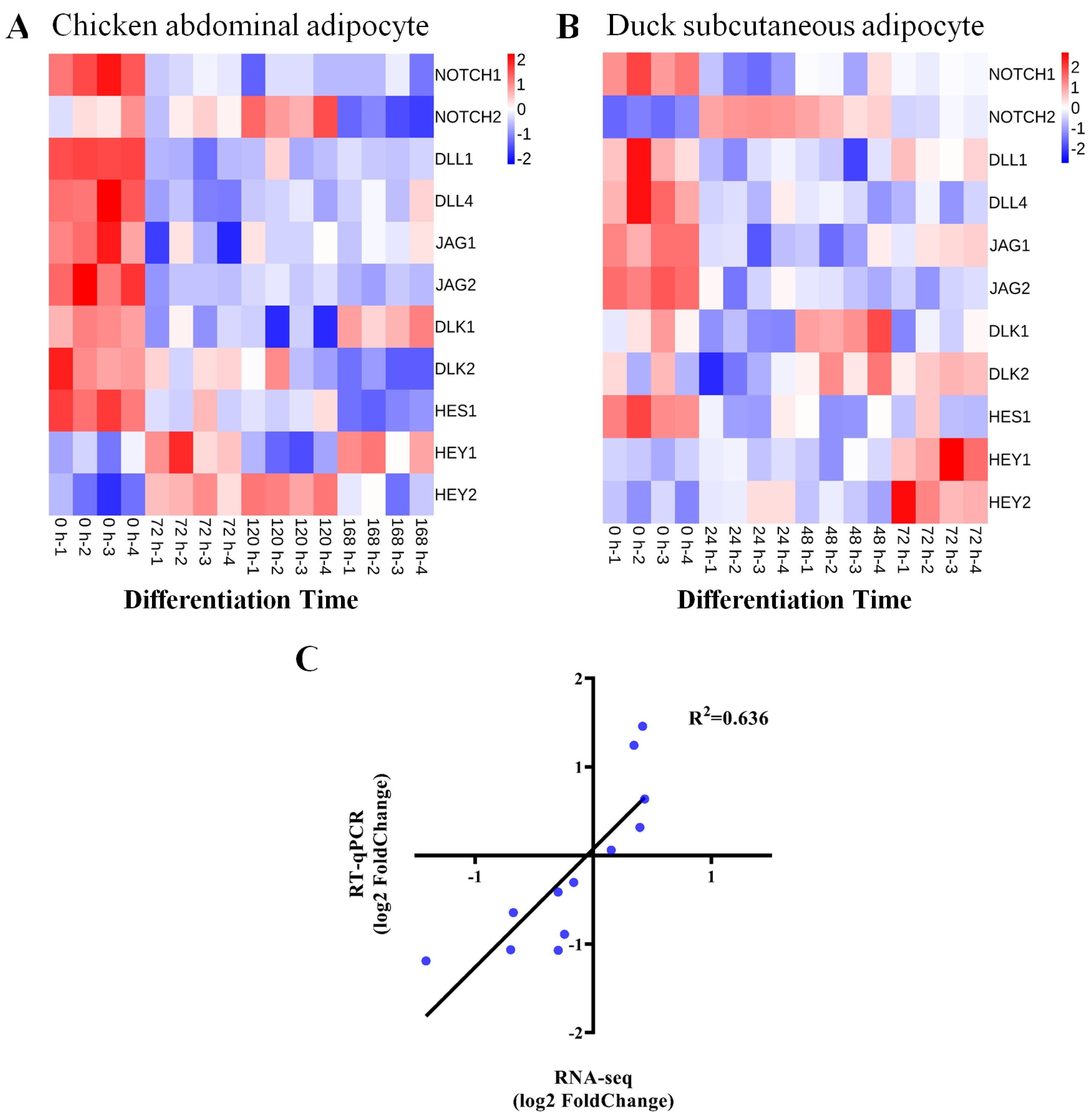
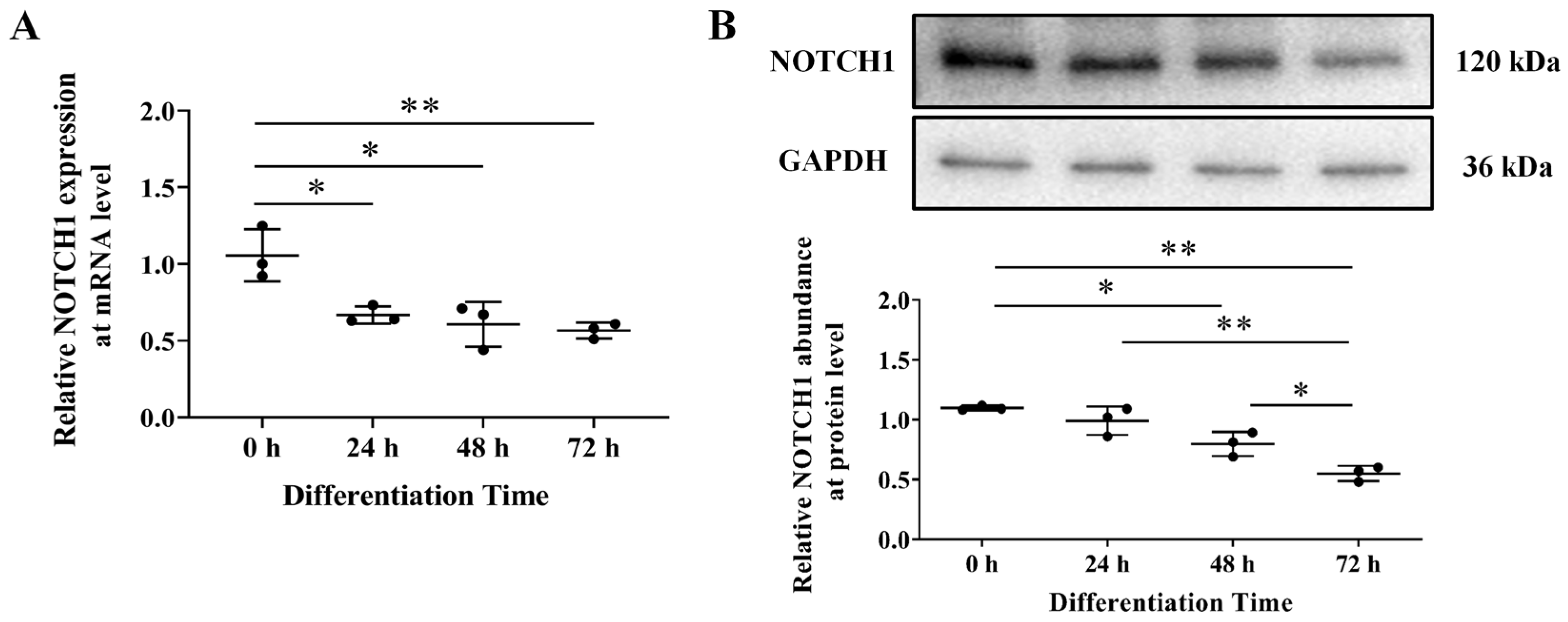
3.1. NOTCH1 Expression during Avian Adipocyte Differentiation

To further investigate the role of the NOTCH signaling in avian adipocytes, we profiled the expression of several signaling members during the differentiation process of chicken abdominal adipocyte line (ICP1 cells) and primary duck subcutaneous adipocyte using two previously published mRNA-seq datasets [9,10]. Interestingly, NOTCH signaling members showed similar expression patterns in both avian species (Figure 1A,B). The expression of NOTCH1 and its ligands DLL1, DLL4, DLK1, JAG1, and JAG2 is significantly decreased during adipocyte differentiation, whereas NOTCH2 shows the opposite expression patterns. Meanwhile, we analyzed the expressions of the downstream transcription factors of NOTCH signaling, including HES1, HEY1, and HEY2. It was clear that the expression of HEY1 and HEY2 were significantly increased after induction of adipogenic differentiation, but the expression of HES1 was significantly decreased (Figure 1A,B). To validate the results of mRNA-seq analysis results, we used RT-qPCR in ICP1 cells to detect the expression of four genes (NOTCH1, HES1, HEY1, and HEY2) at the same differentiation time point. The results showed a good correlation between the fold change measured by mRNA-seq and RT-qPCR (R2 = 0.636; Figure 1C).
The endogenous expression of NOTCH1 was also evaluated by RT–qPCR and Western blot during ICP1 differentiation. We found that NOTCH1 is inhibited during adipocyte differentiation at both the mRNA and protein levels (p < 0.05; Figure 2A,B).

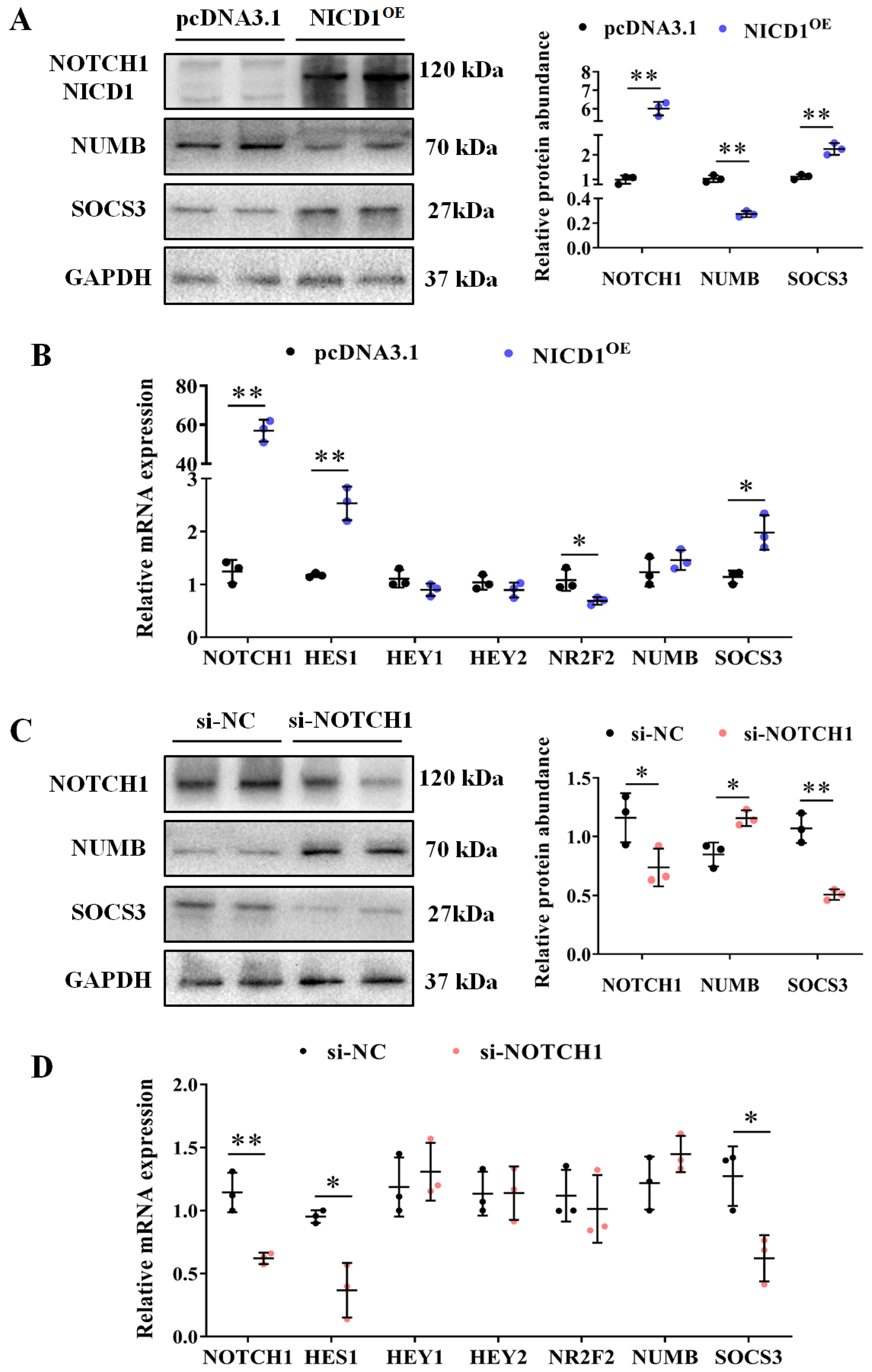
3.2. NOTCH1 Regulates Expression of Signaling-Related Genes

To investigate the effect of NOTCH1 in avian adipocytes, we generated an ICP1 line with endothelial-specific NOTCH intracellular domain (NICD1) overexpression as the constitutively active form of NOTCH1. As expected, NICD1 overexpression induces promoter activity dependent on the RBP–Jκ site (p < 0.01; Supplementary Figure S2). Subsequently, we investigated the expression of several of its targets, including SOCS3, NR2F2, NUMB, etc. The immunoblotting results showed that the protein abundance of NOTCH1 and SOCS3 were increased in NICD1–overexpressing cells, while NUMB, an adaptor protein that promotes NOTCH1 degradation by recruiting the E3 ubiquitin ligase, was decreased (p < 0.05; Figure 3A). In addition, the mRNA levels of downstream genes were quantified by RT–qPCR. The mRNA levels of HES1 and SOCS3 were increased, as well as decreased NR2F2 in NICD1–overexpressing cells (p < 0.05; Figure 3B). Surprisingly, the mRNA level of NUMB was not affected by NICD1 overexpression, implying posttranscriptional regulation of NUMB by NOTCH1. Additionally, the mRNA levels of HEY1 and HEY2 were not affected by NICD1 (Figure 3B).
To further elucidate the regulation of endogenous NOTCH1 in avian adipocytes, we employed siRNA–NOTCH1 to inhibit its expression in cells. The efficiency of the siRNA–mediated NOTCH1 knockdown was validated by RT–qPCR and Western blot (Figure 3C,D). Contrary to the observations in NICD1–overexpressing cells, the downstream genes HES1 and SOCS3 were down–regulated, while NUMB was up–regulated in the NOTCH1 knockdown cells, suggesting that these genes are regulated by NOTCH1 in avian adipocytes (p < 0.05; Figure 3C,D).

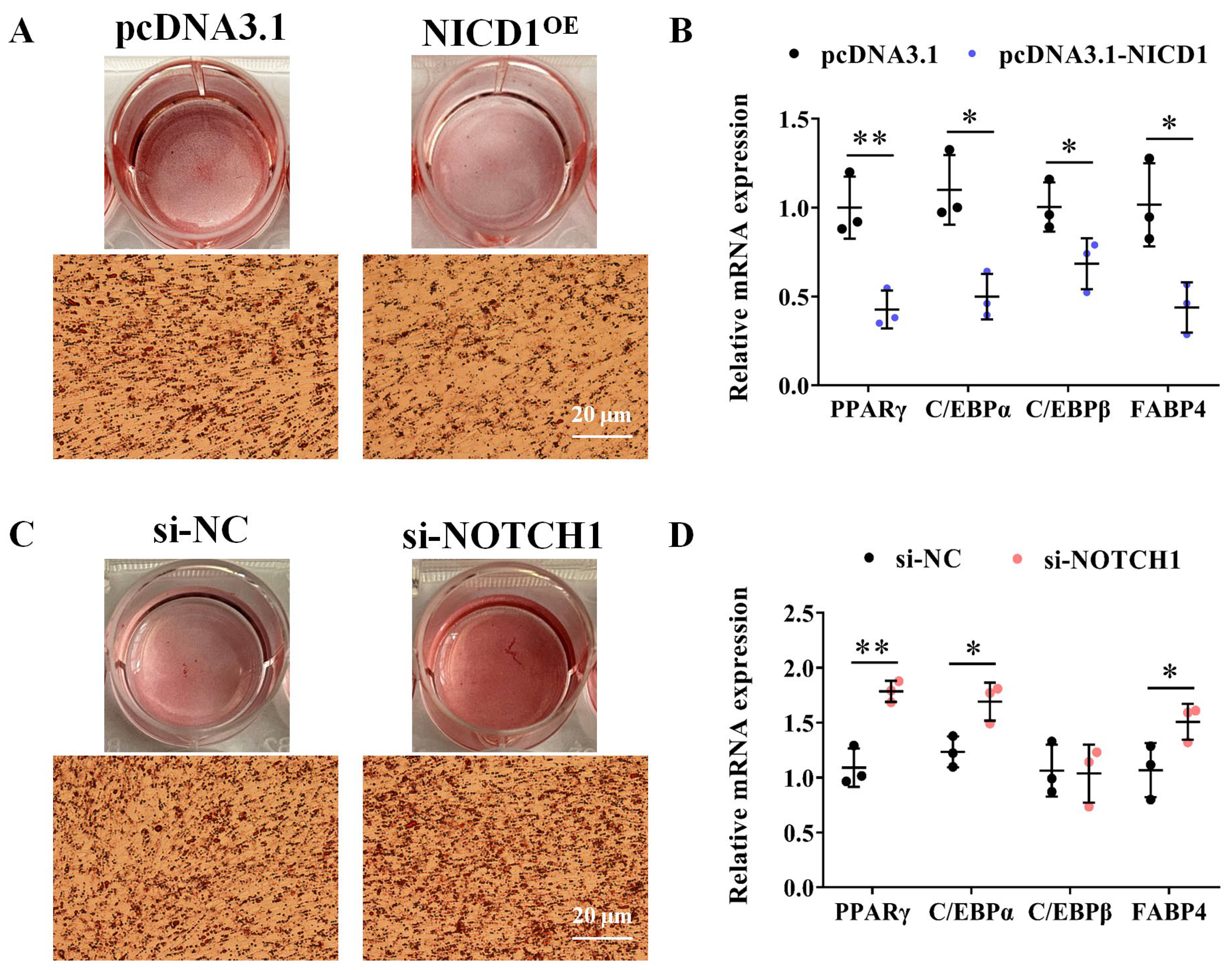
3.3. NOTCH1 Inhibits Avian Adipocyte Differentiation

To specifically assess the potential role of NOTCH1 in adipogenesis, we examined the effects of overexpression of NICD1 on adipogenic differentiation of ICP1 cells. The results indicated that NICD1 overexpression inhibits oleic acid–induced adipogenesis, as shown by Oil Red O (ORO) staining of neutral lipids (p < 0.01; Figures 4A and S3A), and reduces mRNA levels of the pro−adipogenic factors PPARγ, C/EBPα, C/EBPβ, and FABP4 (p < 0.05; Figure 4B), whereas knockdown of NOTCH1 in ICP1 cells exhibited enhanced differentiation into adipocytes, as evidenced by increases in ORO staining, along with elevated mRNA levels of the adipocyte markers PPARγ and C/EBPα (p < 0.05; Figures 4C,D and S3B).

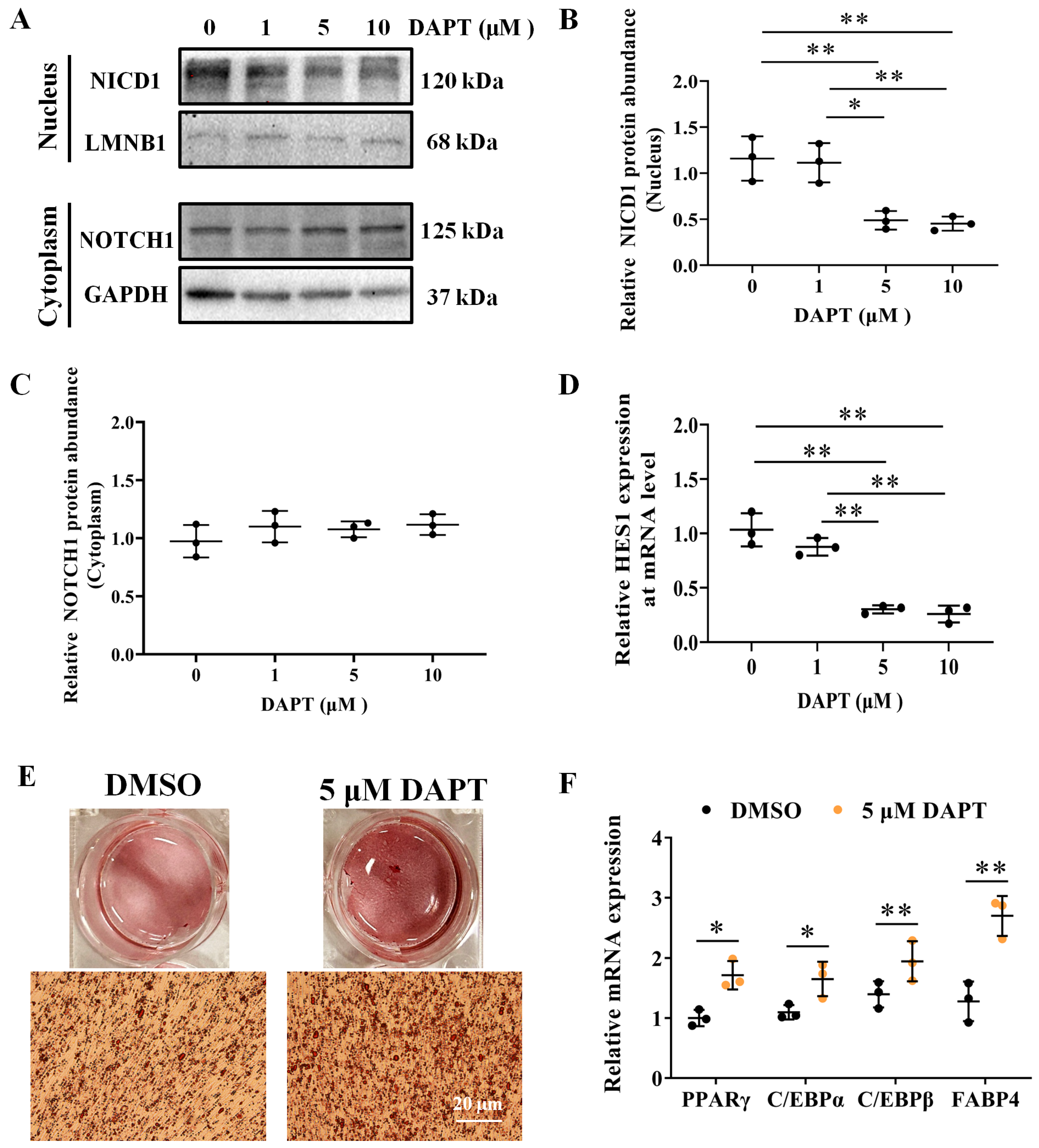
3.4. DAPT, an Inhibitor of NOTCH Signaling, Promotes Adipocyte Differentiation

We also employed DAPT, a γ–Secretase inhibitor, to block NOTCH cleavage and activation. ICP1 cells were treated with DAPT at 0, 1, 5, and 10 μM for 48 h, and the nuclear protein abundance of NICD1 (Figure 5A–C) and mRNA expression of HES1 was detected (Figure 5D). As expected, incubation of the cells with DAPT at 5 and 10 μM significantly decreased the nuclear protein abundance of NICD1 (p < 0.05; Figure 5B) and HES1 mRNA expression (p < 0.01; Figure 5D). Subsequently, ICP1 cells were treated with DAPT at 5 μM or DMSO (0.05% v/v) at 12 h before differentiation. Following adipogenic induction, significantly increased ORO staining (p < 0.01; Figures 5E and S4) and mRNA levels of PPARγ, C/EBPs, and FABP4 were observed in DAPT treated cells compared to cells treated with DMSO (p < 0.05; Figure 5F). These findings demonstrate the crucial role of NOTCH signaling in adipogenesis and NOTCH1 inhibits avian adipocyte differentiation.

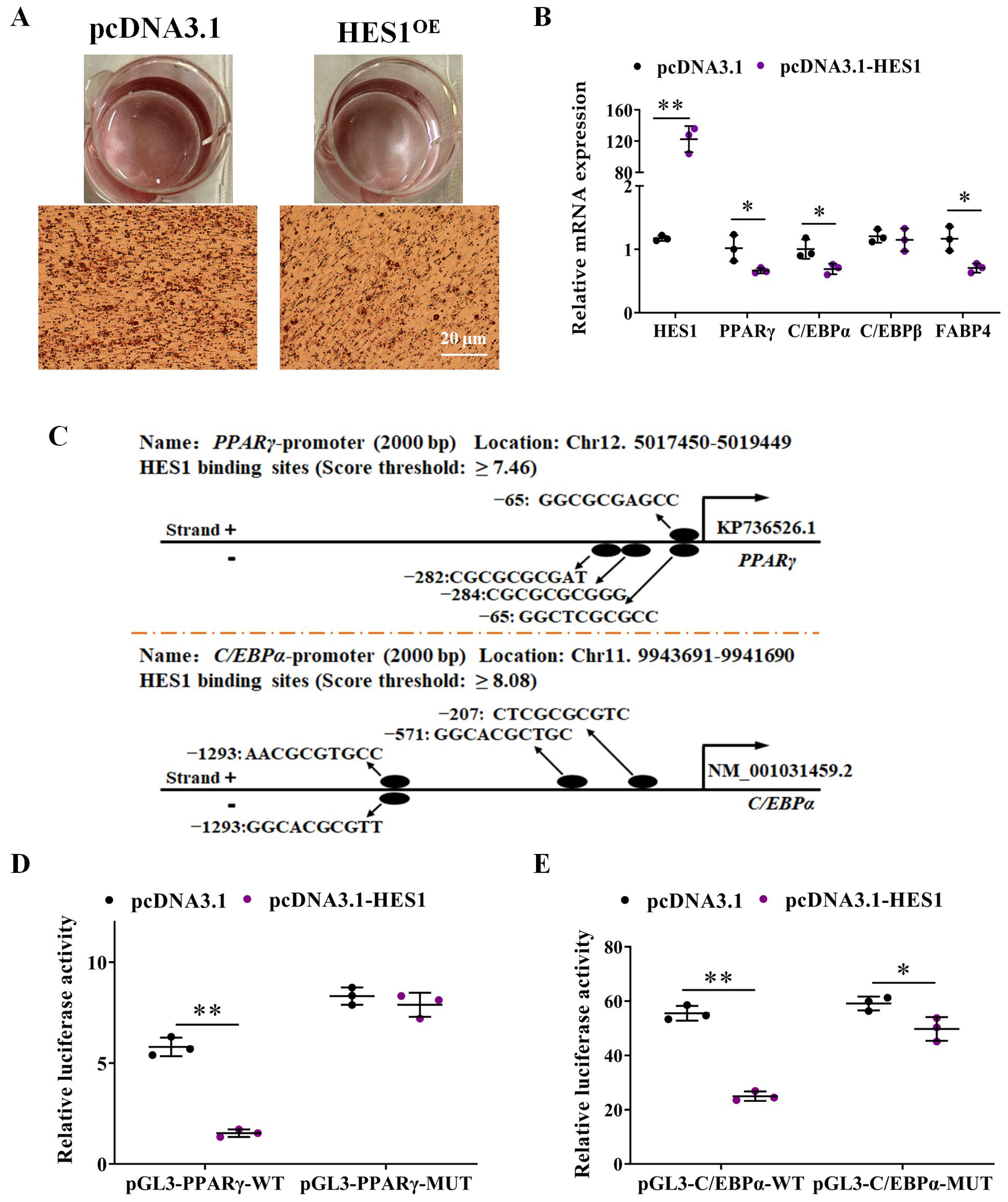
3.5. HES1 Inhibits the Expression of PPARγ and C/EBPα by Directly Binding to Their Upstream Region

Furthermore, we also investigated the effect of HES1, a NOTCH downstream active transcription factor, on avian adipogenesis via a gain-of-function study. In comparison to the control group, HES1–overexpressing ICP1 cells exhibited an approximately 120-fold increase in HES1 mRNA level, as revealed by RT–qPCR analysis. This surge in HES1 levels corresponded to a significant reduction in ORO staining and a decrease in mRNA levels of adipogenic markers, including PPARγ, C/EBPα, and FABP4 (p < 0.05; Figures 6A,B and S5). To strengthen the evidence supporting the direct regulatory connection between HES1 and the adipogenic transcription factor, we employed the JASPAR prediction database. Our analysis revealed multiple potential binding sites for the HES1 transcriptional repressor in the upstream region of PPARγ and C/EBPα (Figure 6C).
To test whether HES1 directly regulates PPARγ and C/EBPα expression via binding to the upstream region, we cloned the 409 bp upstream promoter of PPARγ and the 1548 bp upstream promoter of C/EBPα into the pGL3–Promoter plasmid (pGL3–PPARγ–WT and pGL3–C/EBPα–WT), each of which contained four potential binding sites for HES1. After deleting the four binding sites, two corresponding mutant promoter reporter plasmids were constructed (pGL3–PPARγ–MUT and pGL3–C/EBPα–MUT). The results showed that transfection of pcDNA3.1–HES1 significantly inhibited the wild–type reporters’ activity pGL3–PPARγ–WT and pGL3–C/EBPα–WT compared with control pcDNA3.1 (p < 0.05; Figure 6D,E). On the contrary, the reporter activity of the mutation–type reporter GL3–PPARγ–MUT and pGL3–C/EBPα–MUT were not affected by intracellular HES1 overexpression ((p > 0.05; Figure 6D,E). These results demonstrate the important role of the NOTCH1/HES1 axis in effectively inhibiting avian adipocyte differentiation, thereby presenting a potential target for regulating fat deposition in avian species.

4. Discussion

Excessive fat deposition has a profound impact on both the efficiency and quality of meat production [33]. Fat deposition can be driven either by hyperplasia or hypertrophy, a process known as adipogenesis at the cellular level [4]. Despite substantial research employing various strategies such as genetic selection, feeding supplementation, and multi−omics research to understand the genetic regulation of adipogenesis, the molecular mechanisms underlying avian fat deposition remain largely unknown.
NOTCH1 is a conserved receptor of communication between adjacent cells and broadly involved in cellular differentiation, proliferation, and apoptosis throughout all development stage [11]. Our prior and ongoing research has revealed that the expression of several genes related to the NOTCH signaling pathway undergoes alterations during avian adipocyte differentiation. These findings strongly suggest that these genes may have a pivotal role in regulating avian adipogenesis. However, it remains unclear whether NOTCH1’s role is pro− or anti−adipogenesis. In line with the findings of Song et al. [22] and Wang et al. [34], our findings suggest that NOTCH1 negatively regulates avian adipocyte differentiation.
Our study also revealed a significant association between the mRNA levels of HES1 and NOTCH1 activity, with HES1 expression decreasing following adipogenic differentiation. The functional role of HES1 in adipogenesis was further confirmed through a gain−of−function study in ICP1 cells, resulting in impaired adipogenesis, as evidenced by a decrease in intracellular lipid droplet accumulation. In contrast, previous studies by Ross et al. demonstrated that HES1 is required for 3T3−L1 differentiation by suppressing DLK/PREF1 [20]. Meanwhile, Lei et al. reported that HES1 inhibits adipogenesis of porcine mesenchymal stem cells via transcriptional repression of FAD24 [35]. Moreover, we confirmed that HES1 inhibited the expression and promoter activity of PPARγ and C/EBPα in avian adipocyte by luciferase assay and gene expression analysis.
Chicken PPARγ gene is characterized by five transcript variants (cPPARγ15) generated by alternative promoter usage and alternate splicing, resulting in two protein isoforms (PPARγ1 and PPARγ2) [36]. PPARγ2 protein isoform is distinguished by an extra six amino acids at the N—terminal, which include the activation function—1 domain, compared to PPARγ1 protein isoform [36]. The differences between the two chicken PPARγ protein isoforms in expression patterns, proliferation and differentiation have been well characterized in several studies [6,37]. Given the notably higher expression of cPPARγ1 transcript (encoding PPARγ1 protein isoform) in chicken adipocytes and tissues compared to other transcript variants, our study primarily analyzed the effect of HES1 on the transcriptional activity of the promoter of cPPARγ1 transcript. Additionally, bioinformatics analysis identified several putative HES1 binding sites that existed in the promoter of cPPARγ2 transcript (encoding PPARγ2 protein isoform; Supplementary Table S1), suggesting that chicken PPARγ2 protein isoform may also be regulated by HES1.
Furthermore, increases in HEY1, HEY2, and SOCS3, as well as decreases in NUMB and NR2F2 were detected in the NICD1–overexpressing cells relative to controls, as determined by RT–qPCR or Western blot, implying these genes may be involved in the regulation of avian adipogenesis through NOTCH1. Previous literature has suggested that NR2F2 directly binds to the promoter regions of HEY1 and HEY2, leading to transcriptional repression [38]. Intriguingly, our separate study has reported that NR2F2 is an anti-adipogenic factor in avian adipocytes and its expression also decreased post induction [39]. However, regulatory connection between NOTCH1 and NR2F2 in avian needs additional exploration. The precise understanding of the cascade of these molecular events and the way to regulate them will certainly be crucial to efficiently fight fat deposition.
One limitation of our study is the use of immortalized chicken abdominal adipocytes, ICP1, as an in vitro avian cell culture model for the analyses of the effects of NOTCH1 on adipocyte differentiation and gene expression. However, different depots of fat, such as abdominal fat, subcutaneous fat, and intermuscular fat, differ in their developmental origin, anatomical location, and response to metabolic stimuli [40,41]. Moreover, the adipocytes derived from different depots have shown that they functionally differ in lipolysis, adipogenic potential, and thermogenic activity [42,43]. Therefore, it would be intriguing to reproduce our results in distinct cell subsets from other depots or more detailed functional analyses. Additionally, it is worth investigating the in vivo functions of NOTCH1 in chicken adipogenesis and fat development using different gene editing strategies.

5. Conclusions

In summary, this study not only reaffirms the significance of NOTCH signaling in regulating avian adipogenesis, but also furnishes comprehensive evidence supporting NOTCH1’s role as a repressor of adipocyte differentiation in the avian adipocyte model. Given the pivotal role of adipocyte differentiation in adipose tissue development and function, elucidating the mechanism by which NOTCH signaling modulates adipogenesis becomes imperative. Such understanding is crucial for enabling more efficient selection methods aimed at mitigating excessive fat deposition and thereby enhancing both chicken production efficiency and meat quality.

Supplementary Materials

The following supporting information can be downloaded at: https://www.mdpi.com/article/10.3390/ani14040585/s1. Figure S1: Representative phase contrast images of ICP1 cells approximately 24 h after seeding; Figure S2: NICD1 promoted the relative luciferase activity dependent on RBP−Jκ site; Figure S3: Quantification of ORO staining after NOTCH1 overexpression or knockdown or control; Figure S4: Quantification of ORO staining after DAPT or DMSO treatment; Figure S5: Quantification of ORO staining after HES1 overexpression or control. Table S1: The putative binding sites of transcription factor HES1 within 2000 bp upstream region of cPPARγ2 transcript (Chr12: 5018144− 5020143). The original images of staining cells. The original Western blot figures.

Author Contributions

H.L. and Z.H. conceptualized and designed the experimental plan. Z.W., Y.S., M.Z., Z.M., and J.L. collected samples, conducted the experiments, and performed bioinformatics analyses. Z.W. and H.L. drafted and revised the manuscript. Z.W. and Y.S. contributed equally to this work and share first authorship. All authors have read and agreed to the published version of the manuscript.

Funding

This research was funded by the Fund Project on Application of Basic Research Project of Shanxi Province (202103021224155; 202103021223135), the Excellent doctors come to Shanxi to reward scientific research projects (SXBYKY20222057), the PhD Start−up Foundation of Shanxi Agricultural University (2021BQ114) and the earmarked fund for Shanxi Agriculture Research System (2022−07).

Institutional Review Board Statement

The ICP1 cell line was obtained from the Poultry Breeding Lab of the College of Animal Science and Technology, Northeast Agricultural University.

Informed Consent Statement

Not applicable.

Data Availability Statement

The data presented in this study are available on request from the corresponding author. The mRNA−seq datasets used in this study were taken from previously published paper and is available in NCBI (Accession number: SRX4646736, PRJNA1069883).

Acknowledgments

We thank the Poultry Breeding Lab of the College of Animal Science and Technology, Northeast Agricultural University, for providing the ICP1 cell line.

Conflicts of Interest

The research was conducted in the absence of any commercial or financial relationships that could be construed as potential conflicts of interest.

References

  1. Baeza, E.; Guillier, L.; Petracci, M. Review: Production factors affecting poultry carcass and meat quality attributes. Animal 2018, 16 (Suppl. 1), 100331. [Google Scholar] [CrossRef]
  2. Mathew, H.; Castracane, V.D.; Mantzoros, C. Adipose tissue and reproductive health. Metabolism 2018, 86, 18–32. [Google Scholar] [CrossRef]
  3. Wang, G.; Kim, W.K.; Cline, M.A.; Gilbert, E.R. Factors affecting adipose tissue development in chickens: A review. Poult. Sci. 2017, 96, 3687–3699. [Google Scholar] [CrossRef]
  4. Ghaben, A.L.; Scherer, P.E. Adipogenesis and metabolic health. Nat. Rev. Mol. Cell Biol. 2019, 20, 242–258. [Google Scholar] [CrossRef]
  5. Schwalie, P.C.; Dong, H.; Zachara, M.; Russeil, J.; Alpern, D.; Akchiche, N.; Caprara, C.; Sun, W.; Schlaudraff, K.U.; Soldati, G.; et al. A stromal cell population that inhibits adipogenesis in mammalian fat depots. Nature 2018, 559, 103–108. [Google Scholar] [CrossRef] [PubMed]
  6. Mu, F.; Jing, Y.; Ning, B.; Huang, J.; Cui, T.; Guo, Y.; You, X.; Yan, X.; Li, H.; Wang, N. Peroxisome proliferator-activated receptor gamma isoforms differentially regulate preadipocyte proliferation, apoptosis, and differentiation in chickens. Poult. Sci. 2020, 99, 6410–6421. [Google Scholar] [CrossRef] [PubMed]
  7. Sun, D.; Li, X.; Yin, Z.; Hou, Z. The Full-Length Transcriptome Provides New Insights into the Transcript Complexity of Abdominal Adipose and Subcutaneous Adipose in Pekin Ducks. Front. Physiol. 2021, 12, 767739. [Google Scholar] [CrossRef] [PubMed]
  8. Donati, G.; Proserpio, V.; Lichtenberger, B.M.; Natsuga, K.; Sinclair, R.; Fujiwara, H.; Watt, F.M. Epidermal Wnt/beta-catenin signaling regulates adipocyte differentiation via secretion of adipogenic factors. Proc. Natl. Acad. Sci. USA 2014, 111, E1501–E1509. [Google Scholar] [CrossRef] [PubMed]
  9. Wang, Z.; Zhao, Q.; Li, X.; Yin, Z.; Chen, S.; Wu, S.; Yang, N.; Hou, Z. MYOD1 inhibits avian adipocyte differentiation via miRNA-206/KLF4 axis. J. Anim. Sci. Biotechnol. 2021, 12, 55. [Google Scholar] [CrossRef]
  10. Wang, Z.; Yin, Z.T.; Zhang, F.; Li, X.Q.; Chen, S.R.; Yang, N.; Porter, T.E.; Hou, Z. Dynamics of transcriptome changes during subcutaneous preadipocyte differentiation in ducks. BMC Genom. 2019, 20, 688. [Google Scholar] [CrossRef] [PubMed]
  11. Zohorsky, K.; Mequanint, K. Designing Biomaterials to Modulate Notch Signaling in Tissue Engineering and Regenerative Medicine. Tissue Eng. Part B Rev. 2021, 27, 383–410. [Google Scholar] [CrossRef] [PubMed]
  12. Kopan, R.; Ilagan, M.X. The canonical Notch signaling pathway. unfolding the activation mechanism. Cell 2009, 137, 216–233. [Google Scholar] [CrossRef] [PubMed]
  13. Meurette, O.; Mehlen, P. Notch Signaling in the Tumor Microenvironment. Cancer Cell 2018, 34, 536–548. [Google Scholar] [CrossRef] [PubMed]
  14. Zhou, B.; Lin, W.; Long, Y.; Yang, Y.; Zhang, H.; Wu, K.; Chu, Q. Notch signaling pathway: Architecture, disease, and therapeutics. Signal Transduct. Target. Ther. 2022, 7, 95. [Google Scholar] [CrossRef]
  15. Singh, S.B.; Coffman, C.N.; Carroll-Portillo, A.; Varga, M.G.; Lin, H.C. Notch Signaling Pathway Is Activated by Sulfate Reducing Bacteria. Front. Cell. Infect. Microbiol. 2021, 11, 695299. [Google Scholar] [CrossRef]
  16. Chen, X.; Qin, J.; Cheng, C.M.; Tsai, M.J.; Tsai, S.Y. COUP-TFII is a major regulator of cell cycle and Notch signaling pathways. Mol. Endocrinol. 2012, 26, 1268–1277. [Google Scholar] [CrossRef]
  17. Miao, L.; Lu, Y.; Nusrat, A.; Abdelnasser, H.Y.; Datta, S.; Zhou, B.; Schwartz, R.J.; Wu, M. The Spatiotemporal Expression of Notch1 and Numb and Their Functional Interaction during Cardiac Morphogenesis. Cells 2021, 10, 2192. [Google Scholar] [CrossRef]
  18. Esteves de Lima, J.; Bonnin, M.A.; Birchmeier, C.; Duprez, D. Muscle contraction is required to maintain the pool of muscle progenitors via YAP and NOTCH during fetal myogenesis. eLife 2016, 5, e15593. [Google Scholar] [CrossRef]
  19. Zuo, Q.; Zhang, C.; Jin, K.; Jing, J.; Sun, C.; Ahmed, M.F.; Song, J.; Zhang, Y.; Chen, G.; Li, B. NICD-mediated notch transduction regulates the different fate of chicken primordial germ cells and spermatogonial stem cells. Cell Biosci. 2018, 8, 40. [Google Scholar] [CrossRef]
  20. Ross, D.A.; Rao, P.K.; Kadesch, T. Dual roles for the Notch target gene Hes-1 in the differentiation of 3T3-L1 preadipocytes. Mol. Cell. Biol. 2004, 24, 3505–3513. [Google Scholar] [CrossRef] [PubMed]
  21. Nichols, A.M.; Pan, Y.; Herreman, A.; Hadland, B.K.; De Strooper, B.; Kopan, R.; Huppert, S.S. Notch pathway is dispensable for adipocyte specification. Genesis 2004, 40, 40–44. [Google Scholar] [CrossRef]
  22. Song, B.Q.; Chi, Y.; Li, X.; Du, W.J.; Han, Z.B.; Tian, J.J.; Li, J.J.; Chen, F.; Wu, H.H.; Han, L.X.; et al. Inhibition of Notch Signaling Promotes the Adipogenic Differentiation of Mesenchymal Stem Cells Through Autophagy Activation and PTEN-PI3K/AKT/mTOR Pathway. Cell. Physiol. Biochem. 2015, 36, 1991–2002. [Google Scholar] [CrossRef] [PubMed]
  23. Wang, W.; Zhang, T.; Wu, C.; Wang, S.; Wang, Y.; Li, H.; Wang, N. Immortalization of chicken preadipocytes by retroviral transduction of chicken TERT and TR. PLoS ONE 2017, 12, e0177348. [Google Scholar] [CrossRef] [PubMed]
  24. Ross, D.A.; Kadesch, T. The notch intracellular domain can function as a coactivator for LEF-1. Mol. Cell. Biol. 2001, 21, 7537–7544. [Google Scholar] [CrossRef]
  25. Dovey, H.F.; John, V.; Anderson, J.P.; Chen, L.Z.; de Saint Andrieu, P.; Fang, L.Y.; Freedman, S.B.; Folmer, B.; Goldbach, E.; Holsztynska, E.J.; et al. Functional gamma-secretase inhibitors reduce beta-amyloid peptide levels in brain. J. Neurochem. 2001, 76, 173–181. [Google Scholar] [CrossRef]
  26. Shang, Z.; Guo, L.; Wang, N.; Shi, H.; Wang, Y.; Li, H. Oleate promotes differentiation of chicken primary preadipocytes in vitro. Biosci. Rep. 2014, 34, e00093. [Google Scholar] [CrossRef] [PubMed]
  27. Ramirez-Zacarias, J.L.; Castro-Munozledo, F.; Kuri-Harcuch, W. Quantitation of adipose conversion and triglycerides by staining intracytoplasmic lipids with Oil red O. Histochemistry 1992, 97, 493–497. [Google Scholar] [CrossRef]
  28. Kurien, B.T.; Scofield, R.H. Western blotting. Methods 2006, 38, 283–293. [Google Scholar] [CrossRef]
  29. Castro-Mondragon, J.A.; Riudavets-Puig, R.; Rauluseviciute, I.; Lemma, R.B.; Turchi, L.; Blanc-Mathieu, R.; Lucas, J.; Boddie, P.; Khan, A.; Manosalva Perez, N.; et al. JASPAR 2022: The 9th release of the open-access database of transcription factor binding profiles. Nucleic Acids Res. 2022, 50, D165–D173. [Google Scholar] [CrossRef]
  30. Kim, D.; Paggi, J.M.; Park, C.; Bennett, C.; Salzberg, S.L. Graph-based genome alignment and genotyping with HISAT2 and HISAT-genotype. Nat. Biotechnol. 2019, 37, 907–915. [Google Scholar] [CrossRef]
  31. Anders, S.; Pyl, P.T.; Huber, W. HTSeq—A Python framework to work with high-throughput sequencing data. Bioinformatics 2015, 31, 166–169. [Google Scholar] [CrossRef] [PubMed]
  32. Love, M.I.; Huber, W.; Anders, S. Moderated estimation of fold change and dispersion for RNA-seq data with DESeq2. Genome Biol. 2014, 15, 550. [Google Scholar] [CrossRef] [PubMed]
  33. Li, Y.; He, C.; Ran, L.; Wang, Y.; Xiong, Y.; Wang, Y.; Zhu, J.; Lin, Y. miR-130b duplex (miR-130b-3p/miR-130b-5p) negatively regulates goat intramuscular preadipocyte lipid droplets accumulation by inhibiting Kruppel-like factor 3 expression. J. Anim. Sci. 2023, 101, skad184. [Google Scholar] [CrossRef] [PubMed]
  34. Wang, Y.; Tan, J.; Du, H.; Liu, X.; Wang, S.; Wu, S.; Yuan, Z.; Zhu, X. Notch1 Inhibits Rosiglitazone-Induced Adipogenic Differentiation in Primary Thymic Stromal Cells. Front. Pharmacol. 2018, 9, 1284. [Google Scholar] [CrossRef] [PubMed]
  35. Lei, T.; Bi, Y.; Gao, M.J.; Gao, S.M.; Zhou, L.L.; Zheng, H.L.; Chen, X.D. HES1 inhibits adipogenesis of porcine mesenchymal stem cells via transcriptional repression of FAD24. Domest. Anim. Endocrinol. 2013, 45, 28–32. [Google Scholar] [CrossRef] [PubMed]
  36. Duan, K.; Sun, Y.; Zhang, X.; Zhang, T.; Zhang, W.; Zhang, J.; Wang, G.; Wang, S.; Leng, L.; Li, H.; et al. Identification and characterization of transcript variants of chicken peroxisome proliferator-activated receptor gamma. Poult. Sci. 2015, 94, 2516–2527. [Google Scholar] [CrossRef] [PubMed]
  37. Jing, Y.; Mu, F.; Xing, X.; Huang, J.; Lou, M.; Xu, H.; Ning, B.; Lou, Y.; Gao, Z.; Luo, H.; et al. Knockout and Restoration Reveal Differential Functional Roles of PPARgamma1 and PPARgamma2 in Chicken Adipogenesis. J. Agric. Food Chem. 2022, 70, 14959–14973. [Google Scholar] [CrossRef]
  38. Aranguren, X.L.; Beerens, M.; Coppiello, G.; Wiese, C.; Vandersmissen, I.; Lo Nigro, A.; Verfaillie, C.M.; Gessler, M.; Luttun, A. COUP-TFII orchestrates venous and lymphatic endothelial identity by homo- or hetero-dimerisation with PROX1. J. Cell Sci. 2013, 126 Pt 5, 1164–1175. [Google Scholar] [CrossRef]
  39. Zhu, F.; Yin, Z.T.; Wang, Z.; Smith, J.; Zhang, F.; Martin, F.; Ogeh, D.; Hincke, M.; Lin, F.B.; Burt, D.W.; et al. Three chromosome-level duck genome assemblies provide insights into genomic variation during domestication. Nat. Commun. 2021, 12, 5932. [Google Scholar] [CrossRef]
  40. Hwang, I.; Kim, J.B. Two Faces of White Adipose Tissue with Heterogeneous Adipogenic Progenitors. Diabetes Metab. J. 2019, 43, 752–762. [Google Scholar] [CrossRef]
  41. Nahmgoong, H.; Jeon, Y.G.; Park, E.S.; Choi, Y.H.; Han, S.M.; Park, J.; Ji, Y.; Sohn, J.H.; Han, J.S.; Kim, Y.Y.; et al. Distinct properties of adipose stem cell subpopulations determine fat depot-specific characteristics. Cell Metab. 2022, 34, 458–472e456. [Google Scholar] [CrossRef] [PubMed]
  42. Macotela, Y.; Emanuelli, B.; Mori, M.A.; Gesta, S.; Schulz, T.J.; Tseng, Y.H.; Kahn, C.R. Intrinsic differences in adipocyte precursor cells from different white fat depots. Diabetes 2012, 61, 1691–1699. [Google Scholar] [CrossRef] [PubMed]
  43. Ferrero, R.; Rainer, P.; Deplancke, B. Toward a Consensus View of Mammalian Adipocyte Stem and Progenitor Cell Heterogeneity. Trends Cell Biol. 2020, 30, 937–950. [Google Scholar] [CrossRef] [PubMed]
Figure 1. The expression changes in NOTCH signaling during avian adipocyte differentiation. (A) Heatmap analysis of expression patterns for NOTCH signaling–related genes identified with mRNA–seq analysis of gene expression data from chicken abdominal adipocytes at 0 h, 72 h, 120 h, 168 h post induction (n = 4, Raw in Z–Score). (B) Heatmap analysis of expression patterns for NOTCH signaling–related genes identified with mRNA–seq analysis of gene expression data from Pekin duck subcutaneous adipocytes at 0 h, 24 h, 48 h, 72 h post induction (n = 4, Raw in Z–Score). (C) Comparison between mRNA–seq and RT–qPCR expression measurements for the chosen genes. The graph illustrates the log2 fold changes for both mRNA–seq and RT–qPCR data.
Animals 14 00585 g001
Figure 2. NOTCH1 is inhibited during adipocyte differentiation. (A) ICP1 cells were induced to differentiate with 160 μM oleic acid and total RNA was extracted at specified time points (0 h corresponds to the time of oleic acid supplementation). Subsequently, RT−qPCR was conducted to assess NOTCH1 expression throughout adipocyte differentiation (n = 3). The mRNA expression was normalized to GAPDH mRNA, and the results are presented relative to the mRNA level of NOTCH1 at 0 h, which is set as 1. (B) Western blot analysis of protein abundance of NOTCH1 during the ICP1 cells differentiation; GAPDH was utilized as an internal control. The protein abundance of NOTCH1 and GAPDH were quantitatively compared (n = 3). The data are presented as mean ± SD, and statistical significance was determined through one−way ANOVA. * p < 0.05; ** p < 0.01.
Animals 14 00585 g002
Figure 3. Effect of NOTCH1 on signaling−related genes in ICP1 cells. (A) Western blot analysis of protein abundance of NOTCH1, NUMB, SOCS3, and GAPDH in NICD1 overexpression ICP1 cells and control cells; GAPDH is an internal control. The protein abundance of NOTCH1, NUMB, SOCS3, and GAPDH was quantitatively compared (n = 3). (B) The relative mRNA expression of NOTCH1, HES1, HEY1, HEY2, NR2F2, NUMB, and SOCS3 in NICD1 overexpression ICP1 cells and control cells was determined using RT−qPCR; GAPDH served as an internal control (n = 3). (C) Western blot analysis of protein abundance of NOTCH1, NUMB, SOCS3, and GAPDH in NOTCH1 knockdown ICP1 cells and control cells. The abundance of each protein was quantitatively compared (n = 3). (D) Relative mRNA expression of NOTCH1, HES1, HEY1, HEY2, NR2F2, NUMB, and SOCS3 in NOTCH1 knockdown ICP1 cells and control cells was determined using RT−qPCR (n = 3). Data are shown as mean ± SD, and significance was determined using Student’s t−tests. * p < 0.05; ** p < 0.01.
Animals 14 00585 g003
Figure 4. NOTCH1 inhibits fat accumulation in differentiating adipocyte. (A) Cultured ICP1 cells expressing pcDNA3.1 or pcDNA–NICD1 was induced to differentiate into adipocytes with 160 μM oleic acid. At the end of induction (72 h), cells were subjected to Oil Red O staining to evaluate lipid accumulation. All micrographs were captured at the same magnification, and a 20 μm scale bar is provided in the lower right panel for reference. (B) Relative mRNA expression of mature adipocyte marker genes in NICD1 overexpression ICP1 cells and control cells was determined using RT–qPCR; GAPDH served as an internal control (n = 3). (C) Representative images of NOTCH1 knockdown promoted the lipid accumulation by ORO staining on 72 h post induction. (D) Relative mRNA expression of mature adipocyte marker genes in NOTCH1 knockdown ICP1 cells and control cells was determined using RT–qPCR (n = 3). Data are shown as mean ± SD and significance was determined using Student’s t–tests. * p < 0.05; ** p < 0.01.
Animals 14 00585 g004
Figure 5. DAPT inhibits NOTCH1 and HES1 and promotes adipogenic differentiation. (A) Western blot analysis of protein abundance of NICD1 (nuclear), LMNB1 (nuclear), NOTCH1 (cytoplasm), and GAPDH (cytoplasm) in ICP1 cells treated with various concentrations of DAPT for 48 h; LMNB1 and GAPDH as internal control of nuclear protein and cytoplasmic protein, respectively (n = 3). (B) The protein abundance of NICD1 (nuclear) and LMNB1 (nuclear) was quantitatively compared (n = 3). (C) The protein abundance of NOTCH1 (cytoplasm) and GAPDH (cytoplasm) was quantitatively compared (n = 3). (D) The mRNA level of HES1 in ICP1 cells treated with various concentrations of DAPT for 48 h was determined using RT-qPCR (n = 3). (E) Representative images of 5 μM DAPT promoted the lipid droplet formation in ICP1 cells by ORO staining on 72 h post induction. All micrographs are the same magnification, and a 20 μm scale bar is provided in the lower right panel for reference. (F) Relative mRNA expression of mature adipocyte marker genes in 5 μM DAPT treated with ICP1 cells and alone DMSO treated with cells on 72 h post induction was determined using RT–qPCR (n = 3). Data are shown as mean ± SD and significance was determined using One–way ANOVA or Student’s t–tests. * p < 0.05; ** p < 0.01.
Animals 14 00585 g005
Figure 6. HES1 overexpression inhibits PPARγ and C/EBPα and promotes adipogenic differentiation. (A) Cultured ICP1 cells expressing pcDNA3.1 or pcDNA–HES1 was induced to differentiate into adipocytes with 160 μM oleic acid. At the end of differentiation (72 h), cells were subjected to Oil Red O staining to evaluate lipid accumulation. All micrographs were captured at the same magnification, and a 20 μm scale bar is provided in the lower right panel for reference. (B) Relative mRNA expression of HES1 and mature adipocyte marker genes in HES1 overexpression ICP1 cells and control cells was determined using RT–qPCR; GAPDH served as an internal control (n = 3). (C) Predicted binding sites and putative binding sequence for the HES1 transcription factor in the PPARγ and C/EBPα promoter as identified in the JASPAR database. (D) The cells were co–transfected with pGL3–PPARγ–WT or pGL3–PPARγ–MUT and pcDNA3.1–HES1 or pcDNA3.1, and pRL–TK plasmid (24:24:1). After 72 h of co–transfection, relative luciferase activity was measured. The effect of HES1 overexpression on the luciferase activity of pGL3–PPARγ–WT and pGL3–PPARγ–MUT in ICP1 cells (n = 3). (E) The effect of HES1 overexpression on the luciferase activity of wild type reporter pGL3–C/EBPα–WT and mutation reporter pGL3–C/EBPα–MUT in ICP1 cells (n = 3). Data are shown as mean ± SD and significance was determined using Student’s t–tests. * p < 0.05; ** p < 0.01.
Animals 14 00585 g006
Table 1. Sequences of primers used for RT-qPCR.
Gene SymbolAccession NumbersSequence (5′–3′)Length (bp)
NOTCH1XM_046928731.1F: AACGCTGTGGATGATCTGGG131
R: AGCGGGGTCTCCTCCTTATT
PPARγNM_001001460F: GTGCAATCAAAATGGAGCC170
R: CTTACAACCTTCACATGCAT
FABP4NM_204290F: ATGTGCGACCAGTTTGT143
R: TCACCATTGATGCTGATAG
C/EBPαNM_001031459F: GGAGCAAGCCAACTTCTACG181
R: GTCGATGGAGTGCTCGTTCT
C/EBPβNM_205253F: CGCCCGCCTTTAAATCCATG151
R: GGGCTGAAGTCAATGGCTCT
HES1XM_040679737F: GGACGCGCTGAAGAAGGATA203
R: CTTCGCAGGTGGAGAGGAAC
HEY1XM_040665051F: TGGCTGAAGTGGCTCGATAC175
R: TGAGGGTGATGTCCAAAGGC
HEY2XM_040669098F: GGGCAGCGAGAACAACTACT125
R: CCCGGCGCCTTTTCTCTATA
SOCS3NM_204600.2F: GCCTCAAGACGTTCAGCTCT196
R: GTCTTGACGCTGAGGGTGAA
NUMBAF176086F: ACAAAACCCGTGACAGTGGT168
R: GGCACGGACAGTCTTTGAGA
NR2F2XM_046924463F: TGACCTGGAGCGAGTTGTTC185
R: GTCGACATGCAACGCTTTCA
GAPDHNM_204305F: CCTCTCTGGCAAAGTCCAAG200
R: CATCTGCCCATTTGATGTTG
Disclaimer/Publisher’s Note: The statements, opinions and data contained in all publications are solely those of the individual author(s) and contributor(s) and not of MDPI and/or the editor(s). MDPI and/or the editor(s) disclaim responsibility for any injury to people or property resulting from any ideas, methods, instructions or products referred to in the content.

