Article

Molecular Epidemiological Investigation of Cyclospora spp. in Holstein Cattle in Partial Areas of the Yunnan Province, China

1
Key Laboratory of Veterinary Public Health of Yunnan Province, College of Veterinary Medicine, Yunnan Agricultural University, Kunming 650201, China
2
Key Laboratory of Animal Nutrition and Feed in Yunnan Province, College of Animal Science and Technology, Yunnan Agricultural University, Kunming 650201, China
3
Yunnan New Hope Xuelan Animal Husbandry Technology Co., Ltd., Qujing 650201, China
*
Authors to whom correspondence should be addressed.
These authors contributed equally to this work.
Animals 2023, 13(9), 1527; https://doi.org/10.3390/ani13091527
Submission received: 13 March 2023 / Revised: 21 April 2023 / Accepted: 25 April 2023 / Published: 3 May 2023

Abstract

:

Simple Summary

Cyclospora spp. is an important zoonotic parasite that poses a threat to public health. At present, the research on Cyclospora spp. in cattle of the Yunnan Province remains limited. We firstly reported the prevalence of Cyclospora spp. in Holstein cattle in the Yunnan Province, China. In order to understand the prevalence and genotype of Cyclospora spp. in the Yunnan Province, 524 fecal samples from 4 regions were collected and tested. The results of the present study showed that 13 samples were positive for Cyclospora spp., and the total infection rate of Cyclospora spp. was 2.48%. We analyzed different risk factors, such as regions, sexes, and ages, but the difference was not statistically significant. Phylogenetic analysis showed that five Cyclospora spp. samples were classified into the zoonotic group of Cyclospora cayetanensis, which had zoonotic potential. The results of the present study indicated that Cyclospora spp. in the Holstein cattle of Yunnan Province posed a risk of zoonosis.

Abstract

Cyclospora spp. is a food-borne intestinal protozoan, which is widely distributed in the world and poses the risk of zoonosis. In order to reveal the prevalence of Cyclospora spp. in Holstein cattle in partial areas of the Yunnan Province, 524 fresh fecal samples of Holstein cattle were collected from Dali, Kunming, Chuxiong, and Qujing in Yunnan Province. A nested PCR amplification of the small subunit (SSU) rRNA gene of Cyclospora spp. was carried out, and the products of the nested PCR were further analyzed by restriction fragment length polymorphism (RFLP) using Bsp E Ⅰ. The results of the present study showed that 13 samples were positive for Cyclospora spp., and the total infection rate of Cyclospora sp. was 2.48%. The infection of Cyclospora spp. was detected in Dali, Qujing, and Chuxiong. Chuxiong showed the highest infection rate (5.71%), and infection rate in Dali and Qujing was 2.19% and 3.16%, respectively. Interestingly, the infection of Cyclospora spp. was not detected in Kunming. The infection of Cyclospora spp. showed no significant differences among different regions (p > 0.05). Cyclospora sp. infection was detected in all ages and sexes, but the differences were not significant (p > 0.05). Sequencing and phylogenetic analysis showed that five Cyclospora spp. samples were closely related to the Cyclospora spp. of humans, and the others were closely related to the Cyclospora spp. of bovines. The results of the present study suggested that there was an infection of Cyclospora spp. in Holstein cattle in the Yunnan Province, and the Cyclospora spp. showed a risk of zoonosis. Thus, the prevention and control of Cyclospora spp. should be strengthened in the Yunnan Province, China. The results of this investigation provide data references for the further research of Cyclosporiasis in Holstein cattle in the Yunnan Province.

1. Introduction

Cyclospora spp. belongs to the protozoa subkingdom, subphylum Apicomplexa, subclass Coccidiasina, order Eucoccidiorida, family Eimeriidae, and Cyclospora [1]. Cyclospora spp. is a food-borne zoonotic intestinal protozoan that is widely distributed in the world. It is mainly transmitted through contaminated water or food, and it can also be transmitted through direct contact between humans and animals [2]. Cyclospora spp. mainly infects the epithelial cells of the upper part of the small intestine, especially the jejunum [1]. The clinical symptoms of Cyclospora spp. infection depend on the host’s immunity, and the symptoms of infection include watery diarrhea, nausea, abdominal cramps, weight loss, and even death in severe cases [3,4]. Cyclospora spp. is an opportunistic pathogenic protozoan, and the infection of Cyclospora spp. in normal immunity shows inapparent infection or self-limited diarrhea. However, individuals with immune deficiencies can have persistent diarrhea [5], dehydration diarrhea, and even death in severe cases [6]. There are a large number of oocysts in the feces of patients infected with Cyclospora spp. Thus, feces are an important source of infection. The oocysts of Cyclospora develop into infectious oocysts in the environment, and humans are infected via ingesting water and food contaminated by oocysts [7]. Oocysts enter the host and invade the epithelial cells of host’s small intestine where produces merozoites and oocysts [8]. Cyclospora spp. and Eimeria also have a high similarity in morphology, and Cyclospora spp. was once thought to be a cyanobacterium-like or coccidian-like body (CLB) [9]. With the increase in research, Cyclospora spp. has been clearly classified.
Cyclospora spp. is an emerging pathogen causing worldwide outbreaks of cyclosporiasis. Cyclospora spp. can be found in a wide range of animals, including the Diplopoda, Reptilia, and Insectivora classes, as well as the mammalian orders Rodentia and Primate [10]. Up to now, 20 species of Cyclospora spp. have been reported [2,4,11]. Although most of these Cyclospora spp. species are host-specific, Cyclospora cayetanensis (C. cayetanensis) has been currently confirmed as zoonotic pathogen, and it is the only known Cyclospora species that can infect humans [8]. Humans are thought to be infected by C. cayetanensis through ingesting food or water contaminated with oocysts [12]. Environmental factors such as contaminated fruits, vegetables, water, and soil are considered as the main sources of C. cayetanensis infection in humans [13]. C. cayetanensis DNA can be detected in the fecal samples of various animal species [14], but there are no data to confirm whether animals are involved in the spread of C. cayetanensis [7], and it is generally believed that animals act as mechanical carriers of oocysts.
Cyclospora spp. is an emerging zoonotic intestinal protozoan that poses a threat to human health. Since the discovery of Cyclospora spp., a large number of studies have been conducted to reveal the morphology and molecular characters of Cyclospora spp. in various vertebrates. The oocysts of C.cayetanensis have been identified in the feces of human infants, pupils, and adults in the Anhui Province by using microscopes, and minor sequence polymorphisms were observed in the 18S rRNA gene of C.cayetanensis in the Henan province, China. Three new Cyclospora species from monkeys were identified in Ethiopia by using morphology and molecular characterizations. The 18S rRNA gene fragments of Cyclospora-like organisms have also been detected from non-human primates in China [15,16,17,18,19]. Cyclospora-like oocysts that belong to the group of primate-derived Cyclospora spp. have been found in the fecal specimens of cattle in China [20]. The Yunnan Province is located in the southwest frontier of China and covers an area of 394,100 square kilometers. Yunnan has diverse climates and is suitable for the growth of all kinds of high-quality forage grass, which contribute to the rapid development of dairy farming in Yunnan. Holstein cattle are farmed on a large scale, and parasitic infections may occur during the breeding process. Cyclospora spp. not only affects the development of the farming industry, but it is also a potential threat to the health of humans. However, there is a lack of relevant information about the infection of Cyclospora spp. in Holstein cattle in Yunnan.
In this study, 524 Holstein cattle fecal samples were randomly collected from different Holstein cattle farms in Dali, Kunming, Chuxiong, and Qujing in the Yunnan Province, and all of these collected samples were used for Cyclospora spp. detection. The nested PCR amplification was performed by using primers designed according to the 18S rRNA sequence of the Cyclospora spp. A restriction endonuclease fragment length polymorphism (RFLP) analysis with Bsp E I was used to differentiate the Cyclospora species from cattle Eimeria spp. In this study, the epidemic situation of Cyclospora spp. was investigated, its distribution was clarified, and the risk of zoonotic transmission of Cyclospora spp. in Holstein cattle in the Yunnan Province was also evaluated. At last, the phylogenetic tree was constructed. The results of the present study provide important epidemic data that could support the prevention or control of cyclosporiasis in Holstein cattle in the Yunnan Province.

2. Materials and Methods

2.1. Specimens

In total, 524 fecal samples were collected from Holstein farms in Dali, Kunming, Qujing, and Chuxiong in the Yunnan Province from July to November 2021. The ages of the Holstein cattle ranged from newborn calves to adult cattle. Among them, there were 422 female Holstein cattle feces samples and 102 male Holstein cattle feces samples. Feces samples were collected directly from the rectum using disposable gloves, separately transferred into disposable plastic bags, recorded with their collection date, ages, and geographic information, and stored at 4 °C until used for DNA extraction.

2.2. DNA Extraction and PCR

The fecal sample was added to the beaker, washed with distilled water, filtered, centrifuged at 3000 r/min for 3 min, the supernatant was discarded, and 250 µL of the precipitate was aspirated into a 2 mL centrifuge tube and used for DNA extraction. The DNA of all samples were extracted individually by using the E.Z.N.A.R® Stool DNA Kit (Omega Bio-tek Inc., Norcross, GA, USA), and the extracted DNA were stored at −20 °C until use. Cyclospora spp. in the specimens were genetically characterized by nested PCR amplification of a 501 bp fragment of the small subunit (SSU) rRNA gene. Primers for the fragment (501 bp) were designed as previously described [20].
The primers used for the primary nested amplification of the SSU gene were 18S-F1 (5′-AATGTAAAACCCTTCCAGAGTAAC-3′) and 18S-R1 (5′-GCAATAATCTATCCCCATCACG-3′), 18S-F2 (5′-AATTCCAGCTCCAATAGTGTAT-3′), and 18S-R2 (5′-CAGGAGAAGCCAAGGTAGGCRTTT-3′) were used for secondary PCR amplification.
PCR was conducted in a 25 µL reaction system containing 10× PCR buffer, 200 µM dNTP, 0.4 µM of each primer, 1 unit of TaKaRa Ex-Taq DNA polymerase (TaKaRa Co., Ltd., Tokyo, Japan), and a DNA sample. In the first round of the PCR reaction system, 1 µL of DNA sample was added, but two microliters of primary PCR product were used as the template for the secondary nested PCR. The primary amplification contained 35 cycles (95 °C for 45 s, 55 °C for 45 s and 72 °C for 1 min), with an initial hot start at 94 °C for 7 min and a final extension at 72 °C for 10 min. The secondary cycling parameters were identical to the first-round amplifications with modified extension time, which was increased to 1 min 30 s. After the amplification complete, electrophoresis was performed on 1% agarose gel under 120 V for 30 min. The results of the electrophoresis were photographed in an ultraviolet gel imaging device.

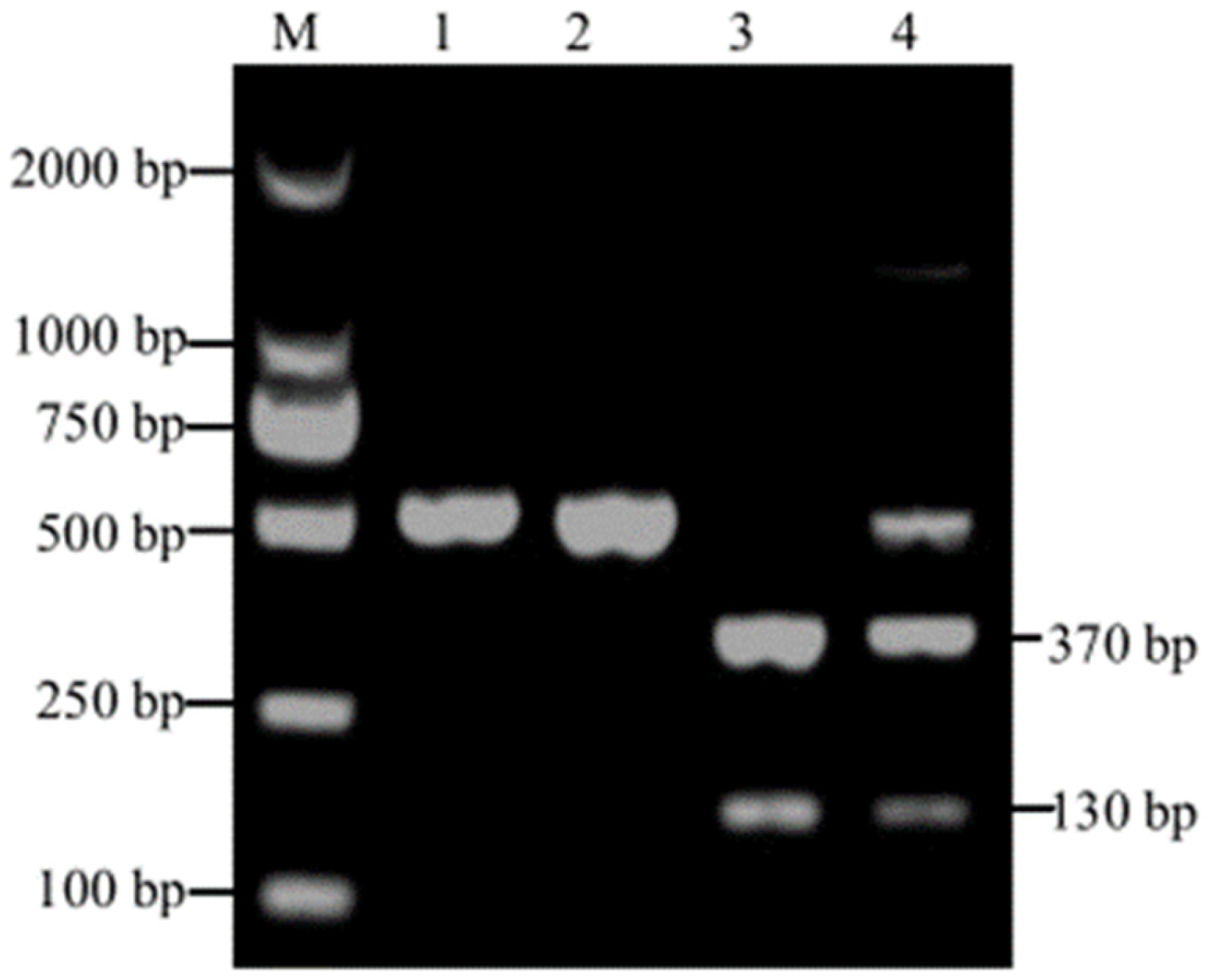
2.3. Restriction Fragment Length Polymorphism (RFLP) Analysis

Positive nested PCR products were further analyzed by RFLP to differentiate Cyclospora spp. from Eimeria spp. The RFLP reaction system contained 1 U volume of restriction endonuclease Bsp E I (10,000 U/mL), 12.5 μL of secondary amplification products, 2.5 μL of 10× NE Buffer 3.1, and 10 μL ddH2O. The samples were incubated at 37 °C for 4–8 h. Then, the products of the enzyme digestion were mixed with 1 µL of 10× loading buffer and separated by using electrophoresis on a 2% agarose gel. The results of electrophoresis were photographed by using a gel imaging system.

2.4. Sequence Analysis and Phylogenetic Analysis

The PCR products that were successfully digested by Bsp E I were sent to Shenggong Bioengineering Co., Ltd. (Shanghai, China) for Bi-directional sequencing. The sequences obtained in this study were further searched for homologous sequences using the GenBank BLAST service. The homologous sequences were downloaded and used as reference data. The sequence alignment analyses were conducted by using the software MEGA 7. The neighbor-joining tree based on sequences of the SSU rRNA gene was constructed using genetic distances of Kimura 2-parameter model. Bootstrap analysis was conducted using 1000 replicates. In this study, those with a node support value higher than 50% were displayed, and the bootstrap analysis was considered significant when the support value greater than 95%. The SSU rRNA gene sequences of Cyclospora spp. obtained in this study had been submitted to GenBank and can be accessed using accession numbers OP268222-OP268234.

2.5. Statistical Analysis

The χ2 test was applied to analyze the differences in the infection rate of Cyclospora spp. in Holstein cattle among different regions, different ages, and different genders. The confidence interval was set as 95%, and p < 0.05 was used as threshold of statistical significance. OR > 1 indicated that the factor is a risk factor; OR < 1 indicated that the factor is a protective factor. The lower limit of 95% CI greater than 1 indicates that this factor is a risk factor; an upper limit < 1 indicated that this factor is a protective factor. All statistical analyses were performed by using the statistical software SPSS 20.0.

3. Results

3.1. PCR Amplification and RFLP Analysis

In this study, the amplified DNA fragments of Cyclospora spp. and Eimeria spp. were both 500 bp. To distinguish Cyclospora spp. from Eimeria spp., RFLP analysis was subsequently performed on these PCR amplicons using the restriction endonuclease Bsp E I. The PCR amplicon of Cyclospora spp. SSU rRNA gene can be digested into 130 bp and 370 bp fragments, but the PCR product amplified from Eimeria spp. can not be digested. In this study, the PCR products amplified from 13 samples were digested into two fragments (130 and 370 bp), and the global positive ratio of Cyclospora spp. was 2.48% (13/524) (Table 1). As shown in Figure 1, the PCR amplicons from Eimeria spp. were not digested (Figure 1, lanes 1 and 2), whereas PCR amplicons from the Cyclospora spp. had been digested into two fragments (130 and 370 bp) (Figure 1, lane 3). One PCR amplicon showed three fragments (130, 370, and 500 bp) (Figure 1, lane 4). It might be the result of the co-infection of Eimeria spp. and Cyclospora spp. A total of three samples showed co-occurrences of three bands.

3.2. Risk Factors of Cyclospora spp. Infection

In this study, the Holstein cattle samples were collected from Dali, Kunming, Qujing, and Chuxiong in Yunnan Province. Although Cyclospora spp. infection was not detected in Kunming, the prevalence of Cyclospora spp. was found in the other three places, among which Chuxiong had the highest infection rate of 5.71% (2/35), followed by Qujing (3.16%, 5/158) and Dali (2.19%, 6/274). There was no significant difference among the four regions (p > 0.05) (Table 1).
In total, 524 samples were divided into four age groups, including 0–3 months, 3–6 months, 6–12 months, and more than one year. The infection of Cyclospora spp. was found in all age groups, and the infection rate in the 0–3 months, 3–6 months, 6–12 months, and more-than-one-year groups was 1.74%, 5.26%, 9.09%, and 2.68%, respectively. The highest infection rate of Cyclospora spp. was found in Holstein cattle aged 6–12 months, whereas the lowest infection rate was found in 0–3 months. A Chi-squared test showed that the prevalence of Cyclospora spp. in four age groups showed no significant difference (p > 0.05) (Table 1). In total, 102 fecal samples from male cows and 422 samples from female cows were collected from Dali, Kunming, Qujing, and Chuxiong. Comparing the infection in different sexes, we found that the infection rate in females and males was 2.37% and 2.94%, respectively. There was no significant difference between the sexes (p > 0.05) (Table 1).

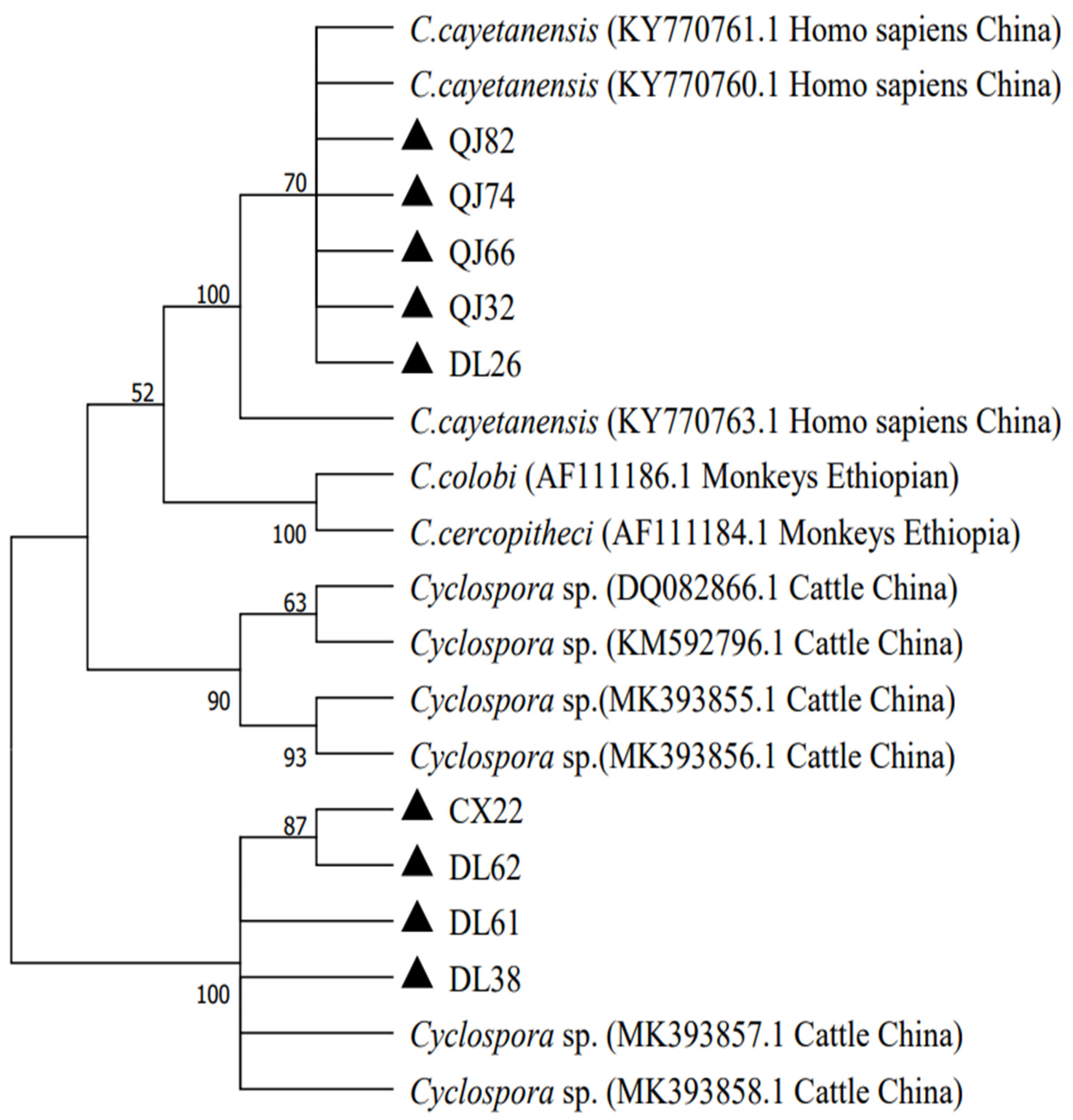
3.3. Phylogenetic Analyses

The PCR products that were successfully digested by the restriction enzyme Bsp E I were bidirectionally sequenced. In the present study, a total of 13 positive samples were successfully sequenced. The sequences of five PCR amplicons showed 99% similarities to the C. cayetanensis sequence in the GenBank database, and the other sequences were more similar to the Cyclospora spp. of the cattle. In the present study, we selected 4, 4, and 1 representative sequences from Dali, Qujing, and Chuxiong to construct phylogenetic relationships. In this study, the Cyclospora spp. were mainly divided into two branches. Four samples from Qujing and one sample from Dali were co-located with C. cayetanesis (KY 7707760) and formed a monophyletic group in the tree, while the other samples formed a separate clade with the Cyclospora spp. of the Bos frontalis in the Yunnan Province. The phylogenetic analysis result of genetic evolution analysis is shown in Figure 2.

4. Discussion

The traditional method for the detection of Cyclospora-like oocysts in animal fecal specimens is through microscopic observation [21]. The morphologies among Eimeria spp., Cyclospora spp., and Cryptosporidium spp. are similar. However, Cyclospora spp. and Cryptosporidium spp. can be distinguished from the size of oocysts and sporulation status in fresh feces. The Cyclospora spp. in fresh feces is not sporulated and is 8–10 μm in diameter, while Cryptosporidium spp. is sporulated and 4–6 μm in diameter. Additionally, modified Ziehl–Neelsen staining can also be used for distinguishing Cryptosporidium from Cyclospora [22]. Unfortunately, the morphology characters of Cyclospora spp. are far more similar to Eimeria spp. than those of Cryptosporidium spp. [4], and we can not distinguish Cyclospora spp. from Eimeria spp. by using microscopic examination. The SSU rRNA gene is highly conserved, and it has been a good genetic marker for distinguishing Cyclospora spp. from other apicomplexan protozoa [23,24]. However, previous studies showed that the primers used for amplifying the small subunit SSU rRNA genes of Cyclospora spp. can also amplify the small subunit SSU rRNAs of Eimeria spp. [23,25]. Restriction fragment length polymorphism (RFLP) analysis was then developed based on the nucleotide differences in the amplified region [26], and it has been a powerful tool in the classification and identification of parasite species, parasite epidemiology research, and genetic differentiation among closely related species. In 1995, Relman et al. [23] used restriction enzyme Mn II to distinguish Cyclospora spp. from Eimeria spp. In 1998, Jinneman KC et al. used the restriction endonuclease Mn II to distinguish Cyclospora spp. from Eimeria spp. [26]. Subsequently, a new PCR- RFLP was developed for ruminants. The restriction endonuclease Kpn I could distinguish the Cyclospora species from Eimeria species in ruminants, and the PCR amplicons of Eimeria spp. can be digested into 80 and 420 bp [20]. However, most of the time, 80 bp can not be clearly observed, and there are certain requirements for marker, agarose, and recognition ability. In this study, the restriction endonuclease Bsp E I was used for enzymatic cleavage, and the positive band of Cyclospora spp. was successfully cleaved into two parts 130 bp and 370 bp, while Eimeria spp. was not. The results of the present study indicate that PCR- RFLP, which was based on Bsp E I, can be an alternative method to distinguish Cyclospora spp. from Eimeria spp. of cattle. Whether this method can distinguish Cyclospora spp. from Eimeria spp. of the other animals remains to be determined.
In China, the research on Cyclospora spp. mainly focuses on human beings, non-human primates, crops and water sources. In 2002, Wang et al. [15] examined the feces of people in the Anhui province and found that the infection rate of C. cayetanensis in the Anhui province was 2.3%. Zhang et al. examined the fresh feces from diarrhea cases in 7 counties/cities in the Yunnan Province and found that infection rate of C. cayetanensis in diarrhea cases in the Yunnan Province was 3.97% [27]. In 2011, Zhou et al. studied the infection of C. cayetanensis in hospitalized patients in Henan and found that the prevalence rate ratio was 0.70% [16]. At present, Cyclospora spp. infection has been detected in non-human primates such as Macaca fascicularis, the primate Rhinopithecus roxellanae, and rhesus monkeys (Macaca mulatta) in China, and the prevalence of Cyclospora spp. in non-human primates ranged from 5.56 to 10.52% [11,19,28]. In addition, Cyclospora spp. was also detected in vegetables, fruits, and water in China. Li et al. found that the positive rate of Cyclospora spp. on the surface of vegetables and fruits in Henan province was 0.2% [29]. Fang et al. found that the Cyclospora contamination rate in wastewater treatment plants and sewer samples in Guangzhou city was 0.42% and 3.41%, respectively [30]. However, there are few studies on the Cyclospora spp. of bovine in China, and information on Cyclospora spp. infection in Holstein cattle in Yunnan is lacking. We studied the infection of Cyclospora spp. in Holstein cattle in Yunnan by performing molecular biology tests on fecal samples of Holstein cattle and genetic evolution analyses.
The total positive rate of Cyclospora spp. in Holstein cattle in the Yunnan Province was 2.48%, and the infection of Cyclospora spp. was detected in Dali, Chuxiong, and Qujing. The infection rates among Dali, Chuxiong, and Qujing were not significant (p > 0.05), suggesting that Cyclospora spp. infections were common in Yunnan and that regional factors did not show significant influences on the infection statuses of Cyclospora spp. Interestingly, Cyclospora spp. infection was not found in Kunming, which may be due to better feeding and environment management in Kunming. Basnett K examined the feces of Indian sheep for studying the infection of Cyclospora spp. and found a 2.14% prevalence of Cyclospora spp. infection in small ruminants [31], which was similar to the results of the present study. In this study, the rate of Cyclospora spp. infection at different ages ranged from 1.74% to 9.09%, and it was found that the rate of Cyclospora spp. infection gradually increased with age, the highest rate of Cyclospora spp. infection was found in Holstein cattle at 6–12 months of age, and the infection rate of Cyclospora spp. gradually decreased as age increased. However, the difference in the rates of Cyclospora spp. infection among different ages was not significant (p > 0.05).
In this study, the infection rate in male was 2.94% and the infection rate in female was 2.37%, and the difference in the infection rate of Cyclospora spp. between the sexes was not significant (p > 0.05), which indicated that the factor of sex had no significant effect on the infection of. Cyclospora spp. in Holstein cattle. Previous studies have suggested that cyclosporiasis is associated with environmental factors, such as season and local climate [7,32,33,34]. This could be due to the fact that Cyclospora oocysts need a sporulation process to become infective, and the oocyst sporulation rate depends on various environmental factors, such as temperature and humidity. Both a room temperature and humid environment is suited for the sporulation of Cyclospora oocysts to form infectious oocysts [35,36,37]. Thus, the infection of Cyclospora spp. is mostly seasonal, and the infection of Cyclospora spp. will increase significantly in the rainy season or summer [7,32,33,34,38,39,40].
An analysis of the 13 sequences of Cyclospora spp. found in this study revealed that five of them were closely related to C. cayetanensis samples isolated from humans in the Henan province (with a 99% similarity), whereas the remaining eight sequences were closely related to the Cyclospora spp. of bovines isolated from Guangzhou, Yunnan, and Henan. Cyclospora spp. can be spread through fruits and vegetables, as well as through water and soil, posing a threat to human life and health [13,36]. Current studies suggest that C. cayetanensis is the only known Cyclospora spp. that can infect humans [2,11]. Additionally, C. cayetanensis can be detected in the feces of other animals. The presence of C. cayetanensis oocysts in the feces of animals does not necessarily indicate infection. It may be due to the fact that animals can act as paratenic hosts, which can aid in the dissemination of oocysts into the environment [4,36].
Domestic animals are both reservoirs and sources of infection for zoonotic pathogens, and they are in frequent contact with breeders closely related to human activities. Infections with Cyclospora spp. could be a public health hazard and a potential threat to human health. Previous studies have detected the presence of Cyclospora spp. in Yunnan, but there has no investigation into the infection of Cyclospora spp. in Holstein cattle in Yunnan. In this study, Cyclospora spp. was detected in the feces of Holstein cattle, which indicated that there was a Cyclospora spp. infection in Holstein cattle in Yunnan. Most importantly, some of the bovine-derived Cyclospora spp. were closely related to human Cyclospora spp. This suggested that there is a potential zoonotic risk for Cyclospora spp. spread via the feces of Holstein cattle in Yunnan. At present, the risk of human infection due to the animal transmission of Cyclospora oocysts has not yet been evaluated [8]. Thus, the use of RFLP coupled with DNA sequencing to confirm the species of Cyclospora spp. in animals is of great importance for public health safety.

5. Conclusions

This is the first study to reveal the prevalence of Cyclospora spp. in Holstein cattle in the Yunnan Province, China (2.48%, 13/524). The infection of Cyclospora spp. is not statistically significant in different regions, sexes, and ages. In this study, our phylogenetic analysis showed that five samples were closely associated with C. cayetanensis in humans and located on the same branch with the C. cayetanensis zoonotic group, which indicated that the Cyclospora spp. infection in Holstein cattle in the Yunnan Province had the risk of zoonosis. It is necessary to strengthen the prevention and control of Cyclospora spp.

Supplementary Materials

The following supporting information can be downloaded at: https://www.mdpi.com/article/10.3390/ani13091527/s1, Figure S1: Original Western Blot Figure.

Author Contributions

H.-M.M. and F.-C.Z. designed the study; J.-F.Y. and Z.-J.H. made substantial contributions to the acquisition of the data and the analysis and interpretation of the data; J.-J.H. and F.-F.S. were involved in revising the content critically for important intellectual content; Y.-S.S. provided experimental samples. All authors have read and agreed to the published version of the manuscript.

Funding

This study was funded by the Scientific and Technological Mission of Dairy Cow Industry in Heqing County, Yunnan Province (Grant no. 202204BI090005); the Veterinary Public Health Innovation Team of the Yunnan Province (Grant no. 202105AE160014); and the Yunnan Expert Workstation (Grant no. 202005AF150041).

Institutional Review Board Statement

The collection of the animal fecal samples was approved by the farmers. The research protocol was reviewed and approved by the Research Ethics Committee of the Yunnan Agricultural University.

Informed Consent Statement

Not applicable.

Data Availability Statement

The data that support the findings are in the possession of the authors. The data of this study are available on request from the corresponding author.

Acknowledgments

The authors would like to thank the managers of the farms involved in this study for providing their assistance during the collection of fecal samples.

Conflicts of Interest

The authors declare no conflict of interest.

References

  1. Shields, J.M.; Olson, B.H. Cyclosporacayetanensis: A review of an emerging parasitic coccidian. Int. J. Parasitol. 2003, 33, 371–391. [Google Scholar] [CrossRef]
  2. Lainson, R. The genus Cyclospora (Apicomplexa: Eimeriidae), with a description of Cyclospora schneideri n.sp. in the snake Anilius scytale scytale (Aniliidae) from Amazonian Brazil—A review. Mem. Inst. Oswaldo Cruz 2005, 100, 103–110. [Google Scholar] [CrossRef]
  3. Insulander, M.; Svenungsson, B.; Lebbad, M.; Karlsson, L.; de Jong, B. A foodborne outbreak of Cyclospora infection in Stockholm, Sweden. Foodborne Pathog. Dis. 2010, 7, 1585–1587. [Google Scholar] [CrossRef]
  4. Ortega, Y.R.; Sanchez, R. Update on Cyclospora cayetanensis, a food-borne and waterborne parasite. Clin. Microbiol. Rev. 2010, 23, 218–234. [Google Scholar] [CrossRef]
  5. Ortega, Y.R.; Nagle, R.; Gilman, R.H.; Watanabe, J.; Miyagui, J.; Quispe, H.; Kanagusuku, P.; Roxas, C.; Sterling, C.R. Pathologic and clinical findings in patients with cyclosporiasis and a description of intracellular parasite life-cycle stages. J. Infect. Dis. 1997, 176, 1584–1589. [Google Scholar] [CrossRef]
  6. Hart, A.S.; Ridinger, M.T.; Soundarajan, R.; Peters, C.S.; Swiatlo, A.L.; Kocka, F.E. Novel organism associated with chronic diarrhoea in AIDS. Lancet 1990, 335, 169–170. [Google Scholar] [CrossRef]
  7. Chacin-Bonilla, L. Transmission of Cyclospora cayetanensis infection: A review focusing on soil-borne cyclosporiasis. Trans. R Soc. Trop. Med. Hyg. 2008, 102, 215–216. [Google Scholar] [CrossRef]
  8. Totton, S.C.; O’Connor, A.M.; Naganathan, T.; Martinez, B.A.F.; Sargeant, J.M. A review of Cyclospora cayetanensis in animals. Zoonoses Public Health 2021, 68, 861–867. [Google Scholar] [CrossRef]
  9. Long, E.G.; White, E.H.; Carmichael, W.W.; Quinlisk, P.M.; Raja, R.; Swisher, B.L.; Daugharty, H.; Cohen, M.T. Morphologic and staining characteristics of a cyanobacterium-like organism associated with diarrhea. J. Infect. Dis. 1991, 164, 199–202. [Google Scholar] [CrossRef]
  10. Onstad, N.H.; Miller, M.R.; Green, M.L.; Witola, W.H.; Davidson, P.C. A Review of Cyclospora cayetanensis Transport in the Environment. Trans. ASABE 2019, 62, 795–802. [Google Scholar] [CrossRef]
  11. Li, N.; Ye, J.; Arrowood, M.J.; Ma, J.; Wang, L.; Xu, H.; Feng, Y.; Xiao, L. Identification and morphologic and molecular characterization of Cyclospora macacae n. sp. from rhesus monkeys in China. Parasitol. Res. 2015, 114, 1811–1816. [Google Scholar] [CrossRef]
  12. Dubey, J.P.; Khan, A.; Rosenthal, B.M. Life Cycle and Transmission of Cyclospora cayetanensis: Knowns and Unknowns. Microorganisms 2022, 10, 118. [Google Scholar] [CrossRef]
  13. Onstad, N.H.; Beever, J.E.; Miller, M.R.; Green, M.L.; Witola, W.H.; Davidson, P.C. CyclosporaCayetanensis Presence in the Environment—A Case Study in the Chicago Metropolitan Area. Environments 2019, 6, 80. [Google Scholar] [CrossRef]
  14. Chu, D.M.; Sherchand, J.B.; Cross, J.H.; Orlandi, P.A. Detection of Cyclospora cayetanensis in animal fecal isolates from Nepal using an FTA filter-base polymerase chain reaction method. Am. J. Trop. Med. Hyg. 2004, 71, 373–379. [Google Scholar] [CrossRef]
  15. Wang, K.X.; Li, C.P.; Wang, J.; Tian, Y. Cyclospore cayetanensis in Anhui, China. World J. Gastroenterol. 2002, 8, 1144–1148. [Google Scholar] [CrossRef]
  16. Zhou, Y.; Lv, B.; Wang, Q.; Wang, R.; Jian, F.; Zhang, L.; Ning, C.; Fu, K.; Wang, Y.; Qi, M.; et al. Prevalence and molecular characterization of Cyclospora cayetanensis, Henan, China. Emerg. Infect. Dis. 2011, 17, 1887–1890. [Google Scholar] [CrossRef]
  17. Eberhard, M.L.; da Silva, A.J.; Lilley, B.G.; Pieniazek, N.J. Morphologic and molecular characterization of new Cyclospora species from Ethiopian monkeys: C. cercopitheci sp.n., C. colobi sp.n., and C. papionis sp.n. Emerg. Infect. Dis. 1999, 5, 651–658. [Google Scholar] [CrossRef]
  18. Lopez, F.A.; Manglicmot, J.; Schmidt, T.M.; Yeh, C.; Smith, H.V.; Relman, D.A. Molecular characterization of Cyclospora-like organisms from baboons. J. Infect. Dis. 1999, 179, 670–676. [Google Scholar] [CrossRef]
  19. Zhao, G.H.; Cong, M.M.; Bian, Q.Q.; Cheng, W.Y.; Wang, R.J.; Qi, M.; Zhang, L.X.; Lin, Q.; Zhu, X.Q. Molecular characterization of Cyclospora-like organisms from golden snub-nosed monkeys in Qinling Mountain in Shaanxi province, northwestern China. PLoS ONE 2013, 8, e58216. [Google Scholar] [CrossRef]
  20. Li, G.; Xiao, S.; Zhou, R.; Li, W.; Wadeh, H. Molecular characterization of Cyclospora-like organism from dairy cattle. Parasitol. Res. 2007, 100, 955–961. [Google Scholar] [CrossRef]
  21. Eberhard, M.L.; Pieniazek, N.J.; Arrowood, M.J. Laboratory diagnosis of Cyclospora infections. Arch. Pathol. Lab. Med. 1997, 121, 792–797. [Google Scholar] [PubMed]
  22. Khanna, V.; Tilak, K.; Ghosh, A.; Mukhopadhyay, C. Modified negative staining of heine for fast and inexpensive screening of Cryptosporidium, Cyclospora, and Cystoisospora spp. Int. Sch. Res. Not. 2014, 2014, 165424. [Google Scholar] [CrossRef]
  23. Relman, D.A.; Schmidt, T.M.; Gajadhar, A.; Sogin, M.; Cross, J.; Yoder, K.; Sethabutr, O.; Echeverria, P. Molecular phylogenetic analysis of Cyclospora, the human intestinal pathogen, suggests that it is closely related to Eimeria species. J. Infect. Dis. 1996, 173, 440–445. [Google Scholar] [CrossRef]
  24. Sulaiman, I.M.; Ortega, Y.; Simpson, S.; Kerdahi, K. Genetic characterization of human-pathogenic Cyclospora cayetanensis parasites from three endemic regions at the 18S ribosomal RNA locus. Infect. Genet. Evol. 2014, 22, 229–234. [Google Scholar] [CrossRef]
  25. Pieniazek, N.J.; Slemenda, S.B.; da Silva, A.J.; Alfano, E.M.; Arrowood, M.J. PCR confirmation of infection with Cyclospora cayetanensis. Emerg. Infect. Dis. 1996, 2, 357–359. [Google Scholar] [CrossRef]
  26. Jinneman, K.C.; Wetherington, J.H.; Hill, W.E.; Adams, A.M.; Johnson, J.M.; Tenge, B.J.; Dang, N.L.; Manger, R.L.; Wekell, M.M. Template preparation for PCR and RFLP of amplification products for the detection and identification of Cyclospora sp. and Eimeria spp. Oocysts directly from raspberries. J. Food Prot. 1998, 61, 1497–1503. [Google Scholar] [CrossRef]
  27. Zhang, B.X.; Yu, H.; Zhang, L.L.; Tao, H.; Li, Y.Z.; Li, Y.; Cao, Z.K.; Bai, Z.M.; He, Y.Q. Prevalence survey on Cyclospora cayetanensis and Cryptosporidium ssp. in diarrhea cases in Yunnan Province. Zhongguo Ji Sheng Chong Xue Yu Ji Sheng Chong Bing Za Zhi 2002, 20, 106–108. [Google Scholar]
  28. Ye, J.; Xiao, L.; Li, J.; Huang, W.; Amer, S.E.; Guo, Y.; Roellig, D.; Feng, Y. Occurrence of human-pathogenic Enterocytozoon bieneusi, Giardia duodenalis and Cryptosporidium genotypes in laboratory macaques in Guangxi, China. Parasitol. Int. 2014, 63, 132–137. [Google Scholar] [CrossRef]
  29. Li, J.; Shi, K.; Sun, F.; Li, T.; Wang, R.; Zhang, S.; Jian, F.; Ning, C.; Zhang, L. Identification of human pathogenic Enterocytozoon bieneusi, Cyclospora cayetanensis, and Cryptosporidium parvum on the surfaces of vegetables and fruits in Henan, China. Int. J. Food Microbiol. 2019, 307, 108292. [Google Scholar] [CrossRef]
  30. Fan, Y.; Wang, X.; Yang, R.; Zhao, W.; Li, N.; Guo, Y.; Xiao, L.; Feng, Y. Molecular characterization of the waterborne pathogens Cryptosporidium spp., Giardia duodenalis, Enterocytozoon bieneusi, Cyclospora cayetanensis and Eimeria spp. in wastewater and sewage in Guangzhou, China. Parasit. Vectors 2021, 14, 66. [Google Scholar] [CrossRef]
  31. Basnett, K.; Nagarajan, K.; Soundararajan, C.; Vairamuthu, S.; Rao, G.V.S. Morphological and molecular identification of Cyclospora species in sheep and goat at Tamil Nadu, India. J. Parasit. Dis. 2018, 42, 604–607. [Google Scholar] [CrossRef]
  32. Helmy, M.M. Cyclospora cayetanensis: A review, focusing on some of the remaining questions about cyclosporiasis. Infect. Disord. Drug. Targets 2010, 10, 368–375. [Google Scholar] [CrossRef]
  33. Li, J.; Wang, R.; Chen, Y.; Xiao, L.; Zhang, L. Cyclospora cayetanensis infection in humans: Biological characteristics, clinical features, epidemiology, detection method and treatment. Parasitology 2020, 147, 160–170. [Google Scholar] [CrossRef]
  34. Frickmann, H.; Alker, J.; Hansen, J.; Dib, J.C.; Aristizabal, A.; Concha, G.; Schotte, U.; Kann, S. Seasonal Differences in Cyclospora cayetanensis Prevalence in Colombian Indigenous People. Microorganisms 2021, 9, 627. [Google Scholar] [CrossRef]
  35. Ortega, Y.R.; Sterling, C.R.; Gilman, R.H. Cyclospora cayetanensis. Adv. Parasitol. 1998, 40, 399–418. [Google Scholar] [CrossRef]
  36. Almeria, S.; Cinar, H.N.; Dubey, J.P. Cyclospora cayetanensis and Cyclosporiasis: An Update. Microorganisms 2019, 7, 317. [Google Scholar] [CrossRef]
  37. Connor, B.A. Cyclospora infection: A review. Ann. Acad. Med. Singap. 1997, 26, 632–636. [Google Scholar]
  38. Hall, R.L.; Jones, J.L.; Hurd, S.; Smith, G.; Mahon, B.E.; Herwaldt, B.L. Population-based active surveillance for Cyclospora infection--United States, Foodborne Diseases Active Surveillance Network (FoodNet), 1997–2009. Clin. Infect. Dis. 2012, 54 (Suppl. S5), S411–S417. [Google Scholar] [CrossRef]
  39. Kaminsky, R.G.; Lagos, J.; Santos, G.R.; Urrutia, S. Marked seasonality of Cyclospora cayetanensis infections: Ten-year observation of hospital cases, Honduras. BMC Infect. Dis. 2016, 16, 66. [Google Scholar] [CrossRef]
  40. Jiang, Y.; Yuan, Z.; Zang, G.; Li, D.; Wang, Y.; Zhang, Y.; Liu, H.; Cao, J.; Shen, Y. Cyclospora cayetanensis infections among diarrheal outpatients in Shanghai: A retrospective case study. Front. Med. 2018, 12, 98–103. [Google Scholar] [CrossRef]
Figure 1. Partial SSU rRNA gene amplicons digested with restriction endonuclease Bsp E I. Lane M: DL 2000 DNA Marker; lanes 1–4: Bsp E I digested products from nested PCR. The original Western Blot Figure is Figure S1.
Animals 13 01527 g001
Figure 2. Phylogenetic relationship of Cyclospora spp. of Holstein cattle in the Yunnan Province. The genotypes identified in this study are marked with ▲.
Animals 13 01527 g002
Table 1. The infection of Cyclospora spp. in Holstein cattle in partial areas of the Yunnan Province.
FactorsCategoryNo. TestedNo. PositiveInfection Rate (%) (95%CI)OR (95%, CI)p-Value
RegionDali27462.19 [0.45–3.93]Reference0.339
Kunmin5700 [0.00–0.00]-
Qujing15853.16 [0.41–5.92]1.46 (0.44–4.86)
Chuxiong3525.71 [0.00–13.80]2.71 (0.53–13.96)
Age (M)0–334461.74 [0.35–3.13]Reference0.204
3–65735.26 [0.00–11.24]3.13 (0.76–12.89)
6–121119.09 [0.00–29.35]5.63 (0.62–51.27)
>1211232.68 [0.00–5.72]1.55 (0.38–6.30)
SexFemale422102.37 [0.91–3.82]Reference0.739
Male10232.94 [0.00–6.28]1.25 (0.34–4.62)
Total524132.48--
No.: number; CI: confidence interval; OR: odds ratio.
Disclaimer/Publisher’s Note: The statements, opinions and data contained in all publications are solely those of the individual author(s) and contributor(s) and not of MDPI and/or the editor(s). MDPI and/or the editor(s) disclaim responsibility for any injury to people or property resulting from any ideas, methods, instructions or products referred to in the content.

