Simple Summary
In mammals, autophagy plays a fundamental role in the defense against intracellular pathogens; however, in fish, this noncanonical function has not been described. In this context, it was proposed to study whether autophagy was modulated/activated in muscle cells challenged with a bacterial pathogen. Muscle cell cultures were performed and challenged with Piscirickettsia salmonis, the main threat to the salmon industry. Genes associated with immune response and autophagy were evaluated. In addition, the protein content of the LC3-II-specific marker of the autophagic process was evaluated via Western blot. Additionally, genes associated with vesicular traffic and endocytosis were evaluated, finding that P. salmonis promotes these processes. The results show a concomitant modulation of the genes associated with the immune response, vesicular trafficking, and autophagy, suggesting an early intracellular response by the muscle cell against this bacterium. Due to the necessity of seeking and discovering new alternatives and strategies to fight intracellular pathogens in the salmon industry, a better understanding of how autophagy participates in immune system responses may lead to the development of new technologies that allow for the effective control of intracellular pathogens, improving animal welfare and contributing to the sustainability of the global fish industry.
Abstract
Autophagy is a fundamental cellular process implicated in the health of the cell, acting as a cytoplasmatic quality control machinery by self-eating unfunctional organelles and protein aggregates. In mammals, autophagy can participate in the clearance of intracellular pathogens from the cell, and the activity of the toll-like receptors mediates its activation. However, in fish, the modulation of autophagy by these receptors in the muscle is unknown. This study describes and characterizes autophagic modulation during the immune response of fish muscle cells after a challenge with intracellular pathogen Piscirickettsia salmonis. For this, primary cultures of muscle cells were challenged with P. salmonis, and the expressions of immune markers il-1β, tnfα, il-8, hepcidin, tlr3, tlr9, mhc-I and mhc-II were analyzed through RT-qPCR. The expressions of several genes involved in autophagy (becn1, atg9, atg5, atg12, lc3, gabarap and atg4) were also evaluated with RT-qPCR to understand the autophagic modulation during an immune response. In addition, LC3-II protein content was measured via Western blot. The challenge of trout muscle cells with P. salmonis triggered a concomitant immune response to the activation of the autophagic process, suggesting a close relationship between these two processes.
1. Introduction
Autophagy is a highly conserved catabolic process essential for cellular homeostasis, degrading cytosolic macromolecules and damaged organelles to guarantee energy supply and nutrient availability for the cell. Furthermore, autophagy can modulate innate and adaptative immunity, contributing to the clearance of pathogens from the cell [1]. Under normal conditions, autophagy occurs at low levels in almost all cells, but is strongly induced under nutrient deprivation, amino acid starvation, cellular stress, cytokine exposure and pathogen infections [2,3]. Additionally, autophagy can act through a noncanonical pathway in antigen processing and presentation for MHC-I and MHC-II, restricting intracellular pathogens and regulating adaptive immunity [4]. At the molecular level, the autophagic pathway involves the concerted action of evolutionarily conserved ATG gene products involved in the initiation (atg9, atg13 and becn1), autophagosome formation (atg5, atg12 and atg16), the elongation of autophagic processes (atg7 and atg4), and autophagosome closure (gabarap and lc3) [5]. Autophagy is triggered by activating a regulatory protein complex that recruits microtubule-associated light chain protein 3 (LC3) to the nascent autophagosome [6]. The conversion of LC3 into its active form, LC3-II, through conjugation to phosphatidylethanolamine (PE), allows for the transformation of the membrane into a closed double-membrane system [7]. Lastly, the autophagosome binds to the endosomal or lysosomal compartment, forming the autolysosome where organelles and macromolecules are degraded by a pool of proteolytic enzymes [8]. In mammals, autophagic activation is associated with immune signals and molecular pathways, involving toll-like receptor (TLR) activation through different pathogen-associated molecular patterns (PAMPs) [9]. For instance, autophagy activated by the endosomal TLR signaling pathway in macrophages plays a key role as a mechanism for Leishmania major infection resistance [10].
Mammalian skeletal muscle, a nonclassical immune tissue, possesses several membrane-bound TLRs with the ability to detect different PAMPs, triggering signaling pathways to activate autophagy [11]. These findings associate TLR signaling and autophagy, providing a potential molecular mechanism for the induction of autophagy in response to pathogen invasion. Therefore, the ability of TLR ligands to stimulate autophagy may be used to treat intracellular pathogens in mammals [12]. Moreover, several proinflammatory cytokines, such as IFN-γ, TNF-α, IL-1, IL-2, IL-6, and TGF-β induce autophagy, while anti-inflammatory cytokines such as IL-4, IL-10, and IL-13 display an inhibitory effect [13]. All these data suggest a close relation between the immune response and autophagy in mammalian skeletal muscle. However, this interaction is unknown in fish, and only a few reports related to autophagy exist. Nevertheless, due to the direct relation of autophagy and growth, a key parameter for fish-industry productivity, autophagic markers have been studied under different diets and nutritional trials. Results showed a direct association between fish’s energy requirement and the activation of autophagy [14]. For instance, the autophagic flux and the overexpression of autophagic genes atg4, atg9, atg12, lc3, gabarap and becn1 was observed in vitro in epithelial gill cell line RT-gill-W1 of rainbow trout under nutrient restriction, and in the muscle of trout challenged with Flavobacterium psychrophilum [15,16]. Nevertheless, the modulation of autophagy in fish muscle cells during an immune-like response against an intracellular pathogen has not been elucidated.
This study aims to describe and characterize autophagic modulation through the immune response of fish muscle cells after a challenge with a critical bacterial pathogen for the salmon industry. Through the immune challenge of primary cultures of muscle cells with Piscirickettsia salmonis, a fastidious intracellular pathogen, we revealed the transcript upregulation and protein production of several immune markers and genes involved in autophagy. Results obtained in the present work give new insight into the role of autophagy in the immune response against bacterial pathogens in muscle cells.
2. Materials and Methods
2.1. Ethics Statement
The present research was approved by the bioethics committee of Pontificia Universidad Católica de Valparaíso (PUCV) BIOEPUCV-B 334-2020 and the Agencia Nacional de Investigación y Desarrollo (ANID) of the Chilean government, and adhered to all animal welfare procedures.
2.2. Fish Husbandry
In total, 24 rainbow trout (Oncorhynchus mykiss) with body weight of 10 ± 1.7 g and body length of 10 ± 1.5 cm were obtained from the Río Blanco Aquaculture Center (Valparaíso, Chile). These fish were kept in 3 tanks at a density of 8 fish per 25-L tank, using 1 tank per trial. Fish were maintained at a temperature of 16 °C, fed daily with commercial feed, and provided with a photoperiod of 13 h of light and 11 h of darkness.
2.3. Primary Culture of Muscle Cells
Muscular cells were prepared from 6-8 g of the skeletal muscle of four rainbow trout as previously reported [17]. Briefly, the dorsal white muscle was collected and disintegrated in Dulbecco’s Modified Eagle’s Medium (DMEM) containing 9 mM NaHCO3, 20 mM HEPES, 10% horse serum at pH 7.4, 100 U/mL penicillin, and 100 µg/mL streptomycin (Sigma, St. Luis, MO, USA). After mechanical dissociation, the muscle was washed with DMEM and then digested with 0.2% collagenase solution in DMEM for 1 h at 18 °C. The suspension was centrifuged at 500 g for 5 min at 15 °C, and the resulting pellet was digested with a 0.1% trypsin solution in DMEM for 30 min at 18 °C. Trypsin was deactivated with complete medium (DMEM containing 10% fetal bovine serum, 100 U/mL penicillin, and 100 µg/mL streptomycin at pH 7.4), the suspension was filtered and centrifuged at 5000× g for 10 min at 18 °C, and 5 mL of DMEM was lastly added to the resulting pellet. The cell suspension was collected to count cells and evaluate viability with Trypan Blue 0.2%.
Cells were seeded at a density of 5 × 10⁵ per well in plates previously treated with poly-L-lysine (2 μg/cm2) and laminin (20 μg/mL). The cells were incubated at 18 °C for 14 days, 7 days in a proliferation medium containing DMEM, 9 mM NaHCO3, 20 mM HEPES, 10% fetal bovine serum, 100 U/mL penicillin, and 100 µg/mL streptomycin at pH 7.4, and 7 days in a differentiation medium composed of DMEM (9 mM NaHCO3, 20 mM HEPES, 100 U/mL penicillin, and 10 mg/mL streptomycin). This procedure was repeated three times (n = 3) per experimental trial described in the next sections.
2.4. Piscirickettsia salmonis Culture and Infection Protocol
A P. salmonis field isolate, Psal-104a, was obtained from the Chilean National Piscirickettsia salmonis Strain Collection at Pontificia Universidad Católica de Valparaíso (PUCV). The bacteria were cultured in basal medium (BM) broth composed of yeast extract (Merck, St. Louis, USA) 2.0 g L−1, peptone from meat (peptic digested, Merck) 2.0 g L−1, (NH4)2SO4 1.32 g L−1, MgSO4·7H2O 0.1 g L−1, K2HPO4 6.3 g L−1, NaCl 9.0 g L−1, CaCl2·2H2O 0.08 g L−1, FeSO4·7H2O 0.02 g L−1, and glucose 5 g L−1 at 18 °C at 100 rpm.
Exponentially growing P. salmonis (O.D. 600 = 0.3) was recovered via centrifugation at 5000× g, washed three times with saline-buffered solution (SBS: 0.15 M NaCl, 7.3 mM KH2PO4, 11.5 mM K2HPO4, pH 6.0) supplemented with 4% lactose, and resuspended in DMEM containing 9 mM NaHCO3, 20 mM HEPES, at pH 7.4. Bacterial cells were counted using a Petroff–Hausser chamber. Muscular cells were infected at a multiplicity of infection (MOI) of 10 for 4, 6, 8, and, 24 h. After the experimental times, the pathogen was removed and then sampled. As a positive control for autophagic activation, muscle cells were treated with rapamycin (50 nm), and DMEM was used as a vehicle for control, sampled at the beginning of the experiment (0 hpi).
2.5. Quantitative Real Time PCR (qPCR)
The total RNA from the primary culture of skeletal muscle was extracted according to the manufacturer’s guidelines for E.Z.N.A total RNA Kit I (Omega Bio-tek, Norcross, GA, USA), and quantified with NanoDrop LITE spectrophotometer (Thermo Scientific, Rockford, IL, USA). Residual genomic DNA was removed, and 1 µg of RNA was used for cDNA synthesis via RevertAid First strain cDNA Synthesis Kit (Thermo Scientific, Rockford, IL, USA) according to the manufacturer’s protocol. Real-time quantitative PCR (qPCR) assays were performed in a AriaMx Real-Time PCR System (Agilent Technologies, Santa Clara, CA, USA) with 15 µL volume reactions, of which 1 µL was cDNA (a 2-fold dilution), 0.2 μl of each primer (250 nM), and 8.8 µL of Takyon master mix (Eurogentec, Seraing, Belgium). The primers used in this study were designed and validated in our laboratory, and the sequence and efficiency of each primer are listed in Supplementary Table S1. Thermal cycling conditions were as follows: initial activation 95 °C for 10 min, 40 cycles of 30 s denaturation at 95 °C, 30 s annealing at 60 °C, and 30 s elongation at 72 °C, and each sample was loaded in duplicate. Relative expression analysis was conducted using geNorm software (https://genorm.cmgg.be/ (accessed on 30 March 2021)), and the results are expressed as fold changes using EF-1α and b-actin as housekeeping reference genes.
2.6. Western Blot
Total protein was extracted from primary culture cells in 150 µL Piercetm RIPA buffer (Thermo) supplemented with 8 mM EDTA, PMSF 1 mM, and a protease inhibitor cocktail (Calbiochem, San Diego, CA, USA), centrifuged at 12,000× g, and solubilized at 4 °C. Protein concentration was determined using a Pierce BCA Protein Assay Kit (Thermo Scientific, Hanover Park, IL, USA). The total protein (10 µg) was loaded into each line, separated with 18% SDS-PAGE, transferred to a nitrocellulose membrane, and blocked for 1 h at 37 °C in Tris-buffered saline (TBS-Tween) with 3% BSA. Primary antibody incubations (LC3A/B (D3U4C) XP® Rabbit mAb #12741, Actine (INVITROGEN), and ant-Rab7a, diluted 1:1000, 1:2000 and 1:500, respectively) were performed overnight at 4 °C. The anti-Rab7a polyclonal antibody was developed in our lab. Briefly, a recombinant Rab7a was used for specific antibody production (Supplementary Figure S1). CF-1 mice (5 weeks old) were subcutaneously injected at 1, 14, and 28 days with 30 μg of peptide diluted 1:1 in FIS, as a T helper cell activator, and in 1:1 Freund’s adjuvant (Thermo Scientific). The antiserum was collected on Day 42 and centrifuged at 700 g for 10 min, and the supernatant was stored at −20 °C. Antibody validation was determined via Western blot (Supplementary Figure S1). Membranes were washed with 1X TBS and incubated for 1 h at room temperature with the appropriate secondary antibody. After washing, the membranes were visualized using commercial kit WESTAR SUPERNOVA from Cyanagen. Then, the membrane was visualized with the ChemiDoc Imaging Systems Bio-Rad system (Bio-Rad Laboratories, Hercules, CA, USA). Digitized images were used for the densitometric analyses of the bands in Image J software(National Institutes of Health, Bethesda, Maryland, USA). Differences are expressed as fold changes in protein content. Western blots were performed for all individual samples per experimental treatment; however, only one representative blot film is shown in the figures (Supplementary Figures S2–S4).
2.7. Immunofluorescence
Adherent cells were washed with 1X PBS and fixed with 4% paraformaldehyde (PFA) (in 1x PBS) for 10 min and permeabilized/blocked with 3% BSA with 0.3% Triton in 1X TBS for 30 min, and the quenching solution (50 mM ammonium chloride) was added for 10 min. Then, the cells were incubated overnight with the anti-LC3 antibody diluted at 1:100 (LC3A/B (D3U4C) XP® Rabbit mAb #12741 in 1% BSA at room temperature. Samples were washed with 1X PBS and 0.02% TBST, and incubated with a commercial secondary antibody (1:750) (Invitrogen, Thermo Scientific) for 90 min at room temperature. Nuclear staining was performed with DAPI (Vector Laboratories) for conventional fluorescence microscopy, following the manufacturer’s instructions. For image capture, the samples were placed in a Leica CTR5000fluorescence microscope (Leica Microsystems, Wetzlar, Germany). Western blots were analyzed using the LC3-II/actin and Rab7a/actin ratios.
2.8. Statistical Analysis
All generated data were evaluated with one-way ANOVA and Student’s t test. Data are expressed as the mean (n = 3 per treatment) ± standard error of the mean (SEM), and p < 0.05 was considered statistically significant. All analyses were performed using general linear models on Graph Prism 7.0 software (GraphPad Company, Boston, MA., USA).
3. Results
3.1. Immune-like Response of Skeletal Muscle Cells after P. salmonis Challenge
To understand and characterize the response of muscle cells against bacterial pathogens, rainbow-trout skeletal muscle cells were challenged with P. salmonis for 6, 8, and 24 h. Next, immune molecular markers tlr3, tlr9, il-1β, tnfα, il-8, hepcidin, mhc-I and mhc-II were measured with RT-qPCR, and results showed a concomitant overexpression of toll-like receptors 3 and 9 after 6 h of challenge (Figure 1A). Proinflammatory cytokines il-1β, tnfα and il-8 were modulated compared to the noninduced controls, where tnfα and il-8 were upregulated significantly after 6 and 8 h after the challenge, respectively (Figure 1B,C). Antimicrobial peptide hepcidin increased after 6, 8, and 24 h after the challenge, but no statistical significance was observed (Figure 1C). No changes were observed in the mRNA levels of mhc-I and mhc-II during the entire trial (Figure 1D).
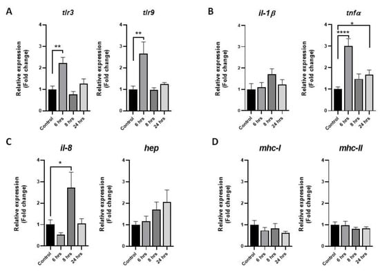
Figure 1.
Expression of immune molecular markers in rainbow-trout muscle cells after Piscirickettsia salmonis challenge. Primary cultures of fish skeletal muscle were challenge with P. salmonis (MOI 10) for 6, 8, and 24 h. As control group, cells were treated with a vehicle (DMEM + 10% FBS). (A) Toll-like receptors 3 and 9. (B) Proinflammatory cytokines il-1β and tnfα. (C) il-8 and the antimicrobial peptide hepcidin. (D) Major complex of histocompatibility I and II (mhc-I and mhc-II). qPCR analyses were normalized with the ef-1α and β-actin genes, and the results are presented as means ± SEMs of triplicates from three independent experiments, using fold change compared to the control values (n = 3). The difference in the transcript levels is denoted by asterisks (*) considering a p-value < 0.05 (*), p-value < 0,01 (**) and p-value < 0.001 (****) significant.
3.2. Autophagic Modulation in Muscle Cells after P. salmonis Challenge
To describe the modulation of autophagy in muscle cells after bacterial challenge, autophagic markers were analyzed via RT-qPCR, Western blot, and immunofluorescence. Rapamycin was used as the positive control of autophagic activation. Autophagic genes associated with the induction of this process were upregulated, particularly becn1, of which the mRNA level was significantly increased after 6 and 24 h of the challenge (Figure 2A). In the case of autophagosome formation genes, the overexpression of atg5 was observed after 6 h of treatment, while the transcript level of atg12 was downregulated after 8 h (Figure 2B). The mRNA levels of atg4, an elongation of an autophagosome molecular marker, was downregulated after 8 h of challenge (Figure 2C). Lastly, the transcript level of lc3 was significantly downregulated after 8 h of challenge (Figure 2D).
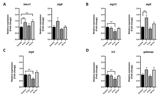
Figure 2.
Effect of P. salmonis challenge on the expression of autophagic genes in rainbow-trout muscle cells. Primary cultures of fish skeletal muscle were infected with P. salmonis at MOI 10 for 6, 8, and 24 h. As the control, cells without bacteria were sampled at the beginning of the trial (time 0). (A) Autophagic genes becn1 and atg9 involved in the induction of this process. (B) Autophagosome formation genes atg12 and atg5. (C) Gene atg4, involved in the elongation of the autophagosome. (D) Vesicle-completion-associated genes gabarap and lc3. The data are presented as means ± SEMs of triplicates from three independent experiments, using fold change compared to the values of the time 0 group (n = 3). qPCR analyses were normalized with the ef-1α and β-actin genes. The difference in transcript levels is denoted by asterisks (**) when the main effects were significant (p < 0.01).
At the protein level, protein autophagic marker LC3-I/LC3-II was evaluated via immunofluorescence and Western blot. Agreeing with the gene expression of autophagic induction gene analysis, the protein content of LC3 showed an increase in fluorescence signal in the muscular cells challenged with P. salmonis compared with the control group at 6 h after challenge (Figure 3A). An increase in the LC3-II protein content was observed through Western blot analysis after 6 h of stimuli (Figure 3B). In addition, the mRNA levels of small GTPase proteins rab5a and rab7a were upregulated at 6 h after challenge, while the transcript level of rab11a was increased after 24 h (Figure 3C). Lastly, the protein content of Rab7 showed an increasing trend through Western blot (Figure 3D).
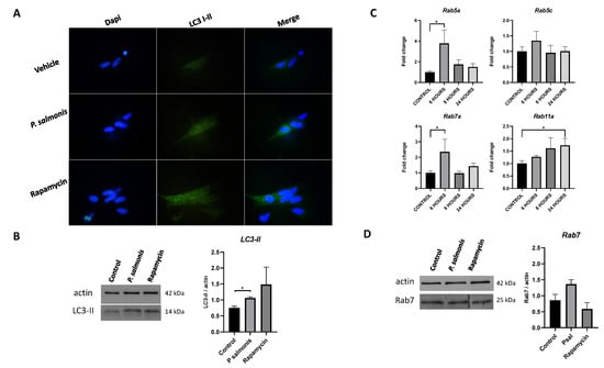
Figure 3.
Effects of P. salmonis on the level of autophagy and Rab protein markers in muscle cells challenged. Fish skeletal muscle cells were cultured and challenge with P. salmonis at MOI 10 (resuspended in differentiation medium) for 6 h. (A) Immunofluorescence of LC3 (LC3-I and LC3-II). (B) Representative Western blot of LC3-II. (C) Rab small GTPases gene expression and, (D) Representative Western blot of Rab7a. The data are presented as the means ± SEMs of triplicates from three independent experiments, using fold change compared to the control values (untreated cells, n = 3). The measure of actin was used as load control for Western blots. qPCR analyses were normalized by the ef-1α and β-actin genes. The difference between the control group and treated groups is denoted by asterisks (*) when the main effects were significant (p < 0.05).
4. Discussion
In mammals, the ability of muscles to respond and participate in the immune response against pathogens and in autoimmune events is well-characterized [18]. This ability is represented by the immunocompetent attributes of muscle cells, such as the expression of costimulatory molecules, antigen-presenting machinery, proinflammatory cytokines, and pathogen recognition receptors [19,20,21,22,23]. Moreover, the interaction between muscle cells and resident immune cells is an essential step for wound healing and damage regeneration in this tissue [24,25].
In fish, the skeletal muscle appears as an active immunological organ, where immune reactions occur after an in vivo challenge with bacterial pathogens [26,27]. However, the ability of a muscle cell to express classical immune-like molecules by itself is still under study, and only few reports address how this type of cells may respond against PAMPs and pathogens [28,29]. For example, rainbow-trout myotubes responded against Piscirickettsia salmonis through the upregulation of tlr1, tlr22, and il-8 at 8 h after a challenge [17]. In the present study, the immune response against P. salmonis generated a concomitant overexpression of tnfα, tlr3, and tlr9 after 6 h of challenge. Toll-like receptors 3 and 9 are associated with an intracellular/antiviral response where these receptors can detect double-strand RNA and CpG DNA, respectively [30]. Reports in mammals showed how these receptors can be involved in diminishing the growth of intracellular bacterium Salmonella typhimurium [31]. Salmon head kidney cells (SHK-1) respond to an infection with P. salmonis through the overexpression of tlr1 and tlr5s [32]. Furthermore, P. salmonis could induce a flagellin-dependent TLR5 activation in Atlantic salmon, which resulted in the upregulation of proinflammatory genes [33]. The overexpression of intracellular receptors suggests that intracellular defense mechanisms could be activated in response to P. salmonis infections. Despite the overexpression of markers associated with an intracellular response, the RNA levels of molecular markers DotA and DotB of P. salmonis were not detected via RT-qPCR (data not shown), which could indicate that, even though the bacterium does not enter the cell, the intracellular defense mechanisms are stimulated. On the other hand, proinflammatory cytokine tnfα represents an early response of muscular cells against bacterial pathogens. Indeed, an increase in NF-κB and TNFα at 48 h after a challenge against Vibrio anguillarum was observed on fish muscle [34]. Additionally, at 8 h after a challenge, the mRNA level of il-8 was upregulated. In higher vertebrates, TNFα induces the expression of IL-8 [35]. This kind of response may be associated with the need for muscle cells to recruit immune cells to the site of infection, since IL-8 is a chemoattractant for leukocytes [36]. These results contribute to existing data confirming that the muscle cell responds to a bacterial challenge by itself. However, communication with the immune resident’s cells is needed to deploy an effective and robust response.
In mammals, the participation of autophagy in the immune response is well-characterized, where autophagy-related gene (ATG) proteins mediate direct pathogen degradation, inflammation restriction, antigen presentation on MHC molecules and survival of memory lymphocyte populations [37]. This process has mainly been studied for its role in the internal quality control of the cell, and for its implication in metabolism and cancer. However, autophagy can participate through a noncanonical pathway to respond against other dangerous stimuli, such as pathogen infection [38]. In fish, the modulation of this process in a pathological context is unclear, and only a few reports show how autophagy is modulated by nutrient restriction and pharmacological treatments [14,39].
Transgenic zebrafish and zebrafish cell lines were used as research tools on the autophagic regulation process, highlighting its role on the protection against pathogen infection, development, and lipid degradation [14]. Autophagy was also linked to development and extracellular matrix formation in zebrafish [40]. Recently, the modulation of autophagic genes atg4, becn1, atg7, lc3, atg12, and gabarap has been evaluated in rainbow-trout liver and muscle under different functional treatments, fasting, and a posteriori F. psychrophilum infection, suggesting a possible role of this process in the resistance against pathogens [16]. In the present work, autophagic genes were up- and downregulated in a time-dependent and coordinated way. The upregulation of becn1 was observed at 6 h after challenge; becn1 is a protein that facilitates the de novo formation of the phagophore, and is critical for the autophagic initiation in embryogenesis and antiviral responses in fish [41,42]. The next step in autophagy is autophagosome formation, where molecules atg5 and atg12 are directly involved [43]. We observed the upregulation of atg5 at 6 h after a challenge with the subsequent downregulation of atg12 after 8 h. Both genes are associated with intracellular immune response against viruses in fish, where atg5 plays crucial roles in viral replication via promoting autophagy in orange-spotted grouper [44], while atg12 can induce both autophagy and the Type I IFN response in large yellow croaker [45]. These two molecules are regulated by RAB5, a small GTPase, which facilitates the early formation of the autophagosome [46]. Six hours after a challenge with P. salmonis, the mRNA levels of rab5a and rab7 increased significantly, suggesting the activation of vesicle traffic in response to the bacteria. In mammals, Rab7a is associated with the endocytic pathway [47]; in fish, this molecule is related with an antiviral response [48], suggesting the activation of different intracellular mechanisms in response to P. salmonis. Lastly, the genes involved in the elongation and closure of the autophagosome were downregulated after 8 h after challenge, particularly atg4 and lc3. Altogether, the present data suggest a tight relation between autophagy and intracellular innate immune response, and are consistent with the used intracellular bacterial pathogen and the evaluated immune markers in our study.
A crosstalk between the immune response and autophagy has been reported in mammals. For example, TLRs were directly involved with autophagic activation after TLR ligand stimulations with poly I:C and LPS, among other PAMPs [9]. In addition, the stimulation of TLR7 with different ligands activated autophagy, eliminating intracellular microorganisms even when the target pathogen had not been canonically associated with TLR7 signaling [12]. In this context, the recognized ability of TLR ligands to stimulate autophagy was proposed as a prophylactic alternative to treat intracellular pathogens [12]. In the present study, tlr3, tlr9, tnfα and autophagic genes becn1 and atg5 were upregulated at the same experimental time (6 h after challenge), suggesting a relation between the autophagy and immune response of muscle cells against P. salmonis. This relation was confirmed in mammals where TNFα and other cytokines could induce autophagy [13]. Lastly, to corroborate the activation of autophagy in rainbow-trout muscle cells, we assessed the protein level of LC3-II, a classical protein marker for autophagy. Finding higher protein content of LC3-II in the treated cells added to the presented overall results and the background information, suggesting that the muscle cell deploys an immunelike response where autophagy may play an important role for the clearance of the pathogen. Deeper functional analysis and characterization are required to corroborate the dialogue and regulation between the two essential cellular functions.
5. Conclusions
The skeletal muscle of fish is a fundamental energy source for the organism and a recurrent site of infection where immunelike responses against different bacterial pathogens occur. The present study described for the first time the modulation of the immune response and its possible connection with autophagy in rainbow-trout muscle cells. The challenge of trout muscle cells with P. salmonis, an intracellular pathogen, triggered an immune response concomitant to the activation of the autophagic process. Results suggest an early response by the muscle cells against the bacteria just after only 6 h of challenge, where both immune and autophagic markers were upregulated. Considering the obtained data and the available research information, two important questions arise regarding the possible implication of P. salmonis challenge in fish muscle: (1) is autophagy acting as an intracellular immune mechanism to fight this pathogen or is the bacterium using this mechanism to enter the cell and evade the immune response? These questions are relevant and represent a new line of research that must be answered. Due to the necessity of seeking and discovering new alternatives and strategies to fight intracellular pathogens in the salmon industry, a better understanding of how autophagy participates in immune system responses may lead to the development of new technologies that allow for the effective control of intracellular pathogens, improving animal welfare and contributing to the sustainability of the global fish industry.
Supplementary Materials
The following supporting information can be downloaded at: https://www.mdpi.com/article/10.3390/ani13050880/s1. Figure S1: characterization of Rab7a from O. mykiss.; Table S1: primers used for qPCR. Supplementary Figure S2. Original Western blot of LC3-II in skeletal muscle cells after 6 h of challenge with P. salmonis. Supplementary Figure S3. Original Western blot of Rab7a in muscle cells after 6 h of challenge with P. salmonis. Original Western blots used for the analysis and representative blot composition (Figure S4).
Author Contributions
Conceptualization, C.A.V.; methodology, C.A.V., C.A.Á., N.O. and M.A.; formal analysis, C.A.V.; investigation, C.A.V.; resources, C.A.V. and L.M.; data curation, C.A.V.; writing—original draft preparation, C.A.V., P.S. and L.M.; writing—review and editing, C.A.V.; visualization, C.A.V.; supervision, C.A.V.; project administration, C.A.V.; funding acquisition, C.A.V. and L.M. All authors have read and agreed to the published version of the manuscript.
Funding
This research was funded by Fondo Nacional de Desarrollo Científico y Tecnológico FONDECYT, postdoctoral grant number 3200172, and FONDECYT, regular grant number 1191763.
Institutional Review Board Statement
The study was approved by the bioethics committee of Pontificia Universidad Católica de Valparaíso (PUCV) BIOEPUCV-B 334-2020.
Informed Consent Statement
Not applicable.
Data Availability Statement
Not applicable.
Acknowledgments
The authors would like to thank German Olivares from the Piscicultura de Río Blanco, Los Andes, Chile for providing the fish for the experiments.
Conflicts of Interest
The authors declare no conflict of interest.
References
- Jagannath, C.; McBride, J.W.; Vergne, I. The autophagy pathway: Bacterial pathogen immunity and evasion. Front. Immunol. 2021, 12, 768935. [Google Scholar] [CrossRef] [PubMed]
- Mizushima, N.; Levine, B.; Cuervo, A.M.; Klionsky, D.J. Autophagy fights disease through cellular selfdigestion. Nature 2008, 451, 1069–1075. [Google Scholar] [CrossRef] [PubMed]
- Belghit, I.; Panserat, S.; Sadoul, B.; Dias, K.; Skiba-Cassy, S.; Seiliez, I. Macronutrient composition of the diet affects the feeding-mediated down regulation of autophagy in muscle of Rainbow Trout (O. mykiss). PLoS ONE 2013, 8, e74308. [Google Scholar] [CrossRef]
- Schmid, D.; Münz, C. Innate and adaptive immunity through autophagy. Immunity 2007, 27, 11–21. [Google Scholar] [CrossRef]
- Virgin, H.W.; Levine, B. Autophagy genes in immunity. Nat. Immunol. 2009, 10, 461–470. [Google Scholar] [CrossRef]
- Boya, P.; Reggiori, F.; Codogno, P. Emerging regulation and functions of autophagy. Nature 2013, 15, 713–720. [Google Scholar] [CrossRef]
- Nath, S.; Dancourt, J.; Shteyn, V.; Puente, G.; Fong, W.M.; Nag, S.; Bewersdorf, J.; Yamamoto, A.; Antonny, B.; Melia, T.J. Lipidation of the LC3/GABARAP family of autophagy proteins relies on a membrane-curvature-sensing domain in Atg3. Nature 2014, 16, 415–424. [Google Scholar] [CrossRef]
- Puleston, D.J.; Simon, A.K. Autophagy in the immune system. Immunology 2014, 141, 1–8. [Google Scholar] [CrossRef]
- Delgado, M.A.; Deretic, V. Toll-like receptors in control of immunological autophagy. Cell Death Differ. 2009, 16, 976–983. [Google Scholar] [CrossRef]
- Franco, L.H.; Fleuri, A.K.; Pellison, N.C.; Quirino, G.F.; Horta, C.V.; de Carvalho, R.V.; Oliveira, S.C.; Zamboni, D.S. Autophagy downstream of endosomal Toll-like receptor signaling in macrophages is a key mechanism for resistance to Leishmania major infection. J. Biol. Chem. 2017, 292, 13087–13096. [Google Scholar] [CrossRef] [PubMed]
- Into, T.; Inomata, M.; Takayama, E.; Takigawa, T. Autophagy in regulation of Toll-like receptor signaling. Cell. Signal. 2012, 24, 1150–1162. [Google Scholar] [CrossRef]
- Delgado, M.A.; Elmaoued, R.A.; Davis, A.S.; Kyei, G.; Deretic, V. Toll-like receptors control autophagy. EMBO J. 2008, 27, 1110–1121. [Google Scholar] [CrossRef]
- Harris, J. Autophagy and cytokines. Cytokine 2011, 56, 140–144. [Google Scholar] [CrossRef] [PubMed]
- Xia, X.; Wang, X.; Qin, W.; Jiang, J.; Cheng, L. Emerging regulatory mechanisms and functions of autophagy in fish. Aquaculture 2019, 511, 734212. [Google Scholar] [CrossRef]
- Balmori-Cedeño, J.; Liu, J.; Misk, E.; Lillie, B.N.; Lumsden, J.S. Autophagy-related genes in rainbow trout Oncorhynchus mykiss (Walbaum) gill epithelial cells and their role in nutrient restriction. J. Fish Dis. 2019, 42, 549–558. [Google Scholar] [CrossRef] [PubMed]
- Liu, J.; Pham, P.H.; Lumsden, J.S. Autophagy modulation in rainbow trout Oncorhynchus mykiss L. and resistance to experimental infection with Flavobacterium psychrophilum. J. Fish Dis. 2022, 45, 535–545. [Google Scholar] [CrossRef]
- Carrizo, V.; Valenzuela, C.A.; Zuloaga, R.; Aros, C.; Altamirano, C.; Valdés, J.A.; Molina, A. Effect of cortisol on the immune-like response of rainbow trout (Oncorhynchus mykiss) myotubes challenged with Piscirickettsia salmonis. Veter-Immunol. Immunopathol. 2021, 237, 110240. [Google Scholar] [CrossRef]
- Afzali, A.M.; Müntefering, T.; Wiendl, H.; Meuth, S.G.; Ruck, T. Skeletal muscle cells actively shape (auto)immune responses. Autoimmun. Rev. 2018, 17, 518–529. [Google Scholar] [CrossRef] [PubMed]
- Wiendl, H.; Hohlfeld, R.; Kieseier, B.C. Immunobiology of muscle: Advances in understanding an immunological microenvironment. Trends Immunol. 2005, 26, 373–380. [Google Scholar] [CrossRef]
- Ding, M.; Huang, T.; Zhu, R.; Gu, R.; Shi, D.; Xiao, J.; Guo, M.; Li, J.; Hu, J.; Liao, H. Immunological behavior analysis of muscle cells under IFN-γ stimulation in vitro and in vivo. Anat. Rec. 2018, 301, 1551–1563. [Google Scholar] [CrossRef] [PubMed]
- Wiendl, H.; Lautwein, A.; Mitsdörffer, M.; Krause, S.; Erfurth, S.; Wienhold, W.; Morgalla, M.; Weber, E.; Overkleeft, H.S.; Lochmüller, H.; et al. Antigen processing and presentation in human muscle: Cathepsin S is critical for MHC class II expression and upregulated in inflammatory myopathies. J. Neuroimmunol. 2003, 138, 132–143. [Google Scholar] [CrossRef]
- Sharma, B.; Dabur, R. Role of pro-inflammatory cytokines in regulation of skeletal muscle metabolism: A systematic review. Curr. Med. Chem. 2020, 27, 2161–2188. [Google Scholar] [CrossRef] [PubMed]
- Dahuja, A.; Dabur, R. Dynamics of toll-like receptors signaling in skeletal muscle atrophy. Curr. Med. Chem. 2021, 28, 5831–5846. [Google Scholar] [CrossRef]
- Tidball, J.G.; Villalta, S.A. Regulatory interactions between muscle and the immune system during muscle regeneration. Am. J. Physiol. Regul. Integr. Comp. Physiol. 2010, 298, R1173–R1187. [Google Scholar] [CrossRef]
- Domingues-Faria, C.; Vasson, M.-P.; Goncalves-Mendes, N.; Boirie, Y.; Walrand, S. Skeletal muscle regeneration and impact of aging and nutrition. Ageing Res. Rev. 2016, 26, 22–36. [Google Scholar] [CrossRef]
- Chettri, J.K.; Raida, M.K.; Kania, P.W.; Buchmann, K. Differential immune response of rainbow trout (Oncorhynchus mykiss) at early developmental stages (larvae and fry) against the bacterial pathogen Yersinia ruckeri. Dev. Comp. Immunol. 2012, 36, 463–474. [Google Scholar] [CrossRef]
- Valenzuela, C.A.; Zuloaga, R.; Poblete-Morales, M.; Vera-Tobar, T.; Mercado, L.; Avendaño-Herrera, R.; Valdés, J.A.; Molina, A. Fish skeletal muscle tissue is an important focus of immune reactions during pathogen infection. Dev. Comp. Immunol. 2017, 73, 1–9. [Google Scholar] [CrossRef]
- Carrizo, V.; Valenzuela, C.A.; Aros, C.; Dettleff, P.; Valenzuela-Muñoz, V.; Gallardo-Escarate, C.; Altamirano, C.; Molina, A.; Valdés, J.A. Transcriptomic analysis reveals a Piscirickettsia salmonis-induced early inflammatory response in rainbow trout skeletal muscle. Comp. Biochem. Physiol. Part D: Genom. Proteom. 2021, 39, 100859. [Google Scholar] [CrossRef] [PubMed]
- Aedo, J.; Reyes, A.; Avendaño-Herrera, R.; Molina, A.; Valdés, J. Bacterial lipopolysaccharide induces rainbow trout myotube atrophy via Akt/FoxO1/Atrogin-1 signaling pathway. Acta Biochim. Biophys. Sin. 2015, 47, 932–937. [Google Scholar] [CrossRef] [PubMed]
- Zhang, J.; Kong, X.; Zhou, C.; Li, L.; Nie, G.; Li, X. Toll-like receptor recognition of bacteria in fish: Ligand specificity and signal pathways. Fish Shellfish Immunol. 2014, 41, 380–388. [Google Scholar] [CrossRef]
- Lee, H.-J.; Lee, S.-H.; Jeon, J.-H.; Kim, H.-J.; Jeong, E.-K.; Kim, M.-J.; Jung, Y.M.; Jung, Y.-J. Stimulation of toll-like receptor 3 diminishes intracellular growth of Salmonella Typhimurium by enhancing autophagy in murine macrophages. Metabolites 2021, 11, 602. [Google Scholar] [CrossRef]
- Salazar, C.; Haussmann, D.; Kausel, G.; Figueroa, J. Molecular cloning of Salmo salar Toll-like receptors (TLR1, TLR22, TLR5M and TLR5S) and expression analysis in SHK-1 cells during Piscirickettsia salmonis infection. J. Fish Dis. 2016, 39, 239–248. [Google Scholar] [CrossRef]
- Rozas-Serri, M.; Peña, A.; Maldonado, L. Transcriptomic profiles of post-smolt Atlantic salmon challenged with Piscirickettsia salmonis reveal a strategy to evade the adaptive immune response and modify cell-autonomous immunity. Dev. Comp. Immunol. 2018, 81, 348–362. [Google Scholar] [CrossRef] [PubMed]
- Yang, J.; Liu, B.; Li, X.; Li, G.; Wen, H.; Qi, X.; Li, Y.; He, F. Immune correlates of NF-κB and TNFα promoter DNA methylation in Japanese flounder (Paralichthys olivaceus) muscle and immune parameters change response to vibrio anguillarum infection. Fish Shellfish Immunol. 2021, 119, 578–586. [Google Scholar] [CrossRef] [PubMed]
- Namba, S.; Nakano, R.; Kitanaka, T.; Kitanaka, N.; Nakayama, T.; Sugiya, H. ERK2 and JNK1 contribute to TNF-α-induced IL-8 expression in synovial fibroblasts. PLoS ONE 2017, 12, e0182923. [Google Scholar] [CrossRef] [PubMed]
- Kim, K.-H.; Kim, H.C.; Park, C.-J.; Park, J.-W.; Lee, Y.M.; Kim, W.-J. Interleukin-8 (IL-8) expression in the olive flounder (Paralichthys olivaceus) against Viral Hemorrhagic Septicemia Virus (VHSV) challenge. Dev. Reprod. 2019, 23, 231–238. [Google Scholar] [CrossRef]
- Münz, C. Autophagy in immunity. Prog. Mol. Biol. Transl. Sci. 2020, 172, 67–85. [Google Scholar] [CrossRef]
- Levine, B.; Mizushima, N.; Virgin, H.W. Autophagy in immunity and inflammation. Nature 2011, 469, 323–335. [Google Scholar] [CrossRef]
- Liu, J.-T.; Balmori-Cedeno, J.; Misk, E.; Lumsden, J.S. Pharmacological and nutritional modulation of autophagy in a rainbow trout (Oncorhynchus mykiss) gill cell line, RTgill-W1. Vitr. Cell. Dev. Biol. Anim. 2020, 56, 659–669. [Google Scholar] [CrossRef]
- Moss, J.J.; Wirth, M.; Tooze, S.A.; Lane, J.D.; Hammond, C.L. Autophagy coordinates chondrocyte development and early joint formation in zebrafish. FASEB J. 2021, 35, e22002. [Google Scholar] [CrossRef]
- Mawed, S.A.; Zhang, J.; Ren, F.; He, Y.; Mei, J. atg7 and beclin1 are essential for energy metabolism and survival during the larval-to-juvenile transition stage of zebrafish. Aquac. Fish. 2021, 7, 359–372. [Google Scholar] [CrossRef]
- Lu, L.-F.; Li, Z.-C.; Zhou, X.-Y.; Zhang, C.; Mou, C.-Y.; Wang, X.-L.; Chen, D.-D.; Zhou, L.; Gui, J.-F.; Li, S. Fish herpesvirus KLP manipulates Beclin1 to selectively degrade MITA through a precise autophagic manner for immune evasion. Water Biol. Secur. 2023, 2, 100092. [Google Scholar] [CrossRef]
- Kuma, A.; Mizushima, N.; Ishihara, N.; Ohsumi, Y. Formation of the ∼350-kDa Apg12-Apg5·Apg16 multimeric complex, mediated by Apg16 oligomerization, is essential for autophagy in yeast. J. Biol. Chem. 2002, 277, 18619–18625. [Google Scholar] [CrossRef] [PubMed]
- Li, C.; Liu, J.; Zhang, X.; Wei, S.; Huang, X.; Huang, Y.; Qin, Q. Fish autophagy protein 5 exerts negative regulation on antiviral immune response against iridovirus and nodavirus. Front. Immunol. 2019, 10, 517. [Google Scholar] [CrossRef] [PubMed]
- Wei, Z.; Wen, Q.; Li, W.; Yuan, X.; Fu, Q.; Cui, Z.; Chen, X. ATG12 is involved in the antiviral immune response in large yellow croaker (Larimichthys crocea). Fish Shellfish Immunol. 2021, 119, 262–271. [Google Scholar] [CrossRef]
- Szatmári, Z.; Sass, M. The autophagic roles of Rab small GTPases and their upstream regulators. Autophagy 2014, 10, 1154–1166. [Google Scholar] [CrossRef]
- Vitelli, R.; Santillo, M.; Lattero, D.; Chiariello, M.; Bifulco, M.; Bruni, C.B.; Bucci, C. Role of the small GTPase RAB7 in the late endocytic pathway. J. Biol. Chem. 1997, 272, 4391–4397. [Google Scholar] [CrossRef]
- Nepal, A.; Wolfson, D.L.; Ahluwalia, B.S.; Jensen, I.; Jørgensen, J.; Iliev, D.B. Intracellular distribution and transcriptional regulation of Atlantic salmon (Salmo salar) Rab5c, 7a and 27a homologs by immune stimuli. Fish Shellfish Immunol. 2020, 99, 119–129. [Google Scholar] [CrossRef]


Disclaimer/Publisher’s Note: The statements, opinions and data contained in all publications are solely those of the individual author(s) and contributor(s) and not of MDPI and/or the editor(s). MDPI and/or the editor(s) disclaim responsibility for any injury to people or property resulting from any ideas, methods, instructions or products referred to in the content. |
© 2023 by the authors. Licensee MDPI, Basel, Switzerland. This article is an open access article distributed under the terms and conditions of the Creative Commons Attribution (CC BY) license (https://creativecommons.org/licenses/by/4.0/).