Article

Edwardsiella tarda in Tambaqui (Colossoma macropomum): A Pathogenicity, Antimicrobial Susceptibility, and Genetic Analysis of Brazilian Isolates

by
Francisco Yan Tavares Reis
1,†,
Victória Pontes Rocha
1,†,
Peter Charrie Janampa-Sarmiento
1,
Henrique Lopes Costa
1,
Renata Catão Egger
1,
Naísa Cristine Passos
1,
Carlos Henrique Santos de Assis
1,
Sarah Portes Carneiro
1,
Ágna Ferreira Santos
2,
Brendhal Almeida Silva
1,
Fernanda Alves Dorella
1,
Márcia Pimenta Leibowitz
1,
Ronald Kennedy Luz
3,
Felipe Pierezan
2,
Sílvia Umeda Gallani
4,
Guilherme Campos Tavares
1 and
Henrique César Pereira Figueiredo
1,*
1
Department of Preventive Veterinary Medicine, School of Veterinary Medicine, Federal University of Minas Gerais, Belo Horizonte 31270-901, MG, Brazil
2
Department of Veterinary Clinics and Surgery, School of Veterinary Medicine, Federal University of Minas Gerais, Belo Horizonte 31270-901, MG, Brazil
3
Department of Animal Science, School of Veterinary Medicine, Federal University of Minas Gerais, Belo Horizonte 31270-901, MG, Brazil
4
Postgraduate Program in Aquaculture, Nilton Lins University, Manaus 69058-030, AM, Brazil
*
Author to whom correspondence should be addressed.
These authors contributed equally to this work.
Animals 2023, 13(18), 2910; https://doi.org/10.3390/ani13182910
Submission received: 17 August 2023 / Revised: 6 September 2023 / Accepted: 8 September 2023 / Published: 14 September 2023
(This article belongs to the Special Issue Bacterial and Viral Diseases in Aquatic Animals)

Abstract

:

Simple Summary

Edwardsiellosis is a disease caused by bacteria of the genus Edwardsiella, like Edwardsiella tarda, which mainly affects fish. In Brazil, this bacterium has been found in many tambaquis since 2014; however, an investigation into how this fish is affected and the potential implications of this has never been conducted. After experimental infection, we found that tambaqui seem clinically healthy; however, their internal organs were replete with histological lesions called granulomas. The bacteria found could also be grouped into three distinct genetic groups and had no reduced susceptibility to florfenicol, norfloxacin, neomycin, erythromycin, or oxytetracycline. This study raises awareness of the occurrence of E. tarda causing subclinical histological lesions in tambaqui, a highly important fish for the Brazilian aquaculture.

Abstract

Edwardsiella tarda is a crucial pathogenic bacterium in tropical aquaculture. This bacterium was recently isolated from tambaqui (Colossoma macropomum), a commercially important fish species in Brazil. This study assessed the antimicrobial susceptibility, pathogenicity, and genetic diversity of the tambaqui-derived E. tarda isolates. Fourteen bacterial isolates isolated from tambaqui were identified as E. tarda by using matrix-assisted laser desorption/ionization–time-of-flight mass spectrometry and dnaJ gene sequencing. Antimicrobial susceptibility tests were conducted against seven drugs using the disc diffusion assay. The pathogenicity test conducted by intraperitoneal injection of 2.4 × 10⁷ colony-forming units (CFU) fish−1 of E. tarda (ED38-17) into tambaqui juveniles eventually revealed that neither clinical signs nor death were present. However, splenomegaly and whitish areas in the spleen and kidneys were observed. The histological investigation also revealed granulomatous splenitis, nephritis, and hepatitis occurring internally. Repetitive extragenic palindromic-PCR fingerprinting separated the 14 isolates into three genetic groups. The antibiogram revealed that all E. tarda isolates were wild-type (WT) to florfenicol (FLO), norfloxacin (NOR), neomycin (NEO), erythromycin (ERY), and oxytetracycline (OXY); however, some were non-wild-type to sulfamethoxazole/trimethoprim (7.1%) and amoxicillin (21.4%). Therefore, through experimental infection, E. tarda ED38-17 could induce pathogenic effects in C. macropomum. Additionally, three distinct genetic types were found, and the E. tarda isolates were WT to FLO, NOR, NEO, ERY, and OXY. These findings raise awareness of a bacteria causing unseen lesions, a pathogen that will potentially impact tambaqui aquaculture in the future.

1. Introduction

Tambaqui (Colossoma macropomum) is an economically important freshwater Amazon fish species. This species is cultured for human consumption in many South American countries, such as Colombia, Peru, Venezuela, Bolivia, Ecuador, and especially in Brazil [1]. Brazilian production reached 101.1 thousand tons in 2019, making tambaqui the second-most-produced fish species in Brazil [2]. A substantial part of production is intended for exportation, with a total of 225 tons having been exported in 2021 [3].
Sanitary issues that occur during the production of this species are largely caused by parasites, such as Neoechinorhynchus buttnerae [4]. Bacterial infections are also observed, but at a smaller scale. Aeromonas hydrophila, Aeromonas jandaei and Flavobacterium columnare are actually the few known bacterial species scientifically established to be pathogens of C. macropomum [5,6].
Edwardsiella tarda is a Gram-negative, facultative anaerobic, rod-shaped bacterium belonging to the Hafniaceae family. Along with E. tarda, four other species belonging to the Edwardsiella genus have been found: E. hoshinae, E. ictaluri, E. piscicida, and E. anguillarum. E. piscicida and E. anguillarum had been misclassified as E. tarda for a long time, since phenotypic differences could not be observed between these species [7,8]. Many methods can be used to distinguish these species, such as matrix-assisted laser desorption/ionization–time-of-flight (MALDI-TOF), repetitive extragenic palindromic-PCR (REP-PCR), gyrB and sodB gene sequencing, and multiplex PCR [9]. E. tarda is known to infect fish, leading to high economic losses [10]; however, it has also been isolated from reptiles [11], birds [12], amphibians [13], and humans [14]. E. tarda has been previously isolated from Myleus micans, a fish closely related to C. macropomum; it leads to a high mortality rate in that species. Moreover, E. tarda showed pathogenicity against Oreochromis spp. and Cyprinus carpio after experimental infection [15]. Although E. tarda has been previously isolated from tambaqui [16], its pathogenicity against this fish has still not been investigated.
E. tarda is susceptible to many antimicrobials; however, due to its facultative intracellular characteristics, higher doses are required to effectively treat E. tarda-infected fish compared to those required to treat infections caused by extracellular pathogens [17]. However, the inappropriate use of antimicrobials in aquaculture has led to the development of resistant bacteria. For instance, Xiao et al. [18] isolated a highly virulent isolate of E. tarda from farmed turbot (Scophthalmus maximus) that was non-wild-type (NWT) to chloramphenicol, tetracycline, streptomycin, and rifampicin. This biological hazard may pose a risk to humans if contaminated raw fish is consumed by humans, as it could potentially lead to persistent gastrointestinal infections [19,20].
This study aimed to investigate isolates of E. tarda found in Brazilian tambaqui farms during routine bacteriological examinations. This is the first report to associate E. tarda as cause of granulomatous lesions in tambaqui without clinical signs. Three distinct genetic groups were within the E. tarda isolates recovered from tambaqui and the antimicrobial susceptibility of these isolates was determined.

2. Materials and Methods

2.1. E. tarda Isolate Collection

During routine bacteriological examinations performed on cultured tambaqui in the Laboratory of Aquatic Animal Diseases (Aquavet-Veterinary School, Federal University of Minas Gerais, Belo Horizonte, Brazil) and the Laboratory of Applied Microbiology of Aquatic Organisms (LAMAO, Nilton Lins University, Manaus, Brazil), 14 isolates of E. tarda were found. These isolates were isolated from Brazilian tambaqui facilities located in Amazonas, Rondônia, and Minas Gerais between 2014 and 2020 (Table 1). Some of these facilities were suffering from fish mortality, which was accompanied by clinical signs of lethargy, melanosis, gill necrosis, and fin and skin erosions.

2.2. Bacteriological Examinations

Bacteriological examination was performed immediately upon the arrival of the fish at the laboratory. First, the fish were euthanized via immersion in 450 mg L−1 benzocaine solution (Sigma-Aldrich, Saint Louis, MO, USA), and any clinical signs of diseases were recorded. The fish were necropsied and swabs of the brain, kidney, liver, intestines, and spleen were aseptically collected, streaked onto brain heart infusion (BHI) agar (KASVI, Pinhais, Brazil) or tryptic soy agar (TSA) (HiMedia, Mumbai, India), and incubated at 28 °C for 72 h to isolate the bacterial pathogens. Thereafter, the isolates were subjected to Gram staining and streaked onto fresh TSA (HiMedia) or BHI (KASVI) agar. After bacterial growth, identification was conducted, and the isolates were stored in tryptic soy broth containing 15% glycerol at −80 °C until use.

2.3. Edwardsiella Tarda Identification

2.3.1. MALDI-TOF MS Real-Time Identification

Real-time identification of the bacterial species using matrix-assisted laser desorption/ionization–time-of-flight mass spectrometry (MALDI-TOF MS) was conducted in the following manner. A single CFU was collected and applied onto one of the 96 spots of a stainless-steel target plate (Bruker Daltonics, Billerica, MA, USA) using a sterile wooden toothpick. Then, 1 µL of 70% formic acid (Sigma-Aldrich) was added and the mixture was air-dried, after which 1 µL of an α-cyano-4-hydroxycinnamic acid (HCCA) matrix (Bruker Daltonics) was applied to the spot and allowed to dry naturally. This process was performed for all of the 14 E. tarda isolates. Spectra were acquired using FlexControl software version 3.4 and a Microflex LT mass spectrometer (Bruker Daltonics), while following the manufacturer’s recommendations. The m/z value range was 1960-20137 Da and the electrical voltages of ion sources 1 and 2 were 19.99 and 18.24 kV, respectively, while the electrical voltage of the lens was 6.0 kV. The bacterial test standard (E. coli DH5 alpha; Bruker Daltonics) was used to calibrate the device. Scores ≥ 2000 indicated species identification.
Additionally, species-specific peptide mass peaks unique to E. tarda (m/z = 4252), E. piscicida (m/z = 8793), and E. anguillarum (m/z = 7628) were searched by analyzing the spectra obtained using FlexAnalysis software version 3.4 (Bruker Daltonics), as recommended by Reichley et al. (2017).

2.3.2. Molecular Confirmation of Identification

All of the selected isolates were thawed, streaked onto MacConkey agar (HiMedia, Mumbai, India), and incubated at 28 °C for 24 h. A Maxwell 16 Tissue DNA purification kit (Promega, Madison, WI, USA) was used to extract the bacterial DNA as recommended by the manufacturer. Then, a Nanodrop spectrophotometer (Thermo Scientific, Wilmington, NC, USA) was used to quantify the extracted DNA and was further stored at −20 °C until use.
To confirm the results obtained for the identification of all 14 E. tarda isolates at the species level using MALDI-TOF, dnaJ gene sequencing was conducted, as previously described by [21], with some modifications. The amplification reaction was carried out using the primers DN1-1F (5′-GATYTRCGHTAYAACATGGA-3′) and DN1-2R (5′-TTCACRCCRTYDAAGAARC-3′). Primers were synthesized and purified by Integrated DNA Technologies (Coralville, IA, USA). PCR was executed using a Hot Start Taq polymerase kit (Qiagen, Valencia, CA, USA) in a final reaction volume of 25 µL. The reaction mixture was composed of 1× PCR buffer, 0.2 mM dNTPs, 2 mM MgCl2, 0.25 mM of each primer, 1.5 U of Taq DNA polymerase, and 100 ng of template DNA. The PCR conditions were as follows: an initial step at 94 °C for 15 min, followed by 40 cycles at 94 °C for 45 s, 51 °C for 45 s, 72 °C for 1 min, and a final elongation at 72 °C for 5 min. Amplification was performed using a Veriti 96-well thermal cycler (Life Technologies, Thermo Scientific, USA), and the PCR amplicons were separated using a QIAxcell Advanced and a QX DNA Screening Kit (Qiagen).
PCR amplicons were purified using Agencourt AMPure XP (Beckman Coulter, Pasadena, CA, USA) as recommended by the manufacturer. A BigDyeTM Terminator Cycle Sequencing kit (Applied Biosystems, Carlsbad, CA, USA), combined with the primers used for the dnaJ PCR procedure, were used to amplify the sequences, which were then evaluated using an ABI 3500 Genetic Analyzer (Life Technologies, Carlsbad, CA, USA). The sequencing products, forward and reverse, were merged using the BioEdit software (Ibis BioSciences, Carlsbad, CA, USA) version 7.2, resulting in contigs. The National Center for Biotechnology Information (NCBI) database was then searched for sequences with high similarity to the generated contigs using the BLAST web server [22].
Also, a phylogenetic tree was constructed with the dnaJ gene sequences of the 14 isolates recovered from the tambaqui and dnaJ gene sequences (719 bp) of E. tarda (NZ_CP023706.1; AB454434.1), E. hoshinae (AB272631.1), E. ictaluri (NC_012779.2), E. anguillarum (CP095163.1), E. piscicida (QCZQ01000004.1), and Serratia rubidaea (LJZP01000034.1), retrieved from NCBI database. MEGA software version 11 [23] was employed to construct the tree. The alignment of sequences was performed with the ClustalW algorithm and the neighbor-joining method with the Jukes–Cantor model was used to construct a tree with 1000 bootstrap pseudoreplicates.

2.4. Genetic Typing Using Repetitive Extragenic Palindromic-PCR (rep-PCR)

To evaluate the genetic diversity of the E. tarda isolates, rep-PCR was performed as previously described by Costa et al. [24]. The amplification reaction was performed using a GTG5 primer (5′-GTGGTGGTGGTGGTG-3′) (Life Technologies, Thermo Scientific, Waltham, MA, USA). PCR was performed using a HotStart Taq polymerase kit (Qiagen) in a final reaction volume of 25 µL. The reaction mixture was composed of 1× PCR buffer, 0.2 µM dNTPs, 1.5 mM MgCl2, 0.5 µM GTG5 primer, 2 U Taq DNA polymerase, and 35 ng of template DNA. The PCR process was carried out using a Veriti 96-well thermal cycler (Applied Biosystems, Waltham, MA, USA). The PCR conditions were as follows: an initial step at 95 °C for 15 min, followed by 30 cycles at 95 °C for 30 s, 45 °C for 1 min, and 72 °C for 4 min. Finally, a final elongation step was performed at 72 °C for 16 min.
The products of the PCR were dissociated via electrophoresis using a 1.5% agarose gel and stained with ethidium bromide (0.5 µg mL−1 for 20 min). Ladders of 1 kb (Promega, USA) were used as the molecular size standards. PCR gels were visualized via UV transillumination, and images were captured using an L-Pix EX digital imaging system (Loccus Biotecnologia, Cotia, Brazil). Rep-PCR gel images were analyzed using BioNumerics version 6.6 (Applied Maths, Kortrijk, Belgium). The Dice coefficient was used to determine the similarities between banding patterns [25], and a dendrogram was created using the unweighted pair group method with the arithmetic mean (UPGMA) approach. To consider the isolates as clonally related, a cutoff based on Dice similarity was proposed, considering the mean value of all clusters minus 1645 per standard deviation (i.e., considering the normal distribution curve, 95% of the similarity values will be above the indicated cutoff). The discriminatory power of rep-PCR was determined using the Simpson diversity index [26].

2.5. Antimicrobial Susceptibility Testing

The disk diffusion assay was conducted in accordance with the VET03 guidelines established by the Clinical and Laboratory Standard Institute [27]. Here, the aim was to address the variability of antimicrobial susceptibility of C. macropomum-derived E. tarda isolates; hence, antimicrobials from different antimicrobial classes were included. Commercially available disks (Oxoid, Basingstoke, UK) containing the antimicrobials florfenicol (FLO, 30 μg), norfloxacin (NOR, 10 μg), neomycin (NEO, 10 μg), erythromycin (ERY, 15 μg), trimethoprim-sulfamethoxazole (SXT, 25 μg), amoxicillin (AMO, 10 μg), and oxytetracycline (OXY, 30 μg) were obtained. However, quality control ranges for NOR, NEO, and AMO were not yet established by CLSI. All of the 14 selected E. tarda isolates were thawed, streaked onto MacConkey agar (HiMedia), and incubated at 28 °C for 24 h. Following incubation, each isolate was collected and suspended in sterile saline to achieve a 625 nm absorbance value in the 0.08–0.13 range, which was measured using a visible spectrophotometer (Spectrum, China). Muller–Hinton agar (Sigma-Aldrich) plates were inoculated with bacteria using sterile swabs. The antimicrobial disks were then placed on agar and the plates were incubated at 28 °C for 24 h. The analysis was performed in triplicate. Moreover, the Escherichia coli isolate ATCC 25922 and Aeromonas salmonicida subsp. salmonicida isolate ATCC 33658 (quality control isolates) were cultured on MacConkey agar (HiMedia) at 28 °C for 24 h. The experimental conditions for these strains were identical to those described above. The diameter of each inhibition zone was measured using a ruler and the averages of the triplicates were used for further analysis. The isolates were classified as either wild-type (WT) or non-wild-type (NWT), based on provisional epidemiological cutoff values (COWT) calculated via normalized resistance interpretation (NRI) [28,29,30]. To meet the minimum requirements of the NRI method [31], the disc diffusion data of the E. tarda isolates isolated from Oreochromis niloticus (n = 2), Arapaima gigas (n = 2), Brycon amazonicus (n = 1), Symphysodon spp. (n = 1), and Pterophyllum scalare (n = 1) from the LAMAO culture collection were included in the calculation of the COWT. NRI analysis was conducted using an online MS Excel spreadsheet program that was made available online by Smith, Finnegan, and Kronvall [32]. NWT isolates categorized thus for at least three antibiotics were classified as multiple-drug reduced-susceptibility bacteria [33].

2.6. Pathogenicity Evaluation

2.6.1. Fish and Experimental Infection

To estimate the pathogenic potential for tambaqui, fish were experimentally infected with a randomly selected E. tarda isolate, ED38-17. The Ethics Committee on Animal Use of the Federal University of Minas Gerais approved all procedures (protocol number 152/2020).
Eighteen C. macropomum juveniles without clinical signs and with an average body weight of 57.68 ± 17.95 g were acquired from the bioterium of the institution. Upon arrival, the fish were acclimated for 15 days in three glass aquaria containing 57 L of dechlorinated water, and half of this volume was renewed every two days. The water temperature was maintained at 28 °C and air stones were used to supply the appropriate level of dissolved oxygen. Commercial fish feed containing 32% protein (Socil, São Paulo, Brazil) was provided to tambaqui juveniles twice per day (3% of body weight day−1).
Prior to infection, six fish were randomly selected and verified to be free of bacterial infection after bacteriological analysis of the aseptically collected brain, kidney, spleen, and liver swabs, which were streaked onto TSA and Hsu–Shotts agar (MHS) and then incubated at 28 °C for 48 h. Also, a bacterial solution of E. tarda (ED38-17) was prepared by adding the bacteria (grown on BHI agar at 28 °C for 24 h) to an Erlenmeyer containing BHI broth, allowing it to grow over six hours at 28 °C and 100 rpm. As subsequently described, two treatments were applied to two different groups: challenged and non-challenged. After 24 h of starvation, sedation of the animals was performed through immersion in a benzocaine solution (100 mg L−1). The fish in the challenged group were subjected to intraperitoneal injection of 0.1 mL of E. tarda (ED38-17) bacterial dose at 2.4 × 10⁸ CFU mL−1, while the fish in the non-challenged group were intraperitoneally injected with the same volume of sterile BHI broth (KASVI). Clinical signs and mortality were recorded four times per day for 21 days. During this period, the water and fish were managed as in the acclimation phase, as previously described. At the end of the challenge period, the surviving fish were euthanized (benzocaine bath; 300 mg L−1), necropsied, and subjected to bacteriological investigation. This was conducted by aseptically sampling the brain, kidney, spleen, and liver; each organ was divided in two segments, so that one of the segments remained intact for the histological analysis, and the other segment was streaked onto TSA and incubated at 28 °C for 48 h. Real-time identification using MALDI-TOF MS was performed as soon as bacterial colony growth was detected (see Section 2.3.).

2.6.2. Histological Analysis

A histological analysis was performed to evaluate the pathogenicity of E. tarda toward C. macropomum at the tissue level. Liver, posterior kidney, and spleen samples were collected from each fish at the end of the infection period and immersed in 10% buffered formalin for 24 h. The organs were dehydrated by immersion in increasing concentrations of ethanol (70–100%), cleared with xylene, and embedded in paraffin wax. Tissue sections (thickness: 4 µm) were obtained from each organ using a semi-automated rotary microtome Leica RM2245 (Leica Biosystems, Wetzlar, Germany) and stained with hematoxylin-eosin (HE) [34]. Sections were observed using a Leica DM4000 B microscope (Leica Biosystems, Nussloch, Germany) and photographed using a Leica DFC 500 digital camera (Leica Biosystems, Nussloch, Germany).

3. Results

3.1. Bacterial Identification

MALDI-TOF MS analysis could identify all 14 isolates as E. tarda at the species level, with scores varying between 2153 and 2600 (Table 1). Moreover, the E. tarda-specific peptide peaks (m/z between 4249.280 and 4254.087) were observed for these isolates. E. piscicida- or E. anguillarum-specific peptide peaks were not detected.
The PCR amplification of dnaJ yielded a product of approximately 729 bp. The sequences obtained from the 14 isolates had a query coverage of 100% and an identity ranging from 97.81% to 100% with other E. tarda sequences deposited in the NCBI database, according to the BLASTn analysis results. Tambaqui-derived E. tarda sequences were also added to NCBI database with the following accession numbers: OP535031 (AM-ED01); OP535032 (AM-ED03); OP535033 (AM-ED05); OP535034 (AM-ED06); OP535035 (AM-ED15); OP535036 (AM-ED36); OP535037 (AM-ED38); OP535038 (AM-ED43); OP535039 (AM-ED45); OP535040 (AM-ED46); OP535041 (ED20-14); OP535042 (ED37-17); OP535043 (ED38-17); and OP535044 (ED48-20). Ultimately, the phylogenetic tree showed that all 14 dnaJ gene sequences were placed within clusters where sequences of known E. tarda were placed (Figure 1), confirming the identification of the isolates as E. tarda.

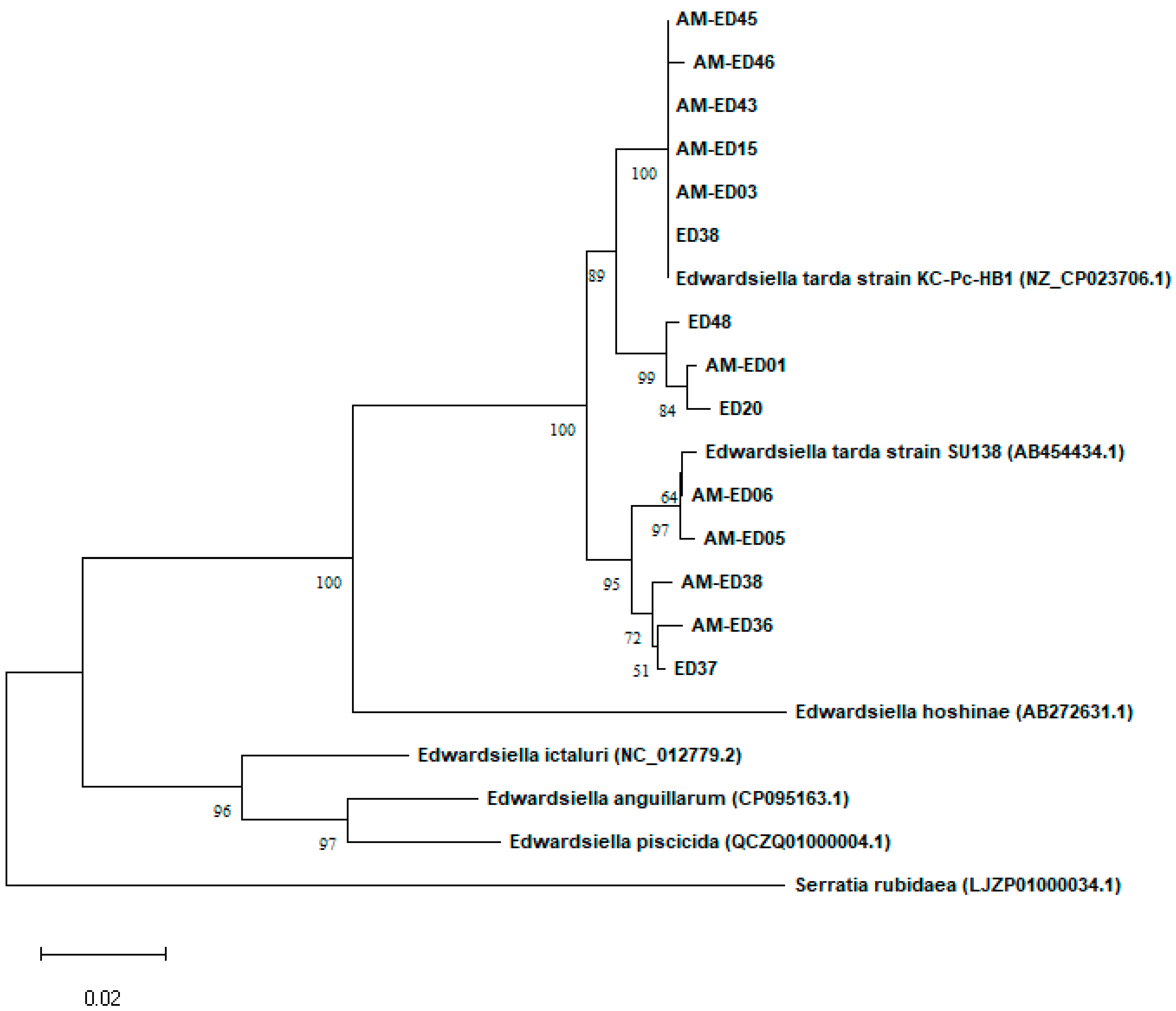
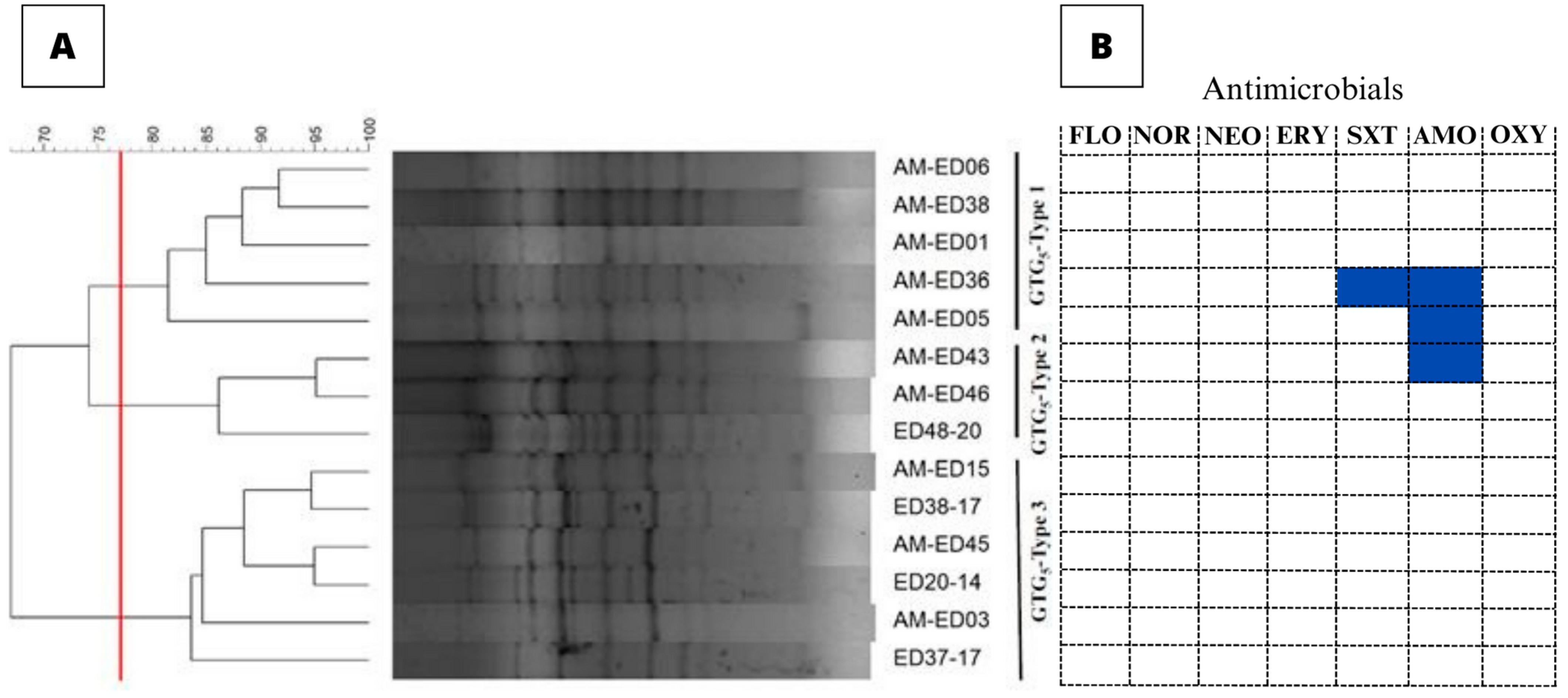
3.2. Genetic Typing of the E. tarda Isolates

Rep-PCR of the 14 E. tarda isolates resulted in the amplification of 8–15 bands, the sizes of which ranged from 400–4000 bp. Three different GTG5 patterns were detected based on a calculated similarity cutoff of 77.08% (Figure 2A). No association was observed between the GTG5-type and the geographic origin of the isolates. The rep-PCR had a discriminatory power of 0.692.

3.3. Antimicrobial Susceptibility

The inhibition zone diameter values obtained for the quality control isolates (Escherichia coli and Aeromonas salmonicida subsp. salmonicida) were within acceptable ranges as determined by the CLSI. Quality control ranges for NOR, NEO, and AMO were not yet established, hence inhibition zone diameter could not be compared. E. tarda isolates exhibited inhibition zones the diameters of which ranged from 6 mm (disc diameter) to 45 mm. The COWT values and the inhibition zone diameters are presented in Table S1. All 14 E. tarda isolates were classified as WT to FLO, NOR, NEO, ERY, and OXY. Nevertheless, 7.1% and 21.4% of the isolates were classified as NWT to SXT and AMO, respectively (Figure 2B). None of the isolates presented multiple-drug reduced susceptibility.

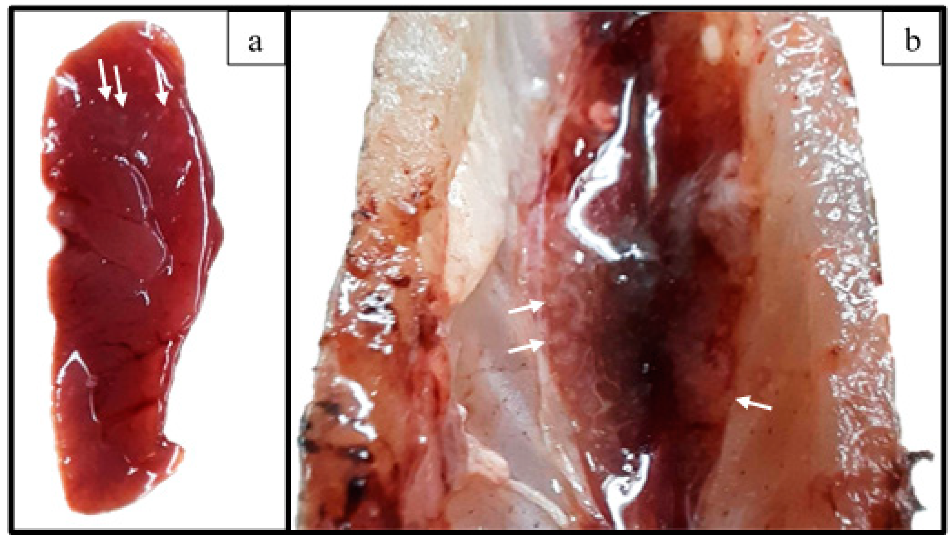
3.4. Challenge Assay

Feed intake did not occur during the first day post-inoculation (dpi) in either the challenged or non-challenged group. However, on the second dpi and afterward, the feed intake of the animals in both groups was normal, as observed during the acclimation phase. All fish from both treatment groups survived throughout the experimental period without the development of clinical signs. Euthanasia of all animals was performed at 21 dpi, followed by necropsy. During inspection of the internal organs, splenomegaly and small, whitish, and round-shaped areas in both the kidney and spleen of two challenged tambaqui juveniles were observed (Figure 3). Bacteriological examination of these challenged animals revealed that E. tarda was recovered from the internal organs of five fish (83.34%). These organs were the spleen (3/6), liver (3/6), and kidney (1/6). Bacterial recovery was not observed from non-challenged fish. Ultimately, the results of the challenge assay satisfied Koch’s postulate.

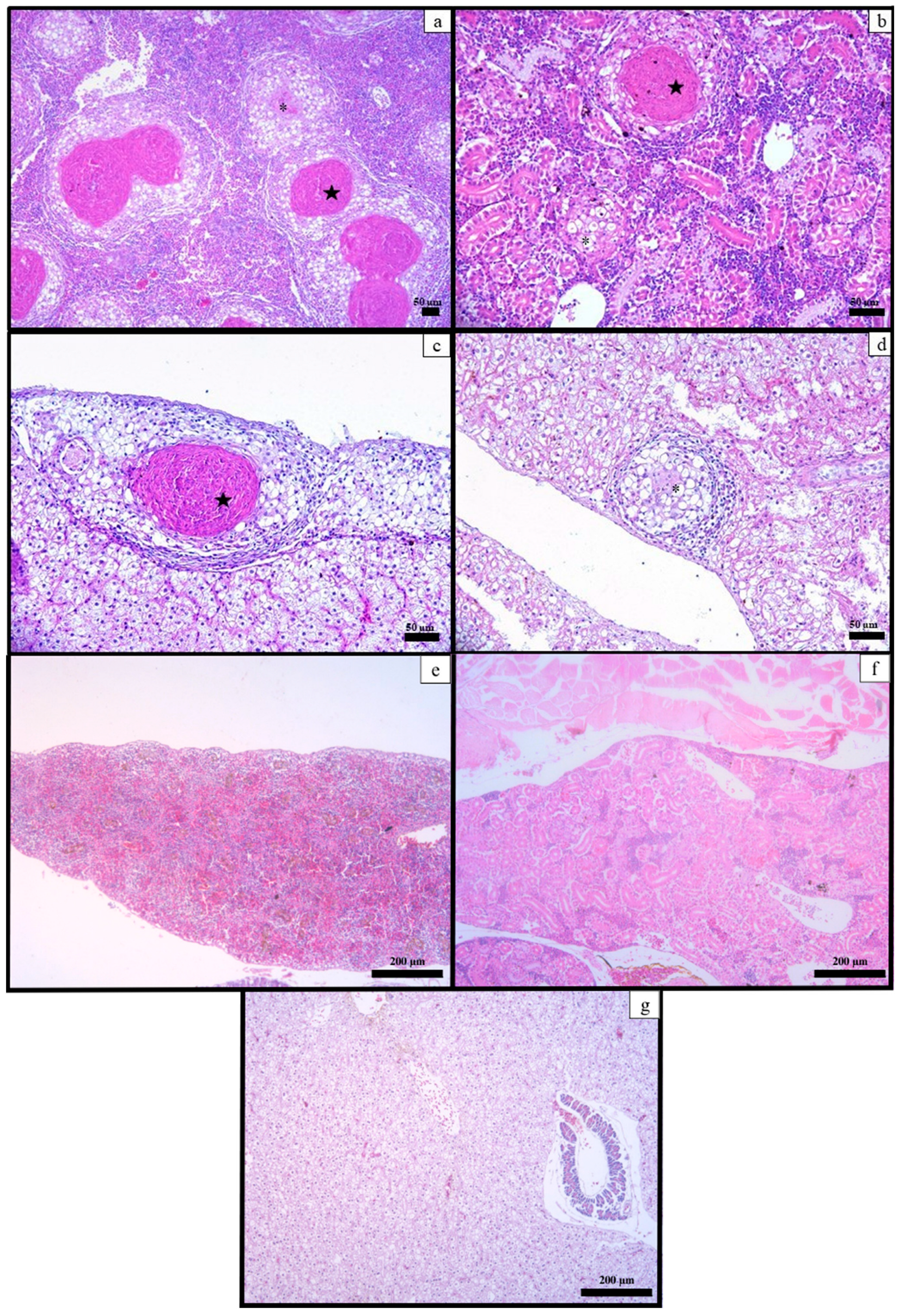
3.5. Histological Examination

Upon histological examination, the non-challenged tambaqui exhibited typical histological features of healthy fish. However, the E. tarda-challenged fish developed several granulomas, mainly composed of epithelioid macrophages and rare lymphocytes. Some granulomas also had necrotic centers. These histopathological alterations were found in the spleen (5/6 fish), liver (5/6 fish), and kidney (1/6 fish), as shown in Figure 4.

4. Discussion

E. tarda is a bacterial pathogen that affects a broad range of fish species [17]. Although previous reports have described the isolation of this pathogen from tambaqui, a clear association with edwardsiellosis could not be found [16]. The higher occurrence of E. tarda in tambaqui over the years reported here is a threat to aquaculture; hence, this subject needs to be further investigated to better understand and address this problem. The missing primordial information was the susceptibility of C. macropomum to E. tarda. Once this was confirmed, the genetic diversity and antimicrobial susceptibility profiles of different E. tarda isolates recovered from tambaqui became relevant to the better comprehension and control of these organisms.
The first isolation of E. tarda from tambaqui collected from Aquavet and LAMAO occurred in 2014. Over the years, the occurrence of this bacterium in tambaqui has increased and, in a few cases, clinical signs were also present. E. tarda-infected fish commonly showed coelomic distension, exophthalmia, rectal prolapse, petechial hemorrhages, swollen internal organs, and granulomas, which were not observed in the cases described here. However, the clinical signs of E. tarda infection in fish may vary after onset [10,17].
During bacteriological examination, E. tarda was isolated from the kidney, spleen, liver, brain, and intestines of C. macropomum. In other fish species, E. tarda has been isolated from a broad range of organs, including the organs where E. tarda was found in tambaqui [12,15,35,36,37]. The identification of the E. tarda isolates was conducted using MALDI-TOF MS followed by confirmation through dnaJ gene sequencing [9,38]. The combination of these methods ensured the precision of the identification. This is important, since it has been reported that E. anguillarum and E. piscicida were mistakenly identified as E. tarda. Additionally, an E. tarda-specific peptide peak was also identified [9].
After challenging C. macropomum with E. tarda, no clinical signs or death were detected, whereas bacterial recovery and histopathological alterations were observed. On the other hand, when E. tarda in similar bacterial doses were experimentally injected in other fish species, both clinical signs and mortality were observed [35,39,40]. E. tarda is known to induce strong clinical signs and high mortality rates (56–90%) in other fish species [35,41,42]; however, E. tarda has been isolated from clinically healthy tambaqui [16]. The differences in clinical status may be related to the characteristics of the isolate or the host, or the presence of predisposing factors. For example, the age of the fish can potentially modulate the results, producing clinical signs or mortality, but further investigations are needed. The use of an E. tarda isolate retrieved from a tambaqui without clinical signs is a limitation of this study. An E. tarda infection with clinical manifestation would result in more precise outcomes. Moreover, a longer observation period after challenge may be necessary for tambaqui to display signs of disease, since granulomas, the main histopathological lesion found in this fish, are usually related to chronic infections.
E. tarda-infected tambaqui showed macroscopic white nodules on the kidney, spleen, and liver, which correlated with the histopathological findings, indicating the establishment of multifocal granulomatous inflammation. Granulomas were also observed in tilapia (Oreochromis spp.) and Japanese snapper (Pagrus major) [43,44], but these differed from the histological lesions found in the Japanese flounder (Paralichthys olivaceus; suppurative inflammation in the kidney and liver), channel catfish (Ictalurus punctatus; necrotizing inflammation in the kidney, liver, and spleen), and seahorse (Hipocampus erectus; enteritis) [35,43,45]. Although the exact cause of granulomas is still unknown; granuloma is the product of a complex process involving the defense mechanisms of the host used to eliminate the pathogen and the strategies of evasion of the bacteria used to resist the immunity [46].
Once the pathogenicity of E. tarda in tambaqui was demonstrated, a rep-PCR analysis was conducted, in which the C. macropomum-derived E. tarda isolates were clustered into three distinct groups, indicating that they were not clonally related. Similar findings were observed in studies analyzing E. tarda isolates derived from a single fish species [47] and from multiple fish species [48], in which two distinct E. tarda groups were found on both occasions. The greater number of genetic variants found among the tambaqui-derived E. tarda isolates may be the outcome of a stress adaptation process triggered by the need to survive in a new host [49].
An antimicrobial susceptibility test was also conducted on C. macropomum-derived E. tarda isolates. The NRI method was employed for statistical validation and standardization for the establishment of the epidemiological cutoff values, resulting in an increase in the accuracy of the resistance data. However, since few isolates were analyzed, the isolates were infecting a new host, and they showed great genetic diversity, possibly leading to different antimicrobial susceptibility patterns from those previously described for other isolates, cutoff values must be considered as only provisional. Also, the antimicrobial susceptibility test data should not be used as a basis for clinical recommendations, since some of these drugs are not approved for use in aquaculture. This analysis revealed that all of the isolates were WT to all antimicrobial agents tested, except for AM-ED36 to SXT and AM-ED05, AM-ED36 and AM-ED43 to AMO. Interestingly, similar cutoff values (Table S1) were found for 52 E. tarda isolates mainly recovered from farmed eels and olive flounder in South Korea [50], which may be related to the use of NRI in both studies. Other studies have shown different levels of agreement with our antimicrobial susceptibility findings [12,51,52,53,54,55,56]. The susceptibility of these isolates to OXY and FLO is of great importance, since these are the only drugs allowed to be used in Brazilian fish farms. However, since E. tarda was not known to infect C. macropomum, no drug has been ever applied in in vivo studies for the treatment of edwardsiellosis in tambaqui.

5. Conclusions

In conclusion, the data presented here show that E. tarda ED38-17 was able to infect and induce tissue lesions in C. macropomum, potentially threatening the production of this Amazonian fish species. The E. tarda isolates recovered from C. macropomum have three distinct genetic types and are WT to FLO, NOR, NEO, ERY, and OXY.

Supplementary Materials

The following supporting information can be downloaded at: https://www.mdpi.com/article/10.3390/ani13182910/s1, Table S1: Antimicrobial susceptibility patterns of the E. tarda isolates. The average inhibition zone diameters (mm) and the provisional epidemiological cutoff (COWT) values (mm) of each isolate are presented. Isolates classified as NWT for a certain antibiotic are highlighted in gray-colored cells.

Author Contributions

F.Y.T.R.: Methodology, investigation, data curation, writing—original draft, writing—review and editing, visualization. V.P.R.: Methodology, investigation, data curation, writing—original draft, writing—review and editing, visualization. P.C.J.-S.: Methodology, investigation, writing—review and editing. H.L.C.: Formal analysis, writing—review and editing. R.C.E.: Investigation, writing—review and editing. N.C.P.: Investigation. C.H.S.d.A.: Investigation. S.P.C.: Investigation. Á.F.S.: Investigation, data curation. B.A.S.: Investigation. F.A.D.: Investigation. M.P.L.: Investigation, validation. R.K.L.: Resources, writing—review and editing. F.P.: Resources, data curation, writing—review and editing. S.U.G.: Resources, data curation, writing—review and editing. G.C.T.: Conceptualization, methodology, data curation, writing—review and editing, visualization, supervision, funding acquisition. H.C.P.F.: Conceptualization, methodology, resources, project administration, writing—review and editing, funding acquisition. All authors have read and agreed to the published version of the manuscript.

Funding

This work was supported by the Coordination for the Improvement of Higher Education Personnel—Brazil (CAPES) through the National Academic Cooperation Program in the Amazon–PROCAD/Amazônia—Finance Code 001 (88881.200614/2018–01), Conselho Nacional de Desenvolvimento Científico e Tecnológico (CNPq, 315995/2021-1), and Fundação de Amparo à Pesquisa do Estado de Minas Gerais (FAPEMIG, PPM-00779-18). LUZ, R.K. received a research grant from the CNPq (308547/2018–7).

Institutional Review Board Statement

The in vivo experiment was approved by the Ethics Committee for Animal Use of the Federal University of Minas Gerais (CEUA-UFMG–152/2020).

Data Availability Statement

DnaJ gene sequences from E. tarda isolated from C. macropomum were included in the NCBI database as follows: AM-ED01: OP535031; AM-ED03: OP535032; AM-ED05: OP535033; AM-ED06): OP535034; AM-ED15: OP535035; AM-ED36: OP535036; AM-ED38: OP535037; AM-ED43: OP535038; AM-ED45: OP535039; AM-ED46: OP535040; ED20: OP535041; ED37: OP535042; ED38: OP535043; ED48: OP535044.

Acknowledgments

The NRI method was used with permission from the patent holder, Bioscand AB, TABY, Sweden (European patent No. 1383913, US patent No. 7,465,559).

Conflicts of Interest

The authors declare no conflict of interest.

References

  1. Valladão, G.M.R.; Gallani, S.U.; Pilarski, F. South American Fish for Continental Aquaculture. Rev. Aquac. 2018, 10, 351–369. [Google Scholar] [CrossRef]
  2. IBGE, Instituto Brasileiro de Geografia e Estatística. Produção Da Pecuária Municipal 2019; IBGE: Rio de Janeiro, Brasil, 2020.
  3. Associação Brasileira da Piscicultura. Anuário Brasileiro Da Piscicultura Peixe BR 2022; Associação Brasileira da Piscicultura: São Paulo, Brasil, 2022. [Google Scholar]
  4. Castro, L.D.A.; Jerônimo, G.T.; da Silva, R.M.; Santos, M.J.; Ramos, C.A.; Porto, S.M.D.A. Occurrence, Pathogenicity, and Control of Acanthocephalosis Caused by Neoechinorhynchus buttnerae: A Review. Rev. Bras. De Parasitol. Veterinária 2020, 29, e008320. [Google Scholar] [CrossRef]
  5. Gallani, S.U.; Valladão, G.M.R.; Assane, I.M.; de Alves, L.O.; Kotzent, S.; Hashimoto, D.T.; Pilarski, F. Motile Aeromonas Septicemia in Tambaqui Colossoma macropomum: Pathogenicity, Lethality and New Insights for Control and Disinfection in Aquaculture. Microb. Pathog. 2020, 149, 104512. [Google Scholar] [CrossRef]
  6. Mielke, T.D.; Francisco, C.J.; Dorella, F.A.; Figueiredo, H.C.P.; Tavares, G.C.; Gallani, S.U. The Strategic Use of Water Additives for Tambaqui Colossoma macropomum Transport: New Insights of Bacteriosis and Productivity Approach. Aquaculture 2022, 558, 738406. [Google Scholar] [CrossRef]
  7. Abayneh, T.; Colquhoun, D.J.; Sørum, H. Edwardsiella piscicida sp. nov., a Novel Species Pathogenic to Fish. J. Appl. Microbiol. 2013, 114, 644–654. [Google Scholar] [CrossRef] [PubMed]
  8. Shao, S.; Lai, Q.; Liu, Q.; Wu, H.; Xiao, J.; Shao, Z.; Wang, Q.; Zhang, Y. Phylogenomics Characterization of a Highly Virulent Edwardsiella Strain ET080813T Encoding Two Distinct T3SS and Three T6SS Gene Clusters: Propose a Novel Species as Edwardsiella anguillarum sp. nov. Syst. Appl. Microbiol. 2015, 38, 36–47. [Google Scholar] [CrossRef]
  9. Reichley, S.R.; Ware, C.; Steadman, J.; Gaunt, P.S.; García, J.C.; LaFrentz, B.R.; Thachil, A.; Waldbieser, G.C.; Stine, C.B.; Buján, N.; et al. Comparative Phenotypic and Genotypic Analysis of Edwardsiella Isolates from Different Hosts and Geographic Origins, with Emphasis on Isolates Formerly Classified as E. tarda, and Evaluation of Diagnostic Methods. J. Clin. Microbiol. 2017, 55, 3466–3491. [Google Scholar] [CrossRef] [PubMed]
  10. Park, S.B.; Aoki, T.; Jung, T.S. Pathogenesis of and Strategies for Preventing Edwardsiella tarda Infection in Fish. Vet. Res. 2012, 43, 67. [Google Scholar] [CrossRef]
  11. Iveson, J.B. Strontium Chloride B and E.E. Enrichment Broth Media for the Isolation of Edwardsiella, Salmonella and Arizona Species from Tiger Snakes. J. Hyg. 1971, 69, 323–330. [Google Scholar] [CrossRef]
  12. Miniero Davies, Y.; Xavier de Oliveira, M.G.; Paulo Vieira Cunha, M.; Soares Franco, L.; Pulecio Santos, S.L.; Zanolli Moreno, L.; Túlio de Moura Gomes, V.; Zanolli Sato, M.I.; Schiavo Nardi, M.; Micke Moreno, A.; et al. Edwardsiella tarda Outbreak Affecting Fishes and Aquatic Birds in Brazil. Vet. Q. 2018, 38, 99–105. [Google Scholar] [CrossRef]
  13. Mauel, M.J.; Miller, D.L.; Frazier, K.S.; Hines, M.E. Bacterial Pathogens Isolated from Cultured Bullfrogs (Rana castesbeiana). J. Vet. Diagn. Investig. 2002, 14, 431–433. [Google Scholar] [CrossRef] [PubMed]
  14. Kamiyama, S.; Kuriyama, A.; Hashimoto, T. Edwardsiella tarda Bacteremia, Okayama, Japan, 2005–2016. Emerg. Infect. Dis. 2019, 25, 1817–1823. [Google Scholar] [CrossRef]
  15. Lima, L.C.; Fernandes, A.A.; Costa, A.A.P.; Velasco, F.O.; Leite, R.C.; Hackett, J.L. Isolation and Characterizaton of Edwardsiella tarda from Pacu Myleus micans. Arq. Bras. Med. Vet. Zootec. 2008, 60, 275–277. [Google Scholar] [CrossRef]
  16. Ramos, E.F.; Sandoval, C.N.; Morales, C.S.; Contreras, S.G.; Manchego, S.A. Lesiones Histopatológicas y Aislamiento Bacteriológico En Gamitanas (Colossoma macropomum) Aparentemente Sanas. Rev. De Investig. Vet. Del Perú 2016, 27, 188–195. [Google Scholar] [CrossRef]
  17. Xu, T.; Zhang, X.H. Edwardsiella tarda: An Intriguing Problem in Aquaculture. Aquaculture 2014, 431, 129–135. [Google Scholar] [CrossRef]
  18. Xiao, J.; Wang, Q.; Liu, Q.; Wang, X.; Liu, H.; Zhang, Y. Isolation and Identification of Fish Pathogen Edwardsiella tarda from Mariculture in China. Aquac. Res. 2008, 40, 13–17. [Google Scholar] [CrossRef]
  19. Suzuki, K.; Yanai, M.; Hayashi, Y.; Otsuka, H.; Kato, K.; Soma, M. Edwardsiella tarda Bacteremia with Psoas and Epidural Abscess as a Food-Borne Infection: A Case Report and Literature Review. Intern. Med. 2018, 57, 893–897. [Google Scholar] [CrossRef]
  20. Michael, J.; Abbott, S.L. Infections Associated with the Genus Edwardsiella: The Role of Edwardsiella tarda in Human Disease. Clin. Infect. Dis. 1993, 17, 742–748. [Google Scholar] [CrossRef] [PubMed]
  21. Hong Nhung, P.; Ohkusu, K.; Mishima, N.; Noda, M.; Monir Shah, M.; Sun, X.; Hayashi, M.; Ezaki, T. Phylogeny and Species Identification of the Family Enterobacteriaceae Based on DnaJ Sequences. Diagn. Microbiol. Infect. Dis. 2007, 58, 153–161. [Google Scholar] [CrossRef]
  22. Sayers, E.W.; Bolton, E.E.; Brister, J.R.; Canese, K.; Chan, J.; Comeau, D.C.; Connor, R.; Funk, K.; Kelly, C.; Kim, S.; et al. Database Resources of the National Center for Biotechnology Information. Nucleic Acids Res. 2022, 50, D20–D26. [Google Scholar] [CrossRef]
  23. Tamura, K.; Stecher, G.; Kumar, S. MEGA11: Molecular Evolutionary Genetics Analysis Version 11. Mol. Biol. Evol. 2021, 38, 3022–3027. [Google Scholar] [CrossRef]
  24. Costa, F.A.A.; Leal, C.A.G.; Leite, R.C.; Figueiredo, H.C.P. Genotyping of Streptococcus dysgalactiae Strains Isolated from Nile Tilapia, Oreochromis niloticus (L.). J. Fish Dis. 2013, 37, 463–469. [Google Scholar] [CrossRef] [PubMed]
  25. Dice, L.R. Measures of the Amount of Ecologic Association between Species. Ecology 1945, 26, 297–302. [Google Scholar] [CrossRef]
  26. Hunter, P.R.; Gaston, M.A. Numerical Index of the Discriminatory Ability of Typing Systems: An Application of Simpson’s Index of Diversity. J. Clin. Microbiol. 1988, 26, 2466. [Google Scholar] [CrossRef]
  27. CLSI, Clinical and Laboratory Standards Institute. VET03 Methods for Antimicrobial Broth Dilution and Disk Diffusion Susceptibility Testing of Bacteria Isolated from Aquatic Animals; Clinical and Laboratory Standards Institute: Wayne, PA, USA, 2020. [Google Scholar]
  28. Kronvall, G.; Smith, P. Normalized Resistance Interpretation, the NRI Method. APMIS 2016, 124, 1023–1030. [Google Scholar] [CrossRef]
  29. Kronvall, G.; Kahlmeter, G.; Myhre, E.; Galas, M.F. A New Method for Normalized Interpretation of Antimicrobial Resistance from Disk Test Results for Comparative Purposes. Clin. Microbiol. Infect. 2003, 9, 120–132. [Google Scholar] [CrossRef] [PubMed]
  30. Smith, P.; Ruane, N.M.; Douglas, I.; Carroll, C.; Kronvall, G.; Fleming, G.T.A. Impact of Inter-Lab Variation on the Estimation of Epidemiological Cut-off Values for Disc Diffusion Susceptibility Test Data for Aeromonas salmonicida. Aquaculture 2007, 272, 168–179. [Google Scholar] [CrossRef]
  31. Smith, P. Eight Rules for Improving the Quality of Papers on the Antimicrobial Susceptibility of Bacteria Isolated from Aquatic Animals. Dis. Aquat. Organ. 2020, 139, 87–92. [Google Scholar] [CrossRef]
  32. Smith, P.; Finnegan, W.; Ngo, T.; Kronvall, G. Influence of Incubation Temperature and Time on the Precision of MIC and Disc Diffusion Antimicrobial Susceptibility Test Data. Aquaculture 2018, 490, 19–24. [Google Scholar] [CrossRef]
  33. Schwarz, S.; Silley, P.; Simjee, S.; Woodford, N.; van Duijkeren, E.; Johnson, A.P.; Gaastra, W. Assessing the Antimicrobial Susceptibility of Bacteria Obtained from Animals. Vet. Microbiol. 2010, 141, 601–604. [Google Scholar] [CrossRef]
  34. Luna, L.G. Manual of Histologic Staining Methods of the Armed Forces Institute of Pathology, 3rd ed.; McGraw-Hill: New York, NY, USA, 1968. [Google Scholar]
  35. Wang, X.; Wang, F.; Chen, G.; Yang, B.; Chen, J.; Fang, Y.; Wang, K.; Hou, Y. Edwardsiella tarda Induces Enteritis in Farmed Seahorses (Hippocampus erectus): An Experimental Model and Its Evaluation. Fish Shellfish Immunol. 2020, 98, 391–400. [Google Scholar] [CrossRef]
  36. Shetty, M.; Maiti, B.; Venugopal, M.N.; Karunasagar, I.; Karunasagar, I. First Isolation and Characterization of Edwardsiella tarda from Diseased Striped Catfish, Pangasianodon hypophthalmus (Sauvage). J. Fish Dis. 2014, 37, 265–271. [Google Scholar] [CrossRef]
  37. Sebastião, F.A.; Furlan, L.R.; Hashimoto, D.T.; Pilarski, F. Identification of Bacterial Fish Pathogens in Brazil by Direct Colony PCR and 16S RRNA Gene Sequencing. Adv. Microbiol. 2015, 5, 409–424. [Google Scholar] [CrossRef]
  38. Buján, N.; Mohammed, H.; Balboa, S.; Romalde, J.L.; Toranzo, A.E.; Arias, C.R.; Magariños, B. Genetic Studies to Re-Affiliate Edwardsiella tarda Fish Isolates to Edwardsiella piscicida and Edwardsiella anguillarum Species. Syst. Appl. Microbiol. 2018, 41, 30–37. [Google Scholar] [CrossRef]
  39. Bera, K.K.; Kumar, S.; Paul, T.; Prasad, K.P.; Shukla, S.P.; Kumar, K. Triclosan Induces Immunosuppression and Reduces Survivability of Striped Catfish Pangasianodon hypophthalmus during the Challenge to a Fish Pathogenic Bacterium Edwardsiella tarda. Environ. Res. 2020, 186, 109575. [Google Scholar] [CrossRef]
  40. Yu, J.E.; Yoo, A.Y.; Choi, K.H.; Cha, J.; Kwak, I.; Kang, H.Y. Identification of Antigenic Edwardsiella tarda Surface Proteins and Their Role in Pathogenesis. Fish Shellfish Immunol. 2013, 34, 673–682. [Google Scholar] [CrossRef]
  41. Hossain, M.M.; Kawai, K.; Oshima, S. Immunogenicity of Pressure Inactivated Edwardsiella tarda Bacterin to Anguilla japonica (Japanese Eel). Pak. J. Biol. Sci. 2011, 14, 755–767. [Google Scholar] [CrossRef]
  42. Qin, L.; Xu, J.; Wang, Y.G. Edwardsiellosis in Farmed Turbot, Scophthalmus maximus (L.), Associated with an Unusual Variant of Edwardsiella tarda: A Clinical, Aetiological and Histopathological Study. J. Fish Dis. 2014, 37, 103–111. [Google Scholar] [CrossRef]
  43. Miyazaki, T.; Kaige, N. Comparative Histopathology of Edwardsiellosis in Fishes. Fish Pathol. 1985, 20, 219–227. [Google Scholar] [CrossRef]
  44. Iregui, C.A.; Guarín, M.; Tibatá, V.M.; Ferguson, H.W. Novel Brain Lesions Caused by Edwardsiella tarda in a Red Tilapia (Oreochromis spp.). J. Vet. Diagn. Investig. 2012, 24, 446–449. [Google Scholar] [CrossRef]
  45. Darwish, A.; Plumb, J.A.; Newton, J.C. Histopathology and Pathogenesis of Experimental Infection with Edwardsiella tarda in Channel Catfish. J. Aquat. Anim. Health 2000, 12, 255–266. [Google Scholar] [CrossRef]
  46. Rajme-Manzur, D.; Gollas-Galván, T.; Vargas-Albores, F.; Martínez-Porchas, M.; Hernández-Oñate, M.Á.; Hernández-López, J. Granulomatous Bacterial Diseases in Fish: An Overview of the Host’s Immune Response. Comp. Biochem. Physiol. A Mol. Integr. Physiol. 2021, 261, 111058. [Google Scholar] [CrossRef]
  47. Castro, N.; Toranzo, A.E.; Bastardo, A.; Barja, J.L.; Magariños, B. Intraspecific Genetic Variability of Edwardsiella tarda Strains from Cultured Turbot. Dis. Aquat. Organ. 2011, 95, 253–258. [Google Scholar] [CrossRef] [PubMed]
  48. Griffin, M.J.; Quiniou, S.M.; Cody, T.; Tabuchi, M.; Ware, C.; Cipriano, R.C.; Mauel, M.J.; Soto, E. Comparative Analysis of Edwardsiella Isolates from Fish in the Eastern United States Identifies Two Distinct Genetic Taxa amongst Organisms Phenotypically Classified as E. tarda. Vet. Microbiol. 2013, 165, 358–372. [Google Scholar] [CrossRef]
  49. Wang, Q.; Yang, M.; Xiao, J.; Wu, H.; Wang, X.; Lv, Y.; Xu, L.; Zheng, H.; Wang, S.; Zhao, G.; et al. Genome Sequence of the Versatile Fish Pathogen Edwardsiella tarda Provides Insights into Its Adaptation to Broad Host Ranges and Intracellular Niches. PLoS ONE 2009, 4, e7646. [Google Scholar] [CrossRef]
  50. Lim, Y.-J.; Kim, D.-H.; Roh, H.J.; Park, M.-A.; Park, C.-I.; Smith, P. Epidemiological Cut-off Values for Disc Diffusion Data Generated by Standard Test Protocols from Edwardsiella tarda and Vibrio harveyi. Aquac. Int. 2016, 24, 1153–1161. [Google Scholar] [CrossRef]
  51. Shin, D.-M.; Hossain, S.; Wimalasena, S.; Heo, G.-J. Antimicrobial Resistance and Virulence Factors of Edwardsiella tarda Isolated from Pet Turtles. Pak. Vet. J. 2017, 37, 85–89. [Google Scholar]
  52. Preena, P.G.; Dharmaratnam, A.; Swaminathan, T.R. Antimicrobial Resistance Analysis of Pathogenic Bacteria Isolated from Freshwater Nile Tilapia (Oreochromis niloticus) Cultured in Kerala, India. Curr. Microbiol. 2020, 77, 3278–3287. [Google Scholar] [CrossRef]
  53. Preena, P.G.; Dharmaratnam, A.; Raj, N.S.; Raja, S.A.; Nair, R.R.; Swaminathan, T.R. Antibiotic-Resistant Enterobacteriaceae from Diseased Freshwater Goldfish. Arch. Microbiol. 2020, 203, 219–231. [Google Scholar] [CrossRef]
  54. Preena, P.G.; Arathi, D.; Raj, N.S.; Kumar, T.V.A.; Raja, S.A.; Reshma, R.N.; Swaminathan, T.R. Diversity of Antimicrobial-Resistant Pathogens from a Freshwater Ornamental Fish Farm. Lett. Appl. Microbiol. 2019, 71, 108–116. [Google Scholar] [CrossRef]
  55. Lee, S.W.; Wendy, W. Antibiotic and Heavy Metal Resistance of Aeromonas hydrophila and Edwardsiella tarda Isolated from Red Hybrid Tilapia (Oreochromis spp.) Coinfected with Motile Aeromonas Septicemia and Edwardsiellosis. Vet. World 2017, 10, 807. [Google Scholar] [CrossRef] [PubMed]
  56. Stock, I.; Wiedemann, B. Natural Antibiotic Susceptibilities of Edwardsiella tarda, E. ictaluri, and E. hoshinae. Antimicrob. Agents Chemother. 2001, 45, 2255. [Google Scholar] [CrossRef] [PubMed]
Figure 1. Phylogenetic tree constructed using the neighbor-joining method based on dnaJ gene sequences (719 bp) from C. macropomum-derived E. tarda isolates. Known E. tarda (NZ CP023706.1; AB454434.1), E. hoshinae (AB272631.1), E. ictaluri (NC 012779.2), E. anguillarum (CP095163.1), E. piscicida (NZ QCZQ1000004.1), and Serratia rubidaea (LJZP01000034.1) are also present. Numbers on the branches indicate bootstrap percentage after 1000 replications in constructing the tree. Scale bar refers to a phylogenetic distance of 14.38 nucleotide substitutions per site.
Animals 13 02910 g001
Figure 2. Genetic diversity and antimicrobial susceptibility of E. tarda isolates recovered from C. macropomum. (A): Rep-PCR results for the 14 E. tarda isolates isolated from C. macropomum. The dendrogram was constructed using the Dice coefficient and the UPGMA method. The red line represents the cutoff defined considering the mean value of all clusters minus 1645 per standard deviation. (B): Antimicrobial susceptibility of C. macropomum-derived E. tarda isolates. Blue boxes represent NWT isolates.
Animals 13 02910 g002
Figure 3. Photographs of the spleen (a) and kidneys (b) of a C. macropomum juvenile experimentally infected with E. tarda ED38-17, showing splenomegaly, as evidenced by the round edges of the organ (a), and the small, whitish, and round-shaped areas (arrows) in the spleen and kidney (b).
Animals 13 02910 g003
Figure 4. Microphotographs of HE staining of the organs from C. macropomum juveniles. Fish experimentally infected with E. tarda ED38-17 exhibited granulomas, both with a necrotic center (star) and without a necrotic center (asterisk), in the spleen (a), kidney (b), and liver (c,d). Spleen (e), kidney (f) and liver (g) from control group fish are depicted without alterations.
Animals 13 02910 g004
Table 1. Metadata of the Colossoma macropomum-derived Edwardsiella tarda isolates.
IsolateCulture CollectionClinical Status of the FishStateYear of IsolationOrganMALDI-TOF Score Value
ED20-14AquavetDiseasedMG2014Kidney2.494
ED37-17AquavetHealthyRO2017Kidney2.358
ED38-17AquavetHealthyRO2017Kidney2.534
AM-ED01LAMAOHealthyAM2018Brain2.210
AM-ED03LAMAOHealthyAM2018Brain2.459
AM-ED05LAMAODiseasedAM2019Kidney2.153
AM-ED06LAMAOHealthyAM2019Brain2.598
AM-ED15LAMAODiseasedAM2019Brain2.521
AM-ED36LAMAOHealthyAM2019Spleen2.539
AM-ED38LAMAODiseasedAM2019Kidney2.600
AM-ED43LAMAODiseasedAM2019Liver2.286
AM-ED45LAMAOHealthyAM2019Intestines2.292
AM-ED46LAMAOHealthyAM2020Kidney2.347
ED48-20AquavetHealthyAM2020Intestines2.316
Disclaimer/Publisher’s Note: The statements, opinions and data contained in all publications are solely those of the individual author(s) and contributor(s) and not of MDPI and/or the editor(s). MDPI and/or the editor(s) disclaim responsibility for any injury to people or property resulting from any ideas, methods, instructions or products referred to in the content.

