
Oak Acorn Poisoning in Cattle during Autumn 2022: A Case Series and Review of the Current Knowledge

 ,
,  , ,
, ,  ,
,
Abstract
Simple Summary
Abstract
1. Introduction
2. Cases Information
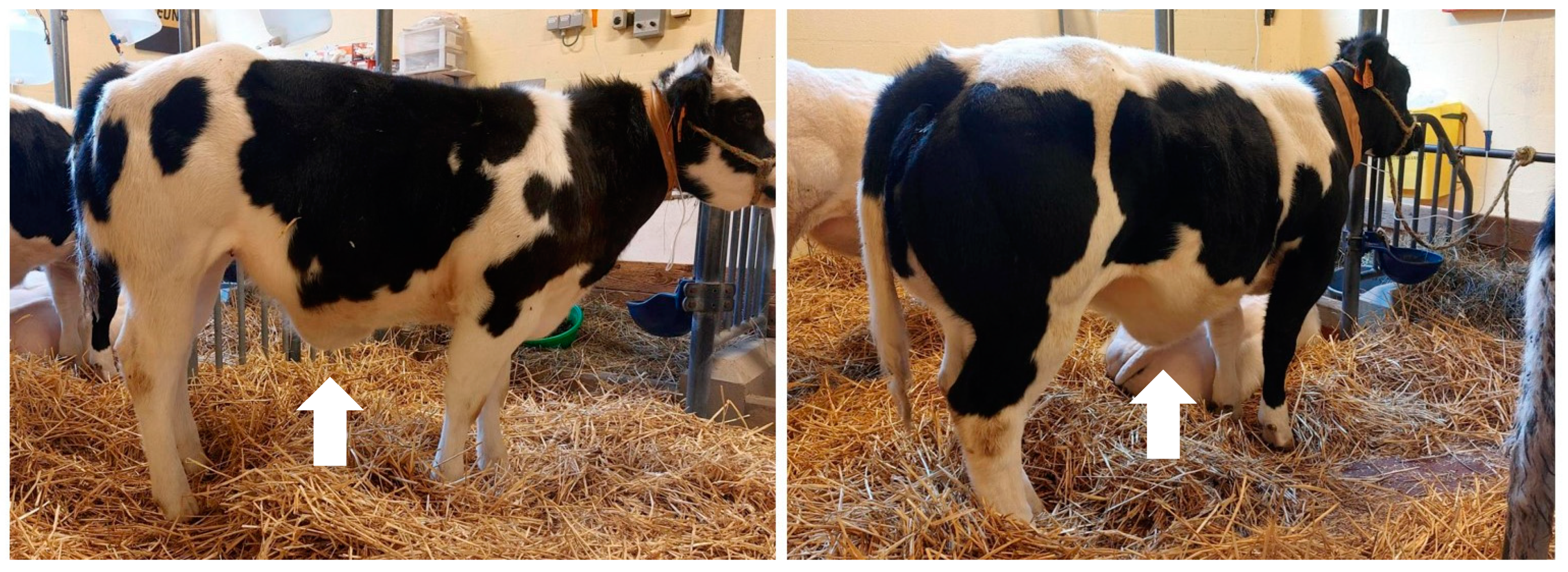
3. Clinical Findings
4. Diagnostic Assessment
5. Therapeutic Intervention
6. Follow Up
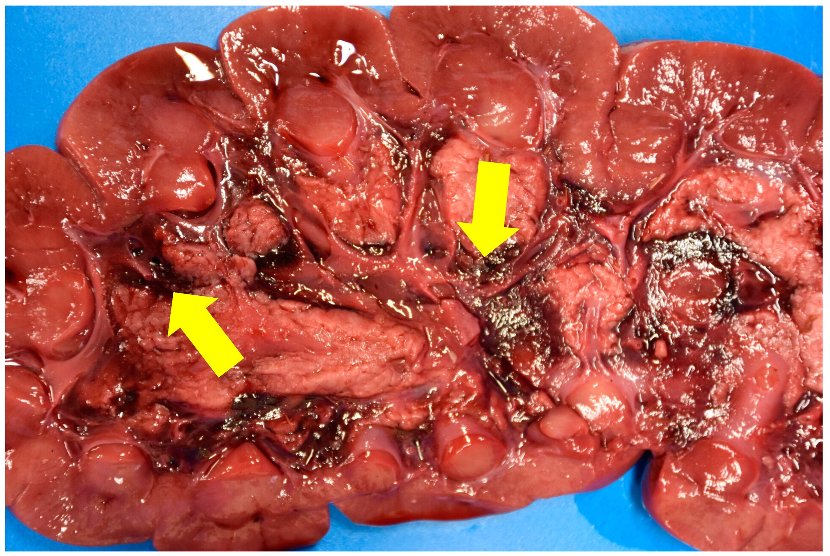
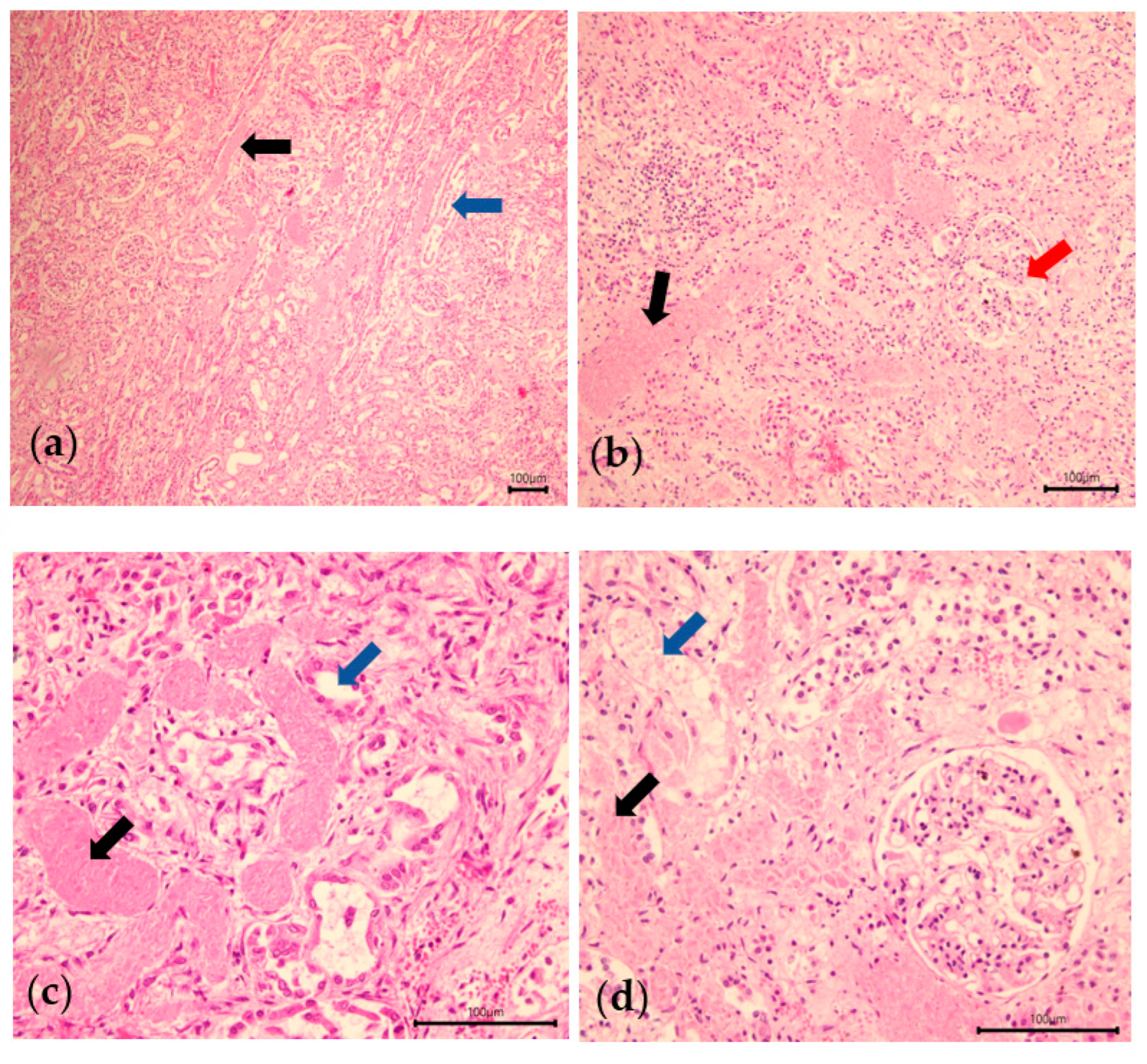
7. Necropsy Findings
8. Outcome
9. Discussion
10. Conclusions
Author Contributions
Funding
Institutional Review Board Statement
Informed Consent Statement
Data Availability Statement
Acknowledgments
Conflicts of Interest
References
- Kremer, A.; Hipp, A.L. Oaks: An Evolutionary Success Story. New Phytol. 2020, 226, 987–1011. [Google Scholar] [CrossRef] [PubMed]
- Bates, N. Autumn Plant Poisoning Hazards. Livestock 2019, 24, 239–245. [Google Scholar] [CrossRef]
- Anadón, A.; Martínez-Larrañaga, M.R.; Castellano, V. Poisonous Plants of Europe. In Veterinary Toxicology: Basic and Clinical Principles; Elsevier: Amsterdam, The Netherlands, 2012; pp. 1080–1094. ISBN 9780123859266. [Google Scholar]
- Plumlee, K.H. Chapter 25—Plants. In Clinical Veterinary Toxicology; Plumlee, K.H., Ed.; Mosby: Saint Louis, MO, USA, 2004; pp. 337–442. ISBN 978-0-323-01125-9. [Google Scholar]
- Pérez, V.; Doce, R.R.; García-Pariente, C.; Hervás, G.; Carmen Ferreras, M.; Mantecón, Á.R.; Frutos, P. Oak Leaf (Quercus Pyrenaica) Poisoning in Cattle. Res. Vet. Sci. 2011, 91, 269–277. [Google Scholar] [CrossRef] [PubMed]
- Dollahite, J.W.; Housholder, G.T.; Camp, B.J. Oak Poisoning In Livestock; Texas Agricultural Experiment Station Bulletin: College Station, TX, USA, 1966; pp. 1–8. [Google Scholar]
- Stober, M.; Ziegler, H.P.; Von Benten, K. Acorn Poisoning in Cattle. Bov. Pract. 1976, 11, 36–41. [Google Scholar] [CrossRef]
- Neser, J.A.; Boomker, J.; Cable, H. Oak (Quercus Rubor) Poisoning In Cattle. J. S. Afr. Vet. Assoc. 1982, 53, 151–155. [Google Scholar] [PubMed]
- Frias, C.; Simões, P.B.A.; Cota, J.; Pissarra, H.; Nunes, T.P.; Hjerpe, C.A.; Lima, M.S.; Ecbhm, D. Case Report-Chronic Oak Toxicity (Quercus Suber) in Beef Cattle in the South of Portugal: 17 Cases (2014–2018). Bov. Pract. 2019, 53, 170–176. [Google Scholar] [CrossRef]
- Marsh, C.D.; Clawson, A.B.; Marsh, H. Oak-Leaf Poisoning of Domestic Animals; Washington Government Printing Office: Washington, DC, USA, 1919; pp. 1–50. [Google Scholar]
- Divers, T.J. Urinary Tract Diseases. In Rebhun’s Diseases of Dairy Cattle: Third Edition; Elsevier: Amsterdam, The Netherlands, 2018; pp. 526–552. ISBN 9780323396622. [Google Scholar]
- Divers, T.J.; Peek, S.F. The Clinical Examination. In Rebhun’s Diseases of Dairy Cattle: Third Edition; Elsevier: Amsterdam, The Netherlands, 2018; pp. 2–16. ISBN 9780323396622. [Google Scholar]
- Jones, M.L.; Allison, R.W. Evaluation of the Ruminant Complete Blood Cell Count. Vet. Clin. North Am.-Food Anim. Pract. 2007, 23, 377–402. [Google Scholar] [CrossRef] [PubMed]
- Boulay, G.; Francoz, D.; Doré, E.; Dufour, S.; Veillette, M.; Badillo, M.; Bélanger, A.M.; Buczinski, S. Preoperative Cow-Side Lactatemia Measurement Predicts Negative Outcome in Holstein Dairy Cattle with Right Abomasal Disorders. J. Dairy Sci. 2014, 97, 212–221. [Google Scholar] [CrossRef] [PubMed]
- Fastré, B. Evaluation Du Poids et de l’état Corporel Chez Les Bovins Blanc-Bleu-Belge (BBB); ULiège: Liège, Belgium, 2012. [Google Scholar]
- Vermeire, L.T.; Wester, D.B. Shinnery Oak Poisoning of Rangeland Cattle: Causes, Effects & Solutions. Rangelands 2001, 23, 19–21. [Google Scholar] [CrossRef][Green Version]
- Vandenbroucke, V.; Van Pelt, H.; De Backer, P.; Croubels, S. Animal Poisonings in Belgium: A Review of the Past Decade. Vlaams Diergeneeskd. Tiijdschrift 2010, 79, 259–268. [Google Scholar] [CrossRef]
- Chamorro, M.F.; Passler, T.; Joiner, K.; Poppenga, R.H.; Bayne, J.; Walz, P.H. Case Report Rapport de Cas Acute Renal Failure in 2 Adult Llamas after Exposure to Oak Trees (Quercus Spp). Can. Vet. J. 2013, 54, 61–64. [Google Scholar] [PubMed]
- Gwaltney-Brant, S.M. Renal Toxicity. In Veterinary Toxicology: Basic and Clinical Principles; Elsevier: Amsterdam, The Netherlands, 2012; pp. 264–277. ISBN 9780123859266. [Google Scholar]
- Davy-Moyle, R.B.; Londoño, L.; Nelson, E.A.; Bandt, C. Treatment of Acute Kidney Injury Secondary to Oak Intoxication with Hemodialysis in a Miniature Zebu. J. Vet. Emerg. Crit. Care 2018, 28, 361–365. [Google Scholar] [CrossRef] [PubMed]
- Boubet, B. L’intoxication Aux Glands. Une Pathologie Souvent Mortelle Chez Les Bovins. Available online: https://www.pleinchamp.com/actualite/l-intoxication-aux-glands-une-pathologie-souvent-mortelle-chez-les-bovins (accessed on 20 March 2023).


| Cattle | 1 | 2 | 3 | 4 | 5 | 6 | 7 | Reference Value |
|---|---|---|---|---|---|---|---|---|
| Sex | male | female | female | female | female | female | male | - |
| Weight (kg) | 200 | 202 | 335 | 275 | 210 | 290 | 127 | - |
| Age (month) | 10 | 9 | 17 | 8 | 7 | 8 | 5 | - |
| Respiratory rate (respiration/min) | 12 | 24 | 54 | 112 | 40 | 32 | 24 | 20–40 |
| Rectal temperature (°C) | 36.7 | 38.1 | 38 | 38.2 | 38.1 | 38.9 | 39.7 | 38.5–39.5 |
| Heart rate (Beats/min) | 76 | 52 | 72 | 108 | 108 | 110 | 72 | 60–84 |
| Capillary refill time (s) | 3–4 | <2 | <2 | <2 | <2 | <2 | 3 | <2 |
| Skin folds (s) | 5 | 3 | 3 | 3 | <2 | <2 | <2 | <2 |
| Enophtalmia (mm) | 3 | 2 | 2 | 0 | 0 | 0 | 0 | 0 |
| Mucous membranes | Pale | Pink | Pink | Pink | Pink | Pink | Congestive | Pink |
| Lymph nodes | Normal | Normal | Normal | Normal | Normal | Normal | Normal | Normal |
| Cattle | 1 | 2 | 3 | 4 | 5 | 6 | 7 | Reference Value |
|---|---|---|---|---|---|---|---|---|
| Urea (mg/dL) | >130 | >130 | 321 | >130 | >130 | >130 | 124 | 10–20 |
| Creatinine (mg/dL) | >13.6 | >13.6 | 3.63 | >13.6 | >13.6 | >13.6 | >13.6 | 0.4–1 |
| Lactate (mmol/L) | 2.6 | 1.3 | 4 | - | - | - | - | <2 |
| Blood pH | 6.96 | 7.24 | 7.22 | - | - | - | 7.35 | 7.35–7.50 |
| HCO3− (mmol/L) | 10.6 | 17.7 | 13.6 | - | - | - | 25.3 | 24–34 |
| Na (mmol/L) | <100 | 122 | 129 | - | - | - | 124 | 134–145 |
| K+ (mmol/L) | 8.4 | 1.9 | 2.3 | - | - | - | 1.4 | 3.9–5.3 |
| Cl− (mmol/L) | 73 | 89 | 94 | - | - | - | 85 | 94–105 |
| Hematocrit (%) | 29.5 | 27.1 | 23.6 | - | - | - | - | 22–33% |
| Hemoglobin (g/dL) | 10.3 | 9.8 | 9 | - | - | - | - | 8–15 |
| Leucocytes (×109/L) | 15.82 | 15.19 | 10.05 | - | - | - | - | 4–12 |
| Neutrophiles (×109/L) | 8.54 | 11.49 | 6.4 | - | - | - | - | 0.6–4 |
| Lymphocytes (×109/L) | 6.54 | 2.85 | 2.64 | - | - | - | - | 2.5–7.5 |
| Monocytes (×109/L) | 0.63 | 0.75 | 0.94 | - | - | - | - | 0.025–0.84 |
| Eosinophiles (×109/L) | 0.09 | 0.04 | 0.03 | - | - | - | - | 0.0–0.24 |
| Basophiles (×109/L) | 0.02 | 0.04 | 0.03 | - | - | - | - | 0.00–0.02 |
| Platelets (K/µL) | 508 | 579 | 426 | - | - | - | - | 100–800 |
| P (mg/L) | >161 | 157 | 154 | - | - | - | - | 42–77 |
| Ca (mg/L) | 48 | 49 | 48 | - | - | - | - | 83–104 |
| Total proteins (g/L) | 68 | 77 | 91 | - | - | - | - | 70–85 |
| Albumin (g/L) | 35 | 40 | 42 | - | - | - | - | 32–42 |
| Gamma-glutamyltransferase (IU/L) | 28 | 26 | 38 | - | - | - | - | 11–39 |
| Cholesterol (g/L) | 0.23 | 0.81 | 0.98 | - | - | - | - | 0.73–2.8 |
| n° | DAY 1 | DAY2 | DAY3 | DAY5 | DAY10 | DAY16 | ||||||
|---|---|---|---|---|---|---|---|---|---|---|---|---|
| U | C | U | C | U | C | U | C | U | C | U | C | |
| 1 | >130 | >13.6 | ||||||||||
| 2 | >130 | >13.6 | ||||||||||
| 3 | 321 | 3.63 | 18 | 1.7 | 29 | 1 | 10 | 5.2 | ||||
| 4 | >130 | >13.6 | >130 | >13.6 | >130 | >13.6 | >130 | >13.6 | ||||
| 5 | >130 | >13.6 | >130 | >13.6 | ||||||||
| 6 | >130 | >13.6 | >130 | >13.6 | ||||||||
| 7 | 124 | >13.6 | 80 | 11.1 | 59 | 7 | 22 | 3.9 | 6 | 2.8 | ||
Disclaimer/Publisher’s Note: The statements, opinions and data contained in all publications are solely those of the individual author(s) and contributor(s) and not of MDPI and/or the editor(s). MDPI and/or the editor(s) disclaim responsibility for any injury to people or property resulting from any ideas, methods, instructions or products referred to in the content. |
© 2023 by the authors. Licensee MDPI, Basel, Switzerland. This article is an open access article distributed under the terms and conditions of the Creative Commons Attribution (CC BY) license (https://creativecommons.org/licenses/by/4.0/).
Share and Cite
Eppe, J.; Bayrou, C.; Casalta, H.; Cassart, D.; Gille, L.; Stipulanti, M.; Versyp, J.; Sartelet, A. Oak Acorn Poisoning in Cattle during Autumn 2022: A Case Series and Review of the Current Knowledge. Animals 2023, 13, 2678. https://doi.org/10.3390/ani13162678
Eppe J, Bayrou C, Casalta H, Cassart D, Gille L, Stipulanti M, Versyp J, Sartelet A. Oak Acorn Poisoning in Cattle during Autumn 2022: A Case Series and Review of the Current Knowledge. Animals. 2023; 13(16):2678. https://doi.org/10.3390/ani13162678
Chicago/Turabian StyleEppe, Justine, Calixte Bayrou, Hélène Casalta, Dominique Cassart, Linde Gille, Margot Stipulanti, Jérôme Versyp, and Arnaud Sartelet. 2023. "Oak Acorn Poisoning in Cattle during Autumn 2022: A Case Series and Review of the Current Knowledge" Animals 13, no. 16: 2678. https://doi.org/10.3390/ani13162678
APA StyleEppe, J., Bayrou, C., Casalta, H., Cassart, D., Gille, L., Stipulanti, M., Versyp, J., & Sartelet, A. (2023). Oak Acorn Poisoning in Cattle during Autumn 2022: A Case Series and Review of the Current Knowledge. Animals, 13(16), 2678. https://doi.org/10.3390/ani13162678