Article

Comparative Label-Free Liquid Chromatography–Mass Spectrometry Milk Proteomic Profiles Highlight Putative Differences between the Autochthon Teramana and Saanen Goat Breeds

1
Department of Bioscience and Technology for Food Agro-Food and Environmental Technology, University of Teramo, 64100 Teramo, Italy
2
National Institute for Cellular Biotechnology, Dublin City University, Dublin 9, Dublin, Ireland
3
School of Biotechnology, Dublin City University, Dublin 9, Dublin, Ireland
*
Author to whom correspondence should be addressed.
Animals 2023, 13(14), 2263; https://doi.org/10.3390/ani13142263
Submission received: 9 June 2023 / Revised: 30 June 2023 / Accepted: 8 July 2023 / Published: 10 July 2023
(This article belongs to the Section Small Ruminants)

Abstract

:

Simple Summary

Milk is a valuable source of proteins and other nutrients. Changes in milk production toward systems that obtain increased milk yield have resulted in a change in milk composition in large and small ruminants, such as goats. A better characterisation of breeds that undergo a limited formal crossbreeding, such as the Teramana goat is pivotal in order to obtain useful data that could be applied for the management of the breed. Proteomic technologies have brought significant advances in the characteristion of new proteins. In this study, this technology was used to compare the proteome of the autochthon Teramana and Saanen breeds, which are commonly used by the industry to allow for the identification of a cohort of proteins that were able to discriminate the two goat breeds. Proteomics offered the potentiality for a deeper investigation of the biological differences of the breeds under study in a substrate easy to obtain.

Abstract

Goat’s milk is an excellent source of nutrients, with greater benefits compared to cow’s milk. Limited information is available on autochthon goat breeds, which are important for biodiversity preservation. In this study, the aim of using label-free quantification was to investigate the milk proteome of two goat breeds, the autochthon Teramana and Saanen breeds, which are commonly used by the industry. Utilising label-free proteomic analysis, 749 and 666 proteins, respectively were identified and quantified from the Teramana and Saanen goat milk. Moreover, utilising statistical analysis, 29 proteins were able to discriminate the two goat breeds, with many of the identified proteins involved in complement and coagulation cascades. This work enhances our understanding of the goat milk proteome and shows differences between the two breeds, leading to an important contribution toward a more detailed molecular-view of this unique substrate. Additionally, charactersation of the milk proteins can help in guiding genetic improvements in the goat herds, and thus increasing its use in human nutrition.

1. Introduction

Milk plays a pivotal role in human nutrition, as it is rich in macro- and micro-nutrients and is an important source of antimicrobial and immunoregulatory agents. Over the last decades, the use of non-bovine milk as an alternative nutrient source has increased, since hypersensitivity to cow’s milk proteins remains one of the major causes of food allergies [1]. For this reason, the milk of small ruminants, such as goats is of particular interest to some consumers as it is easier to digest than milk from cows and may have certain therapeutic values [2]. The major differences between cow’s and goat’s milk are related to the different proportions and classes of caseins [3]. Other differences are the size and structure of the protein micelles and fat globules with a more favourable fat composition in goat’s milk, which is beneficial for digestibility and energy uptake, and makes the goat’s milk less allergenic [4,5,6,7]. Additionally, these properties are beneficial for the cheese manufacturer, as goat milk allows for the development of fermented products with particular characteristics compared to cow’s milk [8]. Moreover, goats are of particular economic interest in developing countries as they can easily adapt to harsh environmental conditions, have the capacity to convert poor quality fibrous feedstuffs into animal proteins, and have a high resistance to diseases, which make the production of this milk a useful strategy to tackle the problems of under-nutrition [9].
Despite the fact that the world goat population has increased during the last decades, many autochthonous breeds have been displaced and are in an endangered status. They even face extinction since they have been replaced by more productive breeds. This is the case for breeds, such as Teramana and Saanen goats. The Teramana goat is an autochthonous breed from the Abruzzo region of Italy, which as a consequence of the marked decrease in fresh goats milk consumption and of the progressive abandoning of low income rural activities by farmers, has become almost extinct [10]. Currently, and according to official national data reported by the Food and Agriculture Organisation of the United Nations (FAO), this breed is listed with a critical risk status. The Saanen goat is a highly productive dairy goat and is one of the most distributed goat breeds in many countries [11].
Proteomics consist of the high-throughput study of the proteome (the protein complement of a genome). The proteome is the entire set of proteins that is produced or modified by a cell, an organism, a body fluid, etc. at a given time under defined conditions [12,13]. Due to these features, proteomics is ideally suited to characterise and map the goat’s milk proteome. Proteomic technologies have brought significant advances in the detection and identification of new proteins in goat’s milk. For many years, gel-based proteomic approaches, such as SDS PAGE [14] and two-dimensional gel electrophoresis (2D PAGE) [15,16] were the main techniques used to investigate the protein composition of goat’s milk. Following, the development of mass spectrometry (MS)-based proteomic methods, such as label-free MS-based quantification has become more popular. This is a method for measuring peptide concentrations in complex samples using a combination of high-performance liquid chromatography (HPLC) and MS, which does not require expensive labelling techniques [17,18]. A variety of milk proteins have been identified by applying this approach with bioinformatic tools [2,19]. Moreover, in our previous study, this method has been successfully applied to discriminate the meat proteome of the Teramana and Saanen goat breeds [10].
Caseins make up 80% of overall protein content of milk [20] and their presence is a limiting factor in studying less abundant proteins that have also been proposed as possible biomarkers for animal health [21]. Prefractionation and further depletion of medium- to high-abundance proteins is required to study minor components [22,23]. A method, such as label-free LC–MS proteomics is ideal for these analyses in order to allow for the study of hydrophobic proteins and proteins with low- or high-molecular weights [17,18].
Studies on goat milk proteomics have mainly focused on the more productive goat breeds [24,25,26]. Interspecies differences can be evaluated using proteomic analysis; however, studies evaluating the milk proteome from different breeds of the same species, particularly among goat breeds, are still scarce [27]. In this context, studies with an emphasis on the comparative proteomic evaluation of goat milk can help in guiding genetic improvements in the goat herds, and thus increasing its use in human nutrition. Therefore, in this study, a label-free proteomic approach was applied to characterise the milk proteome of the Teramana and Saanen goat breeds, which may lead to a better understanding of the biological mechanism of this product.

2. Materials and Methods

2.1. Sample Collection

Five Teramana and five Saanen goats for a total of 10 animals aged 30 to 40 months with no signs of acute mastitis or other clinical diseases were selected from a local farm in Teramo province, Italy. All goats included in this study were reared in the same farm, and the management with regard to their feeding, handling, and period of lactation was the same. Thereafter, approximately 100 mL of milk was collected by milking at one time point from each animal and were used in the study. All animals were representative of goat breeds. After sampling, all milk samples were transferred on ice, and then taken immediately to the laboratory, where they were processed. No permits were required for the described study, which complied with all relevant regulations, since only milk was collected and no animal sacrifice was necessary.

2.2. Sample Preparation

Goat’s milk samples (2 mL) were centrifuged at 3000× g for 30 min, at 4 °C, and the fat layer was carefully removed. Then, 50 µL of the skim milk samples were precipitated by adding 5 µL of sodium acetate (1 N) and 5 µL of acetic acid (10%), and the caseins were removed by centrifugation at 3000× g for 30 min, at 4 °C. Whey proteins were directly precipitated with cold acetone, dried, and stored at −20 °C until further analysis.
The protein content of all samples was determined in triplicate using the Protein Assay Kit (Bio-Rad Labs, Hercules, CA, USA), following the Bradford method using a bovine serum albumin (BSA) standard [28]. Equal concentrations (100 μg) of all protein samples were used for filter-aided sample preparation (FASP) as described below [29].

2.3. Filter-Aided Sample Preparation (FASP)

Equal concentrations (100 μg) of all protein samples were purified and digested according to a slightly modified filter-aided sample preparation (FASP) method [29,30]. Briefly, 100 μg of milk proteins were suspended in 100 μL of lysis buffer [7M Urea (Affymetrix/Thermo Fisher Scientific, Waltham, MA, USA), 2M Thiourea (Affymetrix/Thermo Fisher Scientific, Waltham, MA, USA), 30 mM Tris, 4% CHAPS (Affymetrix/Thermo Fisher Scientific, Waltham, MA, USA), pH 8.5]. Protein samples were reduced and alkylated with dithiothreitol (DTT) and iodoacetamide (IAA; Sigma-Aldrich/Merck, Saint Louis, MO, USA), and then digested with trypsin according to the FASP method [29,30]. Peptides were purified using C18 spin columns (Thermo Fisher Scientific, Waltham, MA, USA), dried under vacuum, and suspended in 2% acetonitrile (ACN) and 0.1% trifluoroacetic acid (TFA) prior to mass spectrometry.

2.4. Mass Spectrometry for Label-Free LC–MS

Nano LC–MS/MS analysis was carried out using an Ultimate 3000 nanoRSLC system (Thermo Scientific) coupled in-line with an Orbitrap Fusion Tribrid™ mass spectrometer (Thermo Scientific). Then, 1 µg of digested protein samples were loaded onto a C18 trap column (C18 PepMap100, 300 μm × 5 mm, 5 μm particle size, 100 Ǻ pore size; Thermo Scientific) and desalted for 3 min using a flow rate of 25 μL/min in 0.1% (v/v) TFA, 2% (v/v) ACN as previously described by Di Luca et al. [10].

2.5. Label-Free LC–MS Quantitative Profiling

Proteome Discoverer v.2.2 (Thermo Fisher Scientific) with the Sequest HT algorithm and Percolator was used to achieve the proteins identification. MS files were searched against the UniProtKB-SwissProt Capra hircus database (downloaded in February 2020 and containing 32,490 sequences). The parameters set for protein identification were essentially as described in detail in our previous study in the same species [10].
Progenesis QI for Proteomics (version 2.0; Nonlinear Dynamics, a Waters company, Newcastle upon Tyne, UK) was used for quantitative label-free data analysis as already described [10]. The steps for the alignment, normalisation which use ratiometric data in log space, along with a median and mean absolute deviation outlier filtering approach, calculation of peptide abundance were as recommended by nonlinear dynamics (WatersTM; www.nonlinear.com) and as described in our previous study [10]. Only peptide ions with charge states +1, +2, and +3 were considered and re-imported back into Progenesis QI software for further analysis. Peptide identifications were imported into the Progenesis QI software and assigned to the matching features. Proteins were considered differentially expressed if they passed the following criteria: (1) ANOVA values with a cut off of p < 0.05, (2) proteins with ≥2 peptides matched, and (3) ≥1.5-fold difference in abundance.

2.6. Functional and Protein Network Analyses

Proteins identified in both groups were submitted to classification analysis using the PANTHER (Protein Analysis Through Evolutionary Relationships) database system, release 14.1 (http://www.pantherdb.org/) [31]. Default parameters were used to carry out the analysis for the categorisation into biological process.
Cytoscape (http://www.cytoscape.org/; accessed on 8 May 2020) [32] using the plug-in ClueGO (http://www.ici.upmc.fr/cluego/; accessed on 8 May 2020) [33] was used for the functional interpretation of the differentially expressed proteins. Gene Ontology (GO) Biological Process (BP) branch (May 2020) was used for the gene enrichment analysis. The parameters used were as described in a previous study [34]. Due to the insufficient protein annotation for goat species, the analysis made use of Bos taurus specific functional annotations. Default parameters were used for the other parameters. GO:BP terms with a Benjamini–Hochberg corrected p-value < 0.05 were considered statistically over-represented. The ClueGO plug-in which integrates and the KEGG pathway database (May 2020) were also used to separately create functionally organised pathway term networks.
In silico protein–protein interaction (PPI) analysis of the proteins that were differentially expressed between goat milk breeds was carried out using the Search Tool for the Retrieval of Interacting Genes/Proteins (STRING v.11) database (https://string-db.org/; accessed on 22 November 2022) [35] as already described [34]. Bos taurus specific interactome was used for the analysis due to insufficient protein annotation for goat species. In the analysis, interactions with high confidence (>0.7) STRING combined score were considered.

3. Results

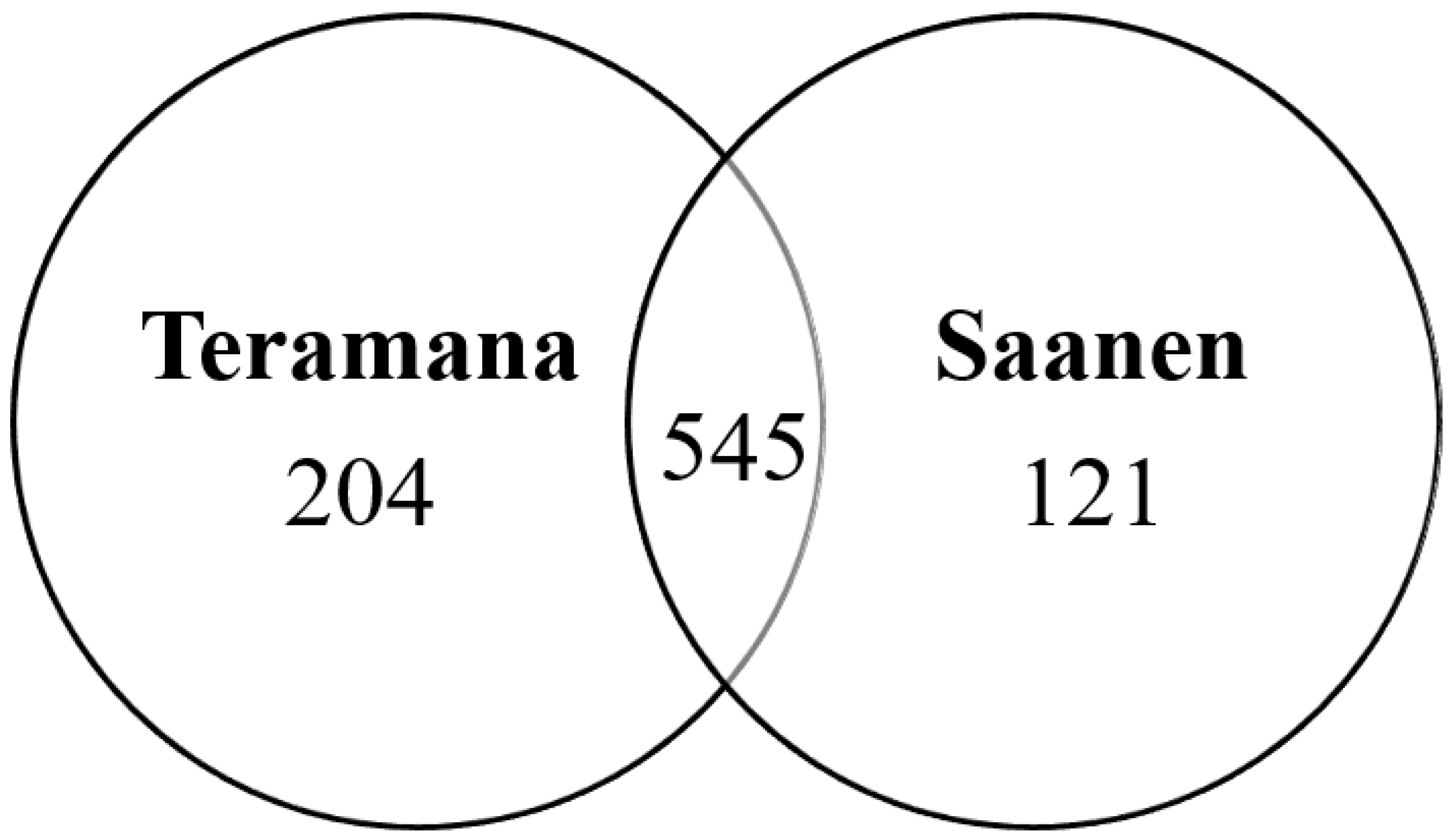
3.1. Identification of Milk Proteins in Goat Breeds

In the present study, 749 (including 3349 unique peptides) and 666 (including 3057 unique peptides) proteins were identified from the Teramana and Saanen goat’s milk, respectively by label-free proteomic analysis (Supplementary Material Tables S1 and S2). Combining these two datasets, this study identified a total of 870 proteins. Among the proteins identified in the Teramana and Saanen goat milk breeds, respectively 204 (23.4%) and 121 (13.9%) proteins were unique to each breed and 545 (62.6%) proteins were common between the two breeds (Figure 1).
The ontology tools in PANTHER indicated that the majority of these proteins (870 proteins) were mainly involved in cellular process 26.9%, in metabolic process 16.3%, biological regulation 11.6%, response to stimulus 8.9%, and cellular component organisation or biogenesis 8.7% (Figure 2). The unique proteins identified in the Teramana were mainly involved in cellular process (29.4%), metabolic process (20%), biological regulation (11.3), cellular component organisation or biogenesis (9.1%), and response to stimulus (8.3%). Whereas the unique proteins identified in the Saanen were mainly involved in cellular process (26.4%), metabolic process (11.7%), biological regulation (11.7), response to stimulus (10.4%), cellular component organisation or biogenesis (8.6%), and signalling (8%) (Supplementary Material Figure S1).

3.2. Label-Free Quantitative Proteomic Analysis of Goat Milk

Label-free quantitative proteomics was used to compare goat milk samples from the Teramana and Saanen breeds. Differences at the proteome level between the two breeds were investigated using the label-free software, Progenesis QI for Proteomics. Normalised proteomics data were used to identify the differentially expressed proteins between the Teramana and Saanen breeds. Differentially expressed proteins were defined as those that showed a fold change cut-off greater than 1.5, a p-value ≤ 0.05 (one-way ANOVA), and a number of unique peptides greater than 2 that matched the protein. Based on these criteria, there were 29 differentially expressed proteins between the Teramana and Saanen goat milk breeds (Table 1), of which 18 (62.1%) were upregulated proteins in the Teramana breed and 11 (37.9%) were upregulated in the Saanen breed. The full list of the 29 proteins identified in this study is shown in Table 1.

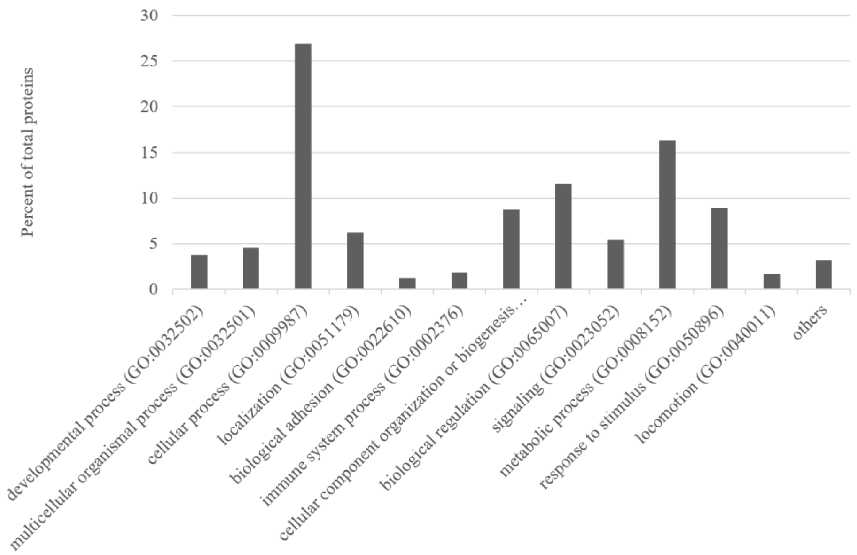
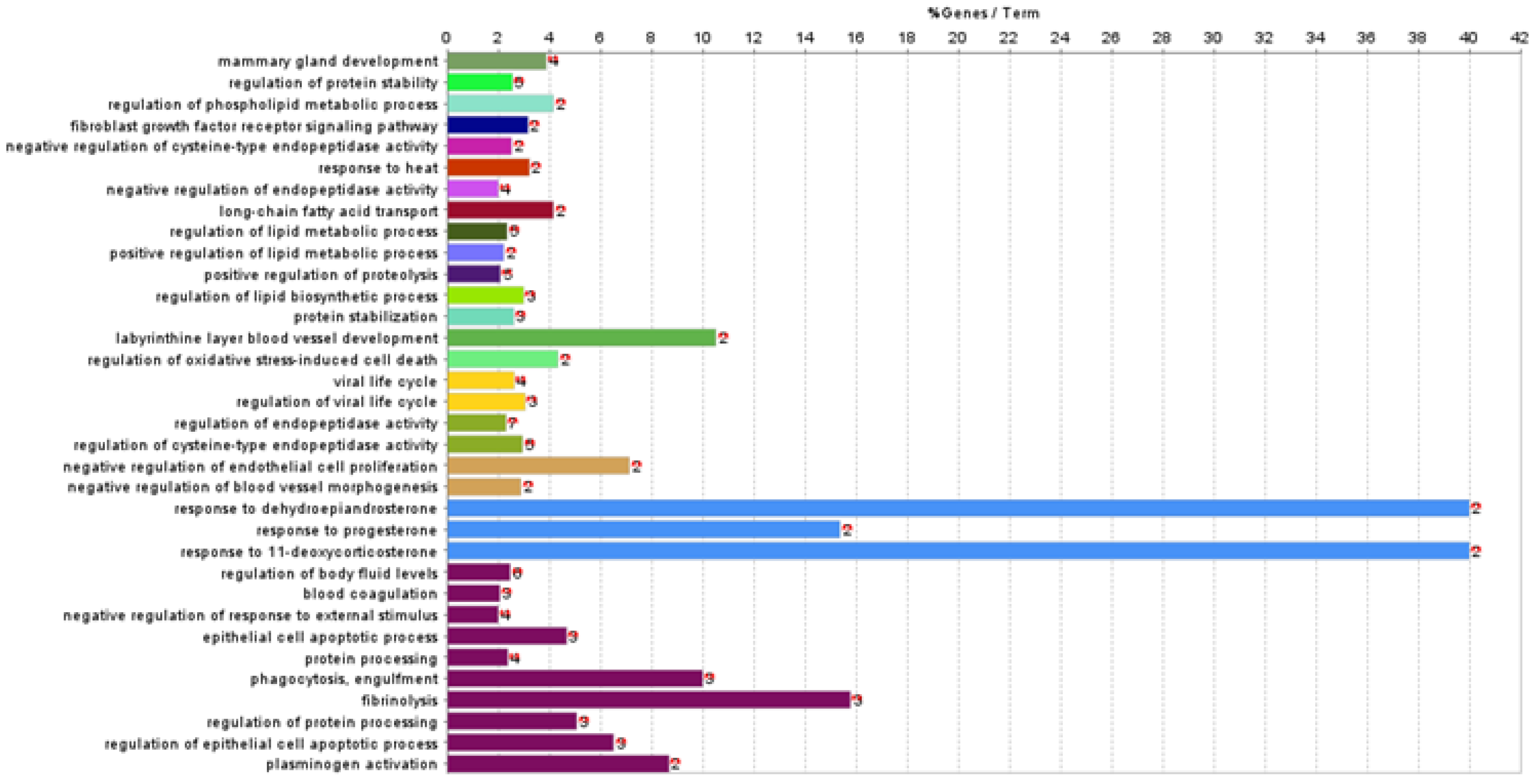
3.3. Functional Association Analysis

Functional association analysis was used to investigate the biological processes and pathways involving the 29 differentially abundant proteins identified. Enrichment analyses were carried out in Cytoscape using the ClueGO plug-in. Analyses were run separately over the GO:BP and KEGG pathway databases. Three proteins (immunoglobulin heavy constant Gamma 4, IGHG4; Ig-like domain-containing protein, rig-5; and cysteine-rich secretory protein 3, Crisp3) were not in the ClueGO annotation sets. A total of 34 GO:BP terms (involving 18 differentially abundant proteins) were retrieved (Table 2, Figure 3). These biological processes can be generally summarised in the following groups: (i) Biological regulation, (ii) cellular and developmental process, (iii) multicellular organismal process, (iv) response to stimulus, (v) signalling.
Over-representation analysis using the KEGG pathway database highlighted a total of four pathways (Table 3, Figure 4) related to the response to stimulus. Eight proteins were involved in these pathways. The differentially abundant proteins involved in the complement and coagulation cascades, as previously shown from the analysis over the GO:BP database, are of higher abundance in the Teramana breed.

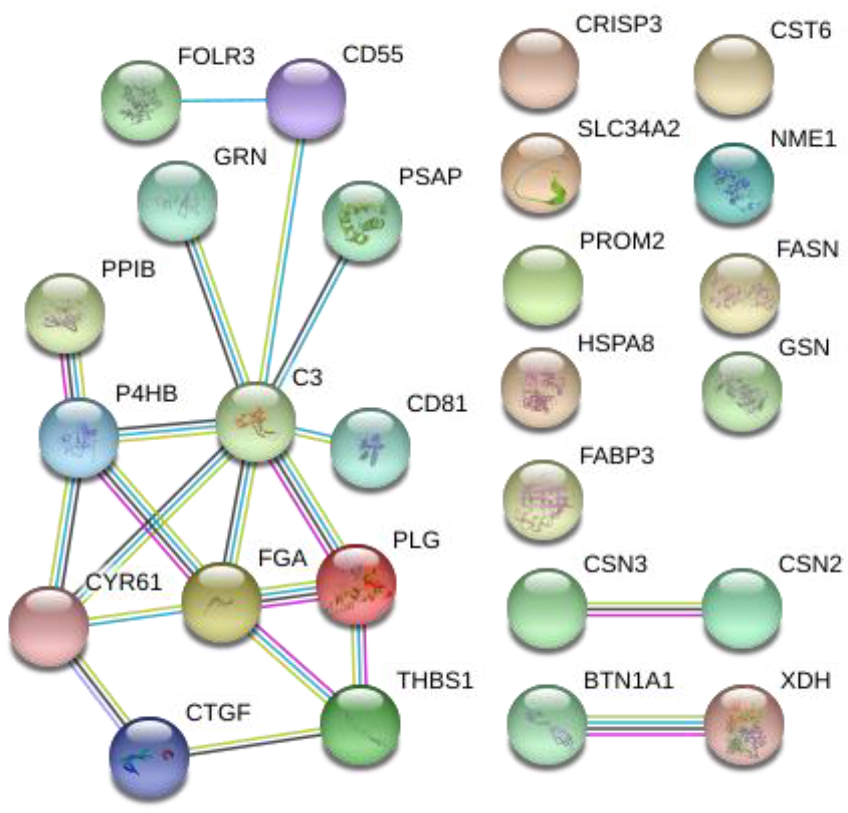
3.4. Protein–Protein Interaction (PPI) Analysis

PPI analysis was performed using STRING with all the significantly different proteins identified in the comparison of two goat breeds (Figure 5). A connected protein network was revealed by the analysis. The protein network was composed of 26 nodes divided in: (i) One big module composed of thirteen nodes (50%), (ii) two small components of two proteins (15.4%), and (iii) nine singletons (34.6%). The resulting network showed a PPI enrichment p-value of 4.32 × 10⁻⁸ (four expected edges vs. twenty detected edges) indicating that proteins are at least partially biologically connected. In this network, most of the proteins interacted with only one or two other partners (average node degree equal to 1.54). Two proteins complement C3 (C3) and fibrinogen alpha chain (FGA) presented the highest degree of connection (eight and five edges, respectively), which may assign to them a role as “hub” proteins playing a putative function of controllers inside biochemical pathways that could potentially lead to cascade of protein expression differences. Both proteins had a higher expression in the Teramana goat breed. Moreover, most of the proteins clustered in the big module were included in the GO and KEGG enrichment processes clearly differentiating the two goat breeds.

4. Discussion

The advancement of electrophoresis and chromatography, along with developments in mass spectrometry technologies, have widened the potential application of proteomics to study milk proteomes from smaller ruminants, such as goats. Milk is an easily accessible body fluid rich in proteins, which are important for tissue growth and cellular functions. Moreover, some proteins can act as hormones, whereas others display antimicrobial properties [1]. These characteristics make milk a promising substrate to investigate proteins and peptides indicative of molecular processes underpinning differences between breeds. Furthermore, this substrate could be easily used for the identification of biomarkers that could enable a genetic improvement in the goat herds and/or to monitor diseases. The aim of the present study was to investigate and compare the whey proteome of the autochthonous Teramana and Saanen goat breeds.
In the last two decades, great efforts have been addressed to increase the study of milk proteomics, especially from bovine. In more recent times, the study of milk proteome from smaller ruminants, such as goat have received great interest, with whole milk, whey, milk fat globule membrane (MFGM) fractions, and casein fractions as the main substrate utilised [2,14,15,16,19,36]. A study of Chen et al. [37] identified a total of 843 proteins by comparing the proteome of goat milk during heated processing using label-free quantification. Using a similar approach, 595 and 486 proteins were characterised in the whey of two autochthon Greek goat breeds [27].
In our study, the label-free LC–MS analysis revealed a total of 870 different proteins in the two goat breeds (Supplementary Material Tables S1 and S2) under study. The comparable and higher number of proteins identified in this study indicates the successful use of proteomic techniques. Many of the identified proteins (e.g., complement C3, fibronectin, plasminogen, calcium and integrin binding 1) were also identified in other studies [26,27,36]. Most of the identified proteins were involved in cellular and metabolic process, biological regulation, response to stimuli, etc. that characterise milk (Figure 2). Comparing the unique proteins identified in each breed, the Teramana breed show a higher number of proteins involved in metabolic process (Supplementary Material Figure S1). A similar class of proteins was also identified in whey from Greek goat and sheep breeds [27]. Furthermore, results are in agreement with those presented in the study of Cunsolo et al. [38], who investigated the goat milk proteome in the Camosciata goat breed.
This work is the first study that applies a label-free method to unravel the milk whey proteome of the Teramana and Saanen goat breeds. Whey proteins show specific characteristics, which reflect the physiological requirements of the animal [1]. Utilising statistical analysis, 29 proteins were differentially expressed between the two breeds, highlighting interspecies difference. To our knowledge, this is the first study to observe proteins that are changing significantly in whey between goat breeds. Using the same method, Zhao al. [39] identified 156 differential MFGM proteins between yak and cow. The higher number of proteins identified in their study compared to ours is mainly due to the different species used for the comparison. As expected, the dominant classes of proteins identified in our substrate were whey proteins; however, residues of caseins were also observed.
Among the 29 proteins changing significantly in milk between the two goat breeds, the top differentially expressed proteins (complement C3, C3; fibrinogen alpha chain, FGA; cellular communication network factor 1, CYR61; plasminogen, PLG; and protein disulfide-isomerase, P4HB) according to their functions, may contribute to explaining in part the phenotypic differences between the Teramana and Saanen goat breeds. Functional analysis of the proteins identified in our study highlights their role in a wide range of regulatory processes, such as lipid metabolic process, regulation of proteolysis, oxidative stress-induced cell death, etc. KEGG pathway shows that most of the whey proteins upregulated in the Teramana breed are involved in complement and coagulation cascades, whereas three pathways were involved in disease-related pathways in both breeds. The complement and coagulation cascades pathway plays an important role in activating innate immunity, in maintaining the balance of the coagulation-fibrinolytic system, and the associated proteins are critical for the health and nutrition of goat kids [40,41,42]. The upregulation observed in the Teramana breed may allude to a greater capacity of this autochthon breed to adapt to harsh environments compared to breeds that are increasingly used from the industry. Whey proteins involved in the same pathways were also highlighted in a study by Sun at al. [26] on the colostrum and mature milk of Xinong Saanen goats.
Complement C3 (C3) was upregulated in the Teramana goat. Complement is a central component of the innate immune system. Its main functions include host defence against agents, facilitating adaptive immune responses and elimination of immune complexes and apoptotic cells [43]. The complement system consists of about 35/40 proteins, which are usually associated with blood cells and blood plasma, but found generally at lower concentrations in other secretions of the body-like milk [44]. Difference of C3 level between Teramana and Saanen breeds might be in-line with a higher defence mechanism of the goat mammary gland against infections of the first breed. It has been shown that the concentration of C3 is significantly higher in preterm human milk than term human milk and it has been postulated that this may be due to the higher requirement of protection from the infants in its early days, as the immune system is less developed than term infants [45]. Similar mechanisms may occur in goat breeds, such as the Teramana that are usually keener to live in harsh environments. These animals are more exposed to pathogens, and thus require a higher protection. Fibrinogen (FGA) is a complex plasma protein required for the last phase of blood coagulation [46]. FGA is one of the acute phase proteins (APPs) which are a serum component whose concentrations vary under external or internal influences, such as inflammation, stress, etc. These proteins are important early diagnostic markers of inflammation in animals [47]. In our study, an upregulation of fibrinogen alpha chain was observed in the Teramana breed whey. It has been shown that the main agent involved in mastitis is Staphylococcus aureus and that molecules, such as fibrinogen can change in abundance during infection [48]. The animals used in this study did not show any signs of mastitis or other clinical diseases and the high concentration of APPs is not always a sign of disease [49], the upregulation of this protein may allude to a stronger resistance of the Teramana breed (namely, in general more rustic compared to commercial breeds) to this kind of disease. Cellular communication network factor 1 (CYR61) and cellular communication network factor 2 (CTGF) (upregulated in the Teramana breed) are cysteine-rich proteins of the CCN family. These proteins mediate many functions, such as cell survival and apoptosis [50,51]. In mammals, a reduced suckling frequency initiates the downregulation of milk synthesis and the induction of apoptotic pathways and structural remodelling of mammary tissue with a corresponding reduction in secretory activity [52]. It has been proposed that CYR61 and CTGF proteins promote apoptosis of mammary epithelial cells, and that their presence points to the mechanisms underlying the lactation inhibition [53]. It is known that the milk production of the Teramana breed is lower compared to the Saanen [11]. The upregulation of these proteins observed in our study in the Teramana breed may suggest a role played by these proteins in the mechanism involved in the lower milk production of the Teramana breed and highlight the potentiality of these proteins as markers to discriminate between the milk of the two breeds. Plasmin is one of the major endogenous protease present in milk that is secreted in its inactive form plasminogen (PLG), which is the predominant form in fresh milk [54]. Plasmin is then activated and inhibited by the levels of plasminogen activators and inhibitors [55]. Theodorou et al. [56] highlighted that among the factors affecting the plasmin-plasminogen system on sheep’s milk, there is the breed factor. Milk contains activins of plasminogen, which can activate fibrinolysis and keep the secretion pipeline unobstructed [26]. In our study, PLG was upregulated in the Teramana breed, which may allude to a stronger resistance of milk duct infections of this breed, namely, in general considered more rustic compared to commercial goat breeds. Moreover, plasmin plays a pivotal role in cheese ripening by breaking down α- and β-caseins to result in flavour and texture development [57]. On the other hand, the activity of this protein has been associated with alteration of mammary epithelium permeability and with an increment in paracellular flow, activities that aggravate the milk quality, coagulation properties, and cheese yield. The presence of mastitis, the increment of the age of the animals, and stage of lactation increase the level of PLG [58]. As the animals used in our study had no signs of mastitis or other clinical diseases and were of a similar age, the higher abundance of PLG observed in the Teramana breed may contribute to the development of a characteristic flavour and texture of the dairy products made by the milk of this autochthon breed. Protein disulfide isomerase (P4HB) is a member of the thioredoxin superfamily of redox proteins that is mainly located in the endoplasmic reticulum. P4HB has multiple roles, is a binding partner of other proteins, acts as a chaperone, is a hormone reservoir, as well as a disulfide isomerase in the formation of disulfide bonds [59]. Given their pivotal role in protein-folding, the loss of P4HB activity and the consequent accumulation of misfolded proteins have been associated with the pathogenesis of numerous disease states [60]. To our knowledge, no other studies observed changes in this protein among dairy breeds, the upregulation observed in the Teramana breed might allude to a stronger resistance to disease of this breed that undergo a limited formal crossbreeding. Indeed, due to the negative genetic correlation evidenced between milk production traits and health traits, the genetic selection for higher milk production has been associated with an increased sensitivity to diseases [61,62].

5. Conclusions

In summary, using label-free proteomics, it was possible to highlight proteomic differences between the whey fraction of the milk from the autochthon Teramana goat breed and Saanen goat breed. Casein and whey proteins are the major proteins of milk, where the casein constitutes approximately 80% of the total protein. An in-depth characterisation of both fractions will benefit dairy productions, will contribute to the conservation on animal biodiversity, and will help in the design of novel products. In this study, we report that the most extensive investigation on the goat whey proteome of a total of 870 proteins was identified. Twenty-nine proteins were able to discriminate between Teramana and Saanen goat breeds with many of the identified proteins involved in complement and coagulation cascades. These findings elucidate the proteome composition of milk whey and quantitative protein profile in the analysed goat’s milk. Moreover, the association between the proteins and milk from the two breeds supports the potential use of proteomic profile as a predictive biomarker in milk substrate that can discriminate between different genetic backgrounds. This may be used to evaluate milk adulteration of specific milk and expand the potential direction for the production of specific milk proteins.

Supplementary Materials

The following supporting information can be downloaded at: https://www.mdpi.com/article/10.3390/ani13142263/s1. Table S1: Full list of the 749 proteins identified by mass spectrometry in the Teramana goat milk determined in Proteome Discoverer using SEQUEST HT algorithm. Table S2: Full list of the 666 proteins identified by mass spectrometry in the Saanen goat milk determined in Proteome Discoverer using SEQUEST HT algorithm. Figure S1: Percentage of milk proteins (over the unique proteins identified in the Teramana and Saanen goat breeds) grouped according to different biological processes.

Author Contributions

Conceptualisation, A.D.L. and G.M.; methodology, A.D.L. and M.H.; formal analysis, A.D.L., M.H., A.I. and F.B.; investigation, A.D.L., A.I. and F.B.; resources, G.M. and P.M.; data curation, A.D.L. and M.H.; writing—original draft preparation, A.D.L.; writing—review and editing, A.I., F.B., L.G., M.H., P.M. and G.M.; supervision, G.M. and A.D.L.; project administration, G.M. and L.G.; funding acquisition, G.M. All authors have read and agreed to the published version of the manuscript.

Funding

This research was funded by the European Union—Next Generation EU. Project Code: ECS00000041; Project CUP: C43C22000380007; Project Title: Innovation, digitalisation, and sustainability for the diffused economy in Central Italy—VITALITY.

Institutional Review Board Statement

No permits were required for the described study, which complied with all relevant regulations, since only milk was collected and no animal sacrifice was necessary.

Informed Consent Statement

Not applicable.

Data Availability Statement

Not applicable.

Acknowledgments

The Orbitrap Fusion Tribrid mass spectrometer was funded under a Science Foundation Ireland (SFI) Infrastructure award to Dublin City University, grant number 16/RI/3701.

Conflicts of Interest

The authors declare no conflict of interest. The funders had no role in the design of the study; in the collection, analyses or interpretation of data; in the writing of the manuscript or in the decision to publish the results.

References

  1. Roncada, P.; Piras, C.; Soggiu, A.; Turk, R.; Urbani, A.; Bonizzi, L. Farm animal milk proteomics. J. Proteom. 2012, 75, 4259–4274. [Google Scholar] [CrossRef] [PubMed]
  2. Cheng, D.; Li, X.Y.; Zhao, X.; Qin, Y.S.; Zhang, X.X.; Li, J.; Wang, J.M.; Wang, C.F. Proteomics and microstructure profiling of goat milk protein after homogenization. J. Dairy Sci. 2019, 102, 3839–3850. [Google Scholar] [CrossRef] [PubMed]
  3. Vargas, M.; Cháfer, M.; Albors, A.; Chiralt, A.; González-Martínez, C. Physicochemical and sensory characteristics of yoghurt produced from mixtures of cows’ and goats’ milk. Int. Dairy J. 2008, 18, 1146–1152. [Google Scholar] [CrossRef]
  4. Ceballos, L.S.; Morales, E.R.; de la Torre Adarve, G.; Castro, J.D.; Martínez, L.P.; Sampelayo, M.R.S. Composition of goat and cow milk produced under similar conditions and analyzed by identical methodology. J. Food Compos. Anal. 2009, 22, 322–329. [Google Scholar] [CrossRef]
  5. Park, Y.W. Hypo-allergenic and therapeutic significance of goat milk. Small Rumin. Res. 1994, 14, 151–159. [Google Scholar] [CrossRef]
  6. Lara-Villoslada, F.; Olivares, M.; Jiménez, J.; Boza, J.; Xaus, J. Goat milk is less immunogenic than cow milk in a murine model of atopy. J. Pediatr. Gastroenterol. Nutr. 2004, 39, 354–360. [Google Scholar] [CrossRef] [Green Version]
  7. Haenlein, G.F.W. Past, present, and future perspectives of small ruminant dairy research. J. Dairy Sci. 2001, 84, 2097–2115. [Google Scholar] [CrossRef]
  8. Faye, B.; Konuspayeva, G. The sustainability challenge to the dairy sector—The growing importance of non-cattle milk production worldwide. Int. Dairy J. 2012, 24, 50–56. [Google Scholar] [CrossRef]
  9. Haenlein, G.F.W. Goat milk in human nutrition. In Small Ruminant Research; Elsevier: Amsterdam, The Netherlands, 2004; Volume 51, pp. 155–163. [Google Scholar]
  10. Di Luca, A.; Ianni, A.; Bennato, F.; Henry, M.; Meleady, P.; Martino, G. A Label-Free Quantitative Analysis for the Search of Proteomic Differences between Goat Breeds. Animals 2022, 12, 3336. [Google Scholar] [CrossRef]
  11. Food and Agriculture Organization of the United Nations Domestic Animal Diversity Information System (DAD-IS). Available online: http://www.fao.org/dad-is (accessed on 23 April 2020).
  12. Auerbach, D.; Thaminy, S.; Hottiger, M.O.; Stagljar, I. The post-genomic era of interactive proteomics: Facts and perspectives. Proteomics 2002, 2, 611. [Google Scholar] [CrossRef]
  13. Lottspeich, F. Proteome analysis: A pathway to the functional analysis of proteins. Angew. Chemie Int. Ed. 1999, 38, 2476. [Google Scholar] [CrossRef]
  14. Cebo, C.; Caillat, H.; Bouvier, F.; Martin, P. Major proteins of the goat milk fat globule membrane. J. Dairy Sci. 2010, 93, 868–876. [Google Scholar] [CrossRef] [PubMed] [Green Version]
  15. Roncada, P.; Gaviraghi, A.; Liberatori, S.; Canas, B.; Bini, L.; Greppi, G.F. Identification of caseins in goat milk. Proteomics 2002, 2, 723–726. [Google Scholar] [CrossRef] [PubMed]
  16. Di Gerlando, R.; Tolone, M.; Sutera, A.M.; Monteleone, G.; Portolano, B.; Sardina, M.T.; Mastrangelo, S. Variation of proteomic profile during lactation in Girgentana goat milk: A preliminary study. Ital. J. Anim. Sci. 2019, 18, 88–97. [Google Scholar] [CrossRef] [Green Version]
  17. Neilson, K.A.; Ali, N.A.; Muralidharan, S.; Mirzaei, M.; Mariani, M.; Assadourian, G.; Lee, A.; van Sluyter, S.C.; Haynes, P.A. Less label, more free: Approaches in label-free quantitative mass spectrometry. Proteomics 2011, 11, 535. [Google Scholar] [CrossRef]
  18. Becker, C.H.; Bern, M. Recent developments in quantitative proteomics. Mutat. Res. 2011, 722, 171. [Google Scholar] [CrossRef] [Green Version]
  19. Lu, J.; Wang, X.; Zhang, W.; Liu, L.; Pang, X.; Zhang, S.; Lv, J. Comparative proteomics of milk fat globule membrane in different species reveals variations in lactation and nutrition. Food Chem. 2016, 196, 665–672. [Google Scholar] [CrossRef]
  20. Ginger, M.R.; Grigor, M.R. Comparative aspects of milk caseins. Comp. Biochem. Physiol.—B Biochem. Mol. Biol. 1999, 124, 133–145. [Google Scholar] [CrossRef]
  21. Fox, P.F.; Kelly, A.L. Developments in the chemistry and technology of milk proteins. 2. Minor milk proteins. Food Australia 2003, 55, 104–108. [Google Scholar]
  22. Affolter, M.; Grass, L.; Vanrobaeys, F.; Casado, B.; Kussmann, M. Qualitative and quantitative profiling of the bovine milk fat globule membrane proteome. J. Proteom. 2010, 73, 1079–1088. [Google Scholar] [CrossRef]
  23. Hogarth, C.J.; Fitzpatrick, J.L.; Nolan, A.M.; Young, F.J.; Pitt, A.; Eckersall, P.D. Differential protein composition of bovine whey: A comparison of whey from healthy animals and from those with clinical mastitis. Proteomics 2004, 4, 2094–2100. [Google Scholar] [CrossRef] [PubMed]
  24. da Costa, W.K.A.; de Souza, E.L.; Beltrão-Filho, E.M.; Vasconcelos, G.K.V.; Santi-Gadelha, T.; de Almeida Gadelha, C.A.; Franco, O.L.; Magnani, M. Comparative Protein Composition Analysis of Goat Milk Produced by the Alpine and Saanen Breeds in Northeastern Brazil and Related Antibacterial Activities. PLoS ONE 2014, 9, e93361. [Google Scholar] [CrossRef] [PubMed] [Green Version]
  25. Ma, Y.; Zhang, L.; Wu, Y.; Zhou, P. Changes in milk fat globule membrane proteome after pasteurization in human, bovine and caprine species. Food Chem. 2019, 279, 209–215. [Google Scholar] [CrossRef] [PubMed]
  26. Sun, Y.; Wang, C.; Sun, X.; Guo, M. Proteomic analysis of whey proteins in the colostrum and mature milk of Xinong Saanen goats. J. Dairy Sci. 2020, 103, 1164–1174. [Google Scholar] [CrossRef]
  27. Anagnostopoulos, A.K.; Katsafadou, A.I.; Pierros, V.; Kontopodis, E.; Fthenakis, G.C.; Arsenos, G.; Karkabounas, S.C.; Tzora, A.; Skoufos, I.; Tsangaris, G.T. Milk of Greek sheep and goat breeds; characterization by means of proteomics. J. Proteom. 2016, 147, 76–84. [Google Scholar] [CrossRef]
  28. Bradford, M.M. A rapid and sensitive method for the quantitation of microgram quantities of protein utilizing the principle of protein-dye binding. Anal. Biochem. 1976, 72, 248–254. [Google Scholar] [CrossRef]
  29. Wisniewski, J.R.; Zougman, A.; Nagaraj, N.; Mann, M. Universal sample preparation method for proteome analysis. Nat. Methods 2009, 6, 359–362. [Google Scholar] [CrossRef]
  30. Coleman, O.; Henry, M.; Clynes, M.; Meleady, P. Filter-Aided Sample Preparation (FASP) for Improved Proteome Analysis of Recombinant Chinese Hamster Ovary Cells. Methods Mol. Biol. 2017, 1603, 187–194. [Google Scholar]
  31. Mi, H.; Muruganujan, A.; Ebert, D.; Huang, X.; Thomas, P.D. PANTHER version 14: More genomes, a new PANTHER GO-slim and improvements in enrichment analysis tools. Nucleic Acids Res. 2018, 47, D419–D426. [Google Scholar] [CrossRef] [Green Version]
  32. Shannon, P.; Markiel, A.; Ozier, O.; Baliga, N.S.; Wang, J.T.; Ramage, D.; Amin, N.; Schwikowski, B.; Ideker, T. Cytoscape: A software environment for integrated models of biomolecular interaction networks. Genome Res. 2003, 13, 2498–2504. [Google Scholar] [CrossRef]
  33. Bindea, G.; Mlecnik, B.; Hackl, H.; Charoentong, P.; Tosolini, M.; Kirilovsky, A.; Fridman, W.-H.; Pagès, F.; Trajanoski, Z.; Galon, J. ClueGO: A Cytoscape plug-in to decipher functionally grouped gene ontology and pathway annotation networks. Bioinformatics 2009, 25, 1091–1093. [Google Scholar] [CrossRef] [PubMed] [Green Version]
  34. Bovo, S.; Di Luca, A.; Galimberti, G.; Dall’Olio, S.; Fontanesi, L. A comparative analysis of label-free liquid chromatography-mass spectrometry liver proteomic profiles highlights metabolic differences between pig breeds. PLoS ONE 2018, 13, e0199649. [Google Scholar] [CrossRef] [PubMed] [Green Version]
  35. Szklarczyk, D.; Gable, A.L.; Lyon, D.; Junge, A.; Wyder, S.; Huerta-Cepas, J.; Simonovic, M.; Doncheva, N.T.; Morris, J.H.; Bork, P.; et al. STRING v11: Protein-protein association networks with increased coverage, supporting functional discovery in genome-wide experimental datasets. Nucleic Acids Res. 2019, 47, D607–D613. [Google Scholar] [CrossRef] [PubMed] [Green Version]
  36. Lu, J.; Liu, L.; Pang, X.; Zhang, S.; Jia, Z.; Ma, C.; Zhao, L.; Lv, J. Comparative proteomics of milk fat globule membrane in goat colostrum and mature milk. Food Chem. 2016, 209, 10–16. [Google Scholar] [CrossRef]
  37. Chen, D.; Li, X.; Zhao, X.; Qin, Y.; Wang, J.; Wang, C. Comparative proteomics of goat milk during heated processing. Food Chem. 2019, 275, 504–514. [Google Scholar] [CrossRef]
  38. Cunsolo, V.; Fasoli, E.; Saletti, R.; Muccilli, V.; Gallina, S.; Righetti, P.G.; Foti, S. Zeus, Aesculapius, Amalthea and the proteome of goat milk. J. Proteom. 2015, 128, 69–82. [Google Scholar] [CrossRef]
  39. Zhao, L.; Du, M.; Gao, J.; Zhan, B.; Mao, X. Label-free quantitative proteomic analysis of milk fat globule membrane proteins of yak and cow and identification of proteins associated with glucose and lipid metabolism. Food Chem. 2019, 275, 59–68. [Google Scholar] [CrossRef]
  40. Keragala, C.B.; Draxler, D.F.; McQuilten, Z.K.; Medcalf, R.L. Haemostasis and innate immunity—A complementary relationship. Br. J. Haematol. 2018, 180, 782–798. [Google Scholar] [CrossRef] [Green Version]
  41. Moreno-Indias, I.; Dodds, A.W.; Argüello, A.; Castro, N.; Sim, R.B. The complement system of the goat: Haemolytic assays and isolation of major proteins. BMC Vet. Res. 2012, 8, 91. [Google Scholar] [CrossRef] [Green Version]
  42. Moreno-Indias, I.; Morales-delaNuez, A.; Hernández-Castellano, L.E.; Sánchez-Macías, D.; Capote, J.; Castro, N.; Argüello, A. Docosahexaenoic acid in the goat kid diet: Effects on immune system and meat quality1. J. Anim. Sci. 2012, 90, 3729–3738. [Google Scholar] [CrossRef]
  43. Janssen, B.J.C.; Huizinga, E.G.; Raaijmakers, H.C.A.; Roos, A.; Daha, M.R.; Nilsson-Ekdahl, K.; Nilsson, B.; Gros, P. Structures of complement component C3 provide insights into the function and evolution of immunity. Nature 2005, 437, 505–511. [Google Scholar] [CrossRef] [PubMed] [Green Version]
  44. Rainard, P. The complement in milk and defense of the bovine mammary gland against infections. Vet. Res. 2003, 34, 647–670. [Google Scholar] [CrossRef] [PubMed] [Green Version]
  45. Tregoat, V.; Montagne, P.; Cuilliere, M.L.; Bene, M.C.; Faure, G.C. Sequential C3 and C4 levels in human milk in relation to prematurity and parity. Clin. Chem. Lab. Med. 2000, 38, 609–613. [Google Scholar] [CrossRef] [PubMed]
  46. Prunkard, D.; Cottingham, I.; Garner, I.; Bruce, S.; Dalrymple, M. High-level expression of recombinant human fibrinogen in the milk of transgenic mice. Nat. Biotechnol. 1996, 14, 867–871. [Google Scholar] [CrossRef]
  47. Petersen, H.H.; Nielsen, J.P.; Heegaard, P.M.H. Application of acute phase protein measurements in veterinary clinical chemistry. Vet. Res. 2004, 35, 163–187. [Google Scholar] [CrossRef] [Green Version]
  48. Vet, T.J.; Sci, A.; Fasulkov, I.; Karadaev, M.; Vasilev, N.; Urumova, V.; Mircheva, T. Determination of plasma fibrinogen and haptoglobin, hematological and blood biochemical changes in Bulgarian local goats with experimentally induced Staphylococcus aureus mastitis. Turkish J. Vet. Anim. Sci. 2014, 38, 439–444. [Google Scholar]
  49. Nagy, O.; Tóthová, C.; Nagyová, V.; Kováč, G.; Pošivák, J. Changes in the serum protein electrophoretic pattern in lambs during the first month of life. Acta Vet. Brno 2014, 83, 187–193. [Google Scholar] [CrossRef] [Green Version]
  50. Yang, G.P.; Lau, L.F. Cell Growth & Differentiation Cyr6l, Product of a Growth Factor-inducible Immediate Early Gene, Is Associated with the Extracellular Matrix and the Cell Surface. Cell Growth Differ. 1991, 2, 351–357. [Google Scholar]
  51. Sherbet, G.V. Connective Tissue Growth Factor. In Growth Factors and Their Receptors in Cell Differentiation, Cancer and Cancer Therapy; Elsevier: Amsterdam, The Netherlands, 2011; pp. 105–110. [Google Scholar]
  52. Stelwagen, K.; Knight, C.H. Effect of unilateral once or twice daily milking of cows on milk yield and udder characteristics in early and late lactation. J. Dairy Res. 1997, 64, 487–494. [Google Scholar] [CrossRef]
  53. Littlejohn, M.D.; Walker, C.G.; Ward, H.E.; Lehnert, K.B.; Snell, R.G.; Verkerk, G.A.; Spelman, R.J.; Clark, D.A.; Davis, S.R. Effects of reduced frequency of milk removal on gene expression in the bovine mammary gland. Physiol. Genom. 2010, 41, 21–32. [Google Scholar] [CrossRef] [Green Version]
  54. Ozen, B.F.; Hayes, K.D.; Mauer, L.J. Measurement of plasminogen concentration and differentiation of plasmin and plasminogen using Fourier-transform infrared spectroscopy. Int. Dairy J. 2003, 13, 441–446. [Google Scholar] [CrossRef]
  55. Ismail, B.; Nielsen, S.S. Invited review: Plasmin protease in milk: Current knowledge and relevance to dairy industry. J. Dairy Sci. 2010, 93, 4999–5009. [Google Scholar] [CrossRef] [Green Version]
  56. Theodorou, G.; Kominakis, A.; Rogdakis, E.; Politis, I. Factors affecting the plasmin-plasminogen system in milk obtained from three Greek dairy sheep breeds with major differences in milk production capacity. J. Dairy Sci. 2007, 90, 3263–3269. [Google Scholar] [CrossRef] [PubMed]
  57. Bastian, E.D.; Brown, R.J. Plasmin in milk and dairy products: An update. Int. Dairy J. 1996, 6, 435–457. [Google Scholar] [CrossRef]
  58. Battacone, G.; Cannas, E.A.; Mazzette, A.; Dimauro, C.; Enne, G. Why does the increase of plasmin worsen the coagulation properties of milk in dairy sheep? Ital. J. Anim. Sci. 2016, 4, 342–344. [Google Scholar] [CrossRef] [Green Version]
  59. Ali Khan, H.; Mutus, B. Protein disulfide isomerase a multifunctional protein with multiple physiological roles. Front. Chem. 2014, 2, 70. [Google Scholar] [CrossRef] [Green Version]
  60. Galligan, J.J.; Petersen, D.R. The human protein disulfide isomerase gene family. Hum. Genom. 2012, 6, 6. [Google Scholar] [CrossRef] [Green Version]
  61. Pryce, J.E.; Royal, M.D.; Garnsworthy, P.C.; Mao, I.L. Fertility in the high-producing dairy cow. In Livestock Production Science; Elsevier: Amsterdam, The Netherlands, 2004; Volume 86, pp. 125–135. [Google Scholar]
  62. Royal, M.D.; Darwash, A.O.; Flint, A.P.F.; Webb, R.; Woolliams, J.A.; Lamming, G.E. Declining fertility in dairy cattle: Changes in traditional and endocrine parameters of fertility. Anim. Sci. 2000, 70, 487–501. [Google Scholar] [CrossRef]
Figure 1. Venn diagrams showing the total number of proteins identified in the Teramana goat milk compared to Saanen goat milk. The full list of proteins are in Supplementary Material Tables S1 and S2.
Animals 13 02263 g001
Figure 2. Percentage of milk proteins (over 870 identified proteins; Supplementary Material Tables S1 and S2) grouped according to different biological processes.
Animals 13 02263 g002
Figure 3. Gene enrichment analyses over the Gene Ontology—Biological Process branch of the 29 differentially abundant milk proteins between the Teramana and Saanen goat breeds. Bars represent the percentage of input proteins found associated with respect to the number of proteins directly annotated with the functional term. The term significance and the number of input proteins related to the term are reported next to each bar. Detailed statistics are reported in Table 2. In each panel, bars sharing a specific colour are clustered in the same functional group (Table 2).
Animals 13 02263 g003
Figure 4. Gene enrichment analyses over the KEGG pathway database of the 29 differentially abundant milk proteins between the Teramana and Saanen goat breeds. Bars represent the percentage of input proteins found associated with respect to the number of proteins directly annotated with the functional term. The term significance and the number of input proteins related to the term are reported next to each bar. Detailed statistics are reported in Table 3.
Animals 13 02263 g004
Figure 5. Protein–protein interaction (PPI) network of the 29 differentially abundant proteins in goat milk. Each node represents a protein, and different line colours represent the types of evidence for the association: Cyan is from curated databases, magenta is experimentally determined, dark green is gene neighborhood, blue is gene co-occurrence, light green is text-mining, black is co-expression, and purple is protein homology.
Animals 13 02263 g005
Table 1. Twenty-nine proteins identified as differentially expressed in goat milk between the Teramana and Saanen breeds following label-free MS/MS analysis (Progenesis QI for proteomics). Proteins are ordered according to the number of peptides identified.
UniProt *Gene NameDescriptionPeptides §Score ¥ANOVA (p)Fold ChangeHighest Condition ǂ
A0A452DX18C3Complement C3 2794.920.00974.03Teramana
A0A452F1G2THBS1Thrombospondin 1 924.440.000724.62Teramana
A0A452DRX5PSAPProsaposin 618.880.00312.63Teramana
A0A452FHJ7GSNGelsolin 620.570.00561.52Teramana
A0A452E7A0PLGPlasminogen 622.230.02412.27Teramana
A0A452FK23CSN2Beta-casein 410.730.00862.06Teramana
A0A452G9D9CSN3Kappa-casein 436.550.01402.53Teramana
A0A452E2Y0CLUClusterin 415.790.02924.47Teramana
D6PX62Crisp3Cysteine-rich secretory protein 3 39.860.002638.63Teramana
A0A452G7V5P4HBProtein disulfide-isomerase 39.500.02452.12Teramana
A0A452EZB5CD55Complement decay-accelerating factor 37.990.02673.89Teramana
A0A452F9Y8PPIBPeptidyl-prolyl cis-trans isomerase 310.260.03411.98Teramana
A0A452GBG4CYR61Cellular communication network factor 1 27.980.000123.17Teramana
A0A452EKN2IGHG4Immunoglobulin heavy constant gamma 426.110.00069.56Teramana
A0A452DZD6rig-5Ig-like domain-containing protein 28.560.00291.51Teramana
A0A452G5W1GRNGranulin precursor 26.710.00343.02Teramana
A0A452F4L3FGAFibrinogen alpha chain 25.470.01942.46Teramana
A0A452G077CTGFCellular communication network factor 2 26.670.04026.69Teramana
A0A452EZW6XDHFAD-binding PCMH-type domain-containing protein 1438.580.00891.96Saanen
A0A452EYF6HSPA8Heat shock cognate 71 kDa protein 515.530.00111.72Saanen
A0A452ERT5SLC34A2Solute carrier family 34 member 2 49.430.00025.17Saanen
A0A452E4K0CST6Cystatin E/M 415.210.00412.86Saanen
Q6S4N9FABP3Fatty acid binding protein 3 37.850.00021.75Saanen
A0A452FN55BTN1A1Butyrophilin subfamily 1 member A138.430.00581.76Saanen
A0A452F4U3FASNFatty acid synthase 310.660.01044.12Saanen
A0A452G9E6FOLR3Folate_rec domain-containing protein38.190.01641.83Saanen
A0A452F3X9PROM2Prominin 225.840.01162.99Saanen
A0A452E0U0CD81Tetraspanin 213.070.02112.28Saanen
A0A452F230NME1Nucleoside diphosphate kinase 24.550.03201.52Saanen
Eighteen proteins were upregulated in milk from the Teramana breed and eleven proteins were upregulated in milk from Saanen goat breed. (*) Accession number in the UniProt database; (§) unique peptides used for quantitation; (¥) SEQUEST score. (ǂ) Indicates whether the proteins were upregulated in the Teramana or in the Saanen goat breeds.
Table 2. Over-represented biological processes (GO:BP) associated with upregulated or downregulated proteins in the goat milk breed comparison.
GOIDDescriptionFunctional Group 1p-Value 2% of Associated
Proteins 3
No. of
Proteins
Upregulated or Downregulated Proteins 4
GO:0030879mammary gland developmentG00.0000573.884CSN2 ↓, CSN3 ↓, FASN ↑, XDH ↑
GO:0031647regulation of protein stabilityG10.0000532.565CD81 ↑, CLU ↓, CSN3 ↓, GSN ↓, PPIB ↓,
GO:1903725regulation of phospholipid metabolic processG20.0031204.172CD81 ↑, FABP3 ↑
GO:0008543fibroblast growth factor receptor signalling pathwayG30.0046633.172CTGF ↓, THBS1 ↓
GO:2000117negative regulation of cysteine-type endopeptidase activityG40.0068242.532CSN2 ↓, THBS1 ↓
GO:0009408response to heatG50.0048203.232CSN2 ↓, PLG ↓
GO:0010951negative regulation of endopeptidase activityG60.0004182.004C3 ↓, CSN2 ↓, CST6 ↑, THBS1 ↓
GO:0015909long-chain fatty acid transportG70.0031204.172FABP3 ↑, THBS1 ↓
GO:0019216regulation of lipid metabolic processG80.0000602.365C3 ↓, CD81 ↑, CYR61 ↓, FABP3 ↑, PSAP ↓
GO:0045834positive regulation of lipid metabolic processG90.0078582.222CD81 ↑, CYR61 ↓
GO:0045862positive regulation of proteolysisG100.0000712.095C3 ↓, CLU ↓, CYR61 ↓, GSN ↓, XDH ↑
GO:0046890regulation of lipid biosynthetic processG110.0007333.003C3 ↓, CYR61 ↓, FABP3 ↑
GO:0050821protein stabilisationG120.0010112.613CLU ↓, CSN3 ↓, PPIB ↓
GO:0060716labyrinthine layer blood vessel developmentG130.00068010.532CYR61 ↓, PLG ↓
GO:1903201regulation of oxidative stress-induced cell deathG140.0030884.352P4HB ↓, PSAP ↓
GO:0019058viral life cycleG150.0001862.634CD81 ↑, GSN ↓, P4HB ↓, PPIB ↓
GO:1903900regulation of viral life cycleG150.0001863.063GSN ↓, P4HB ↓, PPIB ↓
GO:0052548regulation of endopeptidase activityG160.0000042.317C3 ↓, CSN2 ↓, CST6 ↑, CYR61 ↓, GSN ↓, THBS1 ↓, XDH ↑
GO:2000116regulation of cysteine-type endopeptidase activityG160.0000042.965CSN2 ↓, CYR61 ↓, GSN ↓, THBS1 ↓, XDH ↑
GO:0001937negative regulation of endothelial cell proliferationG170.0069287.142THBS1 ↓, XDH ↑
GO:2000181negative regulation of blood vessel morphogenesisG170.0069282.902THBS1 ↓, XDH ↑
GO:1903494response to dehydroepiandrosteroneG180.00039040.002CSN2 ↓, CSN3 ↓
GO:0032570response to progesteroneG180.00039015.382CSN2 ↓, CSN3 ↓
GO:1903496response to 11-deoxycorticosteroneG180.00039040.002CSN2 ↓, CSN3 ↓
GO:0050878regulation of body fluid levelsG190.0000142.486CSN2 ↓, CSN3 ↓, FGA ↓, PLG ↓, THBS1 ↓, XDH ↑
GO:0007596blood coagulationG190.0000142.073FGA ↓, PLG ↓, THBS1 ↓
GO:0032102negative regulation of response to external stimulusG190.0000142.004CSN2 ↓, FGA ↓, PLG ↓, THBS1 ↓
GO:1904019epithelial cell apoptotic processG190.0000144.693FGA ↓, GSN ↓, THBS1 ↓
GO:0016485protein processingG190.0000142.384C3 ↓, FGA ↓, GSN ↓, THBS1 ↓
GO:0006911phagocytosis, engulfmentG190.00001410.003C3 ↓, GSN ↓, THBS1 ↓
GO:0042730fibrinolysisG190.00001415.793FGA ↓, PLG ↓, THBS1 ↓
GO:0070613regulation of protein processingG190.0000145.083C3 ↓, GSN ↓, THBS1 ↓
GO:1904035regulation of epithelial cell apoptotic processG190.0000146.523FGA ↓, GSN ↓, THBS1 ↓
GO:0031639plasminogen activationG190.0000148.702FGA ↓, THBS1 ↓
1 Groups of closely-related terms; 2 Benjamini–Hochberg corrected p-values; 3 percentage of input proteins found associated with respect to the number of proteins directly annotated with the functional term; 4 the symbol ↑ indicates protein abundance higher in Saanen goat milk than Teramana goat milk, while the symbol ↓ indicates protein abundance higher in Teramana goat milk than Saanen goat milk.
Table 3. Over-represented KEGG pathways associated with upregulated or downregulated proteins in the goat breed milk comparison.
GOIDDescriptionFunctional
Group 1
p-Value 2% of Associated
Proteins 3
No. of
Proteins
Upregulated or Downregulated Proteins 4
KEGG:04610Complement and coagulation cascadesG00.0000045.565C3 ↓, CD55 ↓, CLU ↓, FGA ↓, PLG ↓
KEGG:05134LegionellosisG10.0131923.452C3 ↓, HSPA8 ↑
KEGG:05144MalariaG20.0093933.332CD81 ↑, THBS1 ↓
KEGG:05150Staphylococcus aureus infectionG30.0097552.822C3 ↓, PLG ↓
1 Groups of closely-related terms; 2 Benjamini–Hochberg corrected p-values; 3 percentage of input proteins found associated with respect to the number of proteins directly annotated with the functional term; 4 the symbol ↑ indicates protein abundance higher in Saanen goat milk than Teramana goat milk, while the symbol ↓ indicates protein abundance higher in Teramana goat milk than Saanen goat milk.
Disclaimer/Publisher’s Note: The statements, opinions and data contained in all publications are solely those of the individual author(s) and contributor(s) and not of MDPI and/or the editor(s). MDPI and/or the editor(s) disclaim responsibility for any injury to people or property resulting from any ideas, methods, instructions or products referred to in the content.

