Article

Antibiotic Resistance of Enterococcus Species in Ornamental Animal Feed

1
Microbiology and Antibiotic Resistance Team (MicroART), Department of Veterinary Sciences, University of Trás-os-Montes and Alto Douro, 5000-801 Vila Real, Portugal
2
Associated Laboratory for Green Chemistry (LAQV-REQUIMTE), University NOVA of Lisbon, 1099-085 Caparica, Portugal
3
Toxicology Research Unit (TOXRUN), University Institute of Health Sciences, Advanced Polytechnic and University Cooperative (IUCS-CESPU), 4585-116 Gandra, Portugal
4
Department of Zootechnics, University of Trás-os-Montes and Alto Douro, 5000-801 Vila Real, Portugal
5
Veterinary and Animal Research Centre (CECAV), University of Trás-os-Montes and Alto Douro, 5000-801 Vila Real, Portugal
6
Associate Laboratory for Animal and Veterinary Science (AL4AnimalS), University of Trás-os-Montes and Alto Douro, 5000-801 Vila Real, Portugal
7
Department of Genetics and Biotechnology, University of Trás-os-Montes and Alto Douro, 5000-801 Vila Real, Portugal
8
Functional Genomics and Proteomics Unit, University of Trás-os-Montes and Alto Douro, 5000-801 Vila Real, Portugal
*
Author to whom correspondence should be addressed.
These authors contributed equally to this work.
Animals 2023, 13(11), 1761; https://doi.org/10.3390/ani13111761
Submission received: 18 April 2023 / Revised: 21 May 2023 / Accepted: 23 May 2023 / Published: 26 May 2023

Abstract

:

Simple Summary

Numerous studies have already reported the presence of different antibiotic-resistant Enterococcus species in food-producing animals, animal products and pet food. However, studies specifically evaluating antimicrobial resistance in Enterococcus spp. in ornamental animals and their food are scarce. Therefore, this study aimed to identify Enterococcus spp. and their antibiotic-resistant patterns in ornamental animal feed, as this could lead to the spread of antimicrobial resistance to humans due to their close contact with these animals.

Abstract

Enterococcus is a bacterial genus that is strongly associated with nosocomial infections and has a high capacity to transfer and acquire resistance genes. In this study, the main objective was to evaluate the presence of Enterococcus species in ornamental animal feed and characterize their antimicrobial resistance and virulence factors. Antimicrobial susceptibility was determined using 14 antimicrobial agents by the disk diffusion method, complemented by genotypic analysis to identify Enterococcus species and the presence of 14 antimicrobial resistance and 10 virulence genes. From 57 samples of ornamental animal feed, 103 Enterococcus isolates were recovered from 15 bird, 9 fish and 4 reptile feed samples. Enterococcus isolates were highly resistance to rifampicin (78%) and erythromycin (48%), and 48% of isolates were classified as multidrug-resistant. Enterococcus faecalis (36.7%) and E. faecium (31.7%) were the species most frequently identified. Most isolates carried the resistance genes ermB (57%) and tetL (52%) and the virulence genes, cylL (52%) and esp (40%). Enterococcus gallinarum was the species with the highest number of multidrug-resistant isolates (50%) and virulence genes (80%). These results highlight the high levels of antibiotic-resistant Enterococcus spp. present in ornamental animal feed and the growing interaction of these animals with humans as a public health concern.

1. Introduction

Enterococcus is a bacterial genus predominantly found in the intestinal microbiota of humans and animals [1] and is sporadically detected in the oral cavity, upper respiratory system and vaginal tract [2]. These bacteria are often found in food, plants, water and soil due to fecal contamination [3]. Currently, several Enterococcus species are known, such as E. avium, E. asini, E. casseliflavus, E. cecorum, E. dispar, E. durans, E. faecalis, E. faecium and E. gallinarum [4,5]. However, E. faecalis and E. faecium are the two most isolated and studied species. This group of microorganisms is strongly associated with nosocomial infections, such as endocarditis, septicemia, wound and urinary tract infections and meningitis [6,7,8,9]. Infections caused by Enterococcus spp. have identified isolates with resistance to several antimicrobial classes, including aminoglycosides, β-lactams, glycopeptides, streptogramins and lincosamides. Cases of multidrug resistance (resistance to ≥3 antimicrobial classes) have been reported [10]. In addition, Enterococcus demonstrates an enormous capacity to transfer and acquire resistance genes, produce biofilms and virulence factors and facilitate the induction of inflammatory processes and antimicrobial resistance [11,12]. Furthermore, this high capacity to antibiotic resistance of Enterococcus can be acquired through horizontal exchange of genetic material, carriers’ resistance determinants or sporadic mutation of intrinsic genes, or it can be intrinsic to the species. For instance, E. faecalis is intrinsically resistant to several antibiotics like streptogramins and lincosamides [10].
Antimicrobial resistance represents a serious risk to the environment and especially to human and animal health [13], apart from the economic consequences resulting from the increase in healthcare costs [14]. This silent pandemic has been aggravated by the inappropriate and overuse of antibiotics in human and animal medicine [15]. The contact between humans and animals is increasing, either with animals produced for human consumption or with companion animals, including ornamental animals. This proximity may have implications for the epidemiology of these microorganisms and public health. Animals are described as potential reservoirs of antimicrobial resistance, contributing to the dissemination of resistance genes in the environment and/or transfer to other animals and humans. Several studies have already demonstrated the presence of antibiotic-resistant Enterococcus spp., mainly in food-producing animals and animal foods [16,17,18]. Regarding ornamental animals, limited data are available where isolates of Enterococcus spp. have been researched, although the interaction of these animals with humans has increased in recent years. Ahmed et al. [19] highlighted that certain multidrug-resistant and virulent zoonotic bacteria carried by ornamental birds can pose a risk to other birds and to their owners or caretakers [19].
Thus, the objectives of this study were to evaluate the presence of different Enterococcus species in samples of food supplied to ornamental animals and characterize their phenotypic and genotypic antibiotic resistance and virulence genes.

2. Materials and Methods

2.1. Isolates

A total of 57 samples of ornamental animal feed (birds, fish, mammals and reptiles) were obtained from a range of pet food supermarkets between February and December 2020. From each sample of feed, 2 g were collected, while 1 g was enriched in 5 mL of brain heart infusion broth (Liofilchem® s.r.l., Roseto d. Abruzzi, Italy) at 37 °C for 12 h. Enterococcus putative isolates were obtained from plates with Slanetz–Bartley agar (Liofilchem® s.r.l., Roseto d. Abruzzi, Italy), supplemented with vancomycin (4 μg mL−1), after a period of incubation at 37 °C for 24–48 h. Up to four typical colonies per sample with compatible enterococcal morphology were isolated or incubated in kanamycin aesculin azide agar (Liofilchem® s.r.l., Roseto d. Abruzzi, Italy), and their identification was confirmed by standard biochemical tests like Gram staining, catalase test and growth in the presence of 6.5% NaCl [20]. The remaining samples were used to evaluate the total microbial level or viable bacterial growth present in each sample as described in the next point.
For species characterization and based on the origin of the sample and observation of different macroscopic morphology of colonies, 60 representative isolates of all obtained Enterococcus, confirmed by microbiological and biochemical methods, were selected for the detection of the following species: E. faecalis (ddl E. faecalis), E. faecium (ddl E. faecium), E. gallinarum (vanC1) and E. durans (mur-2). DNA extraction was performed using the commercial GRS Genomic DNA Kit (GRISP Research Solutions, Porto, Portugal), following the manufacturer’s instructions. For each species, identification was performed by PCR assays using specific primers [21,22] and conditions, as previously described by Guerrero-Ramos et al. [1]. The results were visualized by agarose gel electrophoresis. Negative (water) and positive (collection of the University of Trás-os-Montes and Alto Douro) controls were used in all PCR assays [1].

2.2. Total Microbial Level

To determine the total microbial level, 1 g of each sample was diluted in sterile distilled water in the proportions 1:10, 1:100 and 1:1000. Then, 100 μL was transferred from each dilution and cultured in non-selective Plant Count Agar (PCA, Liofilchem® s.r.l., Roseto d. Abruzzi, Italy), using an L Seeder. Subsequently, the plates were incubated at 37 °C for 24 h, and after this period, the results were recorded, and colonies from the plates with growth were counted and registered. The microorganism’s concentration of amount was expressed as colony-forming unit per gram (CFU/g) of feed.

2.3. Antimicrobial Susceptibility Testing

For all obtained microbiological isolates, antimicrobial susceptibility was carried out using 14 antimicrobial agents (Liofilchem® s.r.l., Roseto d. Abruzzi, Italy), including ampicilin (10 μg), gentamicin (120 μg), erythromycin (15 μg), fosfomycin (200 μg), ciprofloxacin (5 μg), chloramphenicol (30 μg), linezolid (30 μg), nitrofurantoin (300 μg), quinupristin/ dalfopristin (15 μg), rifampicin (5 μg), streptomycin (300 μg), tetracycline (30 μg), teicoplanin (30 μg), and vancomycin (30 μg). The Kirby–Bauer disk diffusion method was used for testing, according to the Clinical and Laboratory Standards Institute (CLSI) standards [23].
Each Enterococcus isolate was inoculated onto plates with Mueller–Hinton II agar (Oxoid®, Basingstoke, UK) impregnated with various antibiotic disks in different concentrations as described above, at 0.5 McFarland standard. The plates were then incubated at 37 °C for 18–24 h [23], and the zone of inhibition formed around each disk was measured according to the CLSI guidelines [23] and registered as sensitive and resistant, including the intermediate.

2.4. Resistance and Virulence Genes

The presence of antibiotic resistance were tested in the extracted DNA, namely resistance genes for erythromicyn (ermA, ermB and ermC), tetracycline (tetL, tetM, tetK and tetO), quinupristin/dalfopristin (vatD and vatE), gentamicin (aac(6′)-aph(2″)), chloramphenicol (catA), streptomycin (ant(6)-Ia) and vancomycin (vanA and vanB).
For virulence factors, we tested for the presence of 10 genes, such as enterococcal surface protein (esp), accessory colonization factor (ace), gelatinase (gelE), aggregation substance (agg), regulator of the expression of gelE (fsr), pheromone determinant (cpd) and cytolysin (cylA, cylB, cylM and cylL).
The results of the PCR assays, using specific primers [21,24,25,26,27,28,29] and conditions, as previously described by Guerrero-Ramos et al. [1], were visualized by agarose gel electrophoresis. Negative and positive controls were used in all PCR assays from the strain collection of the University of Trás-os-Montes and Alto Douro [1,20].

2.5. Statistical Analysis

The data were statistically analyzed using the SPSS 15® software (SPSS Inc., Chicago, IL, USA). Univariate and quantitative analyses of discrete variables were performed for each variable through absolute and relative frequency measures.
For statistical analysis of association between variables, the chi-square (χ2) independence test was used. A probability level (p) < 0.05 was considered statistically significant in the association of variables.

3. Results

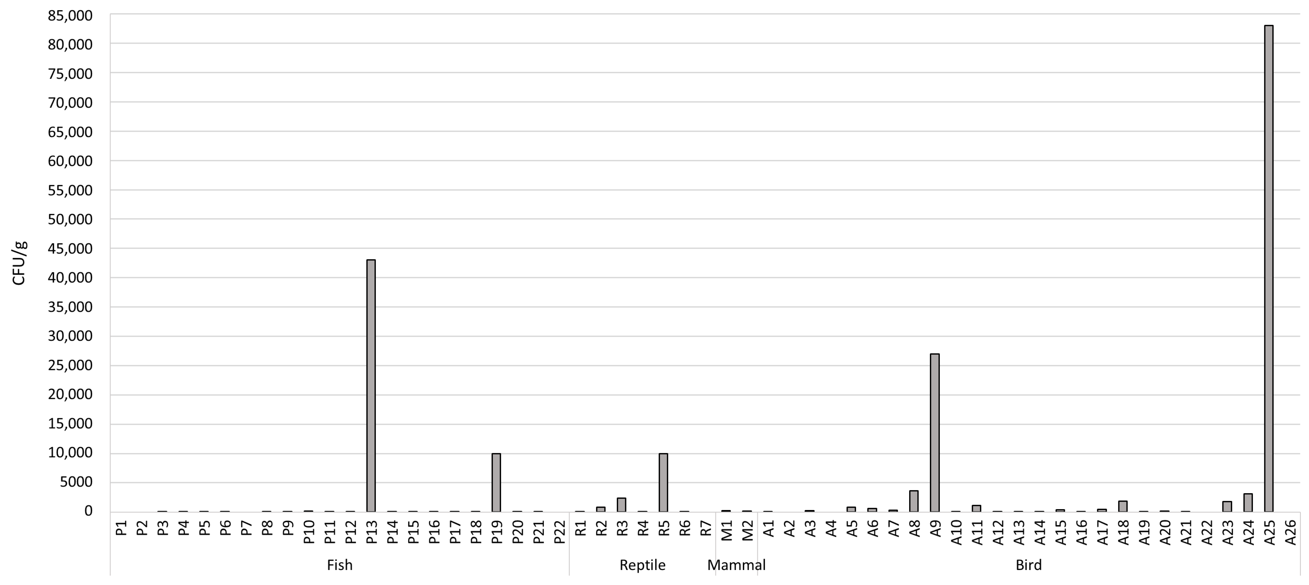
3.1. Total Microbial Level

From the 57 analyzed samples, 84.2% (n = 48) showed microbial growth in PCA, while 15.8% (n = 9) did not (Figure 1 and Table S1). The highest microbial level was observed in a bird food sample (sample ID: A25) and fish food sample (sample ID: P13), with 83,000 CFU/g and 43,000 CFU/g, respectively. The values of microbial growth ranged from 10 to 83,000 CFU/g for bird samples, 10 to 43,000 CFU/g for fish samples, 10 to 10,000 CFU/g for reptiles, and 200 to 260 CFU/g for mammal samples (Table S1).

3.2. Enterococcus Isolates/Species Identification

Microbiological and biochemical tests identified 103 Enterococcus isolates from the 28 of 57 feed samples. Of the 103 isolates, 56.3% (n = 58) were obtained from bird feed; 32% (n = 33) were obtained from fish feed, and 11.7% (n = 12) were obtained from reptile feed (Table 1). No isolates of presumptive Enterococcus were obtained in mammalian samples.
Of the 60 selected enterococci for species confirmation by PCR assay, 56.7% (n = 34) were from bird samples; 31.6% (n = 19) were from fish, and 11.7% (n = 7) were from reptile samples. The following species were identified from these isolates: 36.7% (n = 22) of E. faecalis, 31.7% (n = 19) of E. faecium, 25% (n = 15) of E. gallinarum and 6.7% (n = 4) of E. durans.

3.3. Antimicrobial Susceptibility Characterization

Table 1 shows the phenotypic antibiotic resistance profile for the 103 Enterococcus isolates based on the antimicrobial classes and origin of animal feed samples. A high prevalence of resistance was observed for rifampicin (77.7%). In addition, resistance to erythromycin (48.5%), ciprofloxacin (37.9%), tetracycline (26.2%), linezolid (19.4%), nitrofurantion (18.4%) and fosfomycin (6.8%) was also observed. For the remaining tested antibiotics, the isolates showed a low level of resistance, such as chloramphenicol (3.9%), ampicillin (2.9%), vancomycin (2.9%) and teicoplanin (2.9%). No isolates showed resistance to streptomycin and gentamicin. As expected, all Enterococcus isolates were intrinsically resistant to quinupristin/dalfopristin (Table 1).
Clustering the different tested antibiotics by their classes, the results showed that 47.6% of the isolates were classified as multidrug-resistant (resistance to ≥ 3 antimicrobial classes), of which 17.5% were resistant to five or more classes; 6.8% were resistant to four classes, and 23.3% were resistant to three antimicrobial classes. The remaining 26.2% and 17.5% of enterococcal isolates showed resistance to 2 and 1 antimicrobial classes, respectively, and 8.7% of isolates did not show resistance to any antibiotic (Table S2).

3.4. Genotypic of Antibiotic Resistance and Virulence Genes

From 60 selected enterococcal isolates, the detection of the presence of antibiotic-resistant genes showed that the most prevalent resistance genes were ermB (56.7%), tetL (51.7%), tetM (36.7%) and vanA (38.3%). On the other hand, the least identified resistance genes were ermA, vatD and aac(6″)-aph(2″). No amplification was observed for gene tetO in any of the isolates (Table 2).
The resistant gene to erythromicyn, ermB, was found in E. faecalis (18.2%), E. faecium (52.6%), E. gallinarum (86.7%) and all E. durans isolates. For tetracycline resistance genes in E. faecalis, 18.2% of the isolates contained the tetK gene, and another 18.2% contained the tetM gene, and only 4.5% of isolates showed the tetL gene individually. In addition, the combination tetM + tetL was detected in 13.6% of the isolates, and the combination tetK + tetM was found in 4.5% of the isolates of E. faecalis. In the case of E. faecium, 42.1% of the isolates showed only the tetL gene, and the tetK + tetL combination (26.3%) was also present. The combinations tetM + tetL, tetK + tetM, tetK + tetL and tetK + tetM + tetL were found in 21%, 10.5%, 10.5% and 15.8% of the E. gallinarum isolates, respectively. All E. durans isolates showed resistance to tetracycline, with 50% containing the tetM + tetL combination and the remaining 50% containing the combination tetK + tetM + tetL (Table S2).
Regarding the vancomycin-resistant isolates in E. faecalis, 4.5% showed the presence of the vanA gene; 9% showed the presence of the vanB gene, and 18.2% showed the vanA + vanB combination. In E. faecium, 26.3% of the isolates contained the vanA gene, while 15.8% had the vanB gene. The vanA + vanB combination was identified in one isolated. On the other hand, E. gallinarum demonstrated the presence of vanA in 60% of its isolates, with only one isolate containing the vanB gene and the other the combination vanA + vanB. Likewise, 50% of the E. durans isolates were positive for the presence of the vanA gene.
In contrast to the E. faecalis and E. faecium species, most of the E. gallinarum and E. durans isolates showed the ant(6)-Ia gene (66.7% of E. gallinarum isolates and 100% of E. durans isolates) and the catA gene (60% of E. gallinarum and 50% of E. durans).
The vatD and vatE genes, associated with resistance to quinupristine-dalfopristine, were found in 25% of isolates, and the vatE gene was the most prevalent in the species E. faecalis, E. faecium and E. gallinarum. In E. durans, 50% of the isolates contained the gene vatD, and 25% contained the vatE gene (Table 2).
A total of 46.7% (28/60) of Enterococcus isolates were genotypically classified as multidrug-resistant bacteria. Enterococcus gallinarum, followed by E. faecium, was the species with the most multidrug-resistant isolates, 50% and 22%, respectively.
Virulence genes were also identified, with a greater predominance of cylL and esp genes, identified in 51.7% and 40% isolates, respectively. The ace and fsr genes were not found in any of the species under study (Table 3). In total, 18 isolates did not show any virulence gene. The species E. gallinarum presented the most virulence genes, while in E. faecium, the presence of these genes was residual.
The statistical analysis did not include the E. durans isolates due to their reduced number. The results demonstrated significant associations (p < 0.05) for the ermB, ant(6)-Ia, vanA, tetM and tetL resistance genes. Thus, E. faecalis was more likely to be positive for tetM; E. faecium was more likely to be positive for ermB and tetL, and E. gallinarum was more likely to be positive for ermB, ant(6)-Ia, vanA and tetL. For virulence factors, a significant association was observed for the cylL gene. E. gallinarum was more likely to be positive for cylL, demonstrating a higher probability of this species carrying the cylL gene (Table 4).
Briefly, Table S2 comprises the phenotypic, genotypic and virulence profiles of each of the 60 isolates obtained in this study, as well as the feed origin.

4. Discussion

Enterococci are natural inhabitants of the gastrointestinal tract of mammals, humans, feed and insects. Although enterococci were initially classified as commensal bacteria not pathogenic, they are currently responsible for the most common nosocomial infection in humans and animals [30]. A high level of multidrug resistance in human clinical Enterococcus strains was identified in Slovenia, with 73.3% of E. faecium and 29.6% of E. faecalis showing resistance [14]. The emergence of multidrug resistance in enterococcal species is a public health problem, limiting therapeutic options. However, information in the literature about resistance to antibiotics in ornamental animal feed is still very scarce. In our study, the presence of Enterococcus was identified in 28 of 57 ornamental animal feed samples. Recently, Dolka et al. [31] evaluated the presence of Enterococcus spp. in racing pigeons (Columba livia f. Domestica) and observed that almost all isolates, about 93.1%, were resistant to at least one antibiotic, with E. faecalis being one of the most frequently identified species in these birds. In that study, Enterococcus showed resistance more frequently to teicoplanin (73%) and erythromycin (75.2%). Another study [14] detected resistance to tetracycline (78.9%) and erythromycin (46.5%) in E. faecalis from retail red meat and human clinical samples. In the case of E. faecium, most clinical isolates were resistant to erythromycin (45%), followed by ciprofloxacin (41%) and ampicillin (41%). In comparison with Dolka et al. [31] and Golob et al. [14], our study also showed that Enterococcus is resistant to erythromycin (48.5%) but has a lower percentage of resistance to tetracycline (26.2%) and teicoplanin (2.9%). Another aspect worth reporting is the low resistance to vancomycin, as also described by Marinho [32].
With regard to pets, such as dogs and cats, Trościańczyk et al. [33] recorded results very similar to those mentioned above, with a high prevalence of resistance of Enterococcus to erythromycin (96%), ciprofloxacin (93%) and tetracycline (82%) and a significant number of isolates showing multidrug resistance (78%). In our study, none of the isolates showed resistance to gentamicin and streptomycin, similar to the study of Osman et al. [34]. Freitas et al. [35] reported that 54% of the 55 studied dog food samples contained Enterococcus spp., of which 31% were considered multidrug-resistant. However, the number of multidrug-resistant isolates is higher in our study when compared to Golob et al. [14] and Freitas et al. [35]. E. faecalis and E. faecium were the most frequently identified species, which is consistent with the results obtained in other studies, mainly in studies where Enterococcus spp. were isolated from source food animal [1,14]. E. gallinarium was the Enterococcus species that demonstrated a higher probability of carrying a variety of antibiotic-resistant genes based on statistical analysis.
In general, based on the results of this study, it can be concluded that the ermB gene is the most representative in erythromycin resistance in Enterococcus isolates, just as the tetL and tetM genes are described as the main genes that encode tetracycline resistance. The gene combinations ermB-tetM or ermB-tetM-tetL were present in 27% of the isolates, confirming the fact that the ermB gene is usually linked to the tetM gene in the mobile conjugative transposon Tn1545. Usually, tetracycline resistance genes are found on the same mobile unit as the genes that confer resistance to macrolides [36]. In our study, these combinations were predominant in the species E. durans and E. gallinarum. Alternatively, the gene aac(6′)-aph(2′′) that is normally associated with resistance to gentamicin was identified only in one E. durans isolate and two E. gallinarum isolates, which is consistent with the phenotypic results, where no isolate showed resistance to gentamicin. Freitas et al. [35] identified this gene in E. faecalis and E. faecium from raw dog food samples.
At the genotypic level, 20% of the Enterococcus spp. identified in this study carried the catA gene, which is similar to the results obtained by Osman et al. [34] in Nile tilapia with streptococcosis. In our study, 18.9% of the isolated Enterococcus spp. from birds carried this gene, especially E. faecium. Additionally, the vanA and/or vanB genes were identified in 48% of the isolates, with a higher prevalence of the vanA gene, which is in agreement with the results obtained by Carvalho [20] and Guerrero-Ramos et al. [1] in Miranda donkey and wild game meat, respectively. It should be noted that most of the isolates that presented one or both of these genes belonged to the species E. gallinarum and E. faecium.
The esp gene, which was detected to a lesser extent in the study by Dolka et al. [31], was frequently detected in Enterococcus isolates in the present study, which agrees with the results of Iweriebor et al. [37]. In relation to the gelE gene, which was found to have the highest prevalence, most studies are in agreement [31,37].
Contrary to the studies performed by Carvalho [20] and Guerrero-Ramos et al. [1], in the present study E. faecium showed fewer genes of virulence, particularly the cylL and agg genes. As in our study, Guerrero-Ramos et al. [1] also did not identify the fsr gene in any of the isolates.
In the present study, it was found that most of the isolates contained the esp gene, which is responsible for encoding the surface proteins that make the bacterial colony more persistent. Furthermore, this surface protein is implicated in cases of bacteremia, urinary tract infections and endocarditis [38]. The gene encoding cytolysin, cylL, which was the most commonly detected in this study, is also associated with an increased risk of sudden death in cases of nosocomial bacteremia [39].

5. Conclusions

In conclusion, the results of this study suggest that ornamental pet foods found on the market may contain Enterococcus spp. with high levels of resistance to a variety of antibiotics and the ability to produce virulence factors. These foods can potentially carry epidemiologically important microorganisms and once ingested by ornamental animals can lead to the colonization of pathogenic Enterococcus spp. in the gut microbiota. Consequently, these bacteria, some of which are multidrug-resistant, can cause diseases in these animals with fatal consequences. Moreover, given the close contact with humans, they may cause infections and diseases, making antimicrobial treatment difficult.
Considering the limited information on the presence of multidrug-resistant enterococci in food intended to be fed to ornamental animals, this study highlights the need for greater control in all steps of food processing, as well as the potential risks to the environment and human and animal health. Additionally, this study contributes to the monitoring of antimicrobial resistance in a new, unexplored ecosystem niche, to facilitate the implementation of control measures.

Supplementary Materials

The following supporting information can be downloaded at: https://www.mdpi.com/article/10.3390/ani13111761/s1, Table S1: Total count of microorganisms in bird, fish, reptile and mammal food samples in colony forming unit per gram (CFU/g); Table S2: Phenotypic, genotypic and virulent profile of the 60 selected Enterococcus isolates based on food origin.

Author Contributions

Conceptualization, R.S. and C.M.; methodology, R.S., S.C., Â.M. and L.F.; validation, G.I., C.M., Â.M. and P.P.; writing—original draft preparation, R.S. and C.M.; supervision, G.I. and P.P. All authors have read and agreed to the published version of the manuscript.

Funding

This work was supported by the Associate Laboratory for Green Chemistry—LAQV which is financed by national funds from FCT/MCTES (UIDB/50006/2020 and UIDP/50006/2020). This was also supported by the projects UIDP/CVT/00772/2020 and LA/P/0059/2020 funded by the Portuguese Foundation for Science and Technology (FCT).

Institutional Review Board Statement

Not applicable.

Informed Consent Statement

Not applicable.

Data Availability Statement

The study did not report any data.

Conflicts of Interest

The authors declare no conflict of interest.

References

  1. Guerrero-Ramos, E.; Cordero, J.; Molina-González, D.; Poeta, P.; Igrejas, G.; Alonso-Calleja, C.; Capita, R. Antimicrobial resistance and virulence genes in enterococci from wild game meat in Spain. Food Microbiol. 2016, 53, 156–164. [Google Scholar] [CrossRef] [PubMed]
  2. Strateva, T.; Atanasova, D.; Savov, E.; Petrova, G.; Mitov, I. Incidence of virulence determinants in clinical Enterococcus faecalis and Enterococcus faecium isolates collected in Bulgaria. Braz. J. Infect. Diseas. 2016, 20, 127–133. [Google Scholar] [CrossRef] [PubMed]
  3. García-Solache, M.; Rice, L.B. The Enterococcus: A model of adaptability to its environment. Clin. Microbiol. Rev. 2019, 32, e00058-18. [Google Scholar] [CrossRef]
  4. Abamecha, A.; Wondafrash, B.; Abdissa, A. Antimicrobial resistance profile of Enterococcus species isolated from intestinal tracts of hospitalized patients in Jimma, Ethiopia Microbiology. BMC Res. Not. 2015, 8, 1–7. [Google Scholar]
  5. Martzy, R.; Kolm, C.; Brunner, K.; Mach, R.L.; Krska, R.; Sinkovec, H.; Sommer, R.; Farnleitner, A.H.; Reischer, G.H. A loop-mediated isothermal amplification (LAMP) assay for the rapid detection of Enterococcus spp. in water. Water Res. 2017, 122, 62–69. [Google Scholar] [CrossRef] [PubMed]
  6. Campos, A.C.; Souza, N.R.; Silva, P.H.C.; Santana, A.P. Resistência antimicrobiana em Enterococcus faecalis e Enterococcus faecium isolados de carcaças de frango. Pesq. Vet. Bras. 2013, 33, 575–580. [Google Scholar] [CrossRef]
  7. Thurlow, L.R.; Thomas, V.C.; Narayanan, S.; Olson, S.; Fleming, S.D.; Hancock, L.E. Gelatinase contributes to the pathogenesis of endocarditis caused by Enterococcus faecalis. Infect. Immun. 2010, 78, 4936–4943. [Google Scholar] [CrossRef]
  8. Zhang, Y.; Du, M.; Chang, Y.; Chen, L.; Zhang, Q. Incidence, clinical characteristics, and outcomes of nosocomial Enterococcus spp. bloodstream infections in a tertiary-care hospital in Beijing, China: A four-year retrospective study. Antimicrob. Resist. Infect. Control 2017, 6, 1–11. [Google Scholar] [CrossRef]
  9. Markwart, R.; Willrich, N.; Haller, S.; Noll, I.; Koppe, U.; Werner, G.; Eckmanns, T.; Reuss, A. The rise in vancomycin-resistant Enterococcus faecium in Germany: Data from the german antimicrobial resistance surveillance (ARS). Antimicrob. Resist. Infect. Control 2019, 8, 1–11. [Google Scholar] [CrossRef]
  10. Hollenbeck, B.L.; Rice, L.B. Intrinsic and acquired resistance mechanisms in Enterococcus. Virul 2012, 3, 421–433. [Google Scholar] [CrossRef]
  11. Asmah, N. Molecular aspects of Enterococcus faecalis virulence. J. Syiah Kual Dentis. 2020, 5, 89–94. [Google Scholar] [CrossRef]
  12. Li, B.; Webster, T.J. Bacteria antibiotic resistance: New challenges and opportunities for implant-associated orthopedic infections. J. Orthop. Res. 2018, 36, 22–32. [Google Scholar] [CrossRef]
  13. Radhouani, H.; Silva, N.; Poeta, P.; Torres, C.; Correia, S.; Igrejas, G. Potential impact of antimicrobial resistance in wildlife, environment, and human health. Front. Microbiol. 2014, 5, 23. [Google Scholar] [CrossRef]
  14. Loureiro, R.J.; Roque, F.; Rodrigues, A.T.; Herdeiro, M.T.; Ramalheira, E. Use of antibiotics and bacterial resistances: Brief notes on its evolution. Rev. Port Saud Public 2016, 34, 77–84. [Google Scholar]
  15. Ventola, C.L.M.S. The antibiotic resistance crisis. Pharm Ther. 2015, 40, 277–283. [Google Scholar]
  16. Lee, J.H.; Shin, D.; Lee, B.; Lee, H.; Lee, I.; Jeong, D.W. Genetic Diversity and Antibiotic Resistance of Enterococcus faecalis Isolates from Traditional Korean Fermented Soybean Foods. J. Microbiol. Biotechnol. 2017, 27, 916–924. [Google Scholar] [CrossRef]
  17. Golob, M.; Pate, M.; Kušar, D.; Dermota, U.; Avberšek, J.; Papic, B.; Zdovc, I. Antimicrobial Resistance and Virulence Genes in Enterococcus faecium and Enterococcus faecalis from Humans and Retail Red Meat. BioMed. Res. Intern. 2019, 2019, 14–16. [Google Scholar] [CrossRef]
  18. Elghaieb, H.; Tedim, A.P.; Abbassi, M.S.; Novais, C.; Duarte, B.; Hassen, A.; Peixe, L.; Freitas, A.R. From farm to fork: Identical clones and Tn6674-like elements in linezolid-resistant Enterococcus faecalis from food-producing animals and retail meat. J. Antimicrob. Chemoth. 2020, 75, 30–35. [Google Scholar] [CrossRef]
  19. Ahmed, H.A.; Awad, N.F.S.; El-Hamid, M.I.A.; Shaker, A.; Mohamed, R.E.; Elsohaby, I. Pet birds as potential reservoirs of virulent and antibiotic resistant zoonotic bacteria. Comp. Immun. Microbiol. Infect. Diseas 2021, 75, 101606. [Google Scholar] [CrossRef]
  20. Carvalho, I.; Campo, R.D.; Sousa, M.; Silva, N.; Carrola, J.; Marinho, C.; Santos, T.; Carvalho, S.; Nóvoa, M.; Quaresma, M.; et al. Antimicrobial-resistant Escherichia coli and Enterococcus spp. isolated from Miranda donkey (Equus asinus): An old problem from a new source with a different approach. J. Med. Microbiol. 2017, 66, 191–202. [Google Scholar] [CrossRef]
  21. Dutka-Malen, S.; Evers, S.; Courvalin, P. Detection of Glycopeptide Resistance Genotypes and Identification to the Species Level of Clinically Relevant Enterococci by PCR. J. Clin. Microbiol. 1995, 33, 24–27. [Google Scholar] [CrossRef]
  22. Arias, C.A.; Robredo, B.; Singh, K.V.; Torres, C.; Panesso, D.; Murray, B.E. Rapid identification of Enterococcus hirae and Enterococcus durans by PCR and detection of a homologue of the E. hirae mur-2 Gene in E. durans. J. Clin. Microbiol. 2006, 44, 1567–1570. [Google Scholar] [CrossRef]
  23. CLSI. Performance Standards for Antimicrobial Disk Susceptibility Tests, 13th ed.; CLSI standard M02; Clinical and Laboratory Standards Institute: Wayne, PA, USA, 2018. [Google Scholar]
  24. Sutcliffe, J.; Grebe, T.; Tait-Kamradt, A.; Wondrack, L. Detection of erythromycin-resistant determinants by PCR. Antimicrob. Agents Chemother. 1996, 40, 2562–2566. [Google Scholar] [CrossRef]
  25. Aarestrup, F.M.; Agerso, Y.; Gerner-Smidt, P.; Madsen, M.; Jensen, L.B. Comparison of antimicrobial resistance phenotypes and resistance genes in Enterococcus faecalis and Enterococcus faecium from humans in the community, broilers, and pigs in Denmark. Diagn Microbiol. Infect. Dis. 2000, 37, 127–137. [Google Scholar] [CrossRef]
  26. Robredo, B.; Singh, K.V.; Baquero, F.; Murray, B.E.; Torres, C. Vancomycin-resistant Enterococci isolated from animals and food. Int. J. Food Microbiol. 2000, 54, 197–204. [Google Scholar] [CrossRef]
  27. Del Campo, R.; Tenorio, C.; Rubio, C.; Castillo, J.; Torres, C.; Gómez-Lus, R. Aminoglycoside-modifying enzymes in high-level streptomycin and gentamicin resistant Enterococcus spp. in Spain. Int. J. Antimicrob. Agents 2000, 15, 221–226. [Google Scholar] [CrossRef]
  28. Eaton, T.J.; Gasson, M.J. Molecular screening of Enterococcus virulence determinants and potential for genetic exchange between food and medical isolates. Appl. Environ. Microbiol. 2001, 67, 1628–1635. [Google Scholar] [CrossRef]
  29. Mannu, L.; Paba, A.; Daga, E.; Comunian, R.; Zanetti, S.; Duprè, I.; Sechi, L.A. Comparison of the incidence of virulence determinants and antibiotic resistance between Enterococcus faecium strains of dairy, animal and clinical origin. Int. J. Food Microbiol. 2003, 88, 291–304. [Google Scholar] [CrossRef]
  30. Miranda, C.; Silva, V.; Igrejas, G.; Poeta, P. Impact of European pet antibiotic use on enterococci and staphylococci antimicrobial resistance and human health. Future Microbiol. 2021, 16, 185–201. [Google Scholar] [CrossRef]
  31. Dolka, B.; Czopowicz, M.; Chrobak-Chmiel, D.; Ledwoń, A.; Szeleszczuk, P. Prevalence, antibiotic susceptibility and virulence factors of Enterococcus species in racing pigeons (Columba livia f. domestica). BMC Vet. Res. 2020, 16, 1–14. [Google Scholar] [CrossRef]
  32. Marinho, C. Resistência a antibióticos em Enterococcus spp. e Escherichia coli de equinodermes: Um problema ambiental e de saúde pública. Master’s Thesis, Dissertação de Mestrado em Genética Molecular Comparativa e Tecnológica, University of Trás-os-Montes and Alto Douro, Vila Real, Portugal, 2013; p. 109. Available online: http://hdl.handle.net/10348/5305 (accessed on 26 September 2022).
  33. Trościańczyk, A.; Nowakiewicz, A.; Gnat, S.; Łagowski, D.; Osińska, M. Are dogs and cats a reservoir of resistant and virulent Enterococcus faecalis strains and a potential threat to public health? J. Appl. Microbiol. 2021, 131, 2061–2071. [Google Scholar] [CrossRef]
  34. Osman, K.M.; Al-Maary, K.S.; Mubarak, A.S.; Dawoud, T.M.; Moussa, I.M.I.; Ibrahim, M.D.S.; Hessain, A.M.; Orabi, A.; Fawzy, N.M. Characterization and susceptibility of streptococci and enterococci isolated from Nile tilapia (Oreochromis niloticus) showing septicaemia in aquaculture and wild sites in Egypt. BMC Vet. Res. 2017, 13, 1–10. [Google Scholar] [CrossRef]
  35. Freitas, A.R.; Finisterra, L.; Tedim, A.P.; Duarte, B.; Novais, C.; Peixe, L. Linezolid- And multidrug-resistant enterococci in raw commercial dog food, Europe, 2019–2020. Emerg. Infect. Diseas 2021, 27, 2221–2224. [Google Scholar] [CrossRef]
  36. Santos, T. Proteogenómica de isolados de Enterococcus spp. e Escherichia coli com a utilização do MALDI-TOF MS. Master’s Thesis, Dissertação de Mestrado em Genética Molecular Comparativa e Tecnológica, University of Trás-os-Montes and Alto Douro, Vila Real, Portugal, 2014. Available online: http://hdl.handle.net/10348/5482 (accessed on 26 September 2022).
  37. Iweriebor, B.C.; Obi, L.C.; Okoh, A.I. Virulence and antimicrobial resistance factors of Enterococcus spp. isolated from fecal samples from piggery farms in Eastern Cape, South Africa. BMC Microbiol. 2015, 15, 136. [Google Scholar] [CrossRef]
  38. Andrade, S.D.E. Caracterização fenotípica e genotípica de amostras clínicas e indígenas de Enterococcus isoladas de seres humanos: Diversidade, virulência e resistência a drogas antimicrobianas. Tese de Doutoramento em Microbiologia. Ph.D. Thesis, Instituto de Ciências biológicas, Universidade Federal de Minas Gerais, Belo Horizonte, Brazil, 2018. Available online: http://hdl.handle.net/1843/BUOS-B76JMH (accessed on 26 September 2022).
  39. Van Tyne, D.; Martin, M.J.; Gilmore, M.S. Structure, function, and biology of the 85 Enterococcus faecalis cytolysin. Toxins 2013, 5, 895–911. [Google Scholar] [CrossRef]
Figure 1. Total microbial level obtained in all ornamental feed samples analyzed in this study expressed as colony forming unit per gram (CFU/g) of feed.
Animals 13 01761 g001
Table 1. Number of resistances observed from the antimicrobial susceptibility test of the 103 Enterococcus isolated from the ornamental animal feed.
Antibiotic
Agent
Antimicrobial
Class
Origin of Feed SampleTotal
(%)
Bird (n = 58)Fish (n = 33)Reptile (n = 12)
Ampicilin Penicilins3003 (2.9)
Vancomycin Glycopeptides1203 (2.9)
Teicoplanin Glycopeptides1203 (2.9)
Tetracycline Tetracyclines1610127 (26.2)
Erythromycin Macrolides2918350 (48.5)
Ciprofloxacin Fluoroquinolones248739 (37.9)
Chloramphenicol Phenicols3104 (3.9)
Quinupristin/
Dalfopristin *
Streptogramins583312103 (100)
Nitrofurantoin Nitrofurantoins115319 (18.4)
Rifampicin Ansamycins4824880 (77.7)
Fosfomycin Fosfoycins5207 (6.8)
Gentamicin Aminoglicosides0000 (0.0)
Streptomycin Aminoglicosides0000 (0.0)
Linezolid Oxazolidinones153220 (19.4)
*: intrinsic resistance.
Table 2. Antibiotic-resistant genes in Enterococcus species identified in this study (n = 60).
Antibiotic GeneE. faecalis
(n = 22)
E. faecium
(n = 19)
E. gallinarum
(n = 15)
E. durans
(n = 4)
Total
(%)
ermA
ermB
ermC
0
4
3
3
13
4
1
13
0
0
4
0
4 (6.7)
34 (56.7)
7 (11.7)
tetK
tetM
tetL
5
8
4
5
0
13
7
10
10
2
4
4
19 (31,7)
22 (36.7)
31 (51.7)
aac(6′′)-aph(2′′)00213 (5.0)
catA109212 (20.0)
vatD
vatE
1
4
0
2
1
4
2
1
4 (6.7)
11 (18.3)
vanA
vanB
5
6
6
4
10
2
2
0
23 (38.3)
12 (20.0)
ant(6)-Ia3410421 (35.0)
Table 3. Virulence genes detected in each Enterococcus species analyzed in this study (n = 60).
Virulence GeneE. faecalis
(n = 22)
E. faecium
(n = 19)
E. gallinarum
(n = 15)
E. durans
(n = 4)
Total
(%)
esp10012224 (40.0)
ace00000 (0.0)
gelE4010317 (28.3)
agg627318 (30.0)
fsr00000 (0.0)
cpd308112 (20.0)
cylA10427 (11.7)
cylB20103 (5.0)
cylM803314 (23.3)
cylL8613431 (51.7)
Table 4. Significant associations obtained between resistance and virulence genes and the Enterococcus species.
E. faecalisE. faeciumE. gallinarumχ2p
Antibiotic resistance genes
ermBPositive
Negative
4
18
13
6
13
2
19.370.0001
tetMPositive
Negative
8
14
0
19
10
5
17.380.0002
tetLPositive
Negative
4
18
13
6
10
5
13.100.0014
vanAPositive
Negative
5
17
6
13
10
5
7.780.0205
ant6-IaPositive
Negative
3
19
4
15
10
5
13.040.0015
Virulence factors genes
cylLPositive
Negative
8
14
6
13
13
2
12.270.0022
Disclaimer/Publisher’s Note: The statements, opinions and data contained in all publications are solely those of the individual author(s) and contributor(s) and not of MDPI and/or the editor(s). MDPI and/or the editor(s) disclaim responsibility for any injury to people or property resulting from any ideas, methods, instructions or products referred to in the content.

