Management of Dental Disease in Aardvarks (Orycteropus afer) and Potential Use of Cone-Beam Computed Tomography
by
 ,
,
Jane E. Christman
1,* ,
,
Daniel VanderHart
2,
Ben Colmery
3,
Joy Thompson
1,
Ann E. Duncan
1 and
Wynona C. Shellabarger
1 1
Detroit Zoological Society, 8450 W 10 Mile Rd., Royal Oak, MI 48067, USA
2
VetRad, Veterinary Teleradiology Interpretation Service, 1321 Centerview Circle, Akron, OH 44321, USA
3
Dixboro Veterinary Dental Center, 5740 Plymouth Rd., Ann Arbor, MI 48105, USA
*
Author to whom correspondence should be addressed.
Animals 2022, 12(7), 845; https://doi.org/10.3390/ani12070845
Submission received: 1 March 2022
/
Revised: 24 March 2022
/
Accepted: 25 March 2022
/
Published: 28 March 2022
(This article belongs to the Special Issue Pathology in Zoo Animals and Conservation)
Simple Summary
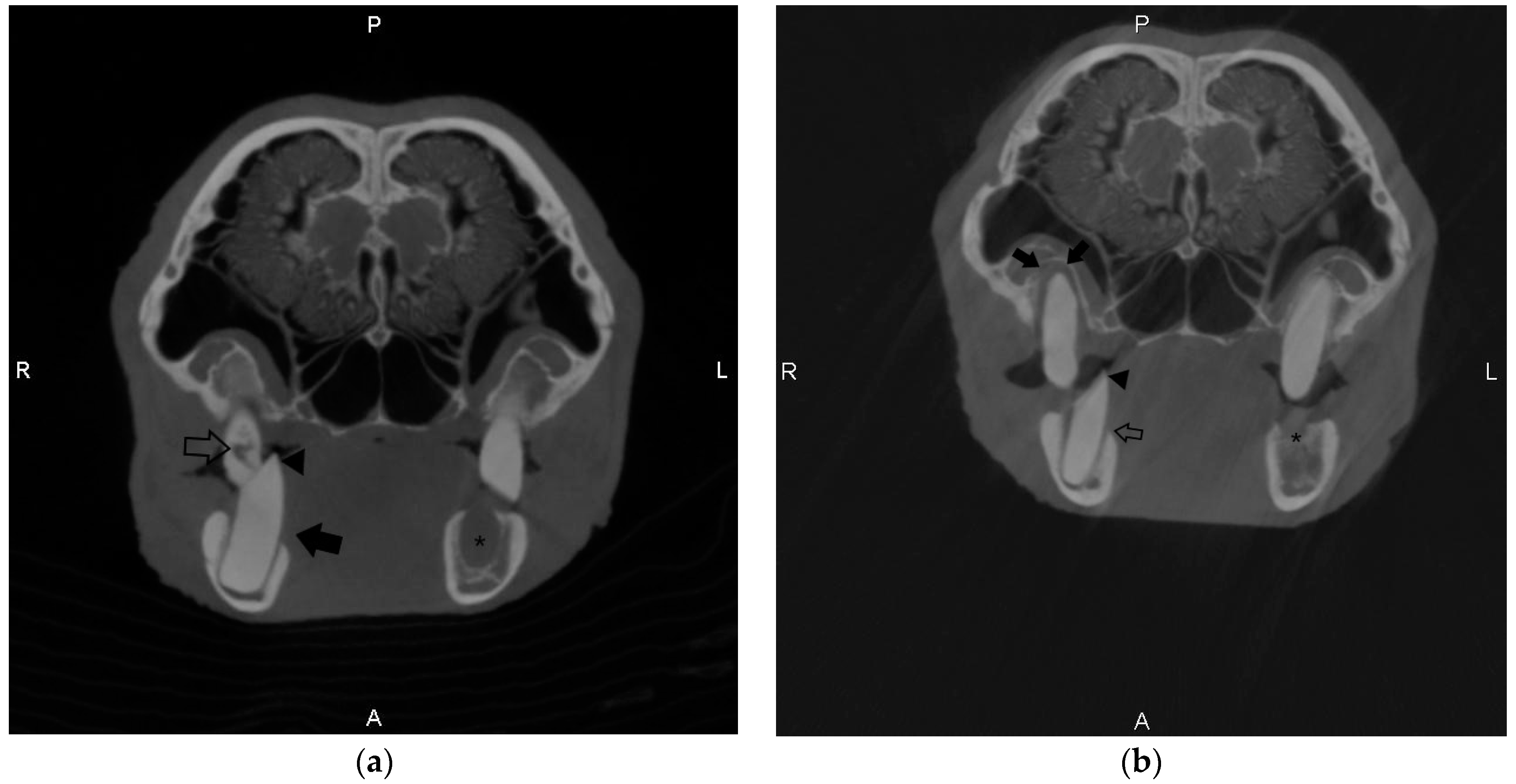
Oral disease involving teeth is a common condition of aardvarks (Orycteropus afer) housed in zoos, although research is limited. Medical records of aardvarks housed at a single zoo in the United States between 1995 and 2021 were evaluated for dental abnormalities. Eight out of ten aardvarks had dental abnormalities, with most cases having mild disease (4/8). Three patients required the removal of abnormal teeth for treatment. Cone-beam computed tomography (CBCT) is a technology used to create a three-dimensional image of the teeth and skull and was used in two aardvarks. This technology was considered helpful in documenting dental abnormalities in patients with severe disease and may be a valuable tool for veterinarians managing oral disease in aardvarks.
Abstract
Oral disease involving teeth is a common cause of morbidity in aardvarks (Orycteropus afer) under managed care. Cases can be challenging due to the species’ unique skull and dental anatomy and limited veterinary literature. A retrospective evaluation was performed on dental examinations in nine aardvarks housed at a single zoological institution in the United States between 1995 and 2021. The prevalence of dental disease in this population was 88%, with most cases categorized as mild (4/8). Clinical signs were only seen in three cases. Facial swelling prior to surgery was the most common clinical sign (3/8). Dental pathology was more common in the mandibular teeth (27/38) compared to the maxillary teeth (11/38). Dental abnormalities found upon intraoral examination included the presence of dental points (7/8), crown elongation (3/8), purulent material within the oral cavity (4/8), loose teeth (2/8), periodontal pockets (2/8), and oronasal fistula (1/8). Three patients required dental extractions with a lateral buccostomy approach. Diagnostic imaging was performed in most cases (7/8), with two cases undergoing cone-beam computed tomography (CBCT) to characterize dental pathology that was difficult to fully evaluate with standard radiography. Tomographic findings are described in both cases. CBCT was found to be a helpful tool for diagnosing and characterizing dental disease in aardvarks.