Article

Comparative Analysis of Milk Microbiomes and Their Association with Bovine Mastitis in Two Farms in Central Russia

1
Laboratory of Microbiology, L.K. Ernst Federal Science Center for Animal Husbandry, Dubrovitsy 142132, Russia
2
Laboratory of Immunochemistry, Shemyakin and Ovchinnikov Institute of Bioorganic Chemistry of the Russian Academy of Sciences, Pushchino 142290, Russia
3
Laboratory of Plasmid Biology, Federal Research Center “Pushchino Scientific Center for Biological Researches”, G.K. Skryabin Institute of Biochemistry & Physiology of Microorganisms of the Russian Academy of Sciences, Pushchino 142290, Russia
4
Laboratory of Cell Genome Functioning Mechanisms, Federal Research Center “Pushchino Scientific Center for Biological Researches”, Institute of Cell Biophysics of the Russian Academy of Sciences, Pushchino 142290, Russia
*
Author to whom correspondence should be addressed.
Animals 2021, 11(5), 1401; https://doi.org/10.3390/ani11051401
Submission received: 2 April 2021 / Revised: 5 May 2021 / Accepted: 10 May 2021 / Published: 14 May 2021
(This article belongs to the Section Cattle)

Abstract

:

Simple Summary

Bovine mastitis is one of the most common diseases in cattle farms in Russia. In this work we investigate a microbial composition of milk samples collected from farms of Russian Central Region. Our data revealed significant dominance of several operational taxonomic units corresponding mostly to groups of Staphylococcus aureus, Aerococcus spp. and Streptococcus spp. We identify interesting fact of Staphylococcus and Aerococcus genera seemed to be antagonistic to each other, and the disappearance of the Aerococcus genus in milk microbiota may represent a marker for its transition to a subclinical stage of mastitis.

Abstract

Bovine mastitis is a widespread infectious disease. In addition to the economic damages associated with reduced milk yield due to mastitis, the problem of food contamination by microorganism metabolites, in particular toxins, is also a concern. Horizontal transfer of microorganisms from animal populations to humans can also be complicated by antibiotic resistance. Therefore, bovine mastitis is relevant to the study of microbiology and veterinary medicine. In this study, we investigated the microbiome of milk samples from healthy cows and cows with different forms of mastitis from individual quarters of the udder of cows during first and second lactation. Total DNA was extracted from milk samples. The V3–V4 regions of the bacterial 16S rRNA genes from each sample were amplified to generate a library via high-throughput sequencing. We revealed significant dominance of several operational taxonomic units (OTUs) corresponding mostly to groups of Staphylococcus aureus, Aerococcus spp., and Streptococcus spp. In addition, we unexpectedly identified Streptococcus thermophilus in samples with high SCC quantities. We found some infectious agents that characterized summer mastitis. We demonstrated that in Central Russia, mastitis is associated with a wide variety of causal organisms. We observed some differences in the diversity of the two investigated farms. However, we did not find any significant difference among healthy, mastitis and subclinical samples according to their SCC status from either farms by principal component analysis. Linear discriminant analysis effect size (LEfSe) confirmed the presence of several indicator genera in farms from Moscow and the Tula Region. These results confirm the complex bacterial etiology of bovine mastitis.

1. Introduction

Bovine mastitis is one of the most economically important diseases in cattle [1]. Mastitis is an inflammatory disease of the mammary glands and similar to all other host-pathogen related diseases, has its own signature of microbiota composition [2]. Several studies has demonstrated that it is not possible to exclude transmission from animal to human [3,4,5,6,7], and dissemination of milk of pathogenic microorganisms may colonize both animals and humans, causing secondary diseases [8,9]. Another important aspect of this disease is the probable presence of toxins in milk produced by the pathogens, as many bacterial toxins are known to facilitate allergy development [10,11,12,13]. A number of studies have attempted to identify the primary cause of mastitis and the major microbial pathogen by analysis of microbiota from healthy animal milk compared to milk from animals with various forms and stages of mastitis [14,15,16,17,18,19,20,21]. Further, mastitis may depend on the season, so summer mastitis may be associated with microbiota transmitted from insects. In winter, mastitis can be caused by hypothermia and use of various mats. Injuries of the udder are another important factor in mastitis [22,23,24].
Interactions between multicellular host organisms and microbiota that colonize various parts of the host organism are currently an actively investigated topic. There is a quest for symbiotic microorganisms that are able to provide protection from various pathogens. Classic culture methods have shown a connection between particular bacterial species and the onset of mastitis. Recently developed omics methods are currently in use for expanding the list of such species and identifying their physiological state [25,26].
The aim of this study was to quantitatively analyze the milk microbiota from healthy animals and animals with various forms and stages of mastitis in farms of Central Russia. In the present work, we investigated bovine milk microbiomes on two farms in different regions (Moscow and the Tula Regions).

2. Materials and Methods

2.1. Sample Collection

Milk samples were collected from Holsteinized black-and-white breed cows from two farms (Moscow and Tula regions) of Central Russia from June to August. Samples were taken 60-70 days after calving from each quarter of the udder separately. Examination of the general condition of the animal by using veterinary medicine methods the mammary gland and inguinal lymph nodes was carried out. To diagnose latent mastitis on a farm, tests of milk samples with dimastin were conducted. Cows had first or second lactation, animals of the same age and lactation were selected. Samples were collected after milking into sterile tubes in compliance with aseptic rules. Classical hygienic procedures included cleaning of teats with an individual paper towel before milking and post milking teat dipping in 0.5% solution of chloramine, after which the udder was dried with an individual paper napkin. All samples were transported to the laboratory under refrigeration (4–8 °C) in cool boxes with ice packs. Analysis of somatic cells was performed by the express method on a Somacount 150 (Bentley Instruments Inc., Chaska, MN, USA) analyzer. According to the veterinary data and the number of quarter somatic cell count (QSCC), all samples were divided into three groups of mastitis form: healthy (H) with QSCC up to 100,000/cm3; mastitic (M) with QSCC over 500,000/cm3, and subclinical (S) the remaining samples. In order to select the most healthy samples we specially lowered the SCC threshold to 100,000. Salt meat broth (HiMedia Laboratories Pvt., Ltd., Mumbai, India) was inoculated with the milk samples at a ratio of 1:9 and stored at 37 °C for 18–24 h. Internationally recognized traditional phenotypic methods, such as Gram-stained colony microscopy, growth in Baird–Parker agar (HiMedia Laboratories Pvt., Ltd.), hemolysis on azide blood agar Pronadisa (Condalab, Madrid, Spain), plasma coagulation, and biochemical identification, were applied to all the isolates. Therefore, 53 milk samples from 31 cows were ultimately used for this study: 18 samples (8 from Moscow rand 10 from Tula regions) derived from clinically healthy, culture negative quarters with an SCC of less than 100,000 cells/cm3 (group 1, (H) healthy); 24 samples (13 from Moscow and 11 from Tula regions) derived from clinical mastitis quarters (group 2, (M) clinical); and 11 samples (5 from Moscow and 6 from Tula regions) derived from clinically healthy quarters with an SCC 100,000–500,000 cells/cm3 (group 3, (S) subclinical). Additional data about milk samples descriptive characteristics can be found in Table S1.

2.2. DNA Extraction and Sequence Library Preparation

Milk aliquots from Tula and MR farms were kept frozen at −70 °C until DNA isolation procedure. The extraction of DNA from milk samples of both datasets was performed by the same researchers’ team in the same laboratory in a laminar flow cabinet using Milk Bacterial DNA Isolation Kit (Norgen Biotek, Thorold, ON, Canada) according to the manufacturer’s instructions. DNA was subsequently used as a template for polymerase chain reaction. The V3–V4 regions of the bacterial 16S rRNA gene from each sample was amplified using a universal primer set (16S Amplicon PCR Forward Primer = 5′-CCTACGGGNGGCWGCAG; 16S Amplicon PCR Reverse Primer = 5′-GACTACHVGGGTATCTAATCC) with corresponding Illumina (Illumina Inc., San Diego, CA, USA) overhang adapters. The KAPA HiFi HotStart DNA Polymerase was used for the DNA amplification. After quantification and purification of the PCR products, a sequencing library was generated using the 16S Metagenomic Sequencing Library Preparation protocol. Finally, the library was sequenced using a MiSeq 2 × 250.

2.3. Bioinformatics Analysis

Generated sequences were processed using the Nephele platform [27], and sequence quality was confirmed with the FastQC toolkit (Babraham Bioinformatics, Cambridge, UK) [28]. The #6494pp sample did not pass quality control and was excluded from further manipulations. Operational taxonomic units (OTUs) were characterized by matching to the SILVA dataset of 16S rRNA sequences [29]. Reads were subsequently mapped back to OTUs to determine OTU abundance for each sample, and differences in the community composition/structure in each sample were analyzed with the Quantitative Insights Into Microbial Ecology (QIIME) package [30].
Diversity was assessed by calculating the Shannon diversity metric, the Chao1 estimate of diversity, and the number of observed species for each sample at various sequencing depths.
Beta diversity estimates were calculated using weighted and unweighted UniFrac distances between samples [31].
Linear discriminant analysis (LDA) effect size (LEfSe) [32] available at https://huttenhower.sph.harvard.edu/galaxy (date accessed 1 March 2021) was used to assess microbial compositional differences between the two groups of samples at the genus or higher taxonomic level.

3. Results

3.1. Taxonomic Profile

To profile the milk microbiome, pools of 16S amplicons were processed to remove low-quality reads, chimeras, and samples with a low depth of coverage. The resultant dataset of 2,870,927 high-quality reads (median reads per sample = 60,413) was rarified to an even depth of 10,885 reads and binned into OTUs at 97% sequence identity. We observed OTUs from 16 bacterial phyla with an average relative abundance of above 0.1%, and six of these phyla (Firmicutes, Proteobacteria, Actinobacteria, Bacteroidetes, Tenericutes, and Fusobacteria) collectively accounted for more than 97% of all sequencing reads (Figure 1).
Core microbiome analysis identified most abundant bacterial genera, including Staphylococcus, Aerococcus, Streptococcus, Enterobacter, Macrococcus, Corynebacterium, Acinetobacter, Psychrobacter, Ignavigranum, and Atopostipes (Figure 2). Many of these dominant core microbiome genera belonged to Firmicutes (especially Bacilli), as well as to Proteobacteria and Actinobacteria.
All milk samples analyzed here revealed great microbial diversity, regardless of their SCC status. In our samples, Staphylococcus spp. and Streptococcus spp. were among the most prevalent genera in all groups. The clinical mastitis group included animals with decreased appetite, depression of the general condition, if at least one quarter of the udder is swollen, affected, enlarged, has compaction, there are clots during milking and pus admixtures, also a positive test with dimastin (analogue of California mastitis test).
In particular, S. aureus was found in all three groups examined. In M samples, its highest abundance varied from 41.19 to 95.82%, while in S samples it did not typically exceed 8.92% (in one sample (#7478lp) it was 20.88%), and in H samples with predominating S. aureus, these values were in the range of 21.0 to 91.15%.
Sequences belonging to the Streptococcus genus were abundant only in samples derived from quarters that had an SCC greater than 430,000 cells/mL, and S. pyogenes (90.88%) was likely associated with clinical mastitis in at least one case (#7029pp). Interestingly, the nonpathogenic S. thermophilus is not a causative agent for infection, but this species was detected in large amounts (up to 96.37%) only in milk samples derived from quarters with clinical mastitis.
Acinetobacter sp. was detected in most subclinical and clinical mastitis milk samples, except for those with predominating Staphylococcus spp. and Streptococcus spp. Other possible causative agents for subclinical mastitis could be Streptococcus spp. (93.95%, #7197pl), Enterobacter sp. (59.32%, #7029zl), and Macrococcus sp. (60.26%, #7284pp). Additionally, one case of clinical mastitis (#7211pp) was presumably associated with the predominance of Mycoplasma spp. (40.09%).
Species level analysis indicated that Aerococcus spp. was the most prevalent genus in 44% of our healthy milk samples.
Sequences representing Atopostipes and Oligella species were found in relatively high quantities (up to 7.09% and 32.8%, correspondingly) only in milk samples derived from the Moscow region. These species were almost absent in Tula milk samples, except for one S sample (#7021pp) with both Atopostipes (6.2%) and Oligella (6.38%) spp. detected.
Corynebacteria were represented in the most milk samples, but notably, larger amounts of Corynebacterium (up to 35.76%) were detected in samples derived from Moscow region cows with second lactation, with greater than 68% of them exhibiting >10% Corynebacterium sequences.

3.2. Diversity Analysis

3.2.1. Alpha Diversity

The alpha diversity of milk microbial communities was assessed by calculating Chao1 and Shannon indices (Figure 3A,B). The Shannon index is a nonparametric diversity index that combines estimates of richness (the total number of OTUs) and evenness (the relative abundance of OTUs). For example, communities with one dominant species have a low index, whereas communities with a more even distribution have a higher index. Chao1 is a nonparametric estimator of the minimum richness (number of OTUs) and is based on the number of rare OTUs (singletons and doublets) within a sample. The median value is represented as the center line of each box, while the lower and upper limits of the box represent the 25th and 75th quantiles, respectively. Error bars extend to the last data point within the ×.
We analyzed diversity and evenness in three groups (H, M, and S) and in two sites (Moscow and Tula). Both metrics showed differences between H, M, and S groups, as well as groups from Tula and Moscow regions.

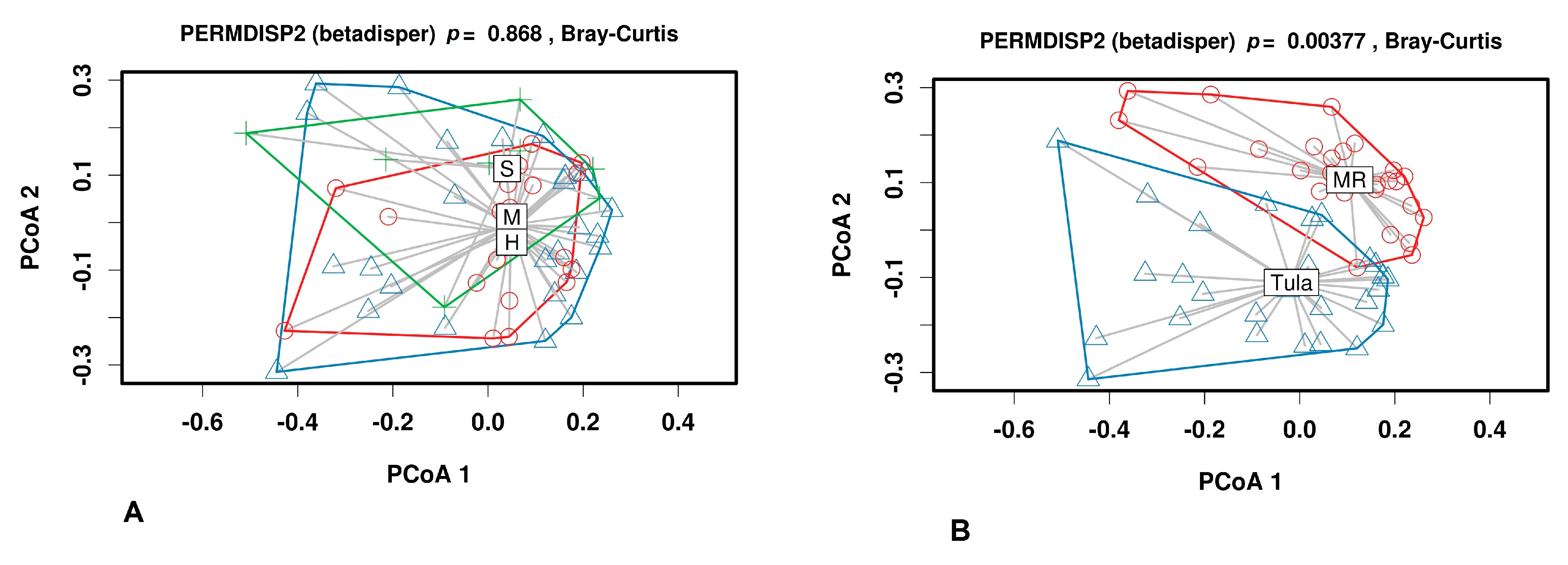
3.2.2. Beta Diversity

Homogeneity of multivariate dispersion (PERMDISP2) was run using Bray–Curtis distances to assess if differences in heterogeneity existed among the samples based on different factors. As shown in Figure 4A, there was no clear separation of milk samples from groups within the plots. The distribution of milk samples from group M revealed an overlap with groups H and S.
However, clustering of the milk samples based on their microbiota made it possible to separate samples from Tula and Moscow regions (Figure 4B).

3.3. Discriminant Analysis

Rarefaction curves for each group of samples and for a cutoff value of 0.03 are presented in Figure S2. For LEfSe analysis, we took sequences with a depth > 50,000 reads and number of observed OTUs > 1000 to avoid the very strong influence of sequences where the minority of numbered OTUs are represented by the ultimate majority of total reads (Figure 5).

4. Discussion

Investigating the problem of mastitis and searching for causes of its development remain relevant today. Omics technologies have been actively used in attempts to find a solution to this problem. [33]. Understanding the causes of mastitis and its pathogens will facilitate creation of preventive measures to reduce the incidence.
In the current work, we analyzed milk microbiotas associated with bovine mastitis in Central Russia. Milk samples were provided from two farms located in two neighboring regions of Central Russia but spaced approximately 200 km apart. We investigated milk microbiomes of individual quarters of mammalian gland isolated from two farms in during the summer.
We identified most abundant bacterial genera, including Staphylococcus, Aerococcus, Streptococcus, Enterobacter, Macrococcus, Corynebacterium, Acinetobacter, Psychrobacter, Ignavigranum, and Atopostipes.
Determination of the most abundant OTUs shown that S. aureus was detected in all types of milk samples (H, M, and S). S. aureus is a major intramammary pathogen associated with mastitis, along with E. coli and Streptococcus uberis [15,19,34,35,36,37]. In previous work, we demonstrated that an acute form of bovine mastitis in Russia during first and second lactations is often characterized by the presence of Staphylococcus species in the milk and Staphylococcus aureus in particular [38]. Moreover, in the current study, we detected the presence of Staphylococcus aureus in huge quantities (21.0 to 91.15%) in five samples from healthy quarters with QSCC < 100,000 cells/cm3. We explain this observation by these conditionally healthy quarters actually belonging to animals with diagnosed clinical mastitis. On the other hand, there are milk samples from mastitis quarters without evident predominating species that could serve as causative agents. These results are consistent with the suggestion that mastitis has a distinct etiology and that dysbacteriosis may be one of the causes of this disease [39].
In addition in this work we specially lowered the SCC level for healthy quarters to 100,000 to analyze healthy samples. In previous works authors suggest that SCC level varies depending on the mastitis pathogen [40]. In this direction, Grispoldi et al. in 2019 [41] discovered the prevalence of S. aureus ranged from 40.74% in milk samples with SCC < 100,000. In addition Oikonomou et al. in 2014 [35] has demonstrated that Staphylococcus spp. and Streptococcus spp. were among the most prevalent genera in all groups with high and low SCC levels.
In this work we pay the special attention to the SCC level < 100,000 to analyze healthy samples and conditionally healthy samples obtained from cows with higher SCC levels in other quarter. As of today the low level of SCC values remains under discussion, different authors consider different values [42].
Interestingly, Staphylococcus and Aerococcus genera seemed to be antagonistic to each other, and the disappearance of the Aerococcus genus in milk microbiota may represent a marker for its transition to a subclinical stage of mastitis. The replacement of Aerococcus with opportunistic microorganisms is clearly visible when comparing healthy milk samples to subclinical and clinical samples.
The next agent identified in this work was Corynebacterium. In recent studies by Porcellato et al. [43] strong negative correlation of Corynebacteriaceae and Staphylococcacea was detected. In contrast, early positive correlations with Staphylococcus were shown for this genus [44]. Here, we did not identify a strong correlation, but most samples contained sequences of Corynebacterium. Similar results on the association of Corynebacterium with mastitis were reported in previous studies [34,45].
Medically relevant members of the genus Acinetobacter are being increasingly recognized as a serious health hazard worldwide. In particular, Acinetobacter baumannii, a member of the ESKAPE group of pathogens (Enterococcus faecium, Staphylococcus aureus, Klebsiella pneumoniae, Acinetobacter baumannii, Pseudomonas aeruginosa, and Enterobacter species), is a leading cause of nosocomial infections [46]. In our study, DNA from Acinetobacter sp. was detected in 82% of subclinical and 74% of mastitic milk samples, except for those with predominating Staphylococcus spp. and Streptococcus spp. However, Pang et al. [45] have shown that Acinetobacter is more prevalent in healthy group samples.
Streptococcus was the second dominant group in our work. This genus, including S. uberis, S. agalactiae and S. dysgalactiae, is a typical mastitis associated pathogen [35,45,47]. However, in our work, we identified nonpathogenic S. thermophilus as a dominant organism in three milk samples with high SCC level (>2,000,000). We speculate that this is due to the ability of increasing somatic cells to stimulate growth of S. thermophilus [48].
The possible role of anaerobic bacteria Fusobacterium spp. and Porphyromonas spp. as opportunistic pathogens in existing mastitis cases was discussed in previous studies [35,49]. In addition, several authors have shown that F. necrophorum may be associated with summer mastitis [50,51], which is in agreement with our period of milk sampling (from June to August). Recently, Gu and coauthors have shown that Fusobacterium causes dysbiosis [52]. We show here that the prevalence of these microorganisms (17.49% and 16.31%, respectively) may be associated with subclinical mastitis in sample #5895zp.
Moreover, one case of clinical mastitis (#7211pp) was presumably associated with the predominance of Mycoplasma sp. (40.09%). Different species of Mycoplasma were determined to be mastitis-caused organisms, and M. bovis was the most prevalent. Mycoplasma sp. stimulates apoptosis and inflammatory cytokine reactions [53] and is one of the reasons for milking reductions [54].
Bovine milk microbiome analysis revealed strong variations between quarters, with some quarters clearly dominated by one taxonomic unit, whereas others displayed a more balanced profile. Similarly, a considerable difference in quarter microbiota from the same cow was described in previous work [43] We show in our study that not only a single group of pathogenic microorganisms but several pathogenic genera and species within milk microbiota might be associated with subclinical and clinical bovine mastitis at the same time within one general area of Central Russia.
Analyzing the microbial diversity of Moscow and Tula regions, we determined that diversity of the Moscow region groups was higher than in the Tula groups.
Analyzing the diversity of H, M and S groups, we detected no significant difference between the Shannon indices of groups M and S. However, the Shannon index of group H was lower compared to previous studies reporting the richness and diversity of microbiota from healthy quarters as significantly higher compared to subclinical and clinical samples [35,45].
We found some differences in OTU with respect to diversity between the farms. Atopostipies and Oligella genera were detected only in the Moscow farm (except for one S sample). Moreover, when comparing the two farms, Corynebacterum was detected in large amounts in the Moscow farm and was associated with second lactation. As shown, early Atopostipies was identified in both mastitis and healthy groups, whereas Corynebacterium was the most abundant genera in mastitic samples [45]. Moreover, Qadri et al. demonstrated that C. piogenes is one causative organism for summer mastitis [55].
Despite the fact that Shannon and Chao1 indices for Moscow region milk samples were higher than in milk samples from Tula, cooccurrence analysis revealed increase marker families in Tula milk microbiomes in contrast to those from the Moscow region. Bacillales, Enterobacteriaceae and Leptotrichiaceae were more prevalent in H Tula samples, while Flavobacteriales, Planctomycetales, Pseudomonadales, and one group of unclassified bacteria were more prevalent in M Tula samples. In the Moscow region, milk samples LEfSe analysis revealed only one significant negative correlation between Xantomonadales in the H group and Neisseriales in the M group.

5. Conclusions

We observed marked difference in certain dominant OTUs in milk samples that have never been reported in related studies. Remarkably, alpha-diversity analysis revealed that the richness and diversity of mastitis microbiota were higher than in healthy and subclinical samples in contrast to previous reports. Beta-diversity analysis showed a clear separation of samples from Moscow and Tula region farms; however, healthy, mastitis and subclinical samples were unable to be distinguished from one another. This indicates that infection-causing agents have an uncertain nature and seem to be common in both farms. We did not identify a significant amount of Escherichia coli OTU in either the Moscow or Tula region farm, despite E. coli being assumed as one of the frequent inflammatory agents in bovine mastitis. In contrast, other common mastitis causative agents, Staphylococcus and Streptococcus spp., were widespread in both healthy and mastitis milk samples. Notably, Streptococcus thermophilus was preferentially detected in milk samples with very high SCC values. The comparison of OTU composition in milk samples revealed that Aerococcus and Acinetobacter could serve as marker genera for healthy cows from the Moscow region farm and in cows with mastitis from the Tula region farm, respectively. The role that unidentified bacilli play in normal healthy microbiota remains unclear and requires further investigation.

Supplementary Materials

The following are available online at https://www.mdpi.com/article/10.3390/ani11051401/s1, Table S1: Descriptive characteristics of milk samples used in the study. https://osf.io/3s9jx (date accessed 15 March 2021); Figure S1: Full heatmap of OTUs abundance in individual milk samples. https://osf.io/pywve (date accessed 15 March 2021); Figure S2: Rarefaction curves for the milk samples. https://osf.io/746dc (date accessed 15 March 2021).

Author Contributions

F.B., S.S., and K.F. designed the study. S.S., A.S. (Anatoly Sorokin), and T.D. performed the bioinformatics analysis. S.S., K.F., and F.B. drafted the manuscript. S.S. and K.F. interpreted the data. O.A., D.N., I.S., M.S., A.S. (Anna Shepelyakovskaya) and E.K. performed the laboratory experiments. F.B. and N.Z. are supervisors of the project. All authors have read and agreed to the published version of the manuscript.

Funding

This research was funded by the Russian Science Foundation, grant number 20-16-00106, and by the Ministry of Education and Science of the Russian Federation (ID: 1021032425419-8; 121022400207-1).

Institutional Review Board Statement

Ethical review and approval were waived for this study, due to all animal manipulation were provided in accordance with the Instruction for Diagnostics, Therapy and Prevention of mastitis # 13-5-2/1948 (http://gov.cap.ru/home/65/aris/bd/vetzac/document/371.html date accessed 15 March 2021) and approved by the Department of Veterinary, Ministry of Agriculture of the Russian Federation; and with the Guidelines for the Microbiological Study of Milk and the Secretion of the Udder of Cows for the Diagnosis of Mastitis (https://standartgost.ru/g/pkey-14293740821 date accessed 15 March 2021), Ministry of Agriculture of the Russian Federation.

Data Availability Statement

Sequences generated in this study were deposited in the NCBI sequence read archive (https://www.ncbi.nlm.nih.gov/sra date accessed 11 January 2021) under accession numbers SRX8374271-SRX8374323.

Acknowledgments

We thank GenoMed (Moscow, Russia) for sequencing.

Conflicts of Interest

The authors declare no conflict of interest. The funders had no role in the design of the study; in the collection, analyses, or interpretation of data; in the writing of the manuscript, or in the decision to publish the results.

References

  1. Heikkilä, A.M.; Nousiainen, J.; Pyorala, S. Costs of clinical mastitis with special reference to premature culling. J. Dairy Sci. 2012, 95, 139–150. [Google Scholar] [CrossRef] [PubMed]
  2. Hahne, J.; Isele, D.; Berning, J.; Lipski, A. The contribution of fast growing, psychrotrophic microorganisms on biodiversity of refrigerated raw cow’s milk with high bacterial counts and their food spoilage potential. Food Microbiol. 2019, 79, 11–19. [Google Scholar] [CrossRef]
  3. Schmidt, T.; Kock, M.M.; Ehlers, M.M. Molecular Characterization of Staphylococcus aureus Isolated from Bovine Mastitis and Close Human Contacts in South African Dairy Herds: Genetic Diversity and Inter-Species Host Transmission. Front. Microbiol. 2017, 8, 511. [Google Scholar] [CrossRef] [PubMed] [Green Version]
  4. Murray, S.; Pascoe, B.; Méric, G.; Mageiros, L.; Yahara, K.; Hitchings, M.D.; Friedmann, Y.; Wilkinson, T.S.; Gormley, F.J.; Mack, D.; et al. Recombination-Mediated Host Adaptation by Avian Staphylococcus aureus. Genome Biol. Evol. 2017, 9, 830–842. [Google Scholar] [CrossRef] [PubMed] [Green Version]
  5. Islam, Z.; Espinosa-Gongora, C.; Damborg, P.; Sieber, R.N.; Munk, R.; Husted, L.; Moodley, A.; Skov, R.; Larsen, J.; Guardabassi, L. Horses in Denmark Are a Reservoir of Diverse Clones of Methicillin-Resistant and -Susceptible Staphylococcus aureus. Front. Microbiol. 2017, 8, 543. [Google Scholar] [CrossRef] [Green Version]
  6. Miragaia, M. Factors Contributing to the Evolution of MecA-Mediated β-Lactam Resistance in Staphylococci: Update and New Insights From Whole Genome Sequencing (WGS). Front Microbiol. 2018, 9, 2723. [Google Scholar] [CrossRef] [Green Version]
  7. Monaco, M.; De Araujo, F.P.; Cruciani, M.; Coccia, E.M.; Pantosti, A. Worldwide Epidemiology and Antibiotic Resistance of Staphylococcus aureus; Springer Science and Business Media LLC: Berlin/Heidelberg, Germany, 2016; pp. 21–56. [Google Scholar]
  8. Akkou, M.; Bouchiat, C.; Antri, K.; Bes, M.; Tristan, A.; Dauwalder, O.; Martins-Simoes, P.; Rasigade, J.P.; Etienne, J.; Vandenesch, F.; et al. New host shift from human to cows within Staphylococcus aureus involved in bovine mastitis and nasal carriage of animal’s caretakers. Vet. Microbiol. 2018, 223, 173–180. [Google Scholar] [CrossRef]
  9. Van Duijkeren, E.; Schwarz, C.; Bouchard, D.; Catry, B.; Pomba, C.; Baptiste, K.E.; A Moreno, M.; Rantala, M.; Ruzauskas, M.; Sanders, P.; et al. The use of aminoglycosides in animals within the EU: Development of resistance in animals and possible impact on human and animal health: A review. J. Antimicrob. Chemother. 2019, 74, 2480–2496. [Google Scholar] [CrossRef]
  10. Profet, M. The Function of Allergy: Immunological Defense Against Toxins. Q. Rev. Biol. 1991, 66, 23–62. [Google Scholar] [CrossRef]
  11. Asao, T.; Kumeda, Y.; Kawai, T.; Shibata, T.; Oda, H.; Haruki, K.; Nakazawa, H.; Kozaki, S. An extensive outbreak of staphylococcal food poisoning due to low-fat milk in Japan: Estimation of enterotoxin A in the incriminated milk and powdered skim milk. Epidemiol. Infect. 2003, 130, 33–40. [Google Scholar] [CrossRef]
  12. Muluk, N.B.; Altın, F.; Cingi, C. Role of Superantigens in Allergic Inflammation: Their Relationship to Allergic Rhinitis, Chronic Rhinosinusitis, Asthma, and Atopic Dermatitis. Am. J. Rhinol. Allergy 2018, 32, 502–517. [Google Scholar] [CrossRef]
  13. Grispoldi, L.; Massetti, L.; Sechi, P.; Iulietto, M.F.; Ceccarelli, M.; Karama, M.; Popescu, P.A.; Pandolfi, F.; Cenci-Goga, B.T. Short communication: Characterization of enterotoxin-producing Staphylococcus aureus isolated from mastitic cows. J. Dairy Sci. 2019, 102, 1059–1065. [Google Scholar] [CrossRef] [Green Version]
  14. Taponen, S.; McGuinness, D.; Hiitiö, H.; Simojoki, H.; Zadoks, R.; Pyörälä, S. Bovine milk microbiome: A more complex issue than expected. Veter Res. 2019, 50, 1–15. [Google Scholar] [CrossRef] [PubMed] [Green Version]
  15. Tong, J.; Zhang, H.; Zhang, Y.; Xiong, B.; Jiang, L. Microbiome and Metabolome Analyses of Milk From Dairy Cows With Subclinical Streptococcus agalactiae Mastitis—Potential Biomarkers. Front. Microbiol. 2019, 10, 2547. [Google Scholar] [CrossRef] [PubMed]
  16. Oikonomou, G.; Addis, M.F.; Chassard, C.; Nader-Macias, M.E.F.; Grant, I.; Delbès, C.; Bogni, C.I.; Le Loir, Y.; Even, S. Milk Microbiota: What Are We Exactly Talking About? Front. Microbiol. 2020, 11, 60. [Google Scholar] [CrossRef] [PubMed] [Green Version]
  17. Herlekar, D.; Shashikant, C.; Gurjar, A.; Jayarao, B. Presence of viral and bacterial organisms in milk and their association with somatic cell counts. J. Dairy Sci. 2013, 96, 6336–6346. [Google Scholar] [CrossRef] [PubMed]
  18. Hertl, J.; Schukken, Y.; Welcome, F.; Tauer, L.; Gröhn, Y. Pathogen-specific effects on milk yield in repeated clinical mastitis episodes in Holstein dairy cows. J. Dairy Sci. 2014, 97, 1465–1480. [Google Scholar] [CrossRef] [Green Version]
  19. Keane, O. Symposium review: Intramammary infections—Major pathogens and strain-associated complexity. J. Dairy Sci. 2019, 102, 4713–4726. [Google Scholar] [CrossRef] [PubMed] [Green Version]
  20. Addis, M.F.; Tanca, A.; Uzzau, S.; Oikonomou, G.; Bicalho, R.C.; Moroni, P. The bovine milk microbiota: Insights and perspectives from -omics studies. Mol. BioSyst. 2016, 12, 2359–2372. [Google Scholar] [CrossRef] [Green Version]
  21. García-García, N.; Tamames, J.; Linz, A.M.; Pedrós-Alió, C.; Puente-Sánchez, F. Microdiversity ensures the maintenance of functional microbial communities under changing environmental conditions. ISME J. 2019, 13, 2969–2983. [Google Scholar] [CrossRef]
  22. Zhang, Z.; Li, X.; Yang, F.; Luo, J.; Wang, X.; Liu, L.; Li, H. Influences of season, parity, lactation, udder area, milk yield, and clinical symptoms on intramammary infection in dairy cows. J. Dairy Sci. 2016, 99, 6484–6493. [Google Scholar] [CrossRef] [PubMed]
  23. Saishu, N.; Morimoto, K.; Yamasato, H.; Ozaki, H.; Murase, T. Characterization of Aerococcus viridans isolated from milk samples from cows with mastitis and manure samples. J. Veter Med. Sci. 2015, 77, 1037–1042. [Google Scholar] [CrossRef] [PubMed]
  24. Agriculture and Horticulture Development Board (AHDB) Knowledge Library. Available online: https://ahdb.org.uk/knowledge-library/mastitis-in-dairy-cows (accessed on 15 March 2021).
  25. Metzger, S.A.; Hernandez, L.L.; Suen, G.; Ruegg, P.L. Understanding the Milk Microbiota. Veter Clin. N. Am. Food Anim. Pr. 2018, 34, 427–438. [Google Scholar] [CrossRef] [PubMed]
  26. Knight, R.; Vrbanac, A.; Taylor, B.C.; Aksenov, A.; Callewaert, C.; Debelius, J.; Gonzalez, A.; Kosciolek, T.; McCall, L.-I.; McDonald, D.; et al. Best practices for analysing microbiomes. Nat. Rev. Genet. 2018, 16, 410–422. [Google Scholar] [CrossRef] [Green Version]
  27. Weber, N.; Liou, D.; Dommer, J.; MacMenamin, P.; Quiñones, M.; Misner, I.; Oler, A.J.; Wan, J.; Kim, L.; McCarthy, M.C.; et al. Nephele: A cloud platform for simplified, standardized and reproducible microbiome data analysis. Bioinformatics 2017, 34, 1411–1413. [Google Scholar] [CrossRef]
  28. Babraham Bioinformatics. FastQC v. 0.11.2. 2014. Available online: http://www.bioinformatics.babraham.ac.uk/projects/fastqc/ (accessed on 10 January 2020).
  29. Yilmaz, P.; Parfrey, L.W.; Yarza, P.; Gerken, J.; Pruesse, E.; Quast, C.; Schweer, T.; Peplies, J.; Ludwig, W.; Glöckner, F.O. The SILVA and “All-species Living Tree Project (LTP)” taxonomic frameworks. Nucleic Acids Res. 2014, 42, D643–D648. [Google Scholar] [CrossRef] [PubMed] [Green Version]
  30. Caporaso, J.G.; Kuczynski, J.; Stombaugh, J.; Bittinger, K.; Bushman, F.D.; Costello, E.K.; Fierer, N.; Peña, A.G.; Goodrich, J.K.; Gordon, J.I.; et al. QIIME Allows Analysis of High-Throughput Community Sequencing data. Nat. Methods 2010, 7, 335–336. [Google Scholar] [CrossRef] [PubMed] [Green Version]
  31. Lozupone, C.; Knight, R. UniFrac: A New Phylogenetic Method for Comparing Microbial Communities. Appl. Environ. Microbiol. 2005, 71, 8228–8235. [Google Scholar] [CrossRef] [Green Version]
  32. Segata, N.; Izard, J.; Waldron, L.; Gevers, D.; Miropolsky, L.; Garrett, W.S.; Huttenhower, C. Metagenomic biomarker discovery and explanation. Genome Biol. 2011, 12, R60. [Google Scholar] [CrossRef] [Green Version]
  33. Metzger, S.A.; Hernandez, L.L.; Skarlupka, J.H.; Walker, T.M.; Suen, G.; Ruegg, P.L. A Cohort Study of the Milk Microbiota of Healthy and Inflamed Bovine Mammary Glands Dryoff Through 150 Days in Milk. Front. Vet. Sci. 2018, 5, 247. [Google Scholar] [CrossRef] [Green Version]
  34. Falentin, H.; Rault, L.; Nicolas, A.; Bouchard, D.S.; Lassalas, J.; Lamberton, P.; Aubry, J.-M.; Marnet, P.G.; Le Loir, Y.; Even, S. Bovine Teat Microbiome Analysis Revealed Reduced Alpha Diversity and Significant Changes in Taxonomic Profiles in Quarters with a History of Mastitis. Front. Microbiol. 2016, 7, 480. [Google Scholar] [CrossRef] [Green Version]
  35. Oikonomou, G.; Bicalho, M.L.; Meira, E.; Rossi, R.E.; Foditsch, C.; Machado, V.S.; Teixeira, A.G.V.; Santisteban, C.; Schukken, Y.H.; Bicalho, R.C. Microbiota of Cow’s Milk; Distinguishing Healthy, Sub-Clinically and Clinically Diseased Quarters. PLoS ONE 2014, 9, e85904. [Google Scholar] [CrossRef] [PubMed] [Green Version]
  36. Dufour, S.; Dohoo, I.; Barkema, H.; Descôteaux, L.; Devries, T.; Reyher, K.; Roy, J.P.; Scholl, D. Epidemiology of coagulase-negative staphylococci intramammary infection in dairy cattle and the effect of bacteriological culture misclassification. J. Dairy Sci. 2012, 95, 3110–3124. [Google Scholar] [CrossRef] [Green Version]
  37. Leimbach, A.; Poehlein, A.; Vollmers, J.; Görlich, D.; Daniel, R.; Dobrindt, U. No evidence for a bovine mastitis Escherichia coli pathotype. BMC Genom. 2017, 18, 359. [Google Scholar] [CrossRef] [PubMed]
  38. Fursova, K.; Sorokin, A.; Sokolov, S.; Dzhelyadin, T.; Shulcheva, I.; Shchannikova, M.; Nikanova, D.; Artem’Eva, O.; Zinovieva, N.; Brovko, F. Virulence Factors and Phylogeny of Staphylococcus aureus Associated With Bovine Mastitis in Russia Based on Genome Sequences. Front. Vet. Sci. 2020, 7, 135. [Google Scholar] [CrossRef] [PubMed]
  39. Fernández, L.; Langa, S.; Martín, V.; Maldonado, A.; Jiménez, E.; Martín, R.; Rodríguez, J.M. The human milk microbiota: Origin and potential roles in health and disease. Pharmacol. Res. 2013, 69, 1–10. [Google Scholar] [CrossRef]
  40. Green, M.; Green, L.; Schukken, Y.; Bradley, A.; Peeler, E.; Barkema, H.; De Haas, Y.; Collis, V.; Medley, G. Somatic Cell Count Distributions During Lactation Predict Clinical Mastitis. J. Dairy Sci. 2004, 87, 1256–1264. [Google Scholar] [CrossRef]
  41. Grispoldi, L.; Karama, M.; Ianni, F.; La Mantia, A.; Pucciarini, L.; Camaioni, E.; Sardella, R.; Sechi, P.; Natalini, B.; Cenci-Goga, B.T. The Relationship between S. aureus and Branched-Chain Amino Acids Content in Composite Cow Milk. Animals 2019, 9, 981. [Google Scholar] [CrossRef] [Green Version]
  42. Rainard, P.; Foucras, G.; Boichard, D.; Rupp, R. Invited review: Low milk somatic cell count and susceptibility to mastitis. J. Dairy Sci. 2018, 101, 6703–6714. [Google Scholar] [CrossRef] [Green Version]
  43. Porcellato, D.; Meisal, R.; Bombelli, A.; Narvhus, J.A. A core microbiota dominates a rich microbial diversity in the bovine udder and may indicate presence of dysbiosis. Sci. Rep. 2020, 10, 1–14. [Google Scholar] [CrossRef]
  44. Ma, Z.; Guan, Q.; Ye, C.; Zhang, C.; Foster, J.A.; Forney, L.J. Network analysis suggests a potentially ‘evil’ alliance of opportunistic pathogens inhibited by a cooperative network in human milk bacterial communities. Sci. Rep. 2015, 5, srep08275. [Google Scholar] [CrossRef] [Green Version]
  45. Pang, M.; Xie, X.; Bao, H.; Sun, L.; He, T.; Zhao, H.; Zhou, Y.; Zhang, L.; Zhang, H.; Wei, R.; et al. Insights Into the Bovine Milk Microbiota in Dairy Farms With Different Incidence Rates of Subclinical Mastitis. Front. Microbiol. 2018, 9, 2379. [Google Scholar] [CrossRef] [PubMed] [Green Version]
  46. Elhosseiny, N.M.; Attia, A.S. Acinetobacter: An emerging pathogen with a versatile secretome. Emerg. Microbes Infect. 2018, 7, 1–15. [Google Scholar] [CrossRef] [Green Version]
  47. Klaas, I.C.; Zadoks, R.N. An update on environmental mastitis: Challenging perceptions. Transbound. Emerg. Dis. 2017, 65, 166–185. [Google Scholar] [CrossRef] [Green Version]
  48. Okello-Uma, I.; Marshall, V.M.E. Influence of mastitis on growth of starter organisms used for the manufacture of fermented milks. J. Dairy Res. 1986, 53, 631–637. [Google Scholar] [CrossRef]
  49. Oikonomou, G.; Machado, V.S.; Santisteban, C.; Schukken, Y.H.; Bicalho, R.C. Microbial Diversity of Bovine Mastitic Milk as Described by Pyrosequencing of Metagenomic 16s rDNA. PLoS ONE 2012, 7, e47671. [Google Scholar] [CrossRef] [Green Version]
  50. Catozzi, C.; Bonastre, A.S.; Francino, O.; Lecchi, C.; De Carlo, E.; Vecchio, D.; Martucciello, A.; Fraulo, P.; Bronzo, V.; Cuscó, A.; et al. The microbiota of water buffalo milk during mastitis. PLoS ONE 2017, 12, e0184710. [Google Scholar] [CrossRef] [PubMed] [Green Version]
  51. Polveiro, R.C.; Vidigal, P.M.P.; Mendes, T.A.D.O.; Yamatogi, R.S.; Lima, M.C.; Moreira, M.A.S. Effects of enrofloxacin treatment on the bacterial microbiota of milk from goats with persistent mastitis. Sci. Rep. 2020, 10, 1–13. [Google Scholar] [CrossRef] [PubMed] [Green Version]
  52. Gu, X.; Song, L.J.; Li, L.X.; Liu, T.; Zhang, M.M.; Li, Z.; Wang, P.; Li, M.; Zuo, X.L. Fusobacterium nucleatum Causes Microbial Dysbiosis and Exacerbates Visceral Hypersensitivity in a Colonization-Independent Manner. Front. Microbiol. 2020, 11, 1281. [Google Scholar] [CrossRef] [PubMed]
  53. Jimbo, S.; Suleman, M.; Maina, T.; Prysliak, T.; Mulongo, M.; Perez-Casal, J. Effect of Mycoplasma bovis on bovine neutrophils. Vet. Immunol. Immunopathol. 2017, 188, 27–33. [Google Scholar] [CrossRef]
  54. Al-Farha, A.A.B.; Hemmatzadeh, F.; Khazandi, M.; Hoare, A.; Petrovski, K. Evaluation of effects of Mycoplasma mastitis on milk composition in dairy cattle from South Australia. BMC Vet. Res. 2017, 13, 351. [Google Scholar] [CrossRef] [PubMed] [Green Version]
  55. Qadri; Ganguly, S.; Para, P.; Praveen, P.; Wakchaure. Summer Mastitis in Cattle: A Review. J. Biol. Chem. Res. 2015, 32, 1006–1009. [Google Scholar]
Figure 1. The relative abundance of major bacterial phyla present in milk samples.
Animals 11 01401 g001
Figure 2. Heatmap of the most abundant OTUs in milk samples. Annotations at the top of the heatmap show clustering of milk samples. The color scale depicts the normalized relative abundance of each OTU. The full OTU heatmap is available as Figure S1. Treatment groups (H, M, and S) are described in Materials and Methods.
Animals 11 01401 g002
Figure 3. Diversity and evenness. (A) Shannon and Chao1 indices for all three groups (H, M, S). (B) Shannon and Chao1 indices for both sites (MR—Moscow region, Tula—Tula region).
Animals 11 01401 g003
Figure 4. PERMDISP2 beta diversity analysis of microbiota. (A) H (red circles), M (blue triangles), and S (green crosses) samples. (B) Tula (blue triangles) and Moscow (red circles) Regions.
Animals 11 01401 g004
Figure 5. Cladograms based on LEfSe analysis (p < 0.05, LDA effect size > 2) of Tula (A) and Moscow region (B) milk sample microbiomes.
Animals 11 01401 g005
Publisher’s Note: MDPI stays neutral with regard to jurisdictional claims in published maps and institutional affiliations.

