Pathological Study of Facial Eczema (Pithomycotoxicosis) in Sheep
by
 , , , , and
Miguel Fernández
1,2,* ,
,
Valentín Pérez
1,2,
Miguel Fuertes
3,
Julio Benavides
2,
José Espinosa
1,2,
Juan Menéndez
4,†,
Ana L. García-Pérez
3 and
M. Carmen Ferreras
1,2 1
Departamento de Sanidad Animal, Facultad de Veterinaria, Universidad de León, C/Prof. Pedro Cármenes s/n, E-24071 León, Spain
2
Instituto de Ganadería de Montaña (IGM), CSIC-Universidad de León, Finca Marzanas s/n, E-24346 León, Spain
3
Department of Animal Health, NEIKER-Basque Institute for Agricultural Research and Development, Basque Research and Technology Alliance (BRTA), Parque Científico y Tecnológico de Bizkaia, P812, E-48160 Derio, Spain
4
Area de Sistemas de Producción Animal, Servicio Regional de Investigación y Desarrollo Agroalimentario, SERIDA, E-33300 Villaviciosa, Asturias, Spain
*
Author to whom correspondence should be addressed.
†
Current address: Centro Veterinario Albéitar, E-33204 Gijón, Spain.
Animals 2021, 11(4), 1070; https://doi.org/10.3390/ani11041070
Submission received: 19 March 2021
/
Revised: 6 April 2021
/
Accepted: 6 April 2021
/
Published: 9 April 2021
(This article belongs to the Special Issue Comparative Pathology and Immunohistochemistry of Veterinary Species)
Simple Summary
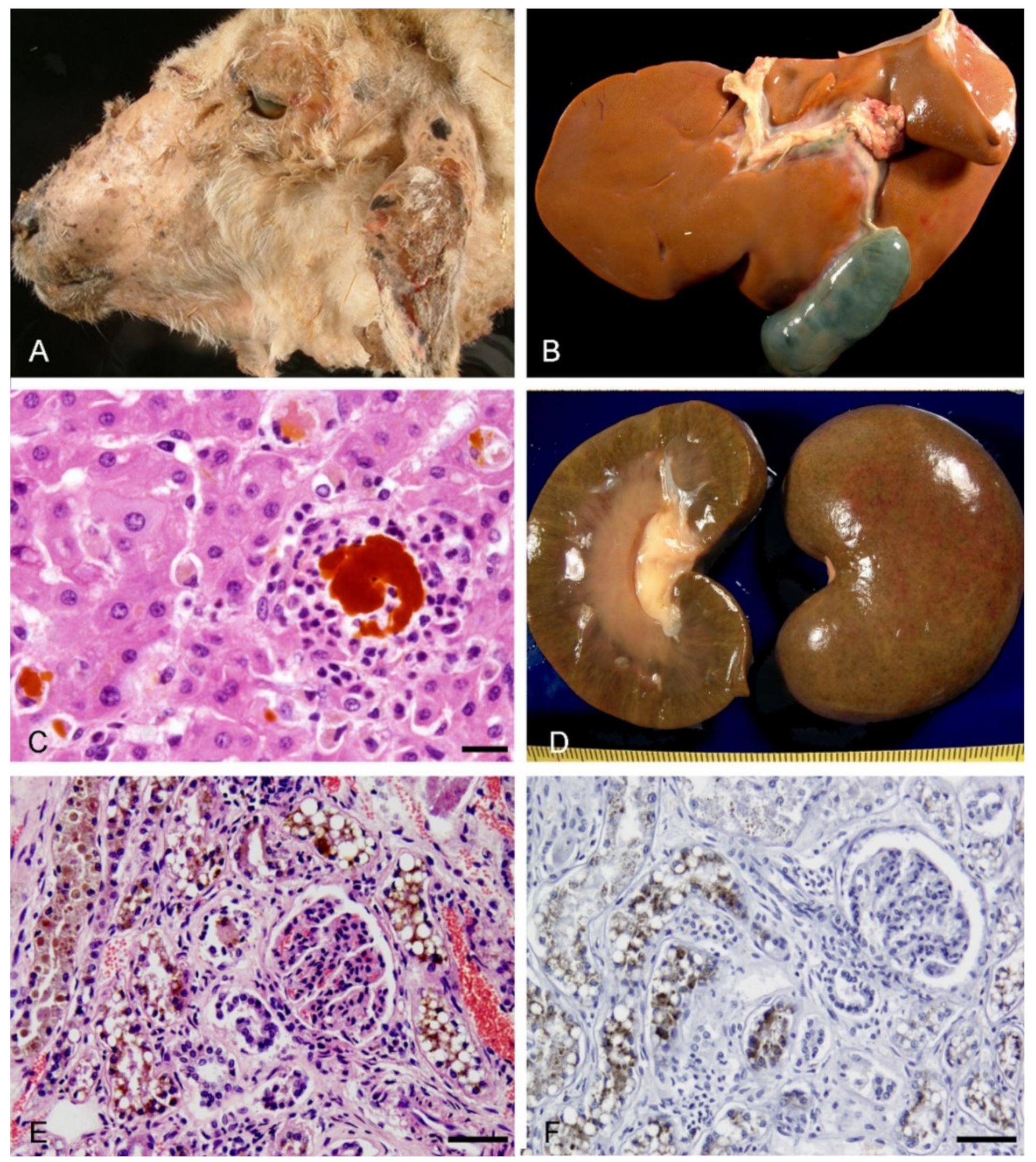
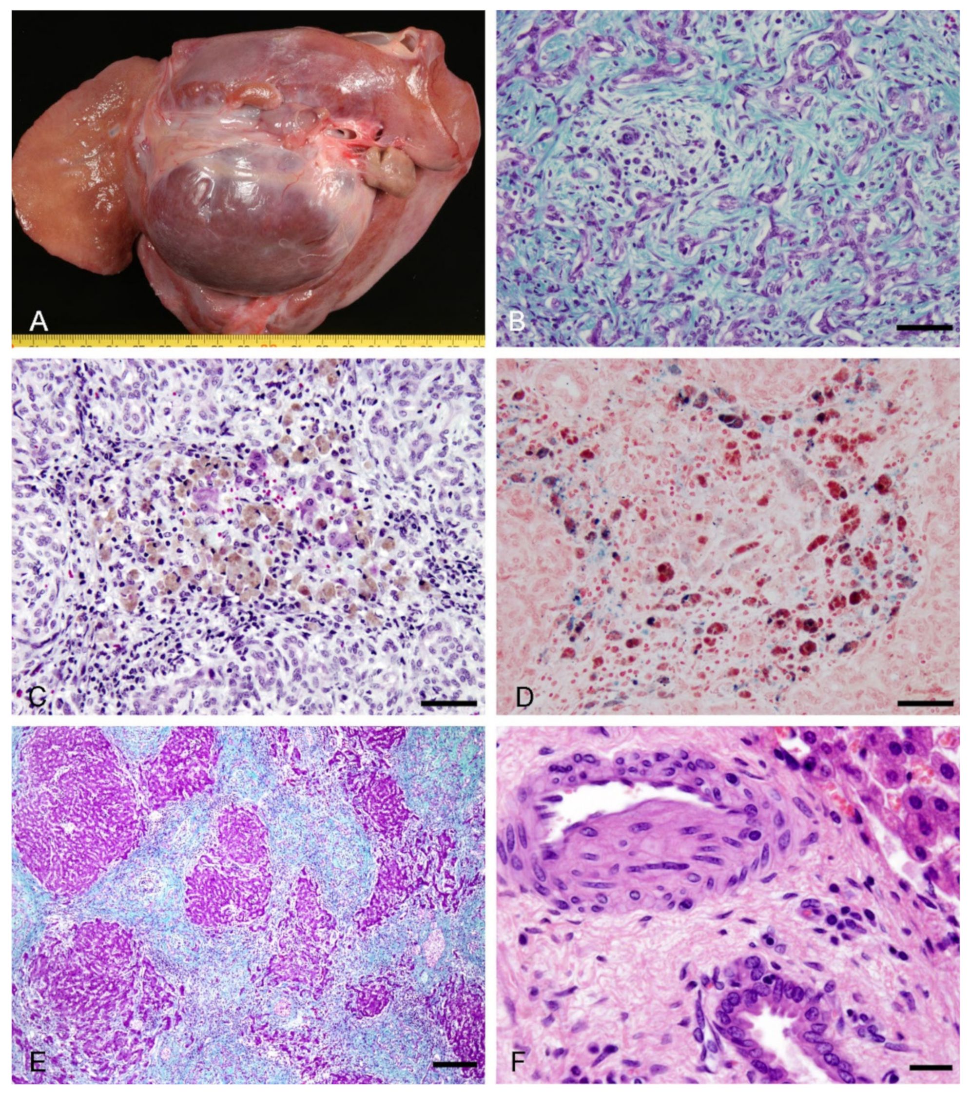
Facial eczema (FE) is a secondary photosensitization disease of farm ruminants caused by the sporidesmin A, present in the spores of the saprophytic fungus Pithomyces chartarum. This study communicates an outbreak of ovine FE in Asturias (Spain) and characterizes the local immune response that may contribute to liver damage promoting cholestasis and progression towards fibrosis and cirrhosis. Animals showed clinical signs of photosensitivity and lower gain of weight, loss of wool and crusting in the head for at least 6 months after the FE outbreak. Some sheep presented acute lesions characterized by subcutaneous edema in the head, cholestasis and nephrosis with macrophages and neutrophils present in areas of canalicular cholestasis. In chronic cases, alopecia and crusting, hepatic atrophy with regenerative nodules, fibrosis and gallstones were seen. The surviving parenchyma persisted with a jigsaw pattern characteristic of biliary cirrhosis. Concentric and eccentric myointimal proliferation was found in arteries near damaged bile ducts, where macrophages and lymphocytes were also observed.
Abstract
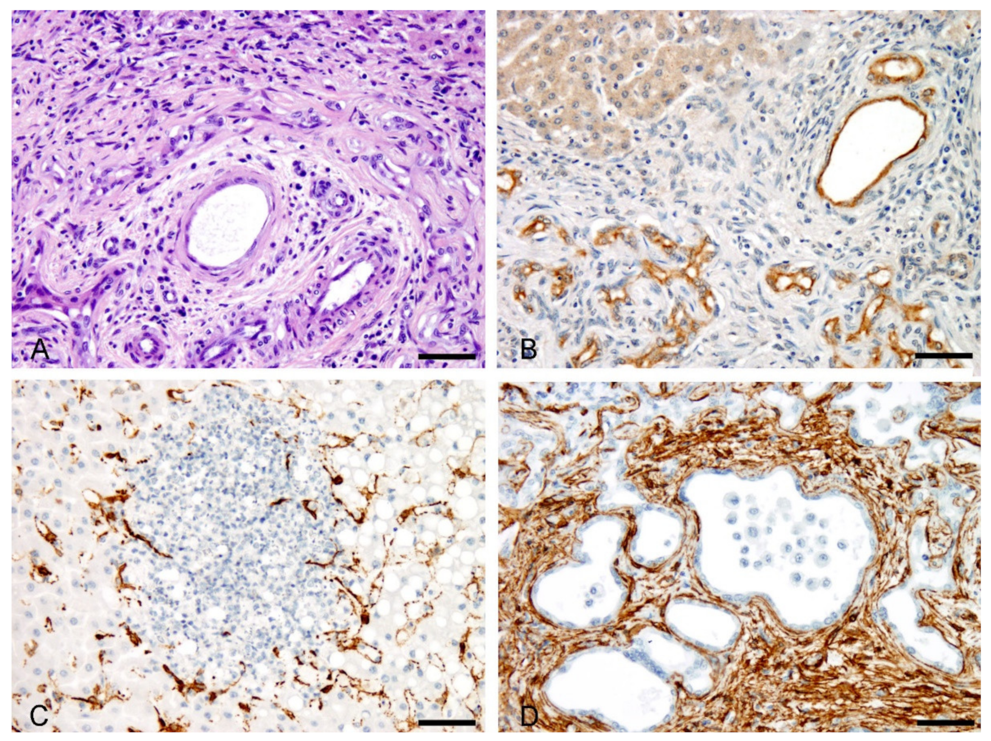
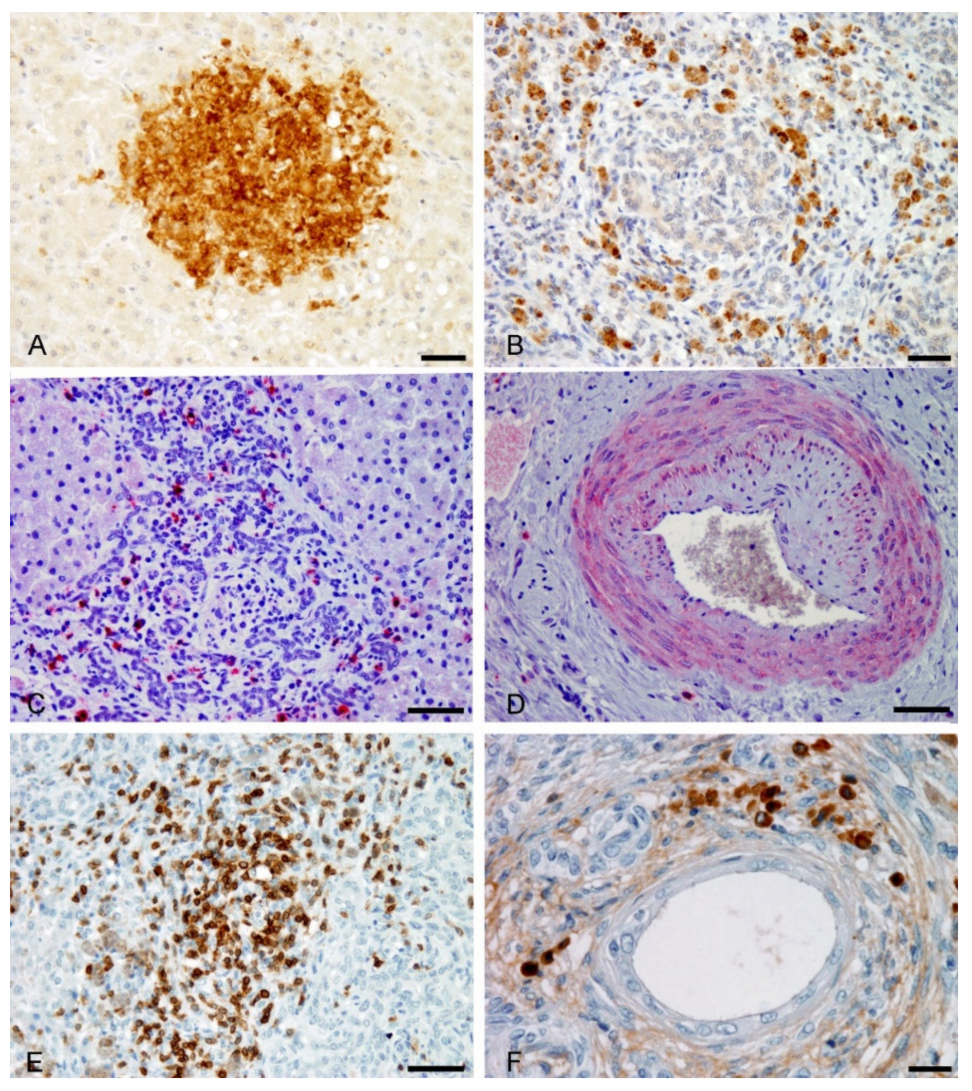
Facial eczema (FE) is a secondary photosensitization disease of farm ruminants caused by the sporidesmin A, produced in the spores of the saprophytic fungus Pithomyces chartarum. This study communicates an outbreak of ovine FE in Asturias (Spain) and characterizes the serum biochemical pattern and the immune response that may contribute to liver damage, favoring cholestasis and the progression to fibrosis and cirrhosis. Animals showed clinical signs of photosensitivity, with decrease of daily weight gain and loss of wool and crusting for at least 6 months after the FE outbreak. Serum activity of γ-glutamyltransferase and alkaline phosphatase were significantly increased in sheep with skin lesions. In the acute phase, edematous skin lesions in the head, hepatocytic and canalicular cholestasis in centrilobular regions, presence of neutrophils in small clumps surrounding deposits of bile pigment, ductular proliferation, as well as cholemic nephrosis, were observed. Macrophages, stained positively for MAC387, were found in areas of canalicular cholestasis. In the chronic phase, areas of alopecia and crusting were seen in the head, and the liver was atrophic with large regeneration nodules and gallstones. Fibrosis around dilated bile ducts, “typical” and “atypical” ductular reaction and an inflammatory infiltrate composed of lymphocytes and pigmented macrophages, with iron deposits and lipofuscin, were found. The surviving parenchyma persisted with a jigsaw pattern characteristic of biliary cirrhosis. Concentric and eccentric myointimal proliferation was found in arteries near damaged bile ducts. In cirrhotic livers, stellated cells, ductular reaction, ectatic bile ducts and presence of M2 macrophages and lymphocytes, were observed in areas of bile ductular reaction.
Keywords:
facial eczema; sheep; pathology; pithomycotoxicosis; immunohistochemistry; cirrhosis; cholestasis; liver