Article

Seroprevalence and Risk Factors of Toxoplasma gondii in Ruminant Meats from Wet Markets in Klang Valley and Abattoirs in Selangor, Malaysia

by
Norhamizah Abdul Hamid
1,
Mohammed Babatunde Sadiq
2,
Siti Zubaidah Ramanoon
2,
Rozaihan Mansor
2,
Malaika Watanabe
3,
Nur Mahiza Md Isa
4,
Juriah Kamaludeen
5 and
Sharifah Salmah Syed-Hussain
1,*
1
Department of Veterinary Clinical Studies, Faculty of Veterinary Medicine, Universiti Putra Malaysia, Serdang 43400 UPM, Selangor, Malaysia
2
Department of Farm and Exotic Animal Medicine and Surgery, Faculty of Veterinary Medicine, Universiti Putra Malaysia, Serdang 43400 UPM, Selangor, Malaysia
3
Department of Companion Animal Medicine and Surgery, Faculty of Veterinary Medicine, Universiti Putra Malaysia, Serdang 43400 UPM, Selangor, Malaysia
4
Department of Veterinary Pathology and Microbiology, Faculty of Veterinary Medicine, Universiti Putra Malaysia, Serdang 43400 UPM, Selangor, Malaysia
5
Department of Animal Science and Fisheries, Faculty of Agriculture and Food Science, Universiti Putra Malaysia Bintulu, Sarawak Campus, Bintulu 97008, Sarawak, Malaysia
*
Author to whom correspondence should be addressed.
Animals 2020, 10(7), 1139; https://doi.org/10.3390/ani10071139
Submission received: 6 May 2020 / Revised: 27 May 2020 / Accepted: 3 June 2020 / Published: 6 July 2020
(This article belongs to the Special Issue Zoonoses: Wild and Domestic Animal Interaction)

Abstract

:

Simple Summary

This study investigated the prevalence of Toxoplasma. gondii in meats of cattle, goat and sheep from wet markets and abattoirs in Selangor, Malaysia. Meat samples from wet markets in various districts and diaphragm samples from abattoirs were analyzed using ELISA to check for T. gondii IgG antibodies. Furthermore, attempts were made to detect T. gondii DNA from meat samples using the nested PCR technique. Twenty-five percent of the samples were positive for T. gondii antibodies, with the highest recorded in goat (55%), followed by sheep (35%) and cattle meat (9%). T. gondii DNA was not detected in any of the meat samples. Being the first report in Malaysia, the findings highlight the need for proper control in reducing exposure of ruminant meats to the parasite, especially those destined for human consumption.

Abstract

(1) Background: The objective of this study was to determine the prevalence of T. gondii in meats of cattle, goat and sheep from wet markets in Klang Valley, and abattoirs in Selangor, Malaysia; (2) Methods: A total of 192 meat samples were purchased from 51 wet markets in six districts in Klang Valley (Gombak, Klang, Kuala Lumpur, Hulu Langat, Petaling and Putrajaya). Meanwhile, a total of 200 diaphragm samples were collected from two government abattoirs located in Shah Alam and Banting, Selangor. All meat juices from samples were subjected to an indirect-ELISA kit for the presence of T. gondii IgG antibodies. Furthermore, all 184 meat samples of goat and sheep were subjected to conventional nested PCR (B1 genes) for the detection of T. gondii DNA; (3) Results: T. gondii antibodies were detected in 25% (n = 98/392) of the samples with seroprevalence of 9.1% (19/208, CI: 5.9%–13.8%) in cattle meat; 54.7% (41/75, 95% CI: 43.5%–65.4%) in goat meat and 34.9% (38/109, CI: 26.6%–44.2%) in sheep meat. No T. gondii DNA was detected in any of the meat samples of goat and sheep. T. gondii seropositivity in wet market samples was higher in goat (OR = 37.1 CI 12.4–110.3) and sheep meat (OR 9.03 CI: 3.28–24.8) compared to cattle meat (OR = 1.0) At univariate level, meat from non-licensed abattoirs (OR = 6.0 CI: 2.9–12.3) and female animals (OR = 6.7; CI 1.9–22.6) had higher risks of being seropositive for T. gondii antibodies than licensed abattoirs and male animals, respectively. (4) Conclusions: This is the first report of seroprevalence of T. gondii in ruminant meats for human consumption in Malaysia. The findings signified high exposure of meat samples from wet markets to T. gondii and the need for control measures to reduce the likelihood of infection when such raw or undercooked meats are consumed.

1. Introduction

Toxoplasma gondii is an apicomplexan protozoan capable of infecting all warm-blooded animals and causing major health concerns to humans, especially to unborn fetuses and immunocompromised individuals. In humans, toxoplasmosis has been shown to cause fever, lymphadenopathy, headache, myalgia, arthralgia, dizziness, and in worst situations, encephalitis, blindness and abortions [1]. Consumption of raw, or undercooked meat or meat products is highly associated with toxoplasmosis in humans. In European countries, it has been estimated that 30–60% of the infection in humans were due to consumption of undercooked meat or meat products [1,2]. Transmission in human may also occur from infected mother to the unborn fetus [3]. A study conducted in the United States had shown that the cost of testing and treating a newborn child due to toxoplasmosis ranges from USD 300,000 to more than USD 3 million for developmental disorder leading to hearing, vision or cognitive losses [4]. In South Australia, it was estimated that toxoplasmosis costs the sheep industry up to AUD 70 million per year [5]. These data indicate that T. gondii causes significant financial loss to animal producers, as well as to the public.
Ruminant animals especially sheep and goats are highly susceptible to T. gondii infection leading to reproductive failures [6]. There are indications of increasing seroprevalence of T. gondii worldwide, as the estimates of 18.6%, 43.9% and 47% were observed in cattle, goats and sheep, respectively [6,7,8]. Ruminant livestock are more likely to be infected due to the nature of grazing in contaminated environment with the oocysts [9]. Thus, the high rate of T. gondii infection reported in small ruminants worldwide, not only affects the economics of ruminant production but also imposes significant zoonotic health hazard in humans consuming infected meat.
In Malaysia, T. gondii seroprevalence among healthy Malaysians ranged from 14% to 30% [10,11], indicating that the disease is highly prevalent. Furthermore, alarming results have indicated that the seroprevalence of T. gondii among pregnant women in South East Asia was highest in Malaysia (42.5%) [12] as compared to the other neighboring countries such as Thailand (28.3%0 [13] and the Philippines (23.8%) [14]. Since T. gondii remains an important zoonotic pathogen worldwide, it is imperative to evaluate the exposure of ruminants to the parasite as well as in meats destined for human consumption in Malaysia. In Malaysia, most works on T. gondii in meat have been conducted in poultry, wild boar and exotic animals [15,16,17] but none in ruminants. Therefore, the aim of the present work was to determine the prevalence of T. gondii in meats of cattle, goat and sheep from wet markets and abattoirs in Klang Valley and Selangor, Malaysia and the risk factors involved.

2. Materials and Methods

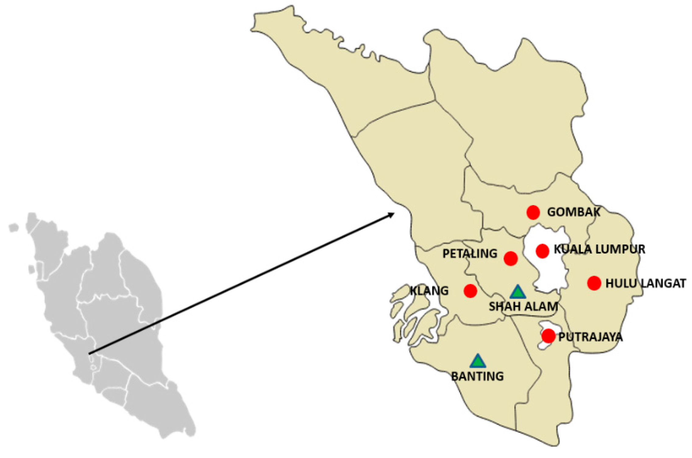
2.1. Study Design, Study Area and Selection of Sample Sites

A cross sectional study was conducted involving the selection of wet markets in Klang Valley and abattoirs in Selangor, Malaysia. The sampling sites were selected based on the availability of fresh meat from animals slaughtered locally in the study area. As shown in Figure 1, Klang Valley consists of four districts within Selangor State territory (Gombak: 3.2535° N, 101.6533° E, Hulu Langat: 2.9936° N, 101.7892° E, Klang: 3.0449° N, 101.4456° E, and Petaling: 3.1846° N, 101.5360° E) and two federal territories (Kuala Lumpur:3.1390° N, 101.6869° E and Putrajaya: 2.9264° N, 101.6964° E).
In Klang Valley, there were three types of wet markets available: (a) “pasar besar”—market located within a building, (b) “pasar tani”—open stalls which operate in the morning, and (c) “pasar malam”—open stalls which operate at night. “Pasar malam” were not included in the sampling, as they only cater for small areas within the districts, and usually locally slaughtered meats would not be available. For the wet markets, only fresh meat samples were collected in the present work. Fresh meat is also locally known as “warm meat”, as opposed to frozen meat. When locally slaughtered meats were not available at markets within the districts, repeated samplings were conducted on the same stall within a two-week interval. Information regarding the meat samples was obtained through informal conversation with the sellers/butchers. The fresh meats were categorized into two groups: with a “Lagenda” tag, and without, based on observation. “Lagenda” tag on the meat indicates that the animal has been approved by the Department of Veterinary Services (DVS), has a Veterinary Health Certificate and Slaughter Permit, and slaughtered at a licensed abattoir. Two licensed abattoirs, (located in Banting and Shah Alam, Selangor) were selected in the present work, since they cater for major ruminant slaughters in Selangor. Animals slaughtered in Banting abattoir were reared locally in Selangor, while those in Shah Alam were imported.
The required sample size was calculated using the EpiTools website (http://epitools.ausvet.com.au) while assuming an expected seroprevalence of 35.5% from a previous study conducted in Malaysia by Chandrawathani et al. [18] at confidence level (CI) of 95%, precision level of 5%, and a target population of ruminants (cattle, goat and sheep) in Selangor as 40,000 animals [19]. The calculated sample size was 350, and they were equally divided into samples from wet markets and abattoirs.

2.2. Meat Sampling

From the wet markets, only meat samples from locally slaughtered animals were purchased. Sirloin meats were selected whenever available and ground meats were not included to prevent mixture of meat from various animal sources. Samples were stored in individual sealed plastic bags and frozen at −20 °C until further analysis. Information on species, source of meat (obtained from licensed or non-licensed abattoir), location of market and stall numbers were recorded. From the abattoirs, diaphragm samples were collected during individual animal slaughtering, stored in individual sealed plastic bags, and frozen at −20 °C until further analysis. Information on the animals such as species and sex were also recorded.

2.3. Serology

All meat and diaphragm samples collected were thawed overnight to collect the meat juice which was then aliquoted into individual micro-centrifuge tubes. These samples were tested using a commercial ELISA test kit (ID Screen® Toxoplasmosis Indirect Multispecies Test Kit, France). This indirect assay uses P30 antigen of T. gondii with anti-multi-species immunoglobulin G (IgG) conjugates that detect antibodies against T. gondii in samples from multiple species including ruminants, cats, dogs and swine. Results were calculated based on a sample-to-positive ratio (S/P), where the S/P percentage (OD sample/OD positive control × 100) was calculated for each sample. Based on the manufacturer’s instructions, samples with S/P ≥ 50% were considered positive, S/P 40 to <49% were considered doubtful, and S/P < 40% were considered negative. In the present work, doubtful results were recorded as negative for T. gondii antibodies.

2.4. Detection of T. gondii DNA in Meat Samples

All meat and diaphragm samples from goat and sheep samples were subjected to nested PCR designed to amplify the B1 gene of T. gondii as described by Jones et al. [20]. The meat and diaphragm from cattle were not subjected to PCR due to the suggested ability of cattle to eliminate T. gondii [3].

2.4.1. DNA Extraction

Approximately 10 g of each meat and diaphragm samples from different parts were finely minced in a Petri dish using a sterile single use scalpel blade. Any connective or fat tissue present was removed. Samples were then processed using the DNeasy Blood and Tissue Kit (Qiagen, Germany) as per manufacture’s instruction. Extracted DNA from meat samples were stored at −20 °C until further analysis.

2.4.2. Polymerase Chain Reaction

The extracted DNA was subjected to a modified conventional nested PCR for the detection of T. gondii DNA. The PCR was designed to amplify the B1 gene (Burg et al., 1989) of T. gondii using the primer sequence as described by Jones et al (2000), and shown in Table 1. DNA extracted from a New Zealand T. gondii isolate was used as the positive control. The DNA was added to the PCR assay, and two negative controls from the first-round amplification were also included in the nested reaction. PCR products were then run on a 3.0 % (w/v) agarose gel (Hydragene, USA) containing SYBR Safe DNA gel stain (Invitrogen, USA) at 100 V for 30 min, and visualized on a trans-illuminator (Biorad, California, US). To confirm that there was no contamination occurring during laboratory testing, no template control (NTC) was also included as processing controls.

2.5. Statistical Analysis

The obtained data were analyzed using SPSS Version 25 (IBM, USA). Descriptive statistics were used to summarize the data and to determine the seroprevalence of T. gondii in the samples collected from wet markets and abattoirs. Separate binary logistic regression models were conducted to determine the association between the potential risk factors and seroprevalence of T. gondii in wet market and abattoir samples. At multivariate level, p < 0.05 was considered for any significant relationship, while odds ratio (OR) and 95% confidence interval (CI: 95%) were used to express the strength of the association.

3. Results

3.1. Descriptive Results

A total of 392 samples (meat, n = 192; diaphragm, n = 200) were collected consisting of cattle, goat and sheep meats from wet markets in Klang Valley, and two main licensed abattoirs in Selangor, as summarized in Table 2. For wet market samples, 51 wet markets (62 stalls selling meat) were visited, but only 42 wet markets (55 stalls) were selling fresh locally slaughtered meat; thus, giving a total of 192 samples. From 192 meat samples, 118 and 74 were with and without “Lagenda” tag, respectively. Majority of the wet market (56%; 108/192) and abattoir (50%; 100/200) meat samples were from cattle, while 61% (118/192) of the former were from licensed abattoirs (Table 2). A higher proportion of the meat samples from the abattoirs were from female (73.5%; 147/200) as compared to male (26.5%; 53/200) animals.

3.2. Seroprevalence of T. gondii in Wet Markets and Abattoirs Meat Samples

The seropositive samples in cattle, goat and sheep purchased from wet markets in Klang Valley, and abattoirs in Selangor are presented in Table 3. T. gondii antibodies was detected in 25% of the meat samples giving an overall seroprevalence of 9.1% (19/208, CI: 5.6–13.9%) in cattle; 54.7% (41/75, CI: 42.7–66.2%) in goats, and 34.9% (38/109, CI: 26–44.6%) in sheep. Seroprevalence of T. gondii from the wet market samples were 6% (6/108, CI: 2.1–11.7%), 69% (24/35, CI: 50.7–83.1%) and 35% (17/49, CI: 21.7–49.6%) in meats of cattle, goat and sheep, respectively, giving an overall seroprevalence of 24.5%. The highest seroprevalence was detected in Klang district at 44% (11/26, CI: 24.4-65.1) followed by Hulu Langat (14/42, CI: 19.6–49.5), and the lowest was in Gombak (7.7%; 1/13, CI: 0.2–36), followed by Kuala Lumpur at 8.3% (2 out of 24, CI: 1.0–27.0). Results from the abattoir samples revealed an overall seroprevalence of T. gondii at 31.6%, with individual results of 13% (13/100, CI: 11.8%–28.1%), 43% (17/40, CI: 27.0–59.1) and 35% (21/60, CI: 22.9–45.2) in meats of cattle, goat and sheep, respectively.

3.3. Risk Factors Associated with T. gondii Seropositivity

As shown in Table 4, results from the wet markets indicated that there was at least one seropositive sample from every district. Repeated sampling from markets in Kajang and Petaling districts revealed that seropositive goat meat samples were consistently found (data not shown). However, no significant associations were found between districts and the number of seropositive T. gondii meat samples detected. Meanwhile, the seroprevalence of samples obtained from the abattoirs were 16% (8/50, CI: 7.2–29.1%) and 36% (55/150, CI: 29.0–44.9%) for Banting and Shah Alam, respectively (Table 5). No association was detected between the location of abattoirs and T. gondii seropositive meat samples.
For the wet market samples, there was a significant association between T. gondii seropositivity and species. At univariate level, sheep (OR = 37.0; CI 12.4–110.2) and goat (OR = 9.0; CI 3.2–24.8) meat samples had higher odds of being seropositive for T. gondii compared to cattle meat (OR = 1.0). Multivariate analysis of samples from abattoirs indicated that meat from goat and sheep were five and four times, respectively more likely to be seropositive for T. gondii than cattle meat. At univariate level, results from the wet markets for the meat source indicated higher odds of meat from non-licensed abattoirs (OR = 6.0 CI: 2.9–12.3) and female animals (OR = 6.7; CI 1.9–22.6) being seropositive for T. gondii antibodies than licensed abattoirs and male animals, respectively.

3.4. Detection of T. gondii DNA in Goat and Meat Samples

The DNA of T. gondii was not detected in any of the 184 goat and sheep meat samples purchased from wet markets and abattoirs.

4. Discussion

The present work aimed to determine the prevalence of T. gondii in meats of cattle, goat and sheep from wet markets and abattoirs in Klang Valley and Selangor, Malaysia, and the risk factors involved. To the best of our knowledge, this is the first attempt to determine the prevalence of T. gondii in ruminant meats in Malaysia, specifically in Selangor.
Wet markets and supermarkets are outlets that play important roles as direct suppliers of meats to consumers. Hence, the need to ensure that such meat is safe and wholesome for consumption. The overall seroprevalence from the wet market samples analyzed in the present work was 24.5%, with higher values in goats (69%) and sheep (35%) as compared to cattle (6%). A similar study conducted on goat’s meat in retail stores in the USA reported a seroprevalence of 53% [21], whereas prevalence of tissue cysts in retail sheep meat in Turkey was 21% [22,23]. These findings are consistent with the results obtained in the present work, suggesting a possible risk of T. gondii transmission to humans.
The current prevalence of T. gondii antibodies found in meats of goats (42.5%) and sheep (35%) from the two abattoirs is similar to that reported in Iran (goats; 48%, sheep; 32.6%) [24] and Pakistan (goats; 42.8%, sheep; 26.2%) [25]. However, the prevalence estimate in the present work is higher than that reported in goats in Myanmar (11.4%) [26], and lower than the estimates in Egypt (62%) [8] and Italy (63.3%) [27]. Such variations could be due to the presence of potential risk factors for exposure to the parasites, types of serological assay and cut-off used, sample size, climate variation, and farm levels of contamination with T. gondii oocysts especially in soil, feed and water trough [28,29].
The overall seroprevalences found in meat samples analyzed in the present work are higher than those previously reported in farms in Malaysia of 55%, 35% and 9% in goats, sheep, and cattle, respectively [18,30,31]. This suggests an increased exposure of ruminants to T. gondii which could be related to increased contamination of the environment by the parasites. Furthermore, the meat samples analyzed in the present work were presumably from adult animals; hence, they were more exposed to the parasites compared to those sampled in the farms [32,33,34]. Studies have shown that age is a risk factor for ovine toxoplasmosis [35,36].
In the present work, indirect ELISA assay was used to detect the presence of T. gondii antibodies in the meat juice from meat samples. The use of ELISA has been widely documented in epidemiological studies for the detection T. gondii antibodies in ruminants [7,8,37,38]. Similarly, the use of meat juice has been reported in various studies involving cattle, goats, sheep, pigs, wild boars, and exotic animals [39,40]. In the present work, the use of meat juice has proven to be convenient and most appropriate especially when dealing with slaughtered animals and testing of meats sold for human consumption.
Various assays and protocols have been applied for the molecular diagnosis of T. gondii by targeting specific DNA sequence of the pathogen with highly conserved regions. This includes the B1 gene, 529 bp repetitive element and 18S rDNA gene sequences [41]. Nested PCR targeting on the B1 gene has been used in various meat studies showing high sensitivity in detecting T. gondii DNA ruminants [42,43]. Similar studies in sheep and cattle revealed 33% to 60% and 16% to 37% sensitivity, respectively, for the detection of T. gondii DNA in the meat samples [35,44,45]. In the present work, however, no T. gondii DNA was detected in any of the meat samples from goats and sheep. Similar results have been reported in other related studies [46]. Favorable sites for T. gondii tissue cysts containing bradyzoites were commonly reported in the brain, liver, heart, diaphragm and skeletal muscles, with naturally infected animals harboring low number of tissue cysts, which could be difficult to detect using direct techniques [3]. In this study, during wet market sampling, it was observed that organs or offal were not commonly available. Thus, only meat samples were selected. Majority of the selected meat samples were sirloin meats (skeletal muscles; Longissimus dorsi muscle) and these body parts have been used in several studies for the detection of T. gondii in meat samples [47,48]. As for the abattoirs, 100 diaphragm samples were negative for T. gondii DNA. The challenges in detecting T. gondii DNA in tissues have been reported in various studies with reasons such as inhomogeneous/uneven distribution of the T. gondii tissue cysts, as well as the relatively small sample size used for the DNA extraction [29,49].
In one study conducted using the same targeted B1 gene and mice bioassays, no T. gondii DNA was detected from 48 pork meat samples [46]. Bioassay in mice or cats is regarded as goal standard in the detection of viable T. gondii, but it is time consuming, laborious and costly, and thus not suitable for epidemiological studies [29].
Factors such as species of ruminants and meat sources were associated with T. gondii seropositivity in the wet market samples analyzed in the present work. Higher odds of seropositivity were recorded in goat and sheep meat samples compared to samples from cattle. This result is consistent with previous studies conducted worldwide reporting significantly higher seroprevalence in small ruminants as compared to cattle [8,32]. Small ruminants, especially sheep, are more susceptible to T. gondii and they suffer from abortion and neonatal losses, but such reports are lacking in cattle [50,51]. The feeding habit of small ruminants, which consume the lower parts of grasses or plants, has been suggested to increase the possibility of ingesting infective oocysts. Therefore, clinical disease occurs more in sheep and goats as compared to in cattle [3]. In addition, cattle have been considered as poor hosts for T. gondii as they have been shown to have the ability to eliminate or reduce the numbers of tissue cysts to undetectable level few weeks post-infection which is suggestive of innate resistance [3].
Sources of meat and locations are closely related factors that could promote T. gondii transmission to meat-producing animals [52]. In this study, the wet market meat samples obtained from non-licensed abattoirs had higher odds of being seropositive as compared to those from licensed abattoirs. Non-licensed or self-slaughter at the farm are farms that might not comply with the local veterinary authority, and that they do not possess the Veterinary Health Certificate and Slaughter Permit. These farms may lack proper farm management such as controlling movement of animals in the farm. Back yard farming with the presence of cats could contribute to the high infection rate of T. gondii. These findings gave an insight on the importance of sampling of meat ready for human consumption as compared to detection at the farm level only.
For the abattoir-based meat samples, the seroprevalence of T. gondii was associated with species and gender. A significant difference in T. gondii seropositivity was observed between the meat samples from two sex groups, with female more likely to be seropositive than male. This finding is in accordance to a recent report by Tilahun et al. [53] who found that female sheep had 2.6 times higher odds of being seropositive to T. gondii compared with male sheep in Ethiopia. Features such as periodic immunosuppression could increase the susceptibility of females to infection by the parasite [54]. In contrast, the higher seroprevalence observed in male sheep in another study was attributed to reduced immunity linked to androgen production [55]. These inconsistent results depict the need for further investigation on the susceptibility to T. gondii between male and female ruminants. Moreover, the uneven sample size distribution between female and male animals sampled at the abattoirs in the present work might have influenced the obtained findings.

5. Conclusions

This is the first study to report the seroprevalence of T. gondii in ruminant meats destined for human consumption in Malaysia. The seroprevalence rate of T. gondii from wet markets and abattoirs in Selangor was found to be high. Despite T. gondii DNA not being detected in the meats of goats and sheep, the result highlights the increasing exposure of slaughtered animals to the parasite and the likelihood of infection when such raw or undercooked meats are consumed.

Author Contributions

Conceptualization, S.S.S.-H. and M.W.; methodology, S.S.S.-H. and N.A.H.; formal analysis, S.S.S.-H.; investigation, S.S.S.-H. and N.A.H.; resources, N.A.H., S.S.S.-H. and N.M.M.I., J.K.; writing—original draft preparation, N.A.H. and M.B.S.; writing—review and editing, S.S.S.-H., M.B.S., M.W., R.M., S.Z.R., J.K., N.M.M.I.; supervision, S.S.S.-H.; project administration, N.A.H. and S.S.S.-H.; funding acquisition, S.S.S.-H. and N.A.H. All authors have read and agreed to the published version of the manuscript.

Funding

This research was funded Universiti Putra Malaysia research grant through the Inisiatif Putra Muda grant scheme (GP-IPM/2016/9509800).

Acknowledgments

The authors would like to thank Dr. Laryssa Howe from School of Veterinary Science, Massey University, New Zealand for providing the positive control used in the present work. The authors appreciate the supports from Universiti Putra Malaysia for funding the present work through the Inisiatif Putra Muda grant scheme (GP-IPM/2016/9509800).

Conflicts of Interest

The authors declare no conflict of interest.

References

  1. Cook, A.J.C.; Holliman, R.; Gilbert, R.E.; Buffolano, W.; Zufferey, J.; Petersen, E.; Dunn, D.T. Sources of Toxoplasma Infection in Pregnant Women: European Multicentre Case-Control Study Commentary: Congenital Toxoplasmosis—Further Thought for Food; BMJ Publisher: London, UK, 2000; Volume 321, pp. 142–147. [Google Scholar]
  2. Dubey, J.P.; Jones, J.L. Toxoplasma gondii infection in humans and animals in the United States. Int. J. Parasitol. 2008, 38, 1257–1278. [Google Scholar] [CrossRef] [PubMed]
  3. Dubey, J.P. Toxoplasmosis of Animals and Humans; CRC Press: Boca Raton, FL, USA, 2016. [Google Scholar]
  4. Stillwaggon, E.; Carrier, C.S.; Sautter, M.; McLeod, R. Maternal serologic screening to prevent congenital toxoplasmosis: A decision-analytic economic model. PLoS Neglect. Trop. Dis. 2011, 5, e1333. [Google Scholar] [CrossRef] [PubMed] [Green Version]
  5. Fowler, C. New Study Estimates Toxoplasmosis Costs Sheep Industry $70 Million per Year in South Australia. 2017. Available online: https://ab.co/2Mgd5px (accessed on 5 May 2020).
  6. Moskwa, B.; Kornacka, A.; Cybulska, A.; Cabaj, W.; Reiterova, K.; Bogdaszewski, M.; Bień, J. Seroprevalence of Toxoplasma gondii and Neospora caninum infection in sheep, goats, and fallow deer farmed on the same area. J. Anim. Sci. 2018, 96, 2468–2473. [Google Scholar] [CrossRef]
  7. Zhou, M.; Cao, S.; Sevinc, F.; Sevinc, M.; Ceylan, O.; Liu, M.; Xuan, X. Enzyme-linked immunosorbent assays using recombinant TgSAG2 and NcSAG1 to detect Toxoplasma gondii and Neospora caninum-specific antibodies in domestic animals in Turkey. J. Vet. Med. Sci. 2017, 78, 1877–1881. [Google Scholar] [CrossRef] [Green Version]
  8. Al-Kappany, Y.M.; Abbas, I.E.; Devleesschauwer, B.; Dorny, P.; Jennes, M.; Cox, E. Seroprevalence of anti-Toxoplasma gondii antibodies in Egyptian sheep and goats. BMC Vet. Res. 2018, 14, 120. [Google Scholar] [CrossRef] [PubMed] [Green Version]
  9. Andreoletti, O.; Budka, H.; Buncic, S.; Colin, P.; Collins, J.D.; De, A.; Vågsholm, I. Surveillance and monitoring of Toxoplasma in humans, food and animals scientific opinion of the panel on biological hazards. EFSA J. 2007, 583, 1–64. [Google Scholar]
  10. Brandon-Mong, G.J.; Che Mat Seri, N.A.; Sharma, R.S.; Andiappan, H.; Tan, T.C.; Lim, Y.A.; Nissapatorn, V. Seroepidemiology of toxoplasmosis among people having close contact with animals. Front. Immunol. 2015, 6, 143. [Google Scholar] [CrossRef] [Green Version]
  11. Nissapatorn, V.; Kamarulzaman, A.; Init, I.; Tan, L.H.; Rohela, M.; Norliza, A.; Quek, K.F. Seroepidemiology of toxoplasmosis among HIV-infected patients and healthy blood donors. Med. J. Malays. 2002, 57, 304–310. [Google Scholar]
  12. Andiappan, H.; Nissapatorn, V.; Sawangjaroen, N.; Nyunt, M.H.; Lau, Y.L.; Khaing, S.L.; bin Mat Adenan, N.A. Comparative study on Toxoplasma infection between Malaysian and Myanmar pregnant women. Parasites Vectors 2014, 7. [Google Scholar] [CrossRef]
  13. Nissapatorn, V.; Suwanrath, C.; Sawangjaroen, N.; Ling, L.Y.; Chandeying, V. Toxoplasmosis-serological evidence and associated risk factors among pregnant women in southern Thailand. Am. J. Trop. Med. Hyg. 2011, 85, 243–247. [Google Scholar] [CrossRef]
  14. Salibay, C.; Dungca, J.; Claveria, F.G. Serological survey of Toxoplasma gondii infection amoug Urban (Manila) and Suburban (Dasmariñas, Cavite) Residents, Philippines. J. Protozool. Res. 2008, 18, 26–33. [Google Scholar]
  15. Fazly, Z.A.; Nurulaini, R.; Shafarin, M.S.; Fariza, N.J.; Zawida, Z.; Muhamad, H.Y.; Adnan, M.; Premaalatha, B.; Erwanas, A.I.; Zaini, C.M.; et al. Zoonotic parasites from exotic meat in Malaysia. Trop. Biomed. 2013, 30, 535–542. [Google Scholar] [PubMed]
  16. Puvanesuaran, V.R.; Noordin, R.; Balakrishnan, V. Isolation and genotyping of Toxoplasma gondii from free-range ducks in Malaysia. Avian Dis. 2013, 57, 128–132. [Google Scholar] [CrossRef]
  17. Puvanesuaran, V.R.; Noordin, R.; Balakrishnan, V. Genotyping of Toxoplasma gondii isolates from wild boars in Peninsular Malaysia. PLoS ONE 2013, 8, e61730. [Google Scholar] [CrossRef] [PubMed] [Green Version]
  18. Chandrawathani, P.; Nurulaini, R.; Zanin, C.M.; Premaalatha, B.; Adnan, M.; Jamnah, O.; Zatil, S.A. Seroprevalence of Toxoplasma gondii antibodies in pigs, goats, cattle, dogs and cats in peninsular Malaysia. Trop. Biomed. 2009, 25, 257–258. [Google Scholar]
  19. Department of Veterinary Services (DVS) Malaysia. Livestock Statistic. 2017. Available online: http://www.dvs.gov.my/index.php/pages/view/2234 (accessed on 5 November 2019).
  20. Jones, C.D.; Okhravi, N.; Adamson, P.; Tasker, S.; Lightman, S. Comparison of PCR detection methods for B1, P30, and 18S rDNA genes of T. gondii in aqueous humor. Investigative Ophthalmol. Vis. Sci. 2000, 41, 634–644. [Google Scholar]
  21. Dubey, J.P.; Rajendran, C.; Ferreira, L.R.; Martins, J.; Kwok, O.C.; Hill, D.E.; Jones, J.L. High prevalence and genotypes of Toxoplasma gondii isolated from goats, from a retail meat store, destined for human consumption in the USA. Inter. J. Parasitol. 2011, 41, 827–833. [Google Scholar] [CrossRef]
  22. Doni, N.Y.; Simsek, Z.; Gurses, G.; Zeyrek, F.Y.; Demir, C. Prevalence and associated risk factors of Toxoplasma gondii in female farmworkers of southeastern Turkey. J. Infect. Dev. Ctries. 2015, 9, 087–093. [Google Scholar] [CrossRef] [Green Version]
  23. Yildiz, K.; Kul, O.; Gökpinar, S.; Atmaca, H.T.; Gencay, Y.E.; Gazyağci, A.N.; Gürcan, İ.S. The relationship between seropositivity and tissue cysts in sheep naturally infected with Toxoplasma gondii. Turk. Turk. J. Vet. Anim. Sci. 2014, 38, 169–175. [Google Scholar] [CrossRef]
  24. Bahrami, S.; Zarei, M.; Ghorbanpour, M.; Karami, S. Toxoplasma gondii in sheep and goat livers: Risks for human consumption. J. Hell. Vet. Med. Soc. 2000, 70, 1387–1392. [Google Scholar] [CrossRef]
  25. Ahmed, H.; Malik, A.; Arshad, M.; Mustafa, I.; Khan, M.R.; Afzal, M.S.; Simsek, S. Seroprevalence and spatial distribution of toxoplasmosis in sheep and goats in North-Eastern region of Pakistan. Korean J. Parasitol. 2016, 54, 439–446. [Google Scholar] [CrossRef] [Green Version]
  26. Bawm, S.; Maung, W.Y.; Win, M.Y.; Thu, M.J.; Chel, H.M.; Khaing, T.A.; Tiwananthagorn, S. Serological survey and factors associated with Toxoplasma gondii infection in domestic goats in Myanmar. Scientifica 2016, 2016, 4794318. [Google Scholar] [CrossRef] [PubMed] [Green Version]
  27. Gazzonis, A.L.; Zanzani, S.A.; Villa, L.; Manfredi, M.T. Toxoplasma gondii in naturally infected goats: Monitoring of specific IgG levels in serum and milk during lactation and parasitic DNA detection in milk. Prev. Vet. Med. 2019, 170, 104738. [Google Scholar] [CrossRef] [PubMed] [Green Version]
  28. Alvarado-Esquivel, C.; Silva-Aguilar, D.; Villena, I.; Dubey, J.P. Seroprevalence and correlates of Toxoplasma gondii infection in domestic sheep in Michoacan State, Mexico. Prev. Vet. Med. 2013, 112, 433–437. [Google Scholar] [CrossRef] [PubMed]
  29. Guo, M.; Dubey, J.P.; Hill, D.; Buchanan, R.L.; Gamble, H.R.; Jones, J.L.; Pradhan, A.K. Prevalence and risk factors for Toxoplasma gondii infection in meat animals and meat products destined for human consumption. J. Food. Prod. 2015, 78, 457–476. [Google Scholar] [CrossRef]
  30. Rajamanickam, C.; Cheah, T.; Paramasvaran, S. Antibodies to Toxoplasma gondii from domestic animals in Malaysia. Trop. Anim. Health Prod. 1990, 22, 61–62. [Google Scholar] [CrossRef]
  31. Rahman, W.; Manimegalai, V.; Chandrawathani, P.; Nurulaini, R.; Zaini, C.; Premaalatha, B. Seroprevalence of Toxoplasma gondii in Malaysian cattle. Malays. J. Vet. Res. 2011, 2, 51–56. [Google Scholar]
  32. Berger-Schoch, A.E.; Herrmann, D.C.; Schares, G.; Muller, N.; Bernet, D.; Gottstein, B.; Frey, C.F. Prevalence and genotypes of Toxoplasma gondii in feline faeces (oocysts) and meat from sheep, cattle and pigs in Switzerland. Vet. Parasitol. 2011, 177, 290–297. [Google Scholar] [CrossRef]
  33. Kamani, J.; Mani, A.U.; Egwu, G.O. Seroprevalence of Toxoplasma gondii infection in domestic sheep and goats in Borno state, Nigeria. Trop. Anim. Health Prod. 2010, 42, 793–797. [Google Scholar] [CrossRef]
  34. Opsteegh, M.; Spano, F.; Aubert, D.; Balea, A.; Burrells, A.; Cherchi, S.; Györke, A. The relationship between the presence of antibodies and direct detection of Toxoplasma gondii in slaughtered calves and cattle in four European countries. Int. J. Parasitol. 2011, 49, 515–522. [Google Scholar] [CrossRef]
  35. Amdouni, Y.; Rjeibi, M.R.; Rouatbi, M.; Amairia, S.; Awadi, S.; Gharbi, M. Molecular detection of Toxoplasma gondii infection in slaughtered ruminants (sheep, goats and cattle) in Northwest Tunisia. Meat Sci. 2017, 133, 180–184. [Google Scholar] [CrossRef] [PubMed]
  36. Hamilton, C.M.; Katzer, F.; Innes, E.A.; Kelly, P.J. Seroprevalence of Toxoplasma gondii in small ruminants from four Caribbean islands. Parasites Vectors 2014, 7. [Google Scholar] [CrossRef] [PubMed] [Green Version]
  37. Armand, B.; Solhjoo, K.; Shabani-Kordshooli, M.; Davami, M.H.; Sadeghi, M. Toxoplasma infection in sheep from south of Iran monitored by serological and molecular methods; risk assessment to meat consumers. Vet. World 2016, 9, 850–855. [Google Scholar] [CrossRef] [Green Version]
  38. Bartova, E.; Kobedova, K.; Lamka, J.; Kotrba, R.; Vodicka, R.; Sedlak, K. Seroprevalence of Neospora caninum and Toxoplasma gondii in exotic ruminants and camelids in the Czech Republic. Parasitol. Res. 2017, 116, 1925–1929. [Google Scholar] [CrossRef] [PubMed]
  39. Berger-Schoch, A.E.; Bernet, D.; Doherr, M.G.; Gottstein, B.; Frey, C.F. Toxoplasma gondii in Switzerland: A serosurvey based on meat juice analysis of slaughtered pigs, wild boar, sheep and cattle. Zoonoses Public Health 2011, 58, 472–478. [Google Scholar] [CrossRef] [PubMed]
  40. Vismarra, A.; Barilli, E.; Miceli, M.; Mangia, C.; Genchi, M.; Brindani, F.; Bacci, C. Toxoplasma gondii in the Cornigliese sheep breed in Italy: Meat juice serology, in vitro isolation and genotyping. Vet. Parasitol. 2017, 243, 125–129. [Google Scholar] [CrossRef] [PubMed]
  41. Bayarri, S.; Gracia, M.J.; Perez-Arquillue, C.; Lazaro, R.; Herrera, A. Toxoplasma gondii in commercially available pork meat and cured ham: A contribution to risk assessment for consumers. J. Food Protect. 2012, 75, 597–600. [Google Scholar] [CrossRef] [Green Version]
  42. Wahab, T.; Edvinsson, B.; Palm, D.; Lindh, J. Comparison of the AF146527 and B1 repeated elements, two real-time PCR targets used for detection of Toxoplasma gondii. J. Clin. Microbiol. 2010, 48, 591–592. [Google Scholar] [CrossRef] [Green Version]
  43. Correia, C.C.; Melo, H.R.; Costa, V.M. Influence of neurotoxoplasmosis characteristics on real-time PCR sensitivity among AIDS patients in Brazil. Trans. R. Soc. Trop. Med. Hyg. 2010, 104, 24–28. [Google Scholar] [CrossRef]
  44. Franco-Hernandez, E.N.; Acosta, A.; Cortes-Vecino, J.; Gomez-Marin, J.E. Survey for Toxoplasma gondii by PCR detection in meat for human consumption in Colombia. Parasitol. Res. 2016, 115, 691–695. [Google Scholar] [CrossRef]
  45. Mahami-Oskouei, M.; Moradi, M.; Fallah, E.; Hamidi, F.; Asl Rahnamaye Akbari, N. Molecular detection and genotyping of Toxoplasma gondii in chicken, beef, and lamb meat consumed in Northwestern Iran. Iran. J. Parasitol. 2017, 12, 38–45. [Google Scholar] [PubMed]
  46. Galván-Ramirez, M.L.; Madriz Elisondo, A.L.; Rico Torres, C.P.; Luna-Pastén, H.; Rodríguez Pérez, L.R.; Rincón-Sánchez, A.R.; Correa, D. Frequency of Toxoplasma gondii in pork meat in Ocotlán, Jalisco, Mexico. J. Food Prot. 2010, 73, 1121–1123. [Google Scholar] [CrossRef] [PubMed]
  47. Hill, D.E.; Chirukandoth, S.; Dubey, J.P.; Lunney, J.K.; Gamble, H.R. Comparison of detection methods for Toxoplasma gondii in naturally and experimentally infected swine. Vet. Parasitol. 2006, 141, 9–17. [Google Scholar] [CrossRef]
  48. Burrells, A.; Taroda, A.; Opsteegh, M.; Schares, G.; Benavides, J.; Dam-Deisz, C.; Bartley, P.M.; Chianini, F.; Villena, I.; van der Giessen, J.; et al. Detection and dissemination of Toxoplasma gondii in experimentally infected calves, a single test does not tell the whole story. Parasites Vectors 2018, 11, 45. [Google Scholar] [CrossRef] [PubMed] [Green Version]
  49. Opsteegh, M.; Teunis, P.; Mensink, M.; Zuchner, L.; Titilincu, A.; Langelaar, M.; van der Giessen, J. Evaluation of ELISA test characteristics and estimation of Toxoplasma gondii seroprevalence in Dutch sheep using mixture models. Prev. Vet. Med. 2010, 96, 232–240. [Google Scholar] [CrossRef]
  50. Dubey, J.P. Toxoplasmosis in sheep—The last 20 years. Vet. Parasitol. 2009, 163, 1–14. [Google Scholar] [CrossRef]
  51. Gutierrez, J.; O’Donovan, J.; Proctor, A.; Brady, C.; Marques, P.X.; Worrall, S.; Maley, S. Application of quantitative real-time polymerase chain reaction for the diagnosis of toxoplasmosis and enzootic abortion of ewes. J. Vet. Diag. Investig. 2012, 24, 846–854. [Google Scholar] [CrossRef] [Green Version]
  52. Gazzonis, A.L.; Veronesi, F.; Di Cerbo, A.R.; Zanzani, S.A.; Molineri, G.; Moretta, I.; Manfredi, M.T. Toxoplasma gondii in small ruminants in Northern Italy—Prevalence and risk factors. Ann. Agric. Environ. Med. 2015, 22, 62–68. [Google Scholar] [CrossRef] [Green Version]
  53. Tilahun, B.; Tolossa, Y.H.; Tilahun, G.; Ashenafi, H.; Shimelis, S. Seroprevalence and risk factors of Toxoplasma gondii infection among domestic ruminants in East Hararghe Zone of Oromia Region, Ethiopia. Vet. Med. Int. 2018, 2018, 4263470. [Google Scholar] [CrossRef] [Green Version]
  54. Carneiro, A.C.; Carneiro, M.; Gouveia, A.M.G.; Vilas-Boas, L.S.; Vitor, R.W.A. Seroprevalence and risk factors of sheep toxoplasmosis in Minas Gerais, Brazil. Rev. Med. Vet. 2009, 160, 527–531. [Google Scholar]
  55. Lashari, M.H.; Tasawar, Z. Seroprevalence of toxoplasmosis in sheep in Southern Punjab, Pakistan. Pak. Vet. J. 2010, 30, 91–94. [Google Scholar]
Figure 1. The map of Peninsular Malaysia (left), and the map of the state of Selangor (right). Red circles denote the sampling areas (wet markets) in six districts in Klang Valley, while green triangles denote the sampling area (abattoirs) in Selangor, Malaysia.
Animals 10 01139 g001
Table 1. Outer (forward and reverse) and inner (forward and reverse) primer sequences for Toxoplasma gondii B1 gene used for the detection of T. gondii DNA in meat samples.
Oligonucleotide PrimerSequenceSequence PositionPCR Product
Outer primer (forward)5′-GGAACTGCATCCGTTCATGAG-3′694–714193 bp
Outer primer (reverse)5′-TCTTTAAAGCGTTCGTGGTC-3′887–868
Inner primer (forward)5′-TGCATAGGTTGCAGTCACTG-3′757–77696 bp
Inner primer (reverse)5′-TGCATAGGTTGCAGTCACTG-3′853–831
Table 2. Description of meat samples purchased from wet markets in Klang Valley, and abattoirs in Selangor.
Wet Market Samples (Meat)Abattoir Samples (Diaphragm)
CategoryNumber of Samples (n)Percentage (%)CategoryNumber of Samples (n)Percentage (%)
Meat Species
Beef10856.3Cattle10050.0
Chevron3518.2Goat4020.0
Mutton4925.5Sheep6030.0
Location Location
Gombak136.7Banting (local)5025.0
Hulu Langat4221.8Shah Alam (imported)15075.0
Klang2513.0
Kuala Lumpur2412.5
Petaling7941.4
Putrajaya94.7
Type of wet markets Sex
Pasar Besar15178.6Male5326.5
Pasar Tani4121.4Female14773.5
Meat source
Licensed abattoir11861.4
Non-licensed abattoir7438.5
Table 3. Seropositive samples in cattle, goat and sheep purchased from wet markets in Klang Valley, and abattoirs in Selangor.
LocationBeefChevonMuttonTotalSeropositive (%)
Wet Market
District
Gombak0/110/01/21/137.7
Hulu Langat0/1310/144/1514/4233.3
Klang0/75/86/1011/2544.0
Kuala Lumpur1/210/01/32/249.1
Petaling3/479/135/1917/7921.5
Putrajaya2/90/00/02/922.2
Total for Wet Markets6/108 (5.6%)24/35 (68.6%)17/49 (34.7%)47/19224.5
Abattoir
Shah Alam (imported)5/5017/4021/6043/15028.6
Banting (local)8/50008/501.6
Total for Abattoirs13/100 (13%)17/40 (42.5%)21/60 (35%)51/20025.5
Total for Wet Markets + Abattoirs19/20841/7538/10998/392
Overall Seropositive (%)9.154.734.9 25.0
Table 4. Seroprevalence estimates with exact 95% confidence limits (CI), regression univariate and multivariate analysis for Toxoplasma gondii antibodies detected in meat samples of cattle, goat and sheep purchased from wet markets in Klang Valley.
Seropositive Samples (n)Univariate ModelMultivariate Model
CategorySamples (n)Positive (n) Prevalence (%)Exact 95%
(CI)
Crude OR
(95% CI)
p-ValueAdjusted OR
(95% CI)
p-Value
Meat
Cattle10865.62.1,11.71.0-1.0-
Goats352468.650.7, 83.137.1(12.5,110.3)<0.00126.0(12.5,94.6)<0.001
Sheep491734.721.7,49.69.1(3.3-24.8)<0.0017.5(2.5,20.8)<0.001
Location
Gombak1317.70.2,36.0----
Hulu Langat421433.319.6,49.5----
Klang251144.024.4, 65.1----
Kuala Lumpur2428.31.0, 27.0----
Petaling791721.513.1, 32.2----
Putrajaya9222.22.8, 60.0----
Meat Source
Licensed abattoir1181411.96.6,19.11.0--0.57
Non-licensed abattoir742949.235.9,62.55.9(2.9,12.3)<0.001--
Table 5. Seroprevalence estimates with exact 95% confidence limits (CI), regression univariate and multivariate analysis for Toxoplasma gondii antibodies detected in diaphragm samples of cattle, goat and sheep purchased from abattoirs in Selangor.
Seropositive Samples (n)Univariate ModelMultivariate Model
CategorySample (n)Positive (n)Prevalence (%)Exact 95%
(CI)
Crude OR
(95% CI)
p-ValueAdjusted OR
(95% CI)
p-Value
Location
Banting 50816.07.2–29.11.00
Shah Alam 1505536.029.0–44.92.110.080-0.372
Species
Cattle1001919.011.8–28.11.00-1.00-
Goat401742.527.0–59.14.9(2.1,11.6)<0.0014.2(1.9,10.4)<0.001
Sheep602133.322.9–45.23.6(1.6–7.9)<0.0013.1(1.3,6.4)<0.001
Sex
Male5335.61.2–15.71.00---
Female1474832.625.2–40.96.7(2.0–22.7)0.0024.4(0.84,18.4)0.06

