Article

Factors Affecting Embryo Recovery Rate, Quality, and Diameter in Andalusian Donkey Jennies

1
Veterinary Reproduction Group, Department of Medicine and Animal Surgery, Faculty of Veterinary Medicine, University of Cordoba, 14071 Cordoba, Spain
2
Equine Reproduction Center, Centro de Selección y Reproducción Animal, (CENSYRA-Extremadura Government), 06007 Badajoz, Spain
*
Author to whom correspondence should be addressed.
Animals 2020, 10(11), 1967; https://doi.org/10.3390/ani10111967
Submission received: 23 September 2020 / Revised: 22 October 2020 / Accepted: 25 October 2020 / Published: 26 October 2020
(This article belongs to the Special Issue Advances in Donkey and Mule Research)

Abstract

:

Simple Summary

Embryo transfer has been successfully used for the conservation of equine endangered species, but a number of factors may affect the outcome of these techniques in mares. However, only a few studies have evaluated these factors in donkeys. The present study was conducted to determine which factors affect the recovery rate, morphological quality, and diameter in embryos from Andalusian donkey jennies. According to our results, the factors affecting embryo recovery rate were donor jenny, donor age, successive cycle within donor, number of flushings, and jack. Day of flushing and number of flushings had an effect on embryo diameter, whereas donor jenny and day of flushing had an effect on embryo quality. The knowledge of these factors is crucial to achieve a higher efficiency of embryo transfer in endangered donkey breeds.

Abstract

Embryo transfer and the vitrification of embryos could be used for the conservation and recovery of endangered donkey breeds. It is important to develop techniques that optimize recovery rates and the cryotolerance of donkey embryos. This study evaluates factors affecting the recovery rate, quality, and diameter of embryos obtained from donor jennies as a starting point for the use of vitrification and embryo transfer in the conservation of the Andalusian donkey. A total of 100 embryos were recovered out of 124 estrous cycles (80.6%). The donor jenny affected the rates of positive flushings (PFR; p = 0.040) and embryo recovery (ERR; p < 0.05) as well as embryo quality (p = 0.004). ERR was also affected by the number of flushings (p < 0.001), donor age (p < 0.05), successive cycle within donor (p < 0.001), and jacks (p < 0.05). Number of flushings (p < 0.001) and jack (p < 0.05) had a significant effect on PFR, whereas the day of flushing influenced the developmental stage (p < 0.001), embryo quality (p < 0.05), and diameter of embryos (p < 0.001). The number of flushings significantly influenced the diameter (p = 0.038) and embryo developmental stage (p = 0.001), whereas the developmental stage was statistically different between herds (p = 0.020). The factors influencing the success of this assisted reproductive technique were donor jenny, donor age, successive cycle within donor, day of flushing, number of flushings, and jack. The identification of these key points is crucial to achieve a higher efficiency of embryo transfer and vitrification processes, before considering their application in the conservation of endangered donkey breeds.

1. Introduction

In the past, domestic donkeys (Equus africanus asinus) were used as pack animals in agricultural activities, commerce, and militia [1,2], mainly due to their easy care, their resistance to diseases, and their physical resistance [3]. However, the mechanization of agriculture in Europe together with the consequent sharp decrease in mule breeding caused a drastic reduction of the donkey population [4,5,6,7]. Currently, all Spanish donkey breeds (Andaluza, Catalana, Balear, Majorera, Asno de las Encartaciones, and Zamorano-Leonés) are considered endangered (Real Decreto 2129/2008, regulation of the National Catalogue of Endangered Species). Although the population size of the Andalusian donkey has increased to 839 animals in 2018, only 14 females were pure breed. Moreover, the number of herds across Spain has decreased (163 herds), and the average herd size is five heads [8], thereby increasing the possibility of mating of related animals. Considering the contribution of donkeys to biodiversity [9], milk and meat products production [3,10], or pet therapy [2], strategies for the preservation of the genetic pool of donkey breeds and for the maintenance of the genetic heterozygosis of equine endangered species is highly advisable.
The conservation of endangered species is an excellent opportunity for applying assisted reproductive technologies such as embryo transfer, embryo cryopreservation, and germplasm cryobanking. Embryo transfer (ET) has been successfully used for the conservation of equine endangered species such as Przewalski´s horses (Equus przewalskii) [11], and numerous studies have been conducted in the past decades to investigate the suitability and efficiency of equine ET [12]. Together with this technique, the cryopreservation of embryos and their storage in embryo banks offer several advantages to the preservation and management of equine endangered species [13,14]. However, in donkeys, the studies on both procedures are scarce and recent [15,16,17,18,19,20,21].
It is known that some factors may affect the embryo recovery rate and embryo diameter and morphological quality in mares, including the day of flushing, number of ovulations, age of the donor, and quality of semen [22], and that morphological embryo quality has a major effect on pregnancy rates [13]. Other factors such as the size and age of embryos and storage of embryos may also affect pregnancy rates after ET in horses [23,24]. In addition, previous studies have demonstrated that early-stage horse embryos (<300 μm) show better survival rates after cryopreservation than large embryos collected at a later day [25,26]. In donkeys, only a few studies have been conducted, and the results could not prove the influence of embryo quality and age on embryo recovery rate [16,17]. Similarly, no effect on embryo recovery rate and quality was observed by Pérez-Marín et al. [20]
The aim of the present study was to determine which factors affect the recovery rate, morphological quality, and diameter in embryos from Andalusian donkey jennies as a prerequisite to improve the success of both embryo transfer and cryopreservation in this endangered donkey breed.

2. Materials and Methods

2.1. Experimental Animals and Study Location

All animal procedures were approved by the Ethical Committee for Animal Experimentation of the University of Cordoba (no. 31/08/2017/105) and are in accordance with Spanish laws for animal welfare and experimentation (Real Decreto 53/2013).
From February to December of three consecutive years (2015–2017), a total of twenty-six healthy Andalusian jennies (3–13 years old), of known fertility, served as embryo donors, and eight Andalusian jacks (6–9 years old) known to be fertile were used to mate the donors. To assess the effect of the age, the donor jennies were divided into three categories: ≤3 (n = 5), 4–9 (n = 17), and ≥10 years old (n = 5).
General health and reproductive history were recorded, and jennies were submitted to a general and reproductive physical examination [23]. Donors were housed, monitored, mated, and flushed in three different herds: the Equine Center for Assisted Reproduction of the Centro de Selección y Reproducción Animal (CENSYRA, Badajoz, Spain), the Centro Rural Malpica (Palma del Río, Cordoba, Spain) or the Centro de Medicina Deportiva Equina (CEMEDE, Cordoba, Spain). The jennies were housed in paddocks, the jacks were housed in stalls, and they were fed with hay, barley, and water ad libitum.

2.2. Oestrus Synchronization and Mating

Ovarian activity was evaluated by transrectal ultrasonography (Aloka SSD 500, ALOKA Co. Ltd., Tokyo, Japan) on a biweekly schedule during diestrus and daily during oestrus until ovulation (Day 0 = day of ovulation). Estrus was induced with one intramuscular injection of 5.25 mg luprostiol (Prosolvin®, Virbac, Barcelona, Spain) in the presence of corpus luteum. Donor jennys received human chorionic gonadotropin (hCG; 1500 IU, intramuscularly; Veterin Corion®, Divasa-Farmavic S.A., Barcelona, Spain) to induce ovulation when a follicle of 35–40 mm was detected. Next day, donor jennies were bred by live cover every other day until ovulation.

2.3. Embryo Recovery and Evaluation

Six to nine days after ovulation, donor jennies were flushed 3 times with a total of 3 L of Lactated Ringer´s solution (B. Braun VetCare S.A., Rubí, Spain), as described by Camillo et al. [
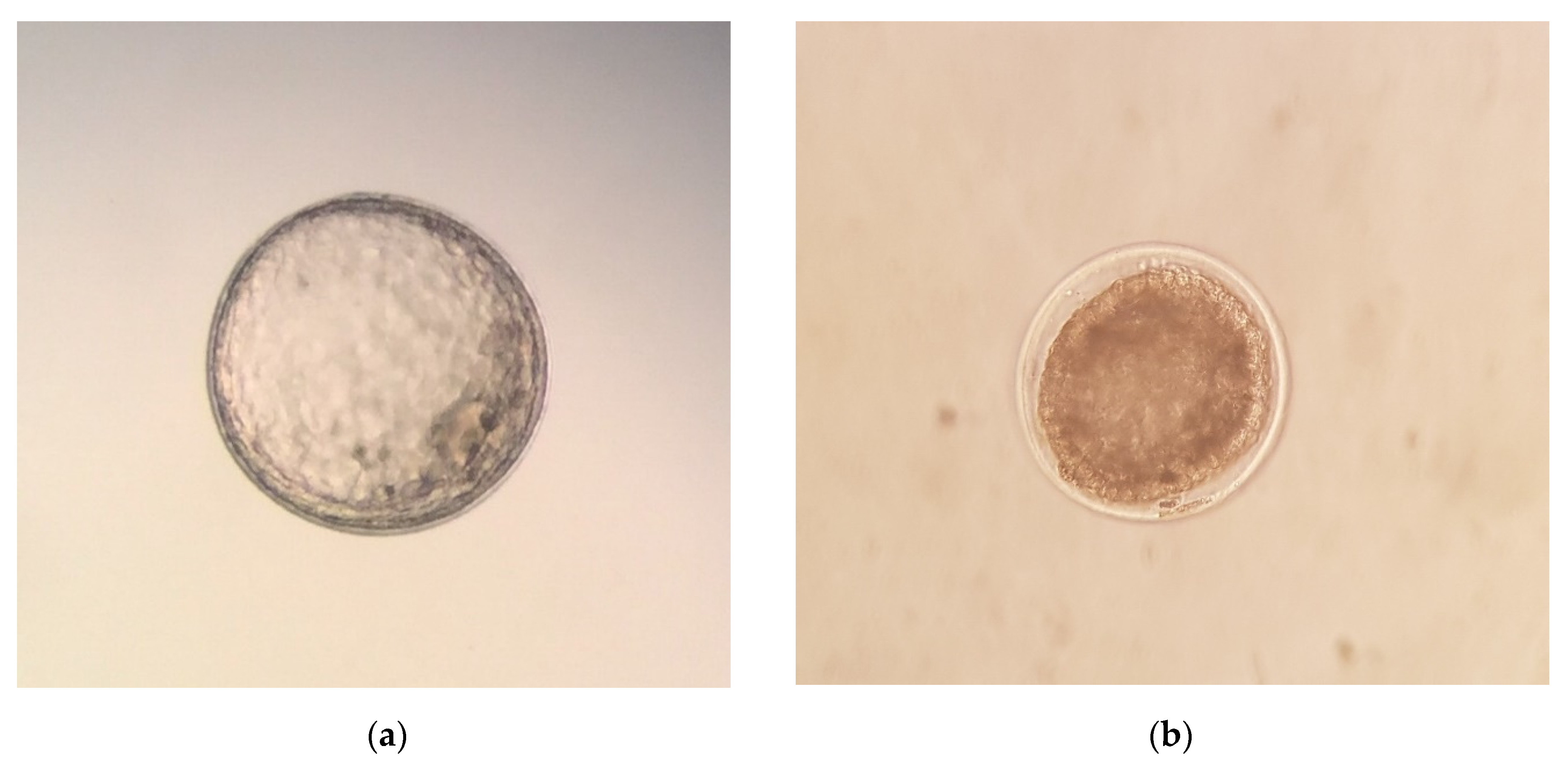
17] for donkeys. After the flushing, luprostiol was administered to donors to induce luteolysis. Recovered embryos were evaluated for developmental stage (morula, early blastocyst, blastocyst, or expanded blastocyst) and morphological quality, and they were graded on a scale of 1–4 [27], 1 being excellent, 2 being good, 3 being fair, and 4 being poor, degenerate, or dead (Figure 1). After the quality evaluation, the embryos were washed ten times in Syngro® holding (Bioniche Animal Health, Washington, DC, USA), as previously described [16]. The diameter of the embryos was measured under bright field conditions (SZ51 Olympus optical, Tokyo, Japan) using an ocular micrometer (scale of 1 mm/100), as previously described [28].

2.4. Statistical Analysis

Descriptive statistical analysis was performed, presenting the qualitative variables as frequencies and percentages, and quantitative as a mean ± standard error of the mean (SEM). The effects of the year in which the study was performed (1–3), season (winter: December 22–March 20; spring: March 21–June 20; summer: June 21–September 22; autumn: September 23–December 21), photoperiod (positive: March–October; negative: November–February), herd (1–3), days of flushing for embryo recovery (6–9), number of flushings (1–3), donor (25 jennies), donor age (≤3; 4–9; ≥10 years old), parity (nulliparous vs. multiparous), successive cycle within donor (1–5; 6–14; 15–23), number of ovulations per cycle (single vs. double), and jack (8 donkeys) on positive uterine flushing rates (PFR; flushing where at least one embryo was recovered), embryo recovery rate (ERR; embryos recovered per cycle), and on ovulation rate (OR; number of ovulations per cycles) were analyzed by the Chi-square test and by the Kruskal–Wallis one-way ANOVA, respectively. When the effect was statistically significant, post-hoc multiple comparisons were made using Chi-square tests for categorical variables and Mann–Whitney U tests for continuous variables.
To evaluate the effects of single factors (year, season, photoperiod, herd, day of flushing, number of flushings, donor, donor age, parity, successive cycle within donor, number of ovulations, and jack) on embryo quality (Grade 1–4), diameter (µm), and developmental stage (morula, early blastocyst, blastocyst, expanded blastocyst) the Kruskal–Wallis one-way ANOVA was performed. Mean values were compared by Duncan’s test.
All analyses were performed using the statistical package SPSS v15.0 (IBM Spain, Madrid, Spain). Differences were considered statistically significant when p < 0.05.

3. Results

The average ovulation rate in donor jennies is shown in Table 1. The average rate per jenny was 1.26 ± 0.04 and varied significantly among jennies (p = 0.01). Results of statistical analysis also showed differences among herds (p = 0.031), while no differences (p > 0.05) were observed among donor age categories, years, photoperiods, and seasons (Table 1). The single ovulation rate was 59.2% (93/157), while for double ovulation, the rate was 40.8% (64/157). Single ovulation occurred with equal frequency (p > 0.05) on both ovaries (left ovary: 42.5%; right ovary: 32.5%). However, the incidence of bilateral double ovulation (18.3%) was significantly higher (p < 0.01) than that of ipsilateral double ovulation (6.7%).
A total of 124 uterine flushings were carried out during the study, of which 92 were positive (PFR: 74.2%; 92/124), and 100 embryos were recovered out of 124 estrous cycles (ERR: 80.6%; 100/124) and 157 ovulations (embryo recovery per ovulation: 63.7%; 100/157).
The embryo diameter and morphological quality score of donkey embryos are shown in Table 2. Overall, 77 of 100 embryos (77%) were classified as Grade 1 (excellent), 17 (17%) were classified as Grade 2 (good) and 6 (6%) were classified as Grade 3 (fair). The most frequent stages of development observed were early blastocyst (37%, 37/100) and expanded blastocyst (36%, 36/100), which were followed by morula (20%, 20/100) and blastocyst (7%, 7/100) stages. The embryo quality score significantly (p < 0.05) varied according to developmental stage and day of recovery, being lower for blastocysts or when flushed at day 8 after ovulation (Table 2). As expected, the embryo diameter was also affected (p < 0.001) by the developmental stage and day of flushing. The mean diameter of embryos was 179.39 ± 9.61 µm (range: 150–300 µm) for morulae, 210.81 ± 7.90 µm (range: 150–325 µm) for early blastocysts, 425.00 ± 45.32 µm (range: 275–600 µm) for blastocysts, and 1022.92 ± 125.22 µm (range: 250–3300 µm) for expanded blastocysts. Moreover, it was observed that embryos recovered at 6 days after ovulation had a diameter of 187.50 ± 15.23 µm (range: 150–300 µm); those collected on day 7 measured 236.48 ± 13.83 µm (range: 150–600 µm); while the mean diameter at 8 and 9 were 806.25 ± 83.31 µm (range: 275–2400 µm) and 2275 ± 300.14 µm (range: 1525–3300 µm), respectively (Table 2).
Presented in Table 3 are the developmental stage and embryo size for Day 6–9 embryos. At Day 6 after ovulation, embryos were mostly at the morula stage (12/13, 92.3%); meanwhile, 67.9% (36/53) of embryos recovered at Day 7 were early blastocyst stage embryos. Expanded blastocysts were recovered on Day 8 (25/28, 89.3%) and 9 (6/6, 100%). At Day 6–7, most of the embryos recovered were small (<200 µm) or medium (200–300 µm) embryos; meanwhile, large embryos (>300 µm) were recovered at Days 8 (27/28, 96.4%) and 9 after ovulation (6/6, 100%; Table 3).
Table 4 shows extrinsic factors that affect the rate of positive flushings (PFR) and the embryo recovery rate (ERR). None of the factors studied affected (p > 0.05) PFR or ERR; however, there was an effect of the number of flushings (1, 2, or 3) on both rates, which were significantly (p < 0.001) reduced in the third flushing (PFR: 15.8%; ERR: 0.16).
The intrinsic factors that affect PFR and ERR are shown in Table 5. PFR did not vary (p > 0.05) with any of the studied variables except the donor (p = 0.040). No differences between parity (p = 0.2610) and number of ovulations (p = 0.0971) were detected for ERR (Table 5). In contrast, ERR not only varies among donors (p < 0.05) but also among donor age groups (p < 0.05) and successive cycles within the donor (p < 0.001). ERR was higher (p < 0.05) in jennies of 4–9 years of age (0.94; 51/54) with respect to the other groups (≤3 years: 0.77 (26/34); ≥10 years: 0.64 (23/36)). With regard to the number of ovulations in the same donor, ERR was significantly higher (p < 0.001) in the first group (1–5 cycles: 1.44; 49/34) with respect to the second (6–14 cycles: 0.78; 39/50) and third (15–23 cycles: 0.30; 12/40) groups.
As shown in Table 6, PFR and ERR varied (p > 0.05) among jacks. Four jacks (numbers 169, 192, 2895, and 4457) that were used in 79.7% of cycles (94/118) showed good results for both rates (PFR: 70.8–100%; ERR: 0.77–1.00). The other three jacks (numbers 95, 148, and 9025) that were used in 18.6% of cycles (22/118) yield a lower PFR (50–58.3%) and ERR (0.50–0.67) than the previous group, but this was not significant statistically (p > 0.05). No embryos were obtained with jack number 232, although he was used only two times.
Developmental stage, embryo quality, and diameter of the embryos recovered in this study are shown in Table 7, Table 8 and Table 9. None of the extrinsic factors studied significantly influenced (p < 0.05) these three variables, except for the day of flushing (6–9), which significantly influenced the developmental stage (p < 0.001), embryo quality (p < 0.05), and diameter of embryos (p < 0.001; Table 7). Similarly, the number of flushings (1–3) significantly influenced the diameter (p = 0.038) and embryo developmental stage (p = 0.001), whereas the developmental stage was statistically different among herds (p = 0.020; Table 7).
Table 8 shows that no influence was detected (p > 0.05) of the intrinsic factors on developmental stage, embryo quality, and diameter of embryos, except for the donor, which affected the embryo quality (p = 0.004). Moreover, no differences (p > 0.05) were detected among jacks for these three variables (Table 9).

4. Discussion

Due to the similarities in the reproductive physiology between horses and donkeys, several assisted reproductive technologies (ARTs) routinely used in horses have been applied directly to donkeys. Hence, previous studies have demonstrated the suitability of mare ET techniques for collecting embryos in jennies [16]. In this line, numerous studies have been conducted to examine the factors that affect embryo recovery, quality, and diameter in mares [13,22,29]. However, in donkeys, these studies have been very scarce [17,20], and they are often performed on a limited number of animals, cycles, or embryos. Therefore, more studies are needed to optimize embryo recovery rates and maximize the success of future ET programs in donkeys.
In this study, in which 26 donor jennies and 124 cycles were used, the average ovulation rate per jenny was 1.26 ± 0.04. This finding was slightly lower than the reported average in spontaneous (1.57 ± 0.06) and prostaglandin F2 alpha (PGF2α)-induced (1.56 ± 0.10) estrus of Andalusian jennies [30]. These small differences could be explained by other factors (such as feeding management, donor age, reproductive status, season of the year, and the use of drugs to induce ovulation) that can affect the incidence of multiple ovulations, as reported in mares [22,31,32,33] and donkeys [30,34].
In jennies, the incidence of multiple ovulation was reported to range between 5.3% and 61% [35,36,37]. In our study, the single ovulation rate was 59.2%, while the double ovulation rate was 40.8%. Double ovulation in jennies was similar to that reported in Catalonian jennies (42.45%) [34] and in the Asinina de Miranda jennies (36.36%) [38]. However, the incidence of double ovulation in this study was lower than that reported in spontaneous (51.7%) and PGF2α-induced cycles (56.5%) in Andalusian jennies [30]. It is interesting to note that single ovulation occurred with equal frequency on both ovaries, as also reported by other authors [30,34]. Similar to that reported by Taberner et al. [34], a minimally greater frequency of ovulation for the left ovary was found (42.5% vs. 32.5%), but the difference was not significant. On the other hand, the incidence of bilateral double ovulation was significantly higher than that of ipsilateral double ovulation, which agrees with findings for mares [31] and jennies [30].
No influence of the age of the donor on the ovulation rate was observed in this study; however, the ovulation rate was numerically (1.36 ± 0.10%), but not significantly higher, in the older jennies (≥10 years old). These results are consistent with previous findings in jennies [20,30] and mares [31]. Although the reason for this fact remains still unclear, it has been suggested that increased ovarian stimulation or enhanced ovarian receptivity to that stimulation may be involved. Thus, multiovulation would be a natural strategy to ensure gestation in older females, which have a reduced ability to become pregnant [32].
The ovulation rate did not vary among years, seasons, and photoperiods, but it tended to be lower in summer (p = 0.095). Similar findings have been reported in previous studies [20,30]. Ginther et al. [39], in a study using different breeds and geographical latitude than our study of donkeys, observed that the incidence of multiple ovulations was not affected by the season of the year. The statistical analysis revealed significant differences among herds (p = 0.031), which could be explained by the individuals comprising each herd. In fact, this study pointed out the existence of significant differences among jennies (p = 0.01).
In our study, in which 100 embryos were used, the overall ERR following non-surgical flushing on Days 6–9 was 80.6%, which was higher than those previously reported in different breeds of donkeys: 53.3% in jennies of unknown breed [40], 63.6% in Poitou jennies [41], 75.9% in Pantesca jennies [17], 50% in Amiata jennies [16], 52.3% in Pega jennies [42], and 40.7% in Andalusian and Zamorano-Leones jennies [20]. The EER obtained in our experiment was also higher than the rates reported in the literature for fertile mares in commercial ET programs [43]—60–77% for fresh, 44% for chilled, and 46% for frozen semen—but similar to that obtained in young fertile mares inseminated with fresh semen, 87% [44]. These results are likely due to the age of the jennies used in the study and the physical and reproductive assessment performed before, including donors in the experimental group.
It is known that the major factor affecting embryo recovery is the donors´ reproductive history. Hence, embryo recovery for old sub-fertile mares can be as low as 30–40% per cycle [45]. Other factors that affect embryo recovery include semen quality and semen type (fresh, cooled, or frozen) [45]. In our study, all donors were selected carefully, based on their reproductive history and clinical examination, and they were mated naturally with jacks of proven fertility. Although the PFR and ERR varied among jacks, seven out of eight jacks showed moderate to good results for both rates (≥50% and ≥0.50, respectively). Only one jack had low fertility (zero out of two positive flushes), but he was used only twice, which could mitigate its negative effect on average PFR and ERR. In addition, hCG was used as the ovulation inductor. Previous studies have clearly demonstrated that ovulation induction can enhance the efficiency of ARTs in domestic animal species, including the donkey [46].
The embryo morphology score is the most common method used to evaluate embryo quality [27]. In line with previous findings [16,17,20,42], 94% of the recovered embryos had a quality grade of excellent (Grade 1) or good (Grade 2).
In our experiment, none of the extrinsic factors analyzed (year of the study, season of the year, photoperiod, herd or day, of flushing) affected significantly PFR or ERR. Consistent with previous findings [17], the ERR obtained in the first year (0.73) was numerically lower than that obtained in the second (0.86) and third (0.82) year, which could be explained by the inexperience with this technology (i.e., ET) in donkeys. The absence of a photoperiod influence on embryo recovery has been previously described in donkeys [17,20]. The study carried out in Pantesca donkeys [17] also noted that the time of the year did not affect PFR and ERR. Considering these results, we could state that seasonality has little impact on reproductive performance of Andalusian jennies, which can get pregnant naturally all year round, as previously reported for other donkey breeds [17]. This fact brings the possibility of applying ET in Andalusian donkeys along the year.
In donkeys, the influence of the day of flushing on ERR has not been well established in the literature [17,20]. Under our experimental conditions, PFR and ERR were not different among Days 6, 7, 8, and 9. Similar findings have been reported in mares [22,47]. However, embryo diameter and developmental stage varied widely depending on the day of recovery, which is consistent with previous studies [16,41]. In line with these findings, poor-quality embryos were collected at Day 8, and ERR tended to be lower (p = 0.08), flushing the uterus 9 days after ovulation. Taken all together, our results emphasize the importance of collecting Day 6 to 7 donkey embryos, smaller than 300 µm in diameter and with good morphological score, to ensure vitrification success, as has been previously suggested [19].
From a practical point of view, another interesting observation was the effect of the number of flushings (1, 2, or 3) on both rates (PFR and ERR), which were significantly reduced in the third flushing (15.8% and 0.16, respectively), indicating that the majority of embryo collections require one or two maximum flushings. In addition, embryo diameter and developmental stage varied among flushings, with larger and older embryos in the first flushing. Thus, our results could suggest that the larger diameter of the older embryos could facilitate their recovery [17].
The embryos recovered in herd 1 were in earlier developmental stages than in the other herds. These results were probably due to the fact that all the embryo rerecovered in this herd (5 embryos) were flushed 6–7 days after ovulation, while in herds 2 (56 embryos) and 3 (39 embryos), flushes were carried out from Day 6 to 9 after ovulation, thus increasing the average diameter and developmental stage of the recovered embryos.
Regarding the intrinsic factors, PFR did not vary with any of the studied variables (i.e., donor age, parity, successive cycle within donor, and number of ovulations) except for the donor jenny. In contrast, ERR not only varied among donors but also among donor age categories, showing higher values in jennies of 4–9 years of age (0.94) compared with all the other categories of age. Moreover, both PFR and ERR were not different between younger (≤3 years old) and older (≥10 years old) jennies. Our findings are in contrast with the results of previous studies [17,20], which reported no effect of donor age and donor jenny on the aforementioned rates. However, the effect on ERR of donor age and donor mare have been reported by many authors [13,22,48,49], in which old age (>15 years old) and a history of sub-fertility were related to a lower ERR. Our results could suggest that embryo donors between 4 and 9 years are the best to be used in an ET program.
The effect of repeated uterine flushings has been previously described in mares [50], which was associated with increased chronic inflammation of the uterus. Although previous studies failed to observe this negative effect in donkeys [17], in our experiment, the ERR on successive cycles from 1 to 5 was higher (1.44) than in attempts from 6 to 14 (0.78) and from 15 to 23 (0.30). The differences observed between studies may be explained by different experimental conditions. Therefore, in our study, young (≤3 years), mature (4–9 years), and old (≥10 years) donor jennies were employed during the entire period of the study, while only young jennies (2–5 years old) were used in this previous work [17]. Moreover, a higher number of donors (10, 8, and 6, respectively) and cycles (63, 45, and 16, respectively) were used in each group.
It has been previously described that the occurrence of multiple ovulations enhances ERR in mares [22,51] and donkeys [17,20], but this effect was not shown in our study. However, despite the absence of statistical significance, ERR after single ovulation tended to be lower than that obtained after double ovulation (0.77 vs. 0.91; p = 0.0971). It has been also reported that ipsilateral double ovulations resulted in a lower ERR than bilateral double ovulations [52], which could be caused by interference between two or more simultaneous ovulations in the limited space of the ovulation fossa [23,53]. In the present study, the incidence of ipsilateral double ovulations was only 6.7%, and no significant difference in ERR was observed between bilateral and ipsilateral ovulations (1.62% vs. 1.88%; p = 0.196).
Conversely, the number of ovulations in the same donor influenced ERR, being significantly higher in the first group (1–5 cycles: 1.44; 49/34), but no effect on PFR was observed. These results partially agree with the findings of Camillo et al. [17], who observed a significant effect on both rates. Finally, we observed that the parity of the donors did not have an effect on PFR and ERR. In cattle, the parity of recipients does not affect pregnancy rates following the transfer of fresh and frozen embryos [54]. However, to our knowledge, no data are available for mare and jenny donors.

5. Conclusions

Based on our results, we can conclude that the donor jenny was the main factor that affects the rate of positive flushings and recovery rates as well as the embryo quality. Other factors that affected embryo recovery rate were the number of flushings, donor age, successive cycle within donor, and jack. Meanwhile, the rate of positive flushings was affected by the number of flushings and the jack. From a practical point of view, these findings could indicate that the majority of embryo collections require one or two maximum flushings per cycle. Moreover, the negative effect of repeated uterine flushings on embryo recovery rate was proven, being lower after six consecutive cycles. On the other hand, the day of flushing had a significant effect on embryo quality and diameter, which emphasizes the importance of collecting Day 6 to 7 donkey embryos, with good morphological score and smaller than 300 µm in diameter, if embryos are going to be cryopreserved.

Author Contributions

Conceptualization, M.H. and J.D.; methodology, M.B., I.O., M.D.-J., B.P., C.C., J.J.C., V.G.-A., A.D. and J.D.; software, M.B. and J.D.; validation, M.H., M.B. and J.D.; formal analysis, M.B., I.O., M.D.-J., B.P. and C.C.; investigation, M.B., I.O., M.D.-J., B.P. and C.C.; resources, M.H. and J.D.; data curation, M.B., M.H. and J.D.; writing—original draft preparation, I.O., M.H. and J.D.; writing—review and editing, I.O., M.H. and J.D.; visualization, M.H. and J.D.; supervision, M.H. and J.D.; project administration, M.H. and J.D.; funding acquisition, M.H. and J.D. All authors have read and agreed to the published version of the manuscript.

Funding

This work has been supported by Grant AGL2013-42726-R (Secretaría de Estado de Investigación, Desarrollo e Innovación, Ministerio de Economía y Competitividad, Spain). I.O. was supported by a Ph.D. fellowship from the ceiA3 (Andalusia, Spain) with funding provided by Banco Santander through its Global Division, Santander Universidades. M.D.-J. and C.C. were supported by a FPU fellowship from Spanish Ministry of Education, Culture, and Sports.

Acknowledgments

We are extremely grateful to the Centro Rural Malpica (Palma del Río, Cordoba, Spain) for providing the animals and facilities.

Conflicts of Interest

The authors declare no conflict of interest. The funders had no role in the design of the study; in the collection, analyses, or interpretation of data; in the writing of the manuscript, or in the decision to publish the results.

References

  1. Gandini, G.C.; Villa, E. Analysis of the cultural value of local livestock breeds: A methodology. J. Anim. Breed Genet. 2003, 120, 1–11. [Google Scholar] [CrossRef]
  2. Miragaya, M.H.; Neild, D.M.; Alonso, A.E. A Review of Reproductive Biology and Biotechnologies in Donkeys. J. Equine Vet Sci. 2018, 65, 55–61. [Google Scholar] [CrossRef]
  3. Kugler, W.; Grunenfelder, H.P.; Broxham, E. Donkey Breeds in Europe: Inventory, Description, Need for Action, Conservation; Report 2007–2008; Monitoring Institute for Rare Breeds and Seeds in Europe: St. Gallen, Switzerland, 2008; p. 124. [Google Scholar]
  4. Quaresma, M.; Martins, A.M.F.; Rodrigues, J.B.; Colaço, J.; Payan-Carreira, R. Viability analyses of an endangered donkey breed: The case of the Asinina de Miranda (Equus asinus). Anim. Prod. Sci. 2015, 55, 1184–1191. [Google Scholar] [CrossRef]
  5. Colli, L.; Perrotta, G.; Negrini, R.; Bomba, L.; Bigi, D.; Zambonelli, P.; Verini Supplizi, A.; Liotta, L.; Ajmone-Marsan, P. Detecting population structure and recent demographic history in endangered livestock breeds: The case of the Italian autochthonous donkeys. Anim. Genet. 2013, 44, 69–78. [Google Scholar] [CrossRef]
  6. Calero-Carretero, R.; Bravo-Delgado, J.A.; Bartolomé-García, P. Razas autóctonas extremeñas en peligro de extinción. In Informe 2007 La Agricultura y la Ganadería Extremeñas; Facultad de Ciencias Económicas y Empresariales, Escuelas de Ingenierías Agrarias, Universidad de Extremadura: Caja de Badajoz, Badajoz, España, 2007; pp. 203–226. [Google Scholar]
  7. Jordana, J.; Folch, P. The endangered catalonian donkey breed: The main ancestor of the American ass or mammoth. J. Equine Vet. Sci. 1996, 16, 436–441. [Google Scholar] [CrossRef]
  8. DAD-IS-FAO. Information System Domestic Animal Diversity. Available online: http://www.fao.org/dad-is/browse-by-country-and-species/en/ (accessed on 21 October 2020).
  9. Scherf, B.D. World Watch List for Domestic Animal Diversity, 3rd ed.; FAO: Rome, Italy, 2000; p. 739. [Google Scholar]
  10. Vita, D.; Passalacqua, G.; Di Pasquale, G.; Caminiti, L.; Crisafulli, G.; Rulli, I.; Pajno, G.B. Ass’s milk in children with atopic dermatitis and cow’s milk allergy: Crossover comparison with goat’s milk. Pediatr. Allergy Immunol. 2007, 18, 594–598. [Google Scholar] [CrossRef]
  11. Summers, P.M.; Shephard, A.M.; Hodges, J.K.; Kydd, J.; Boyle, M.S.; Allen, W.R. Successful transfer of the embryos of Przewalski’s horses (Equus przewalskii) and Grant’s zebra (E. burchelli) to domestic mares (E. caballus). J. Reprod. Fertil. 1987, 80, 13–20. [Google Scholar] [CrossRef] [Green Version]
  12. Hinrichs, K. Assisted reproductive techniques in mares. Reprod. Domest. Anim. 2018, 53, 4–13. [Google Scholar] [CrossRef]
  13. Squires, E.L.; Carnevale, E.M.; McCue, P.M.; Bruemmer, J.E. Embryo technologies in the horse. Theriogenology 2003, 59, 151–170. [Google Scholar] [CrossRef]
  14. Squires, E.L.; McCue, P.M. Cryopreservation of Equine Embryos. J. Equine Vet. Sci. 2016, 41, 7–12. [Google Scholar] [CrossRef]
  15. Pérez-Marín, C.C.; Vizuete, G.; Vazquez-Martinez, R.; Galisteo, J.J. Comparison of different cryopreservation methods for horse and donkey embryos. Equine Vet. J. 2018, 50, 398–404. [Google Scholar] [CrossRef]
  16. Panzani, D.; Rota, A.; Crisci, A.; Kindahl, H.; Govoni, N.; Camillo, F. Embryo quality and transcervical technique are not the limiting factors in donkey embryo transfer outcome. Theriogenology 2012, 77, 563–569. [Google Scholar] [CrossRef]
  17. Camillo, F.; Panzani, D.; Scollo, C.; Rota, A.; Crisci, A.; Vannozzi, I.; Balbo, S. Embryo recovery rate and recipients’ pregnancy rate after nonsurgical embryo transfer in donkeys. Theriogenology 2010, 73, 959–965. [Google Scholar] [CrossRef]
  18. Bottrel, M.; Mogas, T.; Pereira, B.; Ortiz, I.; Díaz-Jiménez, M.; Consuegra, C.; Hidalgo, M.; Morató, R.; Dorado, J. The cryoprotective effect of Ficoll 70 on the post-warming survival and quality of Cryotop-vitrified donkey embryos. Theriogenology 2020, 148, 180–185. [Google Scholar] [CrossRef]
  19. Bottrel, M.; Ortiz, I.; Pereira, B.; Díaz-Jiménez, M.; Hidalgo, M.; Consuegra, C.; Morató, R.; Mogas, T.; Dorado, J. Cryopreservation of donkey embryos by the cryotop method: Effect of developmental stage, embryo quality, diameter and age of embryos. Theriogenology 2019, 125, 242–248. [Google Scholar] [CrossRef]
  20. Pérez-Marín, C.C.; Vizuete, G.; Galisteo, J.J. Embryo recovery results in Hispano-Arabe horse and Spanish donkey breeds. Livest. Sci. 2017, 206, 76–81. [Google Scholar] [CrossRef]
  21. Bottrel, M.; Fortes, T.; Ortiz, I.; Hidalgo, M.; Dorado, J. Establishment and maintenance of donkey-in-mule pregnancy after embryo transfer in a non-cycling mule treated with oestradiol benzoate and long-acting progesterone. Span J. Agric. Res. 2017, 15, e04SC01. [Google Scholar] [CrossRef] [Green Version]
  22. Panzani, D.; Rota, A.; Marmorini, P.; Vannozzi, I.; Camillo, F. Retrospective study of factors affecting multiple ovulations, embryo recovery, quality, and diameter in a commercial equine embryo transfer program. Theriogenology 2014, 82, 807–814. [Google Scholar] [CrossRef]
  23. Carnevale, E.M.; Ramirez, R.J.; Squires, E.L.; Alvarenga, M.A.; Vanderwall, D.K.; McCue, P.M. Factors affecting pregnancy rates and early embryonic death after equine embryo transfer. Theriogenology 2000, 54, 965–979. [Google Scholar] [CrossRef]
  24. Panzani, D.; Vannozzi, I.; Marmorini, P.; Rota, A.; Camillo, F. Factors affecting recipients’ pregnancy, pregnancy loss, and foaling rates in a commercial equine embryo transfer program. J. Equine Vet. Sci. 2016, 37, 17–23. [Google Scholar] [CrossRef]
  25. Eldridge-Panuska, W.D.; Caracciolo Di Brienza, V.; Seidel, G.E., Jr.; Squires, E.L.; Carnevale, E.M. Establishment of pregnancies after serial dilution or direct transfer by vitrified equine embryos. Theriogenology 2005, 63, 1308–1319. [Google Scholar] [CrossRef]
  26. Stout, T. Cryopreservation of Equine Embryos: Current state-of-the-art. Reprod. Domest. Anim. 2012, 47, 84–89. [Google Scholar] [CrossRef] [Green Version]
  27. McKinnon, A.O.; Squires, E.L. Equine embryo transfer. Vet. Clin. N. Am. Equine Pract. 1988, 4, 305–333. [Google Scholar] [CrossRef]
  28. Lagares, M.A.; Castanheira, P.N.; Amaral, D.C.G.; Vasconcelos, A.B.; Veado, J.C.C.; Arantes, R.M.E.; Stahlberg, R. Addition of ficoll and disaccharides to vitrification solutions improve in vitro viability of vitrified equine embryos. Cryo-Letters 2009, 30, 408–413. [Google Scholar]
  29. Hinrichs, K. Assisted reproduction techniques in the horse. Reprod. Fertil. Dev. 2013, 25, 80–93. [Google Scholar] [CrossRef] [PubMed]
  30. Perez-Marin, C.C.; Galisteo, I.; Perez-Rico, A.; Galisteo, J. Effects of breed, age, season, and multiple ovulations on cyclic, PGF2α-induced, and postpartum estrus characteristics in Spanish jennies. Theriogenology 2016, 85, 1045–1052. [Google Scholar] [CrossRef]
  31. Davies Morel, M.C.; O’Sullivan, J.A. Ovulation rate and distribution in the thoroughbred mare, as determined by ultrasonic scanning: The effect of age. Anim. Reprod. Sci. 2001, 66, 59–70. [Google Scholar] [CrossRef]
  32. Morel, M.C.; Newcombe, J.R.; Swindlehurst, J.C. The effect of age on multiple ovulation rates, multiple pregnancy rates and embryonic vesicle diameter in the mare. Theriogenology 2005, 63, 2482–2493. [Google Scholar] [CrossRef]
  33. Fradinho, M.J.; Correia, M.J.; Grácio, V.; Bliebernicht, M.; Farrim, A.; Mateus, L.; Martin-Rosset, W.; Bessa, R.J.B.; Caldeira, R.M.; Ferreira-Dias, G. Effects of body condition and leptin on the reproductive performance of Lusitano mares on extensive systems. Theriogenology 2014, 81, 1214–1222. [Google Scholar] [CrossRef]
  34. Taberner, E.; Medrano, A.; Peña, A.; Rigau, T.; Miró, J. Oestrus cycle characteristics and prediction of ovulation in Catalonian jennies. Theriogenology 2008, 70, 1489–1497. [Google Scholar] [CrossRef]
  35. Henry, M.; Figueiredo, A.E.; Palhares, M.S.; Coryn, M. Clinical and endocrine aspects of the oestrous cycle in donkeys (Equus asinus). J. Reprod. Fertil. Suppl. 1987, 35, 297–303. [Google Scholar]
  36. Blanchard, T.L.; Taylor, T.S.; Love, C.L. Estrous cycle characteristics and response to estrus synchronization in mammoth asses (Equus asinus americanus). Theriogenology 1999, 52, 827–834. [Google Scholar] [CrossRef]
  37. Vandeplassche, G.M.; Wesson, J.A.; Ginther, O.J. Behavioral, follicular and gonadotropin changes during the estrous cycle in donkeys. Theriogenology 1981, 16, 239–249. [Google Scholar] [CrossRef]
  38. Quaresma, M.; Payan-Carreira, R. Characterization of the estrous cycle of Asinina de Miranda jennies (Equus asinus). Theriogenology 2015, 83, 616–624. [Google Scholar] [CrossRef]
  39. Ginther, O.J.; Scraba, S.T.; Bergfelt, D.R. Reproductive seasonality of the jenney. Theriogenology 1987, 27, 587–592. [Google Scholar] [CrossRef]
  40. Allen, W.R.; Kydd, J.; Boyle, M.S.; Antczak, D.F. Between-species transfer of horse and donkey embryos: A valuable research tool. Equine Vet. J. 1985, 17, 53–62. [Google Scholar] [CrossRef]
  41. Vendramini, O.M.; Bruyas, J.F.; Fieni, F.; Battut, I.; Tainturier, D. Embryo transfer in Poitou donkeys, preliminary results. Theriogenology 1997, 47, 409. [Google Scholar] [CrossRef]
  42. Peña-Alfaro, C.E.; Barros, L.O.; Carneiro, G.F.; Gastal, M.O.; Gastal, E.L. Embryo transfer in Pega donkeys (Equus asinus) in Brazil. J. Equine Vet. Sci. 2014, 34, 185. [Google Scholar] [CrossRef]
  43. Squires, E.; Barbacini, S.; Matthews, P.; Byers, W.; Schwenzer, K.; Steiner, J.; Loomis, P. Retrospective study of factors affecting fertility of fresh, cooled and frozen semen. Equine Vet. Educ. 2006, 18, 96–99. [Google Scholar] [CrossRef]
  44. Camillo, F.; Vannozzi, I.; Rota, A.; Romagnoli, S.; Aria, G. Comparison of embryo recovery rates from two years old and mature mares. In Havemeyer Foundation Monograph Series 3, Proceedings of the 5th International Symposium on Equine Embryo Transfer, Saari, Finland, 6–9 July 2000; Katila, T., Wade, J.F., Eds.; R & W Publications (Newmarket) Limited: Suffolk, UK, 2001; p. 86. [Google Scholar]
  45. Squires, E.L. Embryo transfer challenges and perspectives. Rev. Bras. Reprod. Anim. 2013, 37, 105–107. [Google Scholar]
  46. Carluccio, A.; Panzani, S.; Tosi, U.; Faustini, M.; De Amicis, I.; Veronesi, M.C. Efficacy of hCG and GnRH for inducing ovulation in the jenny. Theriogenology 2007, 68, 914–919. [Google Scholar] [CrossRef]
  47. Fleury, J.J.; Alvarenga, M.A. Effects of collection day on embryo recovery and pregnancy rates in a nonsurgical equine embryo transfer program. Theriogenology 1999, 51, 261. [Google Scholar] [CrossRef]
  48. Stout, T.A. Equine embryo transfer: Review of developing potential. Equine Vet J. 2006, 38, 467–478. [Google Scholar] [CrossRef] [PubMed]
  49. Pessoa, M.A.; Cannizza, A.P.; Reghini, M.F.S.; Alvarenga, M.A. Embryo Transfer Efficiency of Quarter Horse Athletic Mares. J. Equine Vet. Sci. 2011, 31, 703–705. [Google Scholar] [CrossRef]
  50. Carnevale, E.M.; Beisner, A.E.; McCue, P.M.; Bass, L.D.; Squires, E.L. Uterine changes associated with repeated inseminations and embryo collections in mares. In Proceedings of the 51st Annual Convention of American Association of Equine Practitioner, Seattle, WA, USA, 3–5 December 2005; pp. 202–203. [Google Scholar]
  51. Losinno, L.; Aguilar, J.; Lisa, H. Impact of multiple ovulations in a commercial equine embryo transfer programme. In Havemeyer Foundation Monograph Series 3, Proceedings of the 5th International Symposium on Equine Embryo Transfer, Saari, Finland, 6–9 July 2000; Katila, T., Wade, J.F., Eds.; R & W Publications (Newmarket) Limited: Suffolk, UK, 2001; pp. 81–83. [Google Scholar]
  52. Riera, F.L.; Roldán, J.E.; Hinrichs, K. Ipsilateral double ovulation is associated with reduced embryo recovery in mares. In Havemeyer Foundation Monograph Series 14, Proceedings of the 6th International Symposium on Equine Embryo Transfer, Rio de Janeiro, Brazil, 4–6 August 2004; Alvarenga, M., Wade, J.F., Eds.; R & W Comunications: Suffolk, UK, 2005; pp. 108–109. [Google Scholar]
  53. Carmo, M.T.; Alvarenga, M.A.; Losinno, L.; Aquilar, J.J. Oocyte transport to the oviduct of superovulated mares. Anim. Reprod. Sci. 2006, 94, 337–339. [Google Scholar]
  54. Ono, T.; Isobe, T.; Morita, Y.; Do, L.T.K.; Tanihara, F.; Taniguchi, M.; Takagi, M.; Otoi, T. Effects of parity and season on pregnancy rates after the transfer of embryos to repeat-breeder Japanese Black beef cattle. Arch. Anim. Breed 2016, 59, 45–49. [Google Scholar] [CrossRef] [Green Version]
Figure 1. Donkey embryos of various developmental stages and quality grades. (a) Expanded blastocyst stage embryo, Grade 1. Note blastocoele cavity and distinct inner cell mass. The zona pellucida has been shed, and the capsule is surrounding the embryo. No morphologic abnormalities are present in this embryo; (b) Blastocyst stage embryo, Grade 2. Note the distinct blastomere cells around the edge of the embryo and the capsule. A blastocoele cavity is just beginning to form within the center of the embryo. Note minor imperfections, such as a few extruded cells, occasional discolored cells, and slight shrinkage of trophoblast from zona pellucida; (c) Early blastocyst stage embryo, Grade 3. Note thick zona pellucida and capsule. Note moderate level of imperfections, such as a high proportion of extruded cells, discoloration of remaining cell mass, and moderate shrinkage of trophoblast from zona pellucida; (d) Expanded blastocyst stage embryo, Grade 4. Note complete collapse of the blastocoele.
Animals 10 01967 g001aAnimals 10 01967 g001b
Table 1. Ovulation rate (mean ± SEM) in 124 cycles of 26 donor jennies according to donor age categories (≤3, 4–9 or ≥10 years old), parity (nulliparous or multiparous), year of the study (first, second or third), season of the year (spring, summer, autumn, or winter), photoperiod (positive or negative), and herd (1, 2, or 3).
VariableNo. of CyclesOvulation Ratep-Values
Donor age
 ≤3 years341.24 ± 0.070.433
 4–9 years651.23 ± 0.05
 ≥10 years251.36 ± 0.10
Parity
 Nulliparous351.26 ± 0.080.988
 Multiparous891.26 ± 0.05
Year
 First331.21 ± 0.070.073
 Second421.17 ± 0.06
 Third491.37 ± 0.07
Season
 Spring441.18 ± 0.060.095
 Summer121.08 ± 0.08
 Autumn521.37 ± 0.07
 Winter161.25 ± 0.11
Photoperiod
 Positive661.20 ± 0.050.099
 Negative581.33 ± 0.06
Herd
 161.33 ± 0.21 ab0.031
 2711.17 ± 0.05 b
 3471.38 ± 0.07 a
Total1241.26 ± 0.04
a,b Values with different superscript differ significantly.
Table 2. Diameters (mean ± SEM) and morphological quality score (1–4) of donkey embryos collected at Days 6 to 9 after ovulation according to their developmental stage (morula, early blastocyst, blastocyst, or expanded blastocyst) and day of recovery (6, 7, 8, or 9).
VariableGradeDiameter (µm)No. (%)Embryo Quality at Collection
Grade 1Grade 2Grade 3
Developmental stages
Morula1.50 ± 0.15 b179.39 ± 9.61 a20 (20%)12 (60%) a6 (30%)2 (10%)
Early blastocyst1.38 ± 0.11 ab210.81 ± 7.90 a37 (37%)26 (70.3%) a8 (21.6%)3 (8.1%)
Blastocyst1.00 ± 0.00 a425.00 ± 45.32 a7 (7%)7 (100%) b0 (0%)0 (0%)
Expanded blastocyst1.14 ± 0.07 ab1022.92 ± 125.22 b36 (36%)32 (88.9%) b3 (8.3%)1 (2.8%)
Day of recovery
61.46 ± 0.22 b187.50 ± 15.23 a13 (13%)9 (69.2%) a2 (15.4%) b2 (15.4%) b
71.38 ± 0.08 ab236.48 ± 13.83 a53 (53%)36 (67.9%) a14 (26.4%) b3 (5.7%) a
81.04 ± 0.04 a806.25 ± 83.31 b28 (28%)27 (96.4%) b1 (3.6%) b0 (0%) a
91.33 ± 0.33 ab2275.00 ± 300.14 c6 (6%)5 (83.3%) a0 (0%) a1 (16.7%) b
Total1.29 ± 0.06515.24 ± 59.95100 (100%)77 (77%)17 (17%)6 (6%)
a–c Values with different superscript differ significantly (p < 0.05).
Table 3. Developmental stage (morula, early blastocyst, blastocyst, or expanded blastocyst) and embryo size (<200 µm, 200–300 µm or >300 µm) of donkey embryos collected at Day 6 to 9 after ovulation.
Day of RecoveryNo. (%)Developmental StageEmbryo Diameter
MorulaEarly BlastocystBlastocystExpanded Blastocyst<200 µm200–300 µm>300 µm
613 (13%)12 (92.3%) a1 (7.7%) b0 (0%) b0 (0%) b9 (69.2%) a4 (30.8%) b0 (0%) c
753 (53%)8 (15.1%) b36 (67.9%) a4 (7.5%) b5 (9.4%) b24 (45.3%) a19 (35.8%) a10 (18.9%) b
828 (28%)0 (0%) b0 (0%) b3 (10.7%) b25 (89.3%) a0 (0%) b1 (3.6%) b27 (96.4%) a
96 (6%)0 (0%) b0 (0%) b0 (0%) b6 (100%) a0 (0%) b0 (0%) b6 (100%) a
Total100 (100%)20 (20%)37 (37%)7 (7%)36 (36%)43 (43%)14 (14%)43 (43%)
a–c Values with different superscript differ significantly (p < 0.05).
Table 4. Extrinsic factors affecting the rate of positive flushings and embryo recovery rate in Andalusian donkeys.
FactorPositive Flushingsp-ValueEmbryo Recovery Ratep-Value
Year
 First21/33 (63.6%)NS24/33 (0.73)NS
 Second33/42 (78.6%) 36/42 (0.86)
 Third38/49 (77.6%) 40/49 (0.82)
Season
 Spring33/44 (75.0%)NS34/44 (0.77)NS
 Summer10/12 (83.3%) 10/12 (0.83)
 Autumn36/52 (69.2%) 42/53 (0.79)
 Winter13/16 (81.3%) 14/15 (0.93)
Photoperiod
 Positive 50/66 (75.8%)NS53/66 (0.80)NS
 Negative 42/58 (72.4%) 47/58 (0.81)
Herd
 14/6 (66.7%)NS5/6 (0.83)NS
 251/71 (71.8%) 56/71 (0.79)
 337/47 (78.7%) 39/47 (0.83)
Day of flushing
 612/16 (75.0%)NS13/16 (0.81)NS
 749/68 (72.1%) 53/68 (0.80)
 825/31 (80.6%) 28/31 (0.90)
 96/9 (66.7%) 6/9 (0.67)
No. of flushings
 166/66 (100%) a0.00173/66 (1.11) a0.001
 220/20 (100%) a 21/20 (1.05) a
 36/38 (15.8%) b 6/38 (0.16) b
Total92/124 (74.2%) 100/124 (0.81)
a,b Values with different superscript differ significantly; NS, not significant.
Table 5. Intrinsic factors affecting the rate of positive flushings and embryo recovery rate in Andalusian donkeys.
FactorPositive Flushingsp–ValueEmbryo Recovery Rate p–Value
Donor
 600/1 (0%) b0.0400/1 (0.00) b0.011
 641/1 (100%) a 1/1 (1.00) a
 1429/10 (90%) a 9/10 (0.90) a
 16710/17(58.8%) ab 11/17 (0.65) a
 1934/5(80%) a 4/5(0.80) a
 11591/2 (50%) ab 1/2 (0.50) ab
 116114/18 (77.8%) a 15/18 (0.83) a
 12201/1 (100%) a 1/1 (1.00) a
 13721/1 (100%) a 1/1 (1.00) a
 18260/1 (0%) b 0/1 (0.00) b
 20891/1 (100%) a 2/1 (2.00) a
 23391/1 (100%) a 1/1 (1.00) a
 26294/8 (50%) ab 4/8 (0.50) ab
 32236/6 (100%) a 6/6 (1.00) a
 39771/1 (100%) a 1/1 (1.00) a
 410312/13 (92.3%) a 12/13 (0.92) a
 53720/1 (0%) b 0/1 (0.00) b
 60691/1 (100%) a 1/1 (1.00) a
 65172/2 (100%) a 2/2 (1.00) a
 714812/15 (80%) a 14/15 (0.93) a
 71712/6 (33.3%) ab 2/6 (0.33) b
 75900/2 (0%) b 0/2 (0.00) b
 83102/2 (100%) a 3/2 (1.50) a
 83116/6 (100%) a 8/6 (1.33) a
 96951/2 (50%) ab 1/2 (0.50) ab
Donor age
 ≤3 years25/34 (73.5%)NS26/34 (0.77) b0.013
 4–9 years48/65 (73.8%) 51/54 (0.94) a
 ≥10 years19/25 (76%) 23/36 (0.64) b
Parity
 Nulliparous 25/35 (71.4%)NS26/35 (0.74)NS
 Multiparous67/89 (75.3%) 74/89 (0.83)
Successive cycle within donor
 1–544/63 (69.8%)NS49/34 (1.44) a0.001
 6–1436/45 (80%) 39/50 (0.78) b
 15–23 12/16 (75%) 12/40 (0.30) c
No. of ovulations
 Single70/92 (76.1%)NS71/92 (0.77)NS
 Double22/32 (68.8%) 29/32 (0.91)
Total92/124 (74.2%) 100/124 (0.81)
a–c Values with different superscript differ significantly; NS, not significant.
Table 6. Variation in the rate of positive flushings and embryo recovery rate between Andalusian jacks.
JackMated DonorsPositive Flushingsp-ValueEmbryo Recovery Ratep-Value
95127/12 (58.3%) ab0.02538/12 (0.67) ab0.0253
1693627/36 (75%) a 29/36 (0.81) a
19233/3 (100%) a 3/3 (1.00) a
23220/2 (0%) b 0/2 (0.00) b
148121/2 (50%) ab 1/2 (0.50) ab
289576/7 (85.7%) a 6/7 (0.86) a
44574834/48 (70.8%) a 37/48 (0.77) a
902584/8 (50%) ab 4/8 (0.50) ab
Total118 *82/118 (69.5%) 88/118 (0.75)
a,b Values with different superscript differ significantly; * Missing data (n = 6).
Table 7. Extrinsic factors affecting the developmental stage, embryo quality, and diameter of embryos in Andalusian donkeys.
FactorNo.Developmental Stagep-ValueEmbryo Gradep-ValueEmbryo Diameterp-Value
Year
First242.25 ± 0.24NS1.25 ± 0.11NS548.91 ± 140.10NS
Second362.69 ± 0.20 1.28 ± 0.10 637.50 ± 124.22
Third402.70 ± 0.18 1.33 ± 0.09 385.83 ± 52.24
Season
Spring342.74 ± 0.20NS1.26 ± 0.09NS660.29 ± 142.21NS
Summer103.20 ± 0.33 1.10 ± 0.10 602.50 ± 176.48
Autumn422.38 ± 0.18 1.40 ± 0.10 410.91 ± 62.66
Winter142.43 ± 0.33 1.14 ± 0.14 405.77 ± 87.90
Photoperiod
Positive532.66 ± 0.16NS1.25 ± 0.07NS619.87 ± 100.27NS
Negative472.51 ± 0.17 1.34 ± 0.09 399.47 ± 56.92
Herd
151.20 ± 0.20 a0.0201.60 ± 0.40NS270.83 ± 30.90NS
2562.61 ± 0.16 b 1.25 ± 0.07 619.20 ± 97.36
3392.74 ± 0.12 b 1.31 ± 0.09 391.03 ± 53.33
Day of flushing
6131.08 ± 0.08 a0.0001.46 ± 0.22 b0.045187.50 ± 15.23 a0.000
7532.11 ± 0.11 b 1.38 ± 0.08 b 236.48 ± 13.83 a
8283.89 ± 0.06 c 1.04 ± 0.04 a 806.25 ± 83.31 b
964.00 ± 0.00 c 1.33 ± 0.33 ab 2275.00 ± 300.14 c
No. of flushings
1732.84 ± 0.14 c0.0011.23 ± 0.06NS604.79 ± 78.25 b0.038
2212.05 ± 0.22 ab 1.48 ± 0.15 290.42 ± 32.19 a
361.50 ± 0.22 a 1.33 ± 0.21 175.00 ± 9.13 a
Total1002.59 ± 0.14 1.29 ± 0.06 515.24 ± 59.95
a–c Values with different superscript differ significantly; NS, not significant.
Table 8. Intrinsic factors affecting the developmental stage, embryo quality, and diameter of embryos in Andalusian donkeys.
FactorNo.Developmental Stagep-ValueEmbryo Gradep-ValueEmbryo Diameterp-Value
Donor
60*-NS-0.004-NS
6411.00 ± - 1.00 ± - -
14292.89 ± 0.39 1.11 ± 0.11 a 631.11 ± 244.42
167112.36 ± 0.34 1.36 ± 0.15 ab 279.55 ± 55.17
19343.00 ± 0.58 1.25 ± 0.25 ab 368.75 ± 110.10
115912.00 ± - 1.00 ± - 200.00 ± -
1161151.50 ± 0.22 1.20 ± 0.15 ab 405.00 ± 101.07
122011.00 ± - 1.00 ± - 175.00 ± -
137211.00 ± - 2.00 ± - 175.00 ± -
1826*- - -
208921.00 ± 0.00 2.00 ± 1.00 b 287.50 ± 12.50
233911.00 ± - 1.00 ± - 175.00 ± -
262943.50 ± 0.50 1.00 ± 0.00 a 1143.75 ± 722.51
322362.67 ± 0.56 1.50 ± 0.34 ab 954.17 ± 440.29
397714.00 ± - 1.00 ± - 1625.00 ± -
4103123.50 ± 0.26 1.17 ± 0.11 a 662.50 ± 181.36
5372*- - -
606911.00 ± - 2.00 ± - 183.33 ± -
651722.00 ± 0.00 3.00 ± 0.00 c 325.00 ± 0.00
7148142.21 ± 0.33 1.43 ± 0.17 ab 360.71 ± 92.94
717122.00 ± 1.00 1.00 ± 0.00 a 262.50 ± 112.50
7590*- - -
831033.00 ± 1.00 1.00 ± 0.00 a 725.00 ± 291.91
831182.63 ± 0.42 1.13 ± 0.13 a 571.88 ± 222.88
969512.00 ± - 1.00 ± - 325.00 ± -
Donor age
≤3 years262.58 ± 0.22NS1.27 ± 0.09NS414.74 ± 91.94NS
4–9 years512.67 ± 0.17 1.29 ± 0.09 579.50 ± 96.80
≥10 years232.43 ± 0.26 1.30 ± 0.12 489.13 ± 108.25
Parity
Nulliparous262.58 ± 0.22NS1.27 ± 0.09NS414.74 ± 91.94NS
Multiparous742.59 ± 0.14 1.30 ± 0.07 551.03 ± 74.30
Successive cycle within donor
1–5492.41 ± 0.17NS1.35 ± 0.09NS556.42 ± 93.53NS
6–14392.72 ± 0.19 1.26 ± 0.09 534.62 ± 97.97
15–23122.92 ± 0.29 1.17 ± 0.11 287.50 ± 37.75
No. of ovulations
Single712.61 ± 0.14NS1.25 ± 0.06NS538.33 ± 79.70NS
Double292.55 ± 0.23 1.38 ± 0.13 459.48 ± 70.70
Total1002.59 ± 0.14 1.29 ± 0.06 515.24 ± 59.95
a–c Values with different superscript differ significantly; NS, not significant; * Missing data.
Table 9. Variation in developmental stage, embryo quality, and diameter of embryos between Andalusian jacks.
JackMated DonorsDevelopmental Stagep-ValueEmbryo Gradep-ValueEmbryo Diameterp-Value
9583.00 ± 0.38NS1.25 ± 0.16NS475.00 ± 140.71NS
169293.03 ± 0.21 1.24 ± 0.08 447.41 ± 67.59
19232.33 ± 0.33 1.67 ± 0.67 258.33 ± 44.10
148111.00 ± - 2.00 ± - 183.33 ± -
289561.83 ± 0.48 1.50 ± 0.22 366.67 ± 177.09
4457372.51 ± 0.19 1.27 ± 0.10 629.05 ± 136.72
902582.25 ± 0.75 1.00 ± 0.00 681.25 ± 412.36
Total88 *2.65 ± 0.12 1.28 ± 0.06 521.97 ± 66.52
NS, not significant; * Missing data (n = 12).
Publisher’s Note: MDPI stays neutral with regard to jurisdictional claims in published maps and institutional affiliations.

