Article

Identification and Isolation Pattern of Globisporangium spp. from a Sanionia Moss Colony in Ny-Ålesund, Spitsbergen Is., Norway from 2006 to 2018

1
Laboratory of Plant Pathology, Graduate School of Life and Environmental Sciences, Osaka Prefecture University, Gakuen-Cho 1-1, Sakai, Osaka 599-8531, Japan
2
Norwegian Institute of Bioeconomy Research (NIBIO), P.O. Box 115, NO-1431 Ås, Norway
3
Department of Life and Environmental Science, Faculty of Engineering, Hachinohe Institute of Technology 88-1, Obiraki, Myo, Hachinohe 031-8501, Japan
4
National Institute of Polar Research (NIPR), 10-3 Midori-cho, Tachikawa, Tokyo 190-8518, Japan
*
Author to whom correspondence should be addressed.
Present address: Plant Protection Station, Haneda Airport, Ohta, Tokyo 144-0041, Japan.
Present address: Education Promotion Division, University office of Osaka Prefecture University, Sakai, Osaka 599-8531, Japan.
§
Present address: Asahi Agria Co., Ltd., Kamikawa-machi, Saitama 367-0394, Japan.
Present address: Kumiai Chemical Industry Co., Ltd., Taito-ku, Tokyo 110-8782, Japan.
Present address: Matsubara-6 Junior High School, Matsubara, Osaka 580-0014, Japan.
Microorganisms 2021, 9(9), 1912; https://doi.org/10.3390/microorganisms9091912
Submission received: 2 August 2021 / Revised: 1 September 2021 / Accepted: 2 September 2021 / Published: 9 September 2021
(This article belongs to the Special Issue Eukaryotic Microorganisms in Cryosphere: Their Diversity and Strategy)

Abstract

:
Globisporangium spp. are soil-inhabiting oomycetes distributed worldwide, including in polar regions. Some species of the genus are known as important plant pathogens. This study aimed to clarify the species construction of Globisporangium spp. and their long-term isolation pattern in Sanionia moss in Ny-Ålesund, Spitsbergen Is., Norway. Globisporangium spp. were isolated at two-year intervals between 2006 and 2018 at a Sanionia moss colony, Ny-Ålesund, Spitsbergen Is., Norway. The isolates were obtained by using three agar media and were identified based on sequences of the rDNA-ITS region and cultural characteristics. Most of the Globisporangium isolates obtained during the survey were identified into six species. All six species were grown at 0 °C on an agar plate and used to infect Sanionia moss at 4 and/or 10 °C under an in vitro inoculation test. The total isolation frequency of Globisporangium gradually decreased throughout the survey period. The isolation frequency varied among the six species, and four of the species that showed a high frequency in 2006 were rarely isolated after 2016. The results suggested that Globisporangium inhabiting Sanionia moss in Ny-Ålesund has a unique composition of species and that most of the species reduced their population over the recent decade.

1. Introduction

Plant pathogenic fungi and oomycetes can affect individual growth and community structure in many wild plants [1]. Warmer temperatures can increase the relative abundance of phytopathogenic fungi and oomycetes [2,3]. Plant pathogens can also occur on mosses and vascular plant species in the polar regions [4]. Svalbard, a High Arctic archipelago, has been investigated for plant pathogenic fungi and oomycetes since the late 19th century [5,6]. Many plant inhabitants have been found, including at least 173 species of fungi and 3 of oomycetes [7]. Fourteen fungal species were also recorded in soil of the archipelago [8]. Some of these fungi and oomycetes are thought to be plant pathogens or potential plant pathogens. The recent climate change is expected to have a significant impact on biological diversity in polar ecosystems [9]. For example, it is expected to lead to an increasing woody plant distribution range and to a decrease in the distribution of mosses and lichens [10]. Consequently, the diversity of mosses and lichens is expected to decrease [11,12]. On the other hand, the biomass of the shrub layer in the tundra is expected to increase [13]. Floristic variation may, in turn, affect microhabitat and microbial diversity. However, there have been very few reports regarding their distribution and pathogenic capacity in natural ecosystems in polar regions.
Mosses play an important role as primary producers of organic matter worldwide, including in polar regions [14]. Sanionia uncinata (Hedw.) is one of the dominant moss species in both the Arctic and Antarctic regions [15,16]. Brown discoloration of stem leaves has been commonly found on moss that inhabits wet ground where water accumulates after snow and ice have melted. It has been reported that Sanionia moss can be infected or actively attacked by ascomycete and basidiomycete fungi [17,18]. Microorganisms, especially fungi, act as decomposers of mosses and higher plants in polar regions [19]. Several studies have been published on fungi actively infecting mosses in the Arctic [18] and Antarctic [17,20,21]. However, there have been few reports of oomycetes as causal agents of damage to mosses [22].
Globisporangium is the major genus of the oomycete segregated from the genus Pythium [23,24]. Globisporangium spp. are cosmopolitan, and many species of this genus can infect a variety of host species [24]. Some species of the genus cause snow rot disease in winter wheat and barley [25]. Globisporangium has also been isolated from the brown discolored moss in the polar region [22]. It has also been identified as a potential plant pathogen on Deschampsia antarctica (Poaceae) in the maritime Antarctic [26]. Although these low-temperature Globisporangium spp. are less freeze-resistant than the low-temperature fungi [27], they can survive under freezing conditions by infecting living plant tissue [28]. However, there are few reports on their distribution and parasitism in natural polar ecosystems compared to those for fungi [4,22,29]. Also, Globisporangium spp. found in the Sanionia moss have not been properly identified and therefore are not in published records to date [22].
The objectives of this study were to clarify long-term population changes of Globisporangium spp. in the Sanionia moss in Ny-Ålesund, Spitsbergen Is., Norway, as well as confirm their species identities and infectivity to the moss.

2. Materials and Methods

2.1. Isolation

Approximately 10 mm-long shoots of the Sanionia moss (Sanionia uncinata (Hedw.) Loeske) were sampled from six 15 cm-square plots in the moss colony at the north side cliff in Ny-Ålesund (78° 55′ 47″ N, 11° 56′ 08″ E, Figure 1), Spitsbergen Is., Norway in July to August in 2006, 2008, 2010, 2012, 2014, 2016. and 2018. The sample amount was limited to less than 4 g per plot to avoid damage to the moss colony. After washing in tap water and air drying, thirty-six shoots of the moss sample were placed on each of water agar (WA) and Globisporangium selective VP3 [30] and NARM [31] media. The moss shoots were incubated at 10 to 15 °C for one week on the media. Mycelia growing on the media were subcultured on corn meal agar (CMA; Becton Dickinson and Company, Franklin Lakes, NJ, USA) and maintained at 10 °C in the dark until use. Experiment was repeated for six 15 cm-square plots close to each other in the single moss colony (Figure 1b).

2.2. rDNA-ITS Analysis

All isolates obtained were compared with known species based on entire rDNA-ITS sequences. Genomic DNA of the obtained Globisporangium isolates was extracted from mycelium grown on V8 broth prepared according to Miller [32]. Mycelia were frozen in liquid nitrogen and ground using pestle and mortar. DNA extraction was performed using the DNeasy Plant kit (Qiagen, Hilden, Germany) following the manufacturer’s instructions, and the DNA was then stored at −20 °C until used.
Sequences of the ITS region containing ITS1 and ITS2 were determined as follows. In the polymerase chain reaction (PCR), primer pairs ITS5 (5′ GGAAGTAAAAGTCGTAACAAGG 3′) and ITS4 (5′ TCCTCCGCTTATTGATATGC 3′) described by White et al. [33] were used. Fifty microliters of PCR reaction mixture contained 25 µL 2×MightyAmp buffer ver. 2, 0.5 µM of each primer, 0.25 µL MightyAmp DNA polymerase (Takara Bio, Shiga, Japan), and 1 µL template DNA. Amplification was carried out in a PerkinElmer 9700 thermal cycler (PerkinElmer Inc., Waltham, MA, USA). The amplification program consisted of a predenaturation at 95 °C for 5 min; 35 cycles of 95 °C for 30 s, 55 °C for 30 s, and 72 °C for 1 min; and a final incubation at 72 °C for 7 min to complete the last extension. PCR products were used for sequence analysis.
The sequence reaction was performed using the primers ITS4 and ITS5. Products of the sequence reaction were analyzed with an ABI 3730 DNA Sequencer (Applied Biosystems). The sequences were aligned with relevant Globisporangium sequences obtained from the GenBank database using BLAST (http://www.ncbi.nlm.nih.gov/blast, accessed on 30 July 2021).
The BLAST search showed that the Globisporangium isolates obtained in this study were divided into six major taxonomic groups. A phylogenetic tree was therefore made based on randomly selected isolates from each of the major taxonomic groups (Figure 2, Table S1). The tree was constructed by MEGA version 5.2.2 [34] based on neighbor-joining (NJ) analysis [35]. To determine the support for each clade, a bootstrap analysis was performed with 1000 replications. Pythium aphanidermatum strain CBS118.80 was used as an outgroup.

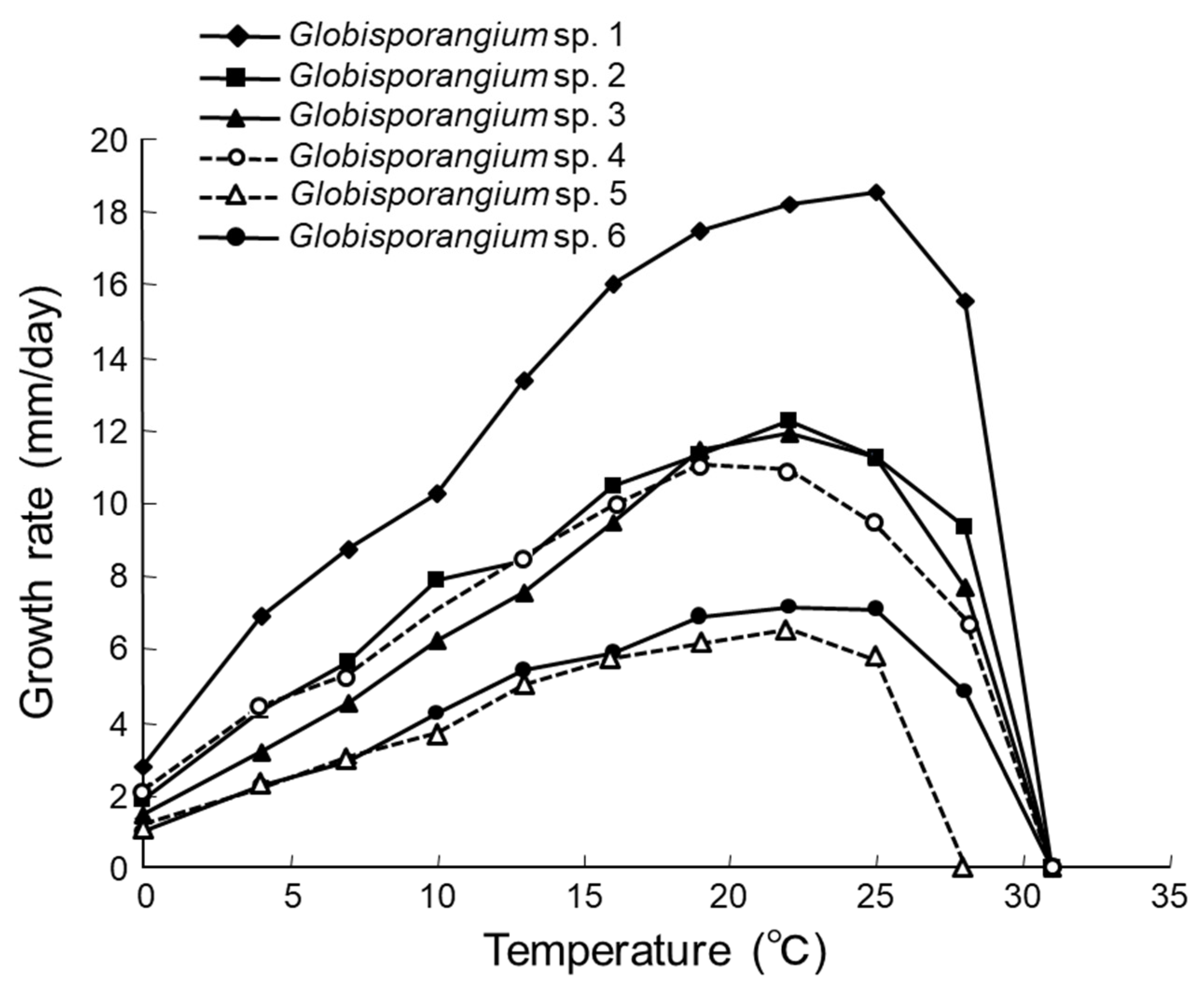
2.3. Characterizations of Morphology and Hyphal Growth Speed

One Globisporangium strain from each of the six major taxonomic groups were used for characterization of morphology and hyphal growth speed. The strains used were 10G16V2, 10G15W, 10C17N1, 10G34N1, 10C12N1, and 10G26N1 (Figure 2, Table S1).
Morphology of the strains was examined in grass-leaf water culture [36]. All strains were grown on CMA, potato dextrose agar (PDA; Becton Dickinson and Company), or V8 juice agar at 4–17 °C. A piece of agar medium was placed in a Petri dish containing a shallow layer of sterilized water, to which some 1–2 cm leaf pieces of gramineous weeds sterilized by autoclave were added. After incubation at 4–17 °C until Globisporangium strains colonized the leaves, sterilized pond water was added. Spore formation and the shape of said spores were examined by optical microscope (Olympus BX 43, Tokyo, Japan).
To determine hyphal growth rates, the strains were incubated on potato carrot agar (PCA) prepared according to van der Plaats-Niterink [37] in Petri dishes at 0, 4, 7, 10, 13, 16, 19, 22, 25, 28, 31, 34, and 37 °C in darkness, and colony diameters were measured. The experiments were repeated three times with one plate per repetition.

2.4. Isolation Pattern

Isolation frequency was determined as the number of the moss shoots that isolated Globisporangium spp. divided by the total number of the moss shoots examined. The isolation frequency was compared yearly by least significant differences based on a Tukey–Kramer Honestly Significant Difference test (p < 0.05) by JMP 13 (SAS Institute, Cary, NC, USA).

2.5. Infectivity to Sanionia Moss

Eleven Globisporangium strains from the six major taxonomic groups were used to test infectivity to Sanionia moss (S. uncinata). The strains used were 10G16V2, 10G15W, 10C17N1, 10G34N1, 10C12N1, 10G26N1, 18G11V1, 18C29N1, 18C32N1, 18C17N2, and 18C14N1 (Figure 2, Table S1). Stem-leaf sections (15 mm long, 0.5 mm wide) of the Sanionia obtained in Ny-Ålesund were placed on plates containing a KNOP agar medium [38] amended with 1.5% agar and were grown in a growth chamber at 10 °C for 3–4 months with continuous light (80 mmol m−2 s−1 measured at the level of the plants). A CMA plug (8 mm diameter) from each strain of Globisporangium, grown at 15 °C for 1 week, was placed in the center of the plate containing the Sanionia moss sections. Uninfected CMA was used as a control. The plates were kept at 0 °C, 4 °C, and 15 °C in darkness for approximately one month in a growth chamber. Infectivity was confirmed by optical microscopic observation. Recovery of the inoculated Globisporangium strains from the infected stem leaves was done using NARM medium. There were 16 replicates for each strain, using one moss segment for each replicate.

3. Results and Discussion

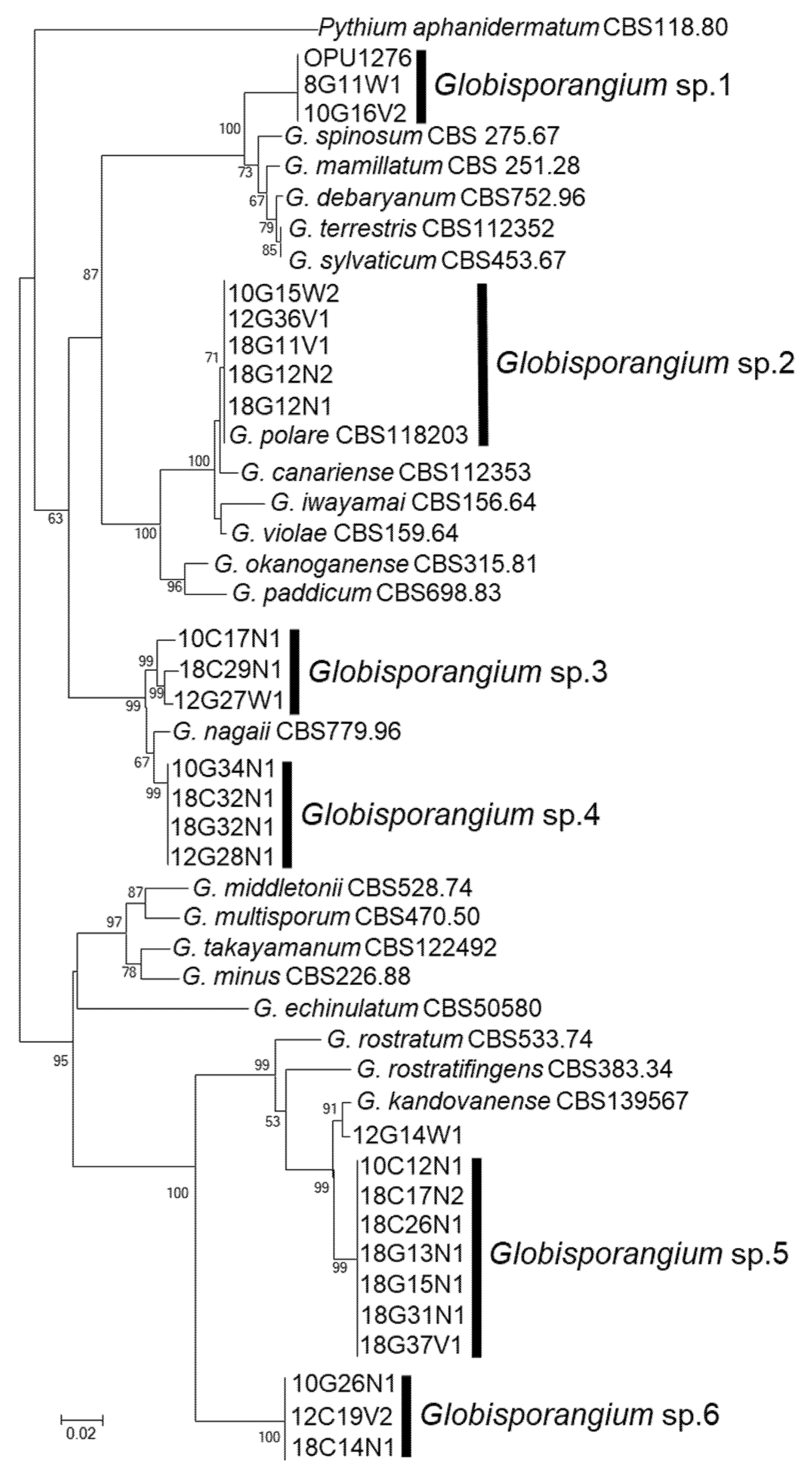
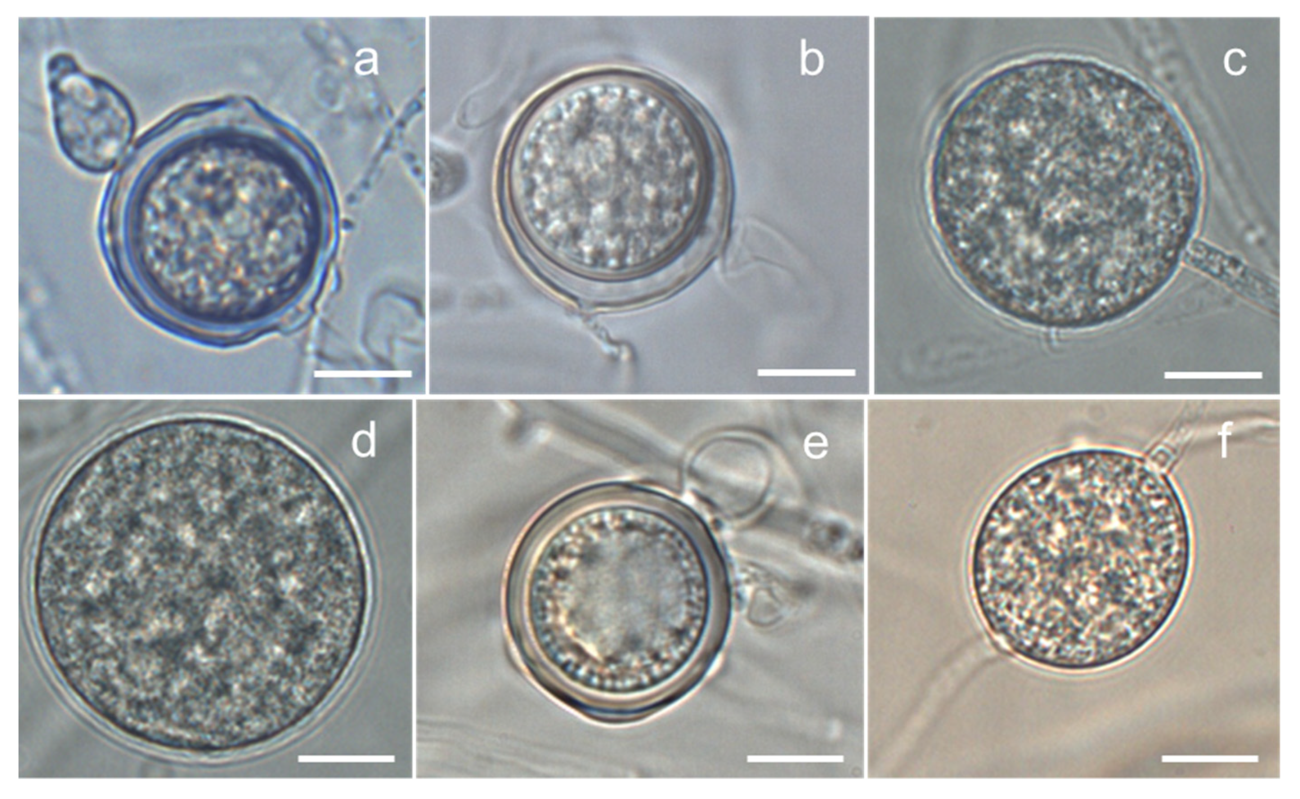
3.1. Isolation and Identification

In total, 434 isolates of Globisporangium spp. were obtained from the Sanionia moss during the 2006–2018 survey. All the isolates obtained were compared with known species based on the entire rDNA-ITS sequences through the GenBank database. The phylogenetic analysis of the sequences revealed that all isolates obtained were divided into six taxonomic groups of Globisporangium spp., which formed each monophyletic clade based on neighbor-joining (NJ) analyses (Figure 2). There was one exception: strain 12G14W1 did not belong to any of the six taxonomic groups. Since the maximum identities of these taxonomic groups against known species [22,39,40,41] were low (86.7 to 96.7%, Table S1), with the exception of one group with 99.8–100% similarity to G. polare (Table S1), they were named as Globisporangium sp. 1, sp. 2 (=G. polare), sp. 3, sp. 4, sp. 5, and sp. 6 (Figure 2). Globisporangium strain 12G14W1 was isolated only once, in 2012. The phylogenetic position of the strain 12G14W1 was the closest to G. kandovanense [41], but further analysis was not conducted because of loss of the strain.
Characteristics of morphology and hyphal growth speed of Globisporangium spp. 1–6 are described below.
Globisporangium sp. 1 strain 10G16V2: Main hyphae were up to 5 µm in diameter. Sporangia were not observed. Hyphal swellings were observed in single culture. Oogonia did not develop in single culture, but developed in dual culture with OPU1276. Strain OPU1276 was isolated from the present study site in July 2003 and showed identical rDNA-ITS sequence with strain 10G16V2 (Figure 2, Table S1). Oogonia were globose (Figure 3a), smooth, terminal sometimes intercalary, and 20.0–24.5 (mean 21.7) µm in diameter. Antheridia were monoclinous, with 1–4 per oogonium. Oospores were aplerotic, globose, smooth, and 16.5–21.5 (mean 18.7) µm in diameter, with one per oogonium. The thickness of the oospore wall was 0.5–1.5 (mean 1.0) µm. The minimum, optimum, and maximum temperatures for growth on PCA were 0 °C, 25 °C, and 28 °C, with daily growth rates at 2.7 mm, 18.3 mm, and 15.7 mm, respectively (Figure 4). The strain did not grow at 31 °C but showed regrowth when the dishes were placed at 22 °C. Globisporangium sp. 1 was closely phylogenetically related to G. spinosum, G. sylvaticum, and P. macrosporum (Figure 2) but was distinguished from these known species by the size and shape of its oogonia.
Globisporangium sp. 2 strain 10G15W2: Main hyphae were up to 6 µm in diameter. Sporangia were terminal and globose or sometimes subglobose. Zoospores were formed at 4–15 °C. Oogonia did not develop in single culture but developed in dual culture with G. polare CBS118202 [22]. Oogonia were globose, smooth, terminal or sometimes intercalary, and 17.3–26.9 (mean 23.1) µm in diameter (Figure 3b). Antheridia were diclinous, with 1–3 per oogonium. Oospores were aplerotic, globose, smooth, and 14.4–24.5 (mean 19.7) µm in diameter, with one per oogonium. The thickness of the oospore wall was 0.7–1.5 (mean 1.0) µm. The minimum, optimum, and maximum temperatures for growth on PCA were 0 °C, 22 °C, and 28 °C, with daily growth rates at 1.7 mm, 12.1 mm, and 9.4 mm, respectively (Figure 4). The growth rate at 25 °C was 11.2 mm per day. Since these taxonomic features matched those of G. polare, Globisporangium sp. 2 was identified as G. polare [22]. The result of the morphological study is in concordance with the result of the phylogenetic study.
Globisporangium sp. 3 strain 10C17N1: Main hyphae were up to 5 µm in diameter. Globose sporangia were observed in single culture (Figure 3c). Sexual reproductive organs did not produce in single or dual culture. The minimum, optimum, and maximum temperatures for growth on PCA were 0 °C, 22 °C, and 28 °C, with daily growth rates at 1.1 mm, 11.9 mm, and 7.8 mm, respectively (Figure 4). The growth rate at 25 °C was 11.2 mm per day. Globisporangium sp. 3 did not grow at 31 °C but showed regrowth at 22 °C.
Globisporangium sp. 4 strain 10G34N1: Main hyphae were up to 5 µm in diameter. Globose sporangia were observed in single culture (Figure 3d). Sexual reproductive organs were not produced in single or dual culture. The minimum, optimum, and maximum temperatures for growth on PCA were 0 °C, 19 °C, and 28 °C, with daily growth rates at 2.1 mm, 11.0 mm, and 6.9 mm, respectively (Figure 4). The growth rate at 25 °C was 9.4 mm per day. The strain did not grow at 31 °C but showed regrowth at 22 °C.
Globisporangium spp. 3 and 4 were closely related to G. nagaii based on rDNA- ITS sequences (Figure 2). Since asexual stages of Globisporangium spp. 3 and 4 were not formed in this study, additional taxonomic study is needed to distinguish Globisporangium spp. 3 and 4 from G. nagaii.
Globisporangium sp. 5 strain 10C12N1: Main hyphae were up to 6 µm in diameter. Globose sporangia, hyphal swellings, and sexual reproductive organs were observed in single culture. Oogonia were globose, smooth, terminal, and 20.0–26.0 (mean 22.9) µm in diameter (Figure 3e). Antheridia were monoclinous or occasionally diclinous, with 1–2 per oogonium. Oospores were aplerotic or occasionally plerotic, globose, smooth, and 19.0–26.0 (mean 22.2) µm in diameter, with one per oogonium. The thickness of the oospore wall was 0.2–2.0 (mean 1.1) µm. The minimum, optimum, and maximum temperatures for growth on PCA were 0 °C, 22 °C, and 25 °C, with daily growth rates at 1.0 mm, 6.3 mm, and 5.7 mm, respectively (Figure 4). Globisporangium sp. 5 was phylogenetically closely related to G. kandovanense, G. rostratifingens, and G. rostratum, but was distinguished from these three related species by the size of its oogonia and the positions of its antheridium. Globisporangium sp. 5 was also distinguished from G. rostratifingens and G. rostratum by growing at 0 °C.
Globisporangium sp. 6 strain 10G26N1: Main hyphae were up to 5 µm in diameter. Sporangia were not observed. Hyphal swellings were observed in single culture (Figure 3f). Sexual reproductive organs and sporangia were formed neither in single nor dual culture. The minimum, optimum, and maximum temperatures for growth on PCA were 0 °C, 22 °C, and 28 °C, with daily growth rates at 0.9 mm, 7.1 mm, and 5.0 mm, respectively (Figure 4). The growth rate at 25 °C was 7.0 mm per day. Like Globisporangium sp. 5, sp. 6 is phylogenetically closely related with G. kandovanense, G. rostratifingens, and G. rostratum. Although the species identity was unclear for Globisporangium sp. 6, this species could be distinguished from G. rostratifingens and G. rostratum by growing at 0 °C. The species also differed from G. kandovanense by not forming sporangia.
All the Globisporangium strains obtained were identified as one of six species, i.e., Globisporangium sp. 1, ibid sp. 2 (=G. polare), ibid sp. 3, ibid sp. 4, ibid sp. 5, and ibid sp. 6, except for the strain 12G14W1. Strains of Globisporangium spp. 1 to 6 grew at 0 °C on agar plates and infected the Sanionia moss at 4 to 10 °C. Among the six species, only Globisporangium sp. 2 was a known species and was G. polare [22]. The other five remained unknown species. G. polare was first described from Sanionia moss with brown discoloration under snow cover in Longyearbyen, Spitsbergen Is., and has been found only in polar regions [22]. The phylogenetic position of the strain 12G14W1 was closest to G. kandovanense which was isolated from Lolium perenne with snow rot symptoms in a natural grassland in East Azerbaijan province, Iran [41]. The present results, together with previous reports, suggest that Globisporangium in Sanionia moss colonies in Ny-Ålesund not only has a unique species composition, but also shows adaptation to cold environments. Further study is needed to describe the new species for the unknown Globisporangium spp.

3.2. Infectivity to Sanionia Moss

Globisporangium spp. 1, 2, 3, 4, and 6 infected the moss cells by penetration and colonization of mycelia at 4 °C and/or 10 °C (Table 1). Only one of the three strains tested of Globisporangium sp. 1 managed to colonize the moss cells, because the other two strains were lost when the test was done. Among the six species, Globisporangium spp. 1–4 consistently formed hyphae, oospores, and sporangia into the stem leaves of the moss cells (Table 1). At least one strain of all six groups produced sporangia or hyphal swellings inside the moss cells (Figure 5). All the strains infected the moss without showing any symptoms such as blight or discoloration of shoots and leaves until about 2 months after inoculation. The Globisporangium spp. were reisolated from the nonsymptomatic moss (Table 1).
Lévesque and de Cock [42] characterized phylogenetic clades of Pythium involving Globisporangium. Based on their clades, the Globisporangium spp. found in this study belong to clades E, F, and G [42]. Globisporangium sp. 1 belonged to clade F. This clade includes important crop pathogens such as G. spinosum, G. irregulare, G. sylvaticum, and G. debaryanum. Globisporangium spp. 2 (=G. polare), 3, and 4 belonged to clade G. This clade also includes important plant pathogens such as G. iwayamai, G. paddicum, and G. okanoganense, which cause snow rot of wheat and barley in Asia and the USA [25]. Globisporangium spp. 5 and 6 belonged to clade E, which includes weak pathogens of many plants [37]. This suggests that Globisporangium spp. 1 to 6 could be potential crop pathogens.

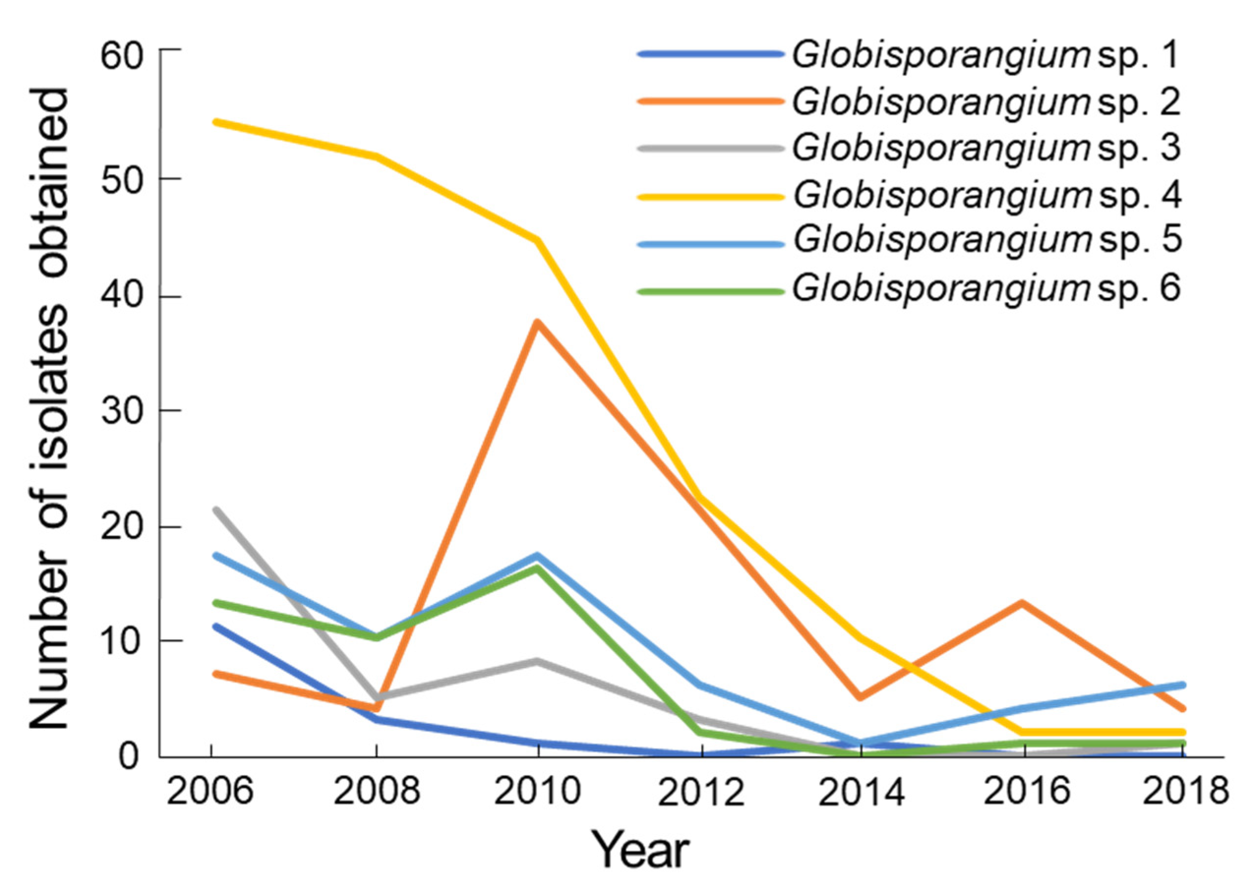
3.3. Isolation Pattern

Isolation frequency of the total population of Globisporangium spp. was maintained between 2006 and 2010, and significantly (p < 0.05) decreased from 2012 to 2018 (Figure 6). The total population was lowest in 2018 during the twelve-year period. The changes in the isolation pattern were different for the six Globisporangium spp. (Figure 7). Globisporangium spp. 1, 3, 4, and 6 consistently decreased from 2012 on. Globisporangium sp. 1 was not recorded in 2012, 2016, and 2018. Globisporangium spp. 2 (=G. polare) and 5 maintained their population, although the population differed from year to year.
Quantitative isolation from 2006 to 2018 demonstrated that total population of Globisporangium significantly decreased during the twelve-year period. Most of the Globisporangium spp. decreased their population. Only Globisporangium spp. 2 (=G. polare) and 5 showed little decreasing. The reason for the population decreasing is difficult to explain, but it may be influenced by climate changes in Arctic regions [43,44]. The influence of climate changes has already been recognized in the species composition and distribution of the Arctic vegetation [45,46]. Globisporangium spp. inhabiting Arctic regions are cold-adapted mesophiles rather than true psychrophiles (cold-loving), because they can grow at 20–25 °C. Mycelia of Globisporangium spp. are less freeze-resistant than those of fungi, even though a few isolates of G. polare are tolerant [28]. However, Globisporangium spp. can be highly tolerant to freezing when they have infected plant tissues [28]. The present in vitro study confirmed consistent infection of wet living moss by all six Globisporangium spp. under cold conditions. Previous and current results suggest that Globisporangium spp. found in the study site mainly increase their population during the summer period by infecting Sanionia moss, although they can grow at 0 °C under snow cover. Since Globisporangium requires wet conditions to produce hyphae, sporangia, and oospores [37], a consistent moist condition during the summer period is necessary to maintain its population. Romero et al. [3] reported that humidity is a primary driving factor for outbreaks of plant diseases caused by fungi and oomycetes. The recent continuous warming in the Arctic regions will decrease the diversity of mosses [11,46], which can be host plants of Globisporangium in the region. Better understanding of taxonomic and ecological features of the Arctic Globisporangium is needed, because they have unique species constructions and are probably vulnerable to climate changes.

4. Conclusions

At least six species of Globisporangium were found in single colony of the Sanionia moss in Ny-Ålesund, Spitsbergen Is., Norway. Among them, G. polare was the only known species, which has only been found in polar regions. The other five were unknown species and remain to be described as new species. All six species grew at 0 °C on an agar plate. All of them infected Sanionia moss under an in vitro inoculation test. Quantitative isolations of Globisporangium spp. from 2006 to 2018 showed that most of the species reduced their population over the recent decade at the study site. Much like other plant-parasitic oomycetes, the present Globisporangium spp. require a consistent moist condition to maintain their population. Recent climate change is influencing humidity in the Arctic region and could become a factor in the population reduction of the Globisporangium spp. Considering the unique species construction of Globisporangium found in this study, further evaluations are needed to provide better understanding of the taxonomic and ecological features of these species.

Supplementary Materials

The following are available online at https://www.mdpi.com/article/10.3390/microorganisms9091912/s1, Table S1: Information for Globisporangium strains used in the phylogenetic tree of Figure 2.

Author Contributions

Conceptualization, M.T., T.H. and M.-L.H.; methodology, M.T.; software, M.T.; validation, M.T.; formal analysis, M.T.; investigation, M.T., N.F., H.Y., Y.Y., K.T., K.K., A.H. and M.U.; resources, M.T., N.F. and Y.Y.; data curation, M.T., H.Y. and N.F.; writing—original draft preparation, M.T.; writing—review and editing, M.T., T.H., M.-L.H. and M.U.; visualization, M.T.; supervision, M.T.; project administration, M.T.; funding acquisition, M.T., T.H. and M.U. All authors have read and agreed to the published version of the manuscript.

Funding

This research was funded by the Japan Society for the Promotion of Science grant-in-aid for scientific research Nos. 15510028, 19510033, 23510032, 15K00626 and 19K12421, and by National Institute of Polar Research (NIPR) through General Collaboration Project Nos. 22-21, 25-25, 28-32 and 31-36 to M.T.; and by the Arctic Challenge for Sustainability (ArCS II) Project for M.T., H.T. and U.M.

Institutional Review Board Statement

Not applicable.

Informed Consent Statement

Not applicable.

Data Availability Statement

The data presented in present paper are available in this article.

Acknowledgments

We thank Koh Aoki of Osaka Prefecture University for his valuable comments on this study. Thanks are also due to Jun Inoue of the National Institute of Polar Research (NIPR) for providing the aerial photograph of the study site and to Ryui Nagano of Osaka Prefecture University for his technical assistance.

Conflicts of Interest

The authors declare no conflict of interest.

References

  1. Gilbert, G.S. Evolutionary ecology of plant disease in natural ecosystems. Annu. Rev. Phytopathol. 2002, 40, 13–43. [Google Scholar] [CrossRef] [Green Version]
  2. Delgado-Baquerizo, M.; Guerra, G.A.; Cano-Díaz, C.; Egidi, E.; Wang, J.; Eisenhauer, N.; Singh, B.K.; Maestre, F.T. The proportion of soil-borne pathogens increases with warming at the global scale. Nat. Clim. Chang. 2020, 10, 550–554. [Google Scholar] [CrossRef]
  3. Romero, F.; Cazzato, S.; Walder, F.; Vogelgsang, S.; Bender, S.F.; van der Heijden, M.G.A. Humidity and high temperature are important for predicting fungal disease outbreaks worldwide. New Phytol. 2021, in press. [Google Scholar] [CrossRef] [PubMed]
  4. Tojo, M.; Newsham, K.K. Snow mould in polar environments. Fungal Ecol. 2012, 5, 395–402. [Google Scholar] [CrossRef]
  5. Karsten, P.A. Fungi in insulis Spetsbergen et Beeren eiland collecti, in Öfvers. Kungliga Vetenskapsakademien. Förhandlingar 1872, 2, 81–108. [Google Scholar]
  6. Lind, J. The Micromycetes of Svalbard. Skr Svalbard Ishavet 1928, 13, 1–61. [Google Scholar]
  7. Tojo, M.; Masumoto, S.; Hoshino, T. Phytopathogenic fungi and fungal-like microbes in Svalbard. In Plant and Microbe Adaptations to Cold in a Changing World; Imai, R., Yoshida, M., Matsumoto, N., Eds.; Springer: New York, NY, USA, 2013; pp. 263–284. [Google Scholar]
  8. Tsuji, M.; Uetake, J.; Tanabe, Y. Changes in the fungal community of Austre Brøggerbreen deglaciation area, Ny-Ålesund, Svalbard, High Arctic. Mycoscience 2016, 57, 448–451. [Google Scholar] [CrossRef]
  9. IPCC Sixth Assessment Report. 2021. Available online: https://www.ipcc.ch/assessment-report/ar6/ (accessed on 7 September 2021).
  10. Cornelissen, J.H.C.; Callaghan, T.V.; Alatalo, J.M.; Michelsen, A.; Graglia, E.; Hartley, A.E.; Hik, D.S.; Hobbie, S.E.; Press, M.C.; Robinson, C.H.; et al. Global change and arctic ecosystems: Is lichen decline a function of increases in vascular plant biomass? Ecol. J. 2001, 89, 984–994. [Google Scholar] [CrossRef]
  11. Hollister, R.D.; Webber, P.J.; Tweedie, C.E. The response of Alaskan arctic tundra to experimental warming: Differences between short-and long-term responses. Glob. Chang. Biol. 2005, 11, 525–536. [Google Scholar] [CrossRef]
  12. Zhang, W.; Miller, P.A.; Smith, B.; Wania, R.; Koenigk, T.; Döscher, R. Tundra shrubification and tree-line advance amplify arctic climate warming: Results from an individual-based dynamic vegetation model. Environ. Res. Lett. 2013, 8, 034023. [Google Scholar] [CrossRef] [Green Version]
  13. Sturm, M.; McFadden, J.P.; Liston, G.E.; Chapin, F.S.; Racine, C.H.; Holmgren, J. Snow–shrub interactions in arctic tundra: A hypothesis with climatic implications. J. Clim. 2001, 14, 336–344. [Google Scholar] [CrossRef] [Green Version]
  14. Miles, J.; Walton, D.W.H. Primary succession revisited. In Primary Succession on Land; Miles, J., Walton, D.W.H., Eds.; Blackwell Science: Oxford, UK, 1993; pp. 295–302. [Google Scholar]
  15. Smith, R.I.L. Terrestrial and freshwater biotic components of the western Antarctic Peninsula. In Foundations of Ecological Research West of the Antarctic Peninsula Antarctic Research Series; Ross, R.M., Hofmann, E.E., Quentin, L.B., Eds.; American Geophysical Union: Washington DC, USA, 1996; Volume 70, pp. 15–59. [Google Scholar]
  16. Virtanen, R.J.; Lundberg, P.A.; Moen, J.; Oksanen, L. Topographic and altitudinal patterns in plant communities on European arctic islands. Polar Biol. 1997, 17, 95–113. [Google Scholar] [CrossRef]
  17. Fenton, J.H.C. Concentric fungal rings in Antarctic moss communities. Trans. Br. Mycol. Soc. 1983, 80, 413–420. [Google Scholar] [CrossRef]
  18. Longton, R.E. The occurrence of radial infection patterns in colonies of polar bryophytes. BAS Bull. 1973, 32, 41–49. [Google Scholar] [CrossRef] [Green Version]
  19. Robinson, C.H.; Wookey, P.A. Microbial ecology, decomposition and nutrient cycling. In Ecology of Arctic Environments; Woodin, S.J., Marquis, M., Eds.; Blackwell Science: Oxford, UK, 1997; pp. 41–68. [Google Scholar]
  20. Rosa, L.H.; de Sousa, J.R.P.; de Menezes, G.C.A.; da Costa Coelho, L.; Carvalho-Silva, M.; Convey, P.; Câmara, P.E.A.S. Opportunistic fungi found in fairy rings are present on different moss species in the Antarctic Peninsula. Polar Biol. 2020, 43, 587–596. [Google Scholar] [CrossRef]
  21. Rosa, L.H.; da Silva, T.H.; Ogaki, M.B.; Pinto, O.H.B.; Stech, M.; Convey, P.; Carvalho-Silva, M.; Rosa, C.A.; Câmara, P.E.A.S. DNA metabarcoding uncovers fungal diversity in soils of protected and non-protected areas on Deception Island, Antarctica. Sci. Rep. 2020, 10, 21986. [Google Scholar] [CrossRef]
  22. Tojo, M.; Van West, P.; Hoshino, T.; Kida, K.; Fujii, H.; Hakoda, H.; Kawaguchi, Y.; Mühlhauser, H.A.; Van den Berg, A.H.; Küpper, F.C.; et al. Pythium polare, a new heterothallic Oomycete causing brown discoloration of Sanionia uncinata in the Arctic and Antarctic. Fungal Biol. 2012, 116, 756–768. [Google Scholar] [CrossRef]
  23. Uzuhashi, S.; Tojo, M.; Kakishima, M. Phylogeny of the genus Pythium and description of new genera. Mycoscience 2010, 51, 337–365. [Google Scholar] [CrossRef]
  24. Molin, C.; Ribeiro, N.R.; Matsumoto, M.N.; Giasson, N.F.; Brollo, J.; Zanardo, B.; Pelissoni, M.; Capitanio, S.; Comín, T.; Deuner, C.C.; et al. Damping-off of soybean in southern Brazil can be associated with different species of Globisporangium spp. and Pythium spp. Plant Pathol. 2021, 70, 1686–1694. [Google Scholar] [CrossRef]
  25. Lipps, P.E.; Bruehl, G.W. Snow rot of winter wheat in Washington. Phytopathology 1978, 68, 1120–1127. [Google Scholar] [CrossRef]
  26. Bridge, P.D.; Newsham, K.K.; Denton, G.J. Snow mould caused by a Pythium sp.: A potential vascular plant pathogen in the maritime Antarctic. Plant Pathol. 2008, 57, 1066–1072. [Google Scholar] [CrossRef]
  27. Hoshino, T.; Terami, F.; Tkachenko, O.B.; Tojo, M.; Matsumoto, N. Mycelial growth of the snow mold fungus, Sclerotinia borealis improved at low water potentials: An adaptation to frozen environment. Mycoscience 2010, 51, 98–102. [Google Scholar] [CrossRef]
  28. Murakami, R.; Yajima, Y.; Kida, K.; Tokura, K.; Tojo, M.; Hoshino, T. Surviving freezing in plant tissues by oomycetous snow molds. Cryobiology 2015, 70, 208–210. [Google Scholar] [CrossRef] [PubMed]
  29. Hoshino, T.; Nakagawa, T.; Yajima, Y.; Uchida, M.; Tojo, M. Note on a snow mold and a fungus-like microbe from Kuujjuarapik-Whapmagoostui, Quebec, subarctic Canada. Polar Sci. 2021, 27, 100559. [Google Scholar] [CrossRef]
  30. Ali-shtayeh, M.S.; Lim-ho, C.L.; Dick, N.W. An improved method and medium for quantitative estimates of populations of Pythium species from soil. Trans. Brit. Mycol. Soc. 1986, 86, 39–47. [Google Scholar] [CrossRef]
  31. Morita, Y.; Tojo, M. Modifications of PARP medium using fluazinam, miconazole and nystatin for detection of Pythium spp. in soil. Plant. Dis. 2007, 91, 1591–1599. [Google Scholar] [CrossRef] [Green Version]
  32. Miller, P.M. V8 juice agar as a general purpose medium for fungi and bacteria. Phytopathology 1955, 45, 461–462. [Google Scholar] [CrossRef] [Green Version]
  33. White, T.J.; Bruns, T.; Lee, S.; Taylor, J. Amplification and direct sequencing of fungal ribosomal RNA gene for phylogenetics. In PCR Protocols: A Guide to Methods and Applications; Innis, M.A., Gelfand, D.H., Sninsky, J.J., White, T.J., Eds.; Academic Press: San Diego, CA, USA, 1990; pp. 315–322. [Google Scholar]
  34. Tamura, K.; Peterson, D.; Peterson, N.; Stecher, G.; Nei, M.; Kumar, S. MEGA5: Molecular evolutionary genetics analysis using maximum likelihood, evolutionary distance, and maximum parsimony methods. Mol. Biol. Evol. 2011, 28, 2731–2739. [Google Scholar] [CrossRef] [Green Version]
  35. Saitou, N.; Nei, M. The neighbor-joining method: A new method for reconstructing phylogenetic trees. Mol. Biol. Evol. 1987, 4, 406–425. [Google Scholar]
  36. Martin, F.N.  Pythium. In Methods for Research on Soilborne Phytopathogenic Fungi; Singleton, L.L., Mihail, J.D., Rush, C.M., Eds.; APS Press: Saint Paul, MN, USA, 1992; pp. 39–49. [Google Scholar]
  37. van der Plaats-Niterink, A.J. Monograph of the genus Pythium. Stud. Mycol. 1981, 21, 1–242. [Google Scholar]
  38. Nehira, K. Germination and protonemata. In Methods in Bryology; Glime, J., Ed.; Hattori Botanical Laboratory: Nichinan, Japan, 1988; pp. 113–117. [Google Scholar] [CrossRef] [Green Version]
  39. Hyde, K.D.; Nilsson, R.H.; Alias, S.A.; Ariyawansa, H.A.; Blair, J.E.; Cai, L.; de Cock, A.W.A.M.; Dissanayake, A.J.; Glockling, S.L.; Goonasekara, I.D.; et al. One stop shop: Backbones trees for important phytopathogenic genera: I. Fungal Divers. 2014, 67, 21–125. [Google Scholar] [CrossRef] [Green Version]
  40. Robideau, G.P.; De Cock, A.W.; Coffey, M.D.; Voglmayr, H.; Brouwer, H.; Bala, K.; Chitty, D.W.; Désaulniers, N.; Eggertson, Q.A.; Gachon, C.M.; et al. DNA barcoding of oomycetes with cytochrome c oxidase subunit I and internal transcribed spacer. Mol. Ecol. Resour. 2011, 11, 1002–1011. [Google Scholar] [CrossRef]
  41. Bouket, A.C.; Arzanlou, M.; Tojo, M.; Babai-Ahari, A. Pythium kandovanense sp. nov., a fungus-like eukaryotic microorganism (Stramenopila, Pythiales) isolated from snow covered ryegrass leaves. Int. J. Syst. Evol. Microbiol. 2015, 65, 2500–2506. [Google Scholar] [CrossRef]
  42. Lévesque, C.A.; de Cock, A.W.A.M. Molecular phylogeny and taxonomy of the genus Pythium. Mycol. Res. 2004, 108, 1363–1383. [Google Scholar] [CrossRef] [PubMed] [Green Version]
  43. Box, J.E.; Colgan, W.T.; Christensen, T.R.; Schmidt, N.M.; Lund, M.; Parmentier, F.J.W.; Brown, R.; Bhatt, U.S.; Euskirchen, E.S.; Romanovsky, V.E.; et al. Key indicators of Arctic climate change: 1971–2017. Environ. Res. Lett. 2019, 14, 045010. [Google Scholar] [CrossRef]
  44. Overland, J.E.; Hanna, E.; Hanssen-Bauer, I.; Kim, S.J.; Walsh, J.E.; Wang, M.; Bhatt, U.S. An RL Surface air temperature. In Arctic Report Card; Richter-Menge, J., Overland, J.E., Mathis, J.T., Osborne, E., Eds.; National Oceanic and Atmospheric Administration (NOAA): Miami, FL, USA, 2017; pp. 5–12. [Google Scholar] [CrossRef]
  45. Pearson, R.G.; Phillips, S.J.; Loranty, M.M.; Beck, P.S.; Damoulas, T.; Knight, S.J.; Goetz, S.J. Shifts in Arctic vegetation and associated feedbacks under climate change. Nat. Clim. Change 2013, 3, 673–677. [Google Scholar] [CrossRef]
  46. Walker, M.D.; Wahren, C.H.; Hollisterc, R.D.; Henry, G.H.R.; Ahlquist, L.E.; Alatalo, J.M.; Bret-Harte, M.S.; Cale, M.P.; Callaghan, T.V.; Carroll, A.B.; et al. Plant community responses to experimental warming across the tundra biome. Proc. Natl. Acad. Sci. USA 2006, 103, 1342–1346. [Google Scholar] [CrossRef] [PubMed] [Green Version]
Figure 1. Study site and plot: (a) location of Ny-Ålesund, Spitsbergen Is., Svalbard Archipelago, Norway; (b) distribution of the six sampling plots (arrow heads) in the Sanionia moss colony at a north side cliff in Ny-Ålesund; (c) the 15 cm-square plot. The yellow threads of the quadrat were put on the moss surface only when the moss was sampled. The aerial photograph was kindly taken by Dr. Jun Inoue of the National Institute of Polar Research (NIPR).
Microorganisms 09 01912 g001
Figure 2. Phylogenetic positions of Globisporangium strains obtained from the Sanionia moss in Ny-Ålesund, Spitsbergen Island, Norway, on a neighbor-joining (NJ) tree of the ITS of the rDNA region. Numbers beside the branches are the bootstrap values (>50%) of 1000 replicates. Pythium aphanidermatum strain CBS118.80 was used as an outgroup. The six major subclades found in this study were named as Globisporangium spp. 1–6.
Microorganisms 09 01912 g002
Figure 3. Oospores or sporangia of Globisporangium spp. isolated from the Sanionia moss in Ny-Ålesund, Spitsbergen Island, Norway: (a) aplerotic oospore of Globisporangium sp. 1 strain 10G16V2; (b) aplerotic oospore of Globisporangium sp. 2 strain 10G15W2 (=G. polare); (c) globose sporangium of Globisporangium sp. 3 strain 10C17N1; (d) globose sporangium of Globisporangium sp. 4 strain 10G34N1; (e) plerotic oospore of Globisporangium sp. 5 strain 10C12N1; and (f) hyphal swelling of Globisporangium sp. 6 strain 10G26N1. Bars = 10 μm.
Microorganisms 09 01912 g003
Figure 4. Mycelial growth rate of Globisporangium spp. isolated from a single colony of Sanionia moss in Ny-Ålesund, Spitsbergen Island, Norway on potato carrot agar at different temperatures in darkness. The strains used were Globisporangium sp. 1 strain 10G16V2, Globisporangium sp. 2 (=G. polare) strain 10G15W2, Globisporangium sp. 3 strain 10C17N1, Globisporangium sp. 4 strain 10G34N1, Globisporangium sp. 5 strain 10C12N1, and Globisporangium sp. 6 strain 10G26N1.
Microorganisms 09 01912 g004
Figure 5. Production of sporangia and hyphal swellings of Globisporangium spp. isolated from a single colony of Sanionia moss in Ny-Ålesund, Spitsbergen Is., Norway in host plant tissues under an in vitro inoculation at 4 to 10 °C: (a) hyphal swelling of Globisporangium sp. 1 strain 10G16V2; (b) sporangium of Globisporangium sp. 2 (=G. polare) strain 18G12N1; (c) sporangium of Globisporangium sp. 3 strain 18C32N1; (d) sporangium of Globisporangium sp. 4 strain 18C32N1; (e) sporangium of Globisporangium sp. 5 strain 18G13N1; and (f) hyphal swelling of Globisporangium sp. 6 strain 18C14N1. Bars = 10 μm.
Microorganisms 09 01912 g005
Figure 6. Changes of isolation frequency for the total population of Globisporangium spp. from a single colony of Sanionia moss in Ny-Ålesund, Spitsbergen Is., Norway from 2006 to 2018. The investigations were conducted in August each year. Isolation frequency was calculated as the number of moss shoots with isolated Globisporangium spp. divided by the total number of moss shoots examined. The average values with SE (N = 6) were shown. Values followed by the same letter are not significantly different according to Tukey’s HSD test (p < 0.05).
Microorganisms 09 01912 g006
Figure 7. Total number of isolates of Globisporangium sp. 1, sp. 2 (=G. polare), sp. 3, sp. 4, sp. 5 and sp. 6 from a single colony of Sanionia moss at the north-side cliff of Ny-Ålesund, Spitsbergen Is., Norway from 2006 to 2018. The investigations were carried out in August each year for the six sampling plots shown in Figure 1.
Microorganisms 09 01912 g007
Table 1. Infectivity of Globisporangium spp. from the Sanionia moss in Ny-Ålesund to Sanionia uncinata in an in vitro inoculation test.
Taxonomic GroupStrainTemperatureInfection into the Host Plant Cells with;Recovery from the Host Plant
(°C)HyphaeOospores or Sporangia
Globisporangium sp. 110G16V24++++
Globisporangium sp. 2 (G. polare)10G15W24+++
18G12N110+++
Globisporangium sp. 310C17N14++++
18C32N110+++
Globisporangium sp. 410G34N14+++
18C32N110+++
Globisporangium sp. 510C12N14
18G13N110+++
Globisporangium sp. 610G26N14++
18C14N110+++
Uninoculated 4
10
++: Infection was found more than 50% of the plant part examined, +: infection was found less than 50% of the plant part examined, −: no infection. Recovery of Globisporangium spp. from stem-leaves was calculated from the number of stem leaves from which Globisporangium was recovered after 4 weeks of incubation at 4 °C.
Publisher’s Note: MDPI stays neutral with regard to jurisdictional claims in published maps and institutional affiliations.

